Introduction
Acute appendicitis (AA) is currently the commonest
acute surgical pathology, with a lifetime risk of 7-9% (1,2).
Although various techniques have been advocated with the aim to
reduce negative appendectomy rates in patients with acute abdominal
pain, these remain between 15 and 50% (3) and seem higher in young female, elderly
and paediatric patients (4). A
safe, cheap, rapid, widely available and accurate diagnostic marker
for AA would be useful, however currently there is no single
diagnostic tool that can lead to a definitive diagnosis of AA
(5,6). In the past, different authors have
studied the diagnostic value of various laboratory tests, but
results are contradictory (4).
White Cell Count (WCC) as well as C-reactive protein (CRP) are two
of the markers that can contribute to the diagnosis of AA, with the
advantages of being relatively inexpensive and carrying no risks,
contrary to other methods such as computerized tomography with
exposure to radiation or laparoscopy with potential surgical
complications. Nevertheless, none of them is diagnostic or
specific. Previous retrospective studies have demonstrated
hyperbilirubinaemia to be a useful predictor of complicated acute
appendicitis and appendiceal perforation (7,8)
although contradiction exists in literature (2,9,10), but
recently others have suggested an association between Total Serum
Bilirubin (TSB) and simple appendicitis (3,6,11).
The present study's aim is to evaluate the
usefulness of biochemical parameters (WCC, CRP, TSB) including a
Receiver Operating Characteristic (ROC) curve analysis in the
diagnosis of AA and to evaluate further specifically the role of
TSB as preoperative diagnostic marker irrespective of the severity
of the disease.
Materials and methods
Patients and parameters
All electronically recorded consecutive cases of
appendicectomies in a 1-year period in a single centre were
reviewed. Demographic data of all patients have been recorded. All
cases in which symptoms were attributed to ovarian pathologies or
other problems identified during surgery and those in which
appendicectomy was not the primary procedure have been excluded
from the study. In the cases in which the study of the surgical
specimen did not confirm acute appendicitis or other pathology the
histology was considered as negative. In cases where the study of
the specimen confirmed acute appendicitis or any other pathology
explaining the clinical findings the histology was considered as
positive.
Median TSB value, as well as median WCC and CRP on
day of admission were statistically studied in relation to the
final histology result. A further comparison has been performed
between patients shown to have negative histology results and
patients demonstrated to have acute appendicitis regarding the
percentage of abnormal and normal values of WCC, CRP and TBS on
admission in each group. Any values of WCC higher than
11x109 wbc/l, CRP higher than 5.5 mg/l and TSB higher
than 21 µmol/l were considered as abnormal, while any values of
WCC, CRP and TSB below 11x109/l, 5.5 mg/l and 21 µmol/l
respectively were considered as normal. CRP given as <5 mg/l was
accounted as 4 mg/l.
As all cases in the current study derive from a
single centre, the diagnostic work up and clinical management
across the patients, such as history taking, clinical examination
and blood tests are homogeneous. Based on the judgment of the
responsible clinician, as well as on other factors such as
patient's gender, age, availability of techniques, timing etc., the
imaging method, if any, has varied among the patients (ultrasound
scan, computerized tomography, magnetic resonance imaging or none).
Conduction of this work is in full compliance with local Ethical
Regulations and Anonymization standards. Approval from local
ethical committee was not required as this was not an
interventional study, involving only retrospective analysis of
clinical data associated with diagnostic and therapeutic techniques
performed without any deviation from institute's local guidelines.
Data were analysed retrospectively thus informed consent from the
patients prior to their inclusion in the study was not required
according to local policy. All patients have signed an informed
consent form prior to their operations.
Statistical analysis
Bivariate associations between scale and binomial
variables were assessed using Mann-Whitney U test. Association of
categorical variables in 4-fold tables were assessed using Fisher's
exact test (2-tailed). P<0.05 was considered to indicate a
statistically significant difference. Sensitivity and specificity
analysis was performed, and subsequent ROC curve was produced. All
aforementioned were carried out using SPSS v20 software (IBM
Corporation).
Results
Descriptive statistics
Out of 311 appendicectomies, either open or
laparoscopic, recorded in the study period, eleven cases have been
excluded from the study. Median age at time of surgery was 27
years. 48.3% of cases were male patients. The negative
appendicectomy rate reached 23% (69 patients). Acute appendicitis
was confirmed in 77% of cases. Median values of WCC, CRP and TSB on
admission were 12.9, 25 and 12, respectively (Table I).
 | Table IDemographics, values on admission and
main outcome. |
Table I
Demographics, values on admission and
main outcome.
| Parameters | Values |
|---|
| Male sexa | 145 (48.3) |
| Age at time of
surgeryb | 27 (6-93) |
| WCC on
admissionb | 12.9 (3.4-26.1) |
| CRP on
admissionb | 25 (4-615) |
| TSB on
admissionb | 12 (4-263) |
| Positive final
histologya | 231(77) |
Comparisons
All median TSB, WCC and CRP on admission were
significantly higher in patients with acute appendicitis compared
to patients with negative histology (9 vs. 13, 9 vs. 13.9 and 4 vs.
37, respectively; P<0.001 for all three parameters) (Table II). Furthermore, after comparing
the respective rates of normal and abnormal TSB, WCC and CRP on
admission between the two groups of positive and negative
histology, these were overall found to have a significant
statistical association with final histology (P<0.014,
P<0.001 and P<0.001, respectively) (Table III). The relative diagnostic
values are shown in Table IV, with
TSB demonstrating compared to WCC and CRP a clearly higher
specificity for AA (0.88 vs. 0.67 for WCC and 0.42 for CRP), but
with lower sensitivity (0.26 vs. 0.77 and 0.89, respectively) and
overall accuracy (0.40 vs. 0.74 and 0.78, respectively).
| Table IIComparison of values on admission with
final histology. |
Table II
Comparison of values on admission with
final histology.
| | Final histology | |
|---|
| Biochemical
parameters | Negative | Positive | P-value |
|---|
| WCC on admission | 9 (3.8-18.3) | 13.9 (5.4-26.1) | <0.001 |
| CRP on admission | 4 (4-256) | 37 (4-615) | <0.001 |
| TSB on admission | 9 (4-71) | 13 (4-263) | <0.001 |
| Table IIIComparison of patients with normal and
abnormal values, with final histology results. |
Table III
Comparison of patients with normal and
abnormal values, with final histology results.
| | Final histology | |
|---|
| Biochemical
markers | Negative | Positive | P-value |
|---|
| WCC | | | <0.001 |
|
Normal | 46 (66.7) | 54 (23.4) | |
|
Raised | 23 (33.3) | 177 (76.6) | |
| CRP | | | <0.001 |
|
Normal | 29 (42.0) | 25 (19.1) | |
|
Raised | 40 (58.0) | 206 (80.9) | |
| TSB | | | 0.014 |
|
Normal | 61 (88.4) | 172 (74.5) | |
|
Raised | 8 (11.6) | 59 (25.5) | |
| Table IVStatistic values of WCC, CRP and TSB
in predicting acute appendicitis. |
Table IV
Statistic values of WCC, CRP and TSB
in predicting acute appendicitis.
| Statistic
values | WCCa | CRPa | TSBa |
|---|
| Sensitivity | 0.77
(0.71-0.82) | 0.89
(0.85-0.93) | 0.26
(0.20-0.31) |
| Specificity | 0.67
(0.56-0.78) | 0.42
(0.30-0.54) | 0.88
(0.81-0.96) |
| Positive predictive
value | 0.89
(0.84-0.93) | 0.84
(0.79-0.88) | 0.88
(0.80-0.96) |
| Negative predictive
value | 0.46
(0.36-0.56) | 0.54
(0.40-0.67) | 0.26
(0.21-0.32) |
| Odds ratio | 7.70
(4.98-11.88) | 5.15
(3.67-7.23) | 2.62
(1.18-5.79) |
| Diagnostic
accuracy | 0.74
(0.69-0.79) | 0.78
(0.74-0.83) | 0.40
(0.35-0.46) |
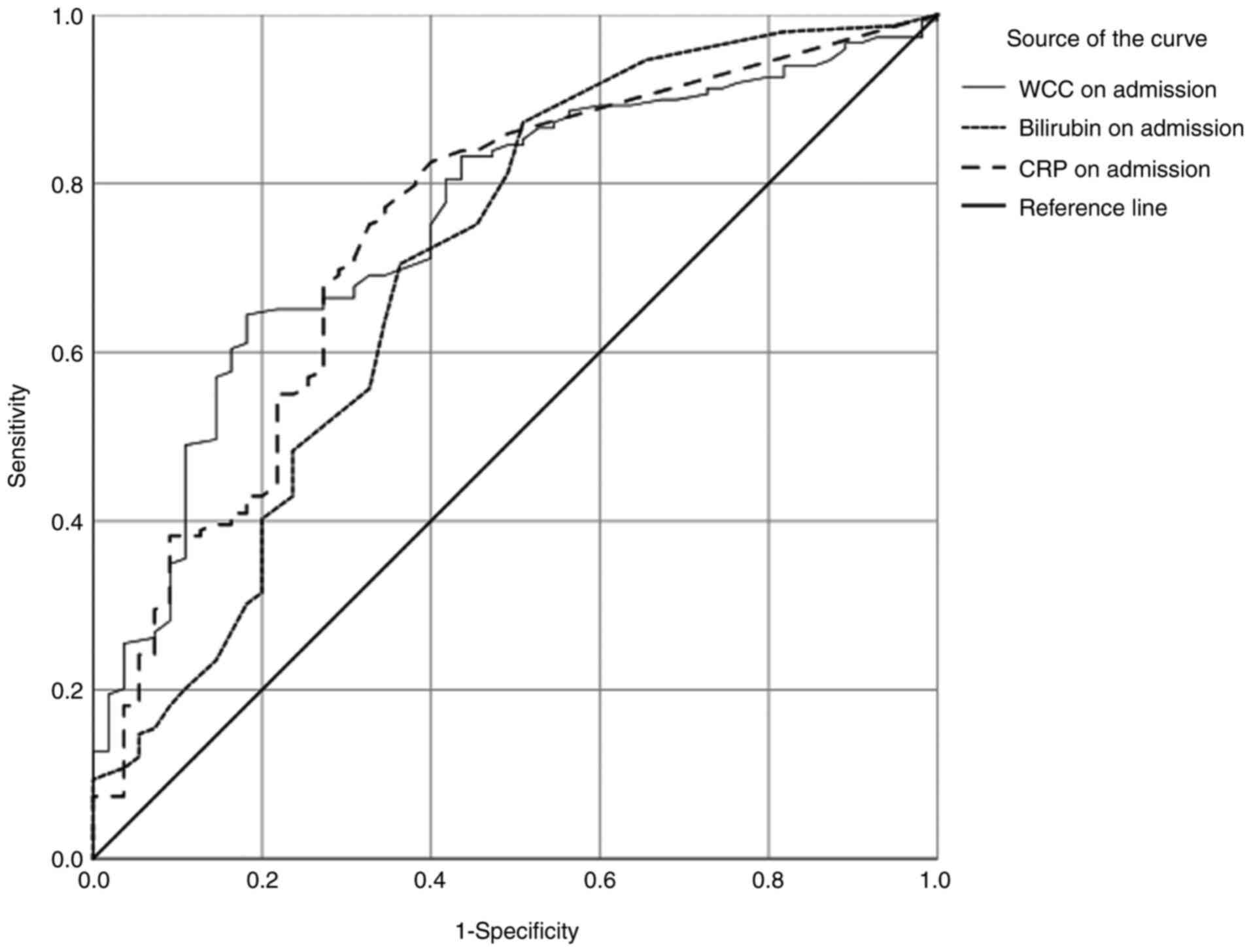
ROC analysis
Fig. 1 presents
cumulatively the ROC curves for the three biochemical predictors,
namely WCC, CRP and TSB. Area under curve was 0.743, 0.755 and
0.703, standard error was 0.04, 0.037 and 0.045, respectively
(P<0.001 for all three variables) (referring to equal
probabilities).
Discussion
The relation between hyperbilirubinaemia and severe
acute appendicitis has been reported since 1969(12). It is postulated that elevated serum
bilirubin occurs as a result of portal sepsis or empyema resulting
in liver hepatocytes dysfunction or damage. This is thought to be
caused by bacterial endotoxins or cytokines. The result is either a
direct damage to hepatocytes, cholestasis, or both leading to
hyperbilirubinaemia. Furthermore, endotoxins are shown to result in
haemolysis, which then adds further increase in bilirubin levels.
Other authors have reported that the elevated load of bacteria in
appendicitis causes either direct invasion or translocation into
the portal venous system. Direct invasion of bacteria into the
hepatic parenchyma interferes with the excretion of bilirubin into
the bile canaliculi by a mechanism that is thought to be caused by
the bacterial endotoxin and is biochemical in nature rather than
obstructive. On the other hand bacteria may transmigrate and
produce portal bacteraemia, hepatocellular dysfunction or pyogenic
liver abscess. The depression of hepatocellular function in the
early, hyper-dynamic stage of sepsis does not appear to be due to
reduction in hepatic perfusion but is associated with elevated
levels of circulating pro-inflammatory cytokines such as TNF and
IL-6. Thus up regulation of TNF and/or IL-6 may be responsible for
producing hepatocellular dysfunction during the early hyper-dynamic
stage of sepsis (13). Various
studies have correlated elevated serum bilirubin and acute
appendicitis, but most of them refer only to perforated
appendicitis (14). While
hyperbilirubinaemia in these cases can contribute to the diagnosis,
the clinical presentation is more diagnostic, therefore the role of
elevated serum bilirubin is probably limited. In the present study
a significant relation between TSB and AA was demonstrated. The
increase of TSB was significantly overall noted in patients with AA
irrespective of perforation. Even though median TSB was within
normal range in both groups with positive and negative histology,
like in other studies (2,15), the values of median TSB varied
significantly between these two groups, in line with other authors
(3,15). Furthermore, similarly to other
reports, the incidence of abnormal bilirubin among patients in the
positive histology group was more than double compared to the
negative appendicectomy group (3,16). As
Table III demonstrates, TSB was
significantly associated with the histology results. Interestingly,
although presenting relatively low sensitivity and overall
diagnostic accuracy, TSB was found in the current study to have a
high specificity, as well as a high Positive Predictive Value (PPV)
and this is in line with other publications that demonstrate a high
specificity for TSB in acute appendicitis, yet with considered low
sensitivity. The rates of sensitivity, specificity and PPV in
literature range respectively 8-80, 56-100 and 80-100%, in
accordance with the present results (2,3,6,7,11,15,17-20).
The present study significantly contributes to current scientific
knowledge by providing additional data and evidence to further
support the findings of the aforementioned studies and furthermore
it makes a step forward by presenting a ROC curve analysis that
clearly visualizes the advantages of the use of bilirubin.
WCC and CRP have been considered significant
parameters for the diagnosis of AA (18) with comparable sensitivity,
specificity, PPV, and NPV (4) and
although they have been studied extensively for the diagnosis of
AA, they lack sufficient specificity, either alone or in
combination (4,21). There are studies that demonstrate
clear differences in these values between patients with
non-surgical abdominal pain or early appendicitis and patients with
phlegmons or perforated appendicitis (3,4,18),
however the generally low sensitivity and specificity reported,
does not allow for them to be considered as optimal indicators for
AA (4,18). This is in line with the current
study, with results generally comparable to the rates of
sensitivity and specificity reported in literature for WCC (43-93
and 50-87% respectively) (4,15,17,18,22,23)
and CRP (53-97.2 and 46-99.3% respectively) (15,24-27).
Elevated WCC demonstrates an acceptable sensitivity and at the same
time a relatively low specificity as it can be triggered by various
conditions (4). CRP levels have
been reported as significantly higher in patients with perforated
appendicitis (4,6,15) and
CRP has been considered a more sensitive test in discriminating the
pathological severity of appendicitis (24,28),
with higher accuracy than WCC (15,27).
Nevertheless, still its relative specificity is low as revealed by
the present study and unable to enhance the role of CRP in the
diagnosis of AA, unless integrated to other parameters.
Limitations of the study include its retrospective
nature, the inclusion only of patients who underwent
appendicectomies which may cause an increase of the specificities
of the study and the non-discrimination of the severity of disease
across the cases. Furthermore, the possibility of inclusion of
patients with Gilbert's syndrome in the cohort studied should be
considered, although it represents an uncommon condition reported
in 3-10% the population (3). The
cohort consisted of all consecutive patients operated for suspected
appendicitis, therefore there were obviously patients that were
eventually proven not to have appendicitis; thus cases were not
previously confirmed. The negative rate is similar to that reported
in literature, as discussed, so it is considered representative.
Although specificity is indeed of less importance to diagnose the
disease, this is the actual clinical question addressed, i.e., a
way to reduce operations in the absence of appendicitis. The
authors of this study agree that a diagnostic test should not be
implemented solely on the basis of high specificity, but in this
particular context, where current diagnostic algorithms have
achieved good sensitivity, such a test could be a useful adjunct
for the reduction of unnecessary operations. A comprehensive
assessment of the whole extent of diagnostic usefulness of TSB
would require performing the full diagnostic algorithm to all
patients that attended emergency department with the cardinal
symptom, but that would righteously raise the concern of running
expensive tests for patients where history taking and objective
examination have excluded the diagnosis of appendicitis. Therefore,
the authors believe that this study accurately represents the
population and the relevant conditions in which the clinical
question arises.
With the present study, the known association of WCC
and CRP with acute appendicitis is again highlighted although the
diagnostic accuracy of each single factor is limited. On the other
hand, although hyperbilirubinaemia in the assessment of AA is not
widely used in daily clinical practice, TSB had a clearly higher
specificity than CRP and WCC overall in patients with appendicitis,
not only in the complicated cases. This highlights the role of TSB
as a diagnostic parameter irrespective of the severity of the
disease and probably indicates that surgeons do not ignore TSB
level in the diagnostic work up as it could improve specificity.
This suggested addition of TSB and the anticipated increase in the
accuracy of the overall diagnostic approach can lead to decrease of
unnecessary operations with a subsequent significant benefit for
patients and healthcare systems.
In conclusion, the low sensitivity and overall
diagnostic accuracy of TSB compared to WCC and CRP indicates that
the single diagnostic laboratory parameters should not been
considered independently but in combination to each other, along
with the clinical picture and eventual radiological adjuncts.
Because of its high specificity, TSB can be considered useful to
confirm rather than exclude the diagnosis of AA. A potential role
as independent diagnostic marker could be investigated further with
larger multicentre prospective studies considering clinical scoring
systems too.
Acknowledgements
Not applicable.
Funding
Funding: No funding was received.
Availability of data and materials
The datasets used and/or analysed during the current
study are available from the corresponding author on reasonable
request.
Author's contributions
DZ, PML, JB, GS, EAC and VS made substantial
contributions to conception and design. DZ, JB, GS and EAC
contributed to acquisition of data. DZ, GS, PML and VS contributed
to analysis and interpretation of data. DZ, JB, GS, and EAC
participated in drafting the article. DZ, PML and VS participated
in revising the article critically for important intellectual
content. DZ, PML, GS, JB, EAC and VS have given final approval of
the version to be submitted and any revised version. All authors
read and approved the final manuscript.
Ethics approval and consent to
participate
Conduction of this work is in full compliance with
local Ethical Regulations and Anonymization standards. Approval
from local ethical committee was not required as this was not an
interventional study, involving only retrospective analysis of
clinical data associated with diagnostic and therapeutic techniques
performed without any deviation from institute's local guidelines.
The study analysed data retrospectively thus informed consent from
the patients prior to their inclusion was not required according to
local policy.
Patient consent for publication
Not applicable.
Competing interests
The authors declare that they have no competing
interests.
References
|
1
|
Slotboom T, Hamminga JT, Hofker HS,
Heineman E and Haveman JW: Apple Study Group A and Laparoscopic
Evaluation. Intraoperative motive for performing a laparoscopic
appendectomy on a postoperative histological proven normal
appendix. Scand J Surg. 103:245–248. 2014.PubMed/NCBI View Article : Google Scholar
|
|
2
|
Adams HL and Jaunoo SS:
Hyperbilirubinaemia in appendicitis: The diagnostic value for
prediction of appendicitis and appendiceal perforation. Eur J
Trauma Emerg Surg. 42:249–252. 2016.PubMed/NCBI View Article : Google Scholar
|
|
3
|
Al-Abed YA, Alobaid N and Myint F:
Diagnostic markers in acute appendicitis. Am J Surg. 209:1043–1047.
2015.PubMed/NCBI View Article : Google Scholar
|
|
4
|
Sack U, Biereder B, Elouahidi T, Bauer K,
Keller T and Trobs RB: Diagnostic value of blood inflammatory
markers for detection of acute appendicitis in children. BMC Surg.
6(15)2006.PubMed/NCBI View Article : Google Scholar
|
|
5
|
Eryigit V, Mahsanlar Y, Demirtas Y and
Parlak I: The value of ultrasonography, leukocyte count and
clinical results in diagnosis of acute appendicitis and the
duration of stay of the patients in emergency department. Turk J
Emerg Med. 14:20–24. 2014.PubMed/NCBI View Article : Google Scholar
|
|
6
|
D'Souza N, D'Souza C, Grant D, Royston E
and Farouk M: The value of ultrasonography in the diagnosis of
appendicitis. Int J Surg. 13:165–169. 2015.PubMed/NCBI View Article : Google Scholar
|
|
7
|
Emmanuel A, Murchan P, Wilson I and Balfe
P: The value of hyperbilirubinaemia in the diagnosis of acute
appendicitis. Ann R Coll Surg Engl. 93:213–217. 2011.PubMed/NCBI View Article : Google Scholar
|
|
8
|
Ramasamy Ramu T, Chinnakkulam Kandhasamy
S, Andappan A and Sankar TB: A prospective study on the diagnostic
value of hyperbilirubinemia as a predictive factor for appendicular
perforation in acute appendicitis. Cureus. 10(e3214)2018.PubMed/NCBI View Article : Google Scholar
|
|
9
|
Bonadio W, Bruno S, Attaway D, Dharmar L,
Tam D and Homel P: Lack of utility of measuring serum bilirubin
concentration in distinguishing perforation status of pediatric
appendicitis. Am J Emerg Med. 35:885–888. 2017.PubMed/NCBI View Article : Google Scholar
|
|
10
|
Silva FR, da Rosa MI, Silva BR, Simon C,
Alexandre MC, Medeiros LR, Bitencourt FS and dos Reis ME:
Hyperbilirubinaemia alone cannot distinguish a perforation in acute
appendicitis. ANZ J Surg. 86:255–259. 2016.PubMed/NCBI View Article : Google Scholar
|
|
11
|
Sandstrom A and Grieve DA:
Hyperbilirubinaemia: Its utility in non-perforated appendicitis.
ANZ J Surg. 87:587–590. 2017.PubMed/NCBI View Article : Google Scholar
|
|
12
|
Miller DF and Irvine RW: Jaundice in acute
appendicitis. Lancet. 1:321–323. 1969.PubMed/NCBI View Article : Google Scholar
|
|
13
|
Chaudhary P, Kumar A, Saxena N and Biswal
UC: Hyperbilirubinemia as a predictor of gangrenous/perforated
appendicitis: A prospective study. Ann Gastroenterol. 26:325–331.
2013.PubMed/NCBI
|
|
14
|
Sand M, Bechara FG, Holland-Letz T, Sand
D, Mehnert G and Mann B: Diagnostic value of hyperbilirubinemia as
a predictive factor for appendiceal perforation in acute
appendicitis. Am J Surg. 198:193–198. 2009.PubMed/NCBI View Article : Google Scholar
|
|
15
|
Panagiotopoulou IG, Parashar D, Lin R,
Antonowicz S, Wells AD, Bajwa FM and Krijgsman B: The diagnostic
value of white cell count, C-reactive protein and bilirubin in
acute appendicitis and its complications. Ann R Coll Surg Engl.
95:215–221. 2013.PubMed/NCBI View Article : Google Scholar
|
|
16
|
Muller S, Falch C, Axt S, Wilhelm P, Hein
D, Konigsrainer A and Kirschniak A: Diagnostic accuracy of
hyperbilirubinaemia in anticipating appendicitis and its severity.
Emerg Med J. 32:698–702. 2015.PubMed/NCBI View Article : Google Scholar
|
|
17
|
Khan S: The Diagnostic value of
hyperbilirubinemia and total leucocyte count in the evaluation of
acute appendicitis. J Clin Diagn Res. 3:1647–1652. 2009.
|
|
18
|
Sevinc MM, Kinaci E, Cakar E, Bayrak S,
Ozakay A, Aren A and Sari S: Diagnostic value of basic laboratory
parameters for simple and perforated acute appendicitis: An
analysis of 3392 cases. Ulus Travma Acil Cerrahi Derg. 22:155–162.
2016.PubMed/NCBI View Article : Google Scholar
|
|
19
|
Farooqui W, Pommergaard HC, Burcharth J
and Eriksen JR: The diagnostic value of a panel of serological
markers in acute appendicitis. Scand J Surg. 104:72–78.
2015.PubMed/NCBI View Article : Google Scholar
|
|
20
|
D'Souza N, Karim D and Sunthareswaran R:
Bilirubin; a diagnostic marker for appendicitis. Int J Surg.
11:1114–1117. 2013.PubMed/NCBI View Article : Google Scholar
|
|
21
|
Mostbeck G, Adam EJ, Nielsen MB, Claudon
M, Clevert D, Nicolau C, Nyhsen C and Owens CM: How to diagnose
acute appendicitis: Ultrasound first. Insights Imaging. 7:255–263.
2016.PubMed/NCBI View Article : Google Scholar
|
|
22
|
Kamran H, Naveed D, Asad S, Hameed M and
Khan U: Evaluation of modified Alvarado score for frequency of
negative appendicectomies. J Ayub Med Coll Abbottabad. 22:46–49.
2010.PubMed/NCBI
|
|
23
|
Markar SR, Karthikesalingam A, Falzon A
and Kan Y: The diagnostic value of neutrophil: Lymphocyte ratio in
adults with suspected acute appendicitis. Acta Chir Belg.
110:543–547. 2010.PubMed/NCBI
|
|
24
|
Yokoyama S, Takifuji K, Hotta T, Matsuda
K, Nasu T, Nakamori M, Hirabayashi N, Kinoshita H and Yamaue H:
C-Reactive protein is an independent surgical indication marker for
appendicitis: A retrospective study. World J Emerg Surg.
4(36)2009.PubMed/NCBI View Article : Google Scholar
|
|
25
|
Sengupta A, Bax G and Paterson-Brown S:
White cell count and C-reactive protein measurement in patients
with possible appendicitis. Ann R Coll Surg Engl. 91:113–115.
2009.PubMed/NCBI View Article : Google Scholar
|
|
26
|
Chung JL, Kong MS, Lin SL, Lin TY, Huang
CS, Lou CC and Lin JN: Diagnostic value of C-reactive protein in
children with perforated appendicitis. Eur J Pediatr. 155:529–531.
1996.PubMed/NCBI View Article : Google Scholar
|
|
27
|
Yu CW, Juan LI, Wu MH, Shen CJ, Wu JY and
Lee CC: Systematic review and meta-analysis of the diagnostic
accuracy of procalcitonin, C-reactive protein and white blood cell
count for suspected acute appendicitis. Br J Surg. 100:322–329.
2013.PubMed/NCBI View
Article : Google Scholar
|
|
28
|
Shindoh J, Niwa H, Kawai K, Ohata K,
Ishihara Y, Takabayashi N, Kobayashi R and Hiramatsu T: Diagnostic
power of inflammatory markers in predicting severity of
appendicitis. Hepatogastroenterology. 58:2003–2006. 2011.PubMed/NCBI View
Article : Google Scholar
|