Introduction
In the last few decades, significant progress has
been made in the quality of imaging techniques in gynaecology that
has consistently contributed to the improvement and refining of
diagnoses. Despite competition from magnetic resonance imaging
(MRI) and computer tomography (CT), ultrasound remains the
first-line modality for gynaecologic applications. The introduction
of three-dimensional (3D) ultrasound in the early 1990s was an
important step, and numerous studies have been conducted to
determine the impact of 3D ultrasound (1). Currently, 3D is widely accepted in the
diagnosis of foetal abnormalities and in the assessment of the main
complications of pregnancy (2,3). Few
studies have been published in the field of gynaecologic
applications of 3D ultrasound, rendering the present study a vital
one.
The aim of the present study was to evaluate the
advantages of 3D ultrasound over two-dimensional (2D) ultrasound in
uterine pathology and to establish the optimal time point, with
regards to the menstrual cycle, to perform 3D ultrasound
examination in order to attain the maximum of useful
information.
Patients and methods
Patient details
A cross-sectional study was performed in which 200
patients [age range, 19-76 years; mean, 40.38 years; inclusion and
exclusion criteria provided in Table
I] underwent gynaecologic and ultrasound examinations at Medis
Medical Centre Iasi, Romania, between November 2019 and October
2020, comprising standard 2D followed by 3D ultrasound. For both
examinations, a 3D endo-vaginal ultrasound probe [4-8 megahertz
(MHz), Voluson E8; GE Healthcare] was used. The ultrasound
examination began with a conventional 2D scan in order to establish
the uterus position and its dimensions (longitudinal and
antero-posterior diameters and the width of the corpus).
Endometrial thickness was measured in the longitudinal plane,
including the two layers at the level of its maximum thickness. Any
pathological findings were recorded. The adnexal region was then
scanned in order to identify the ovaries, the fallopian tube and a
possible adnexal pathology. After the 2D ultrasound examination and
Doppler assessment a diagnosis was established. The same
sonographer, longitudinally along the length of the uterus,
obtained the 3D volume. The coronal plane was obtained and then
volume contrast imaging was gently applied (4). Both 2D images and 3D volumes were
saved on the ultrasound machine's hard drive. The findings observed
by 2D and 3D ultrasound were analysed by sonographers and compared.
In cases with endometrial pathology, in order to confirm the
diagnosis, a hysteroscopy (considered gold standard) was
performed.
 | Table IIndications for the ultrasound scan
(N=200). |
Table I
Indications for the ultrasound scan
(N=200).
| | Data |
|---|
| Indication | No. of patients | Percentage |
|---|
| On demand at a
routine gynaecological consult | 51 | 25.5 |
| IUD control | 10 | 5.0 |
| Abdominal pain,
dysmenorrhea | 38 | 19.0 |
| Menorrhagia | 33 | 16.5 |
| Infertility | 39 | 19.5 |
| Menstrual
disorders | 13 | 6.5 |
| Myoma, cyst | 16 | 8.0 |
| Total | 200 | 100 |
A comparison was made between the patients who
benefitted from the reconstructed coronal view of the uterus vs.
those who did not, based on the indication for scanning, and
sonographic findings on the 2D ultrasonography.
Ethics
Each patient was properly informed regarding the
conditions of the study and provided informed consent. Ethics
approval was obtained from the Institutional Board of the ‘Medis’
Medical Centre (Iasi, Romania).
Statistical analysis
Data were analysed using the statistical package
SPSS 26 (IBM Corp.) and JASP 0.13 (University of Amsterdam). The
statistical tests used were: Chi Square, Shapiro-Wilk,
Kruskal-Wallis, Levene and binomial test. The cut-off value of
P<0.05 was considered to indicate a statistically significant
difference. The confidence interval was designated at 95%.
Results
The age of the patients ranged between 19 and 76
years with a mean of 40.38 years (SD, 9.133). The indications for
scanning for the 200 patients are presented in Table I.
In 71 cases (35.5%) (CI 28.9-42.6%) the ultrasound
scan (both, 2D and 3D) revealed no uterine abnormalities. In 129
cases, different conditions were encountered. The results are
presented in Table II.
| Table IIThe results provided by the ultrasound
scan (N=200). |
Table II
The results provided by the ultrasound
scan (N=200).
| | Data |
|---|
| Diagnosis | No. of patients | Percentage |
|---|
| No uterine
abnormalities | 71 | 35.5 |
| Uterine
myoma/myomas | 52 | 26 |
| Uterine congenital
anomalies | 10 | 5.0 |
| IUD displacement -
Cooper IUD | 1 | 0.5 |
| IUD displacement -
levonorgestrel IUD | 2 | 1 |
| Endometrial
polyps | 17 | 8.5 |
| Isthmocele | 15 | 7.5 |
| Adenomyosiys | 22 | 11 |
| Endometrial
hyperplasia | 10 | 5 |
| Total | 200 | 100 |
The 3D ultrasound changed the diagnosis established
by 2D ultrasound in 6 cases (3.0%) (CI 1.1-6.7%): Four cases with
uterine congenital anomalies and two cases with IUD malposition in
the uterine cavity.
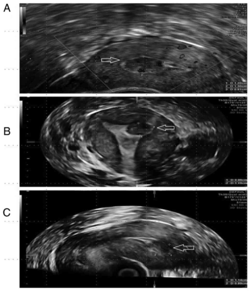
In 53 cases (26.5%) (CI 20.5-33.2%), although the
diagnosis was not changed it was completed by adding 3D and coronal
plane examination. In cases with uterine myoma, the 3D ultrasound
proved to be useful when myoma was situated intracavitary (total or
partial) by showing the type of myoma (G0, 1 or 2 type), (Fig. 1A-C).
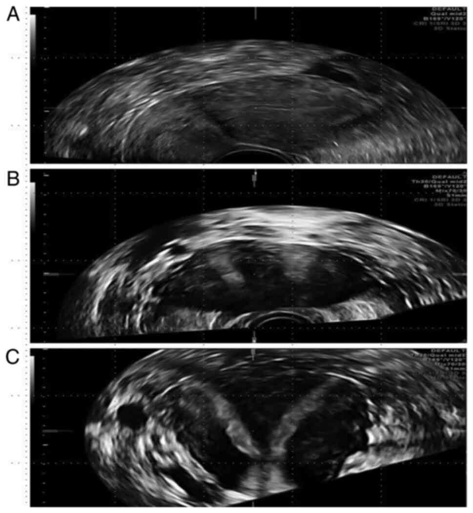
Uterine anomalies, classified as per the ESHRE/ESGE
criteria (5) suspected by 2D, but
completely diagnosed by 3D ultrasound were as follows: Arcuate
uterus, 3 cases; septate uterus, 4 cases (Fig. 2A-C); bicorn uterus, 1 case and
unicorn uterus, 1 case.
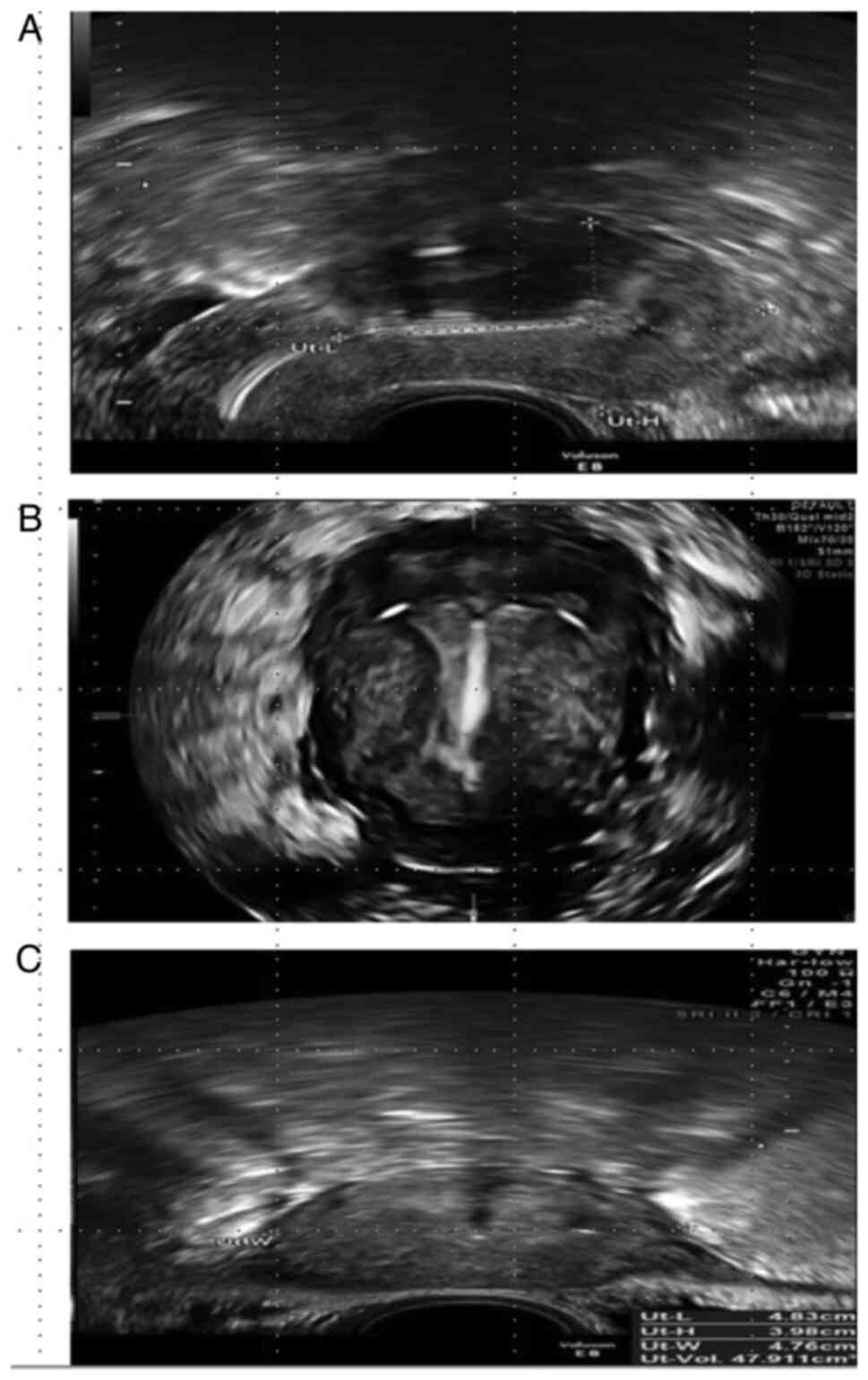
In two cases with IUD, the diagnosis of misplaced or
incongruence between IUD and uterine cavity was missed by 2D
ultrasound (Fig. 3A-C).
High level of agreement was found between 2D and 3D
in cases of uterine myoma and adenomyoss, a κ value of 0.8 and
0.74, respectively. A moderate level of agreement was observed in
cases of polyps (κ -0.67) and a low one in IUD (κ -0.44).
In order to establish the optimal time point for
performing a 3D scan with regards to menstrual cycle, results were
compared in relation to the endometrial thickness. The quality of
3D images was favorable in 149 cases (74.5%) (CI, 67.9-80.4%) and
inadequate in 51 cases (25.5%) (CI: 19.6-32.1%). The quality of the
image was quantified by the possibility of obtaining a clear
picture of the uterine cavity and endometro-myometrial junction
(EMJ). The mean endometrial thickness was 7.86 mm (SD, 2.903) with
values between 2 and 19 mm. A significant statistical relationship
existed between the quality of the image obtained and the thickness
of the endometrium (P=0.622). A thin endometrium was associated
with a poor 3D image. The present study showed a high probability
to achieve a good image if the endometrium was ≥7.38 mm (value
calculated using the Kruskal-Wallis non-parametric test).
Discussion
Ultrasonography is the most frequently used imaging
technique in the assessment of the female genital tract. Usually
the uterus and ovaries are evaluated using a 2D endovaginal
ultrasonography. The main disadvantage of the 2D ultrasound scan
consists in the difficulty of obtaining the coronal plane of the
uterus. Adding a 3D ultrasound scan to the conventional examination
may be beneficial as the coronal plane of the uterus can be
obtained easily using the reconstruction 3D technique.
The present study revealed that from a total number
of 200 examinations, 3D examination changed the diagnosis in 6
cases (3.0%) (CI: 1.1-6.7%) and added useful information or
reinforced the diagnosis in 53 cases (26.5%) (CI: 20.5-33.2%).
The most useful information was obtained in patients
referred to for ultrasonic scan for the following indications:
Infertility, uterine haemorrhage and IUD placement follow-up.
Concerning the accuracy of the final diagnosis, the most useful
information was obtained in patients with uterine pathology:
Uterine congenital anomalies, IUD misplacement, adenomyosis, and
submucous myomas. Neither 3D nor volume contrast imaging (VCI) were
useful in cases with normal uterus.
Although conventional 2D ultrasound has a good
accuracy in diagnosing congenital uterine anomalies, it is highly
dependent on the expertise of the examiner and is limited by the
difficulty in obtaining the coronal plane of the uterus. Several
studies have demonstrated the advantages of 3D ultrasound in
diagnosing uterine anomalies (6-8).
Comparing 3D ultrasound to laparoscopy and hysteroscopy, Mohamed
et al recorded a sensitivity of 97%, specificity of 96%,
positive predictive value of 92% and negative predictive value of
99% in the diagnosis of uterine anomalies (9). Ghi et al reported both a
sensitivity and a specificity of 100% in the diagnosis of uterine
malformations and 96% concordance between ultrasound and endoscopy
with respect to the type of anomaly diagnosed (10). In this study, 3D was mandatory for
the final diagnosis in all cases with uterine congenital anomalies.
As the coronal plane enables the visualization of not only the
endometrial cavity, but also of the uterine fundus, 3D scan was
necessary for the differential diagnosis between bicornuate and
septate uterus. Moreover, in these cases, 3D proved to be superior
to hysteroscopy. Hysteroscopy is able to confirm an anomaly of the
uterine cavity but is not able to provide any information regarding
the external contour of the uterus. In addition, in the coronal
plane of the uterus obtained with 3D ultrasound it is possible to
measure the size of the septum and classify the anomaly according
to the ESHRE/ESGE classification system of female genital anomalies
(5). Although MRI is considered the
gold standard for congenital uterine anomalies, 3D can provide the
same type of information (11).
However, in doubtful or complex cases, MRI should be performed,
particularly for the assessment of the cervix and vagina (12). Thus, 3D ultrasound may be associated
with 2D ultrasound in the diagnosis of uterine malformations.
Analysing the IUD cases from the current study, 3D
was useful for both types: Copper IUD and levonorgestrel (LNG IUD).
VCI proved to be particularly useful especially in cases with LNG
IUD. Observations of this study confirm those of several studies
performed in order to assess the role of 3D ultrasound in patients
with IUD (13-16).
These studies agree that the coronal view shows the entire device
and its position within the uterus, in this way helping to identify
the cause of pelvic pain and (or) bleeding in patients with an
embedded IUD (14). Hösli et
al demonstrated that 3D ultrasound offers the advantage of a
better visualisation of LNG IUD and at the same time the assessment
of uterine anomalies (13). The
coronal plane is useful not only to detect the exact position of
the IUD, but also to measure the uterine cavity and thus to
establish the proper size of the IUD to be inserted in the uterus
(17).
One interesting issue in gynaecology is the
assessment of submucous myomas. It is important when diagnosing a
myoma to know its level of extension in the uterine cavity.
Moreover, before planning a hysteroscopic resection of a submucous
myoma, it is necessary to know the type of myoma (G0, 1 or 2 type).
Salim et al demonstrated that three-dimensional
sonohysterography (3D-SIS) had a similar accuracy as hysteroscopy
in classifying submucous fibroids (18). Lee et al demonstrated that
3D-SIS is reproducible among different observers for quantification
of the percentage of a submucous fibroid protruding into the
uterine cavity (19). Mavrelos
et al demonstrated that 3D-SIS may be useful in predicting
complete hysteroscopic resection of submucous myomas (20). Although findings of the present
study show a high level of agreement between 2D and 3D ultrasound
in the diagnosis of myoma (κ=0.8), reconstructed coronal plane
added useful information for planning the surgical management of
the case.
Regarding adenomyosis, 2D ultrasound was useful in
establishing the diagnosis by revealing the classical ultrasound
signs including: Anechoic foci, heterogeneous myometrium,
asymmetrical uterine wall, uterine enlargement and hyperechogenic
striations (21). However, 2D
ultrasound cannot provide information regarding the
endometro-myometrial junction (EMJ). The changes in EMJ are
considered a key element in diagnosing adenomyosis and
traditionally the assessment of the EMJ has been part of the MRI
evaluation of the uterus (21). 3D
US enables visualisation of the coronal plane of the uterus and
consequently provides a clear image of the EMJ. Due to the
possibility of visualising the EMJ, 3D ultrasound opens up new
horizons in the diagnosis of adenomyosis. Adding VCI to the coronal
plane provides a clearer view of the EMJ. 3D ultrasound allows
visualisation of the lateral and fundal EMJ, which is almost
impossible to observe only with 2D imaging. Thanks to the clearer
view provided by the VCI, this procedure is considered as the best
in analysing and measuring the junctional zone (22). In our cases, 3D and VCI identified
changes in EMJ as different size of the EMJ in different parts of
the cavity and the penetration of the endometrium into the
myometrium. There is some supposition that adenomyosis began with
EMJ alteration and therefore 3D and VCI would be useful for an
early diagnosis of the disease (23).
In the present study, the 2D ultrasound scan
diagnosed endometrial polyps in 17 cases (8.5%). In all cases the
diagnosis was suspected by 2D ultrasound and confirmed by 3D and
VCI. During the 2D ultrasound scan, a search for the pedicle artery
sign was conducted in order to detect the position of the stalk of
the polyp (24). 3D and VCI proved
to be useful in 2 of 17 cases by providing a clearer image
regarding the topography of the polyps. At the same time, the
coronal plane was useful in the patient's understanding of the
disease. Identical size of the polyps using 2D and 3D scans was
obtained. Based on longitudinal and coronal plane and using Doppler
mode we localised the base of the polyps and the place of its
stalk. In all cases of polyps, hysteroscopy confirmed the
diagnosis. 3D scan seems not to be superior to 2D in diagnosing
endometrial polyps, but it helps in reinforcing the diagnosis and
is very useful for the patient's understanding of the disease.
Another important issue concerns the question if
there is a specific time frame regarding the endometrial cycle to
perform a 3D ultrasound scan for obtaining the ideal image of the
coronal plane. In relation to the size of the endometrium, the
coronal plane was difficult to obtain if the size was <7.38 mm.
Difficulties in obtaining a proper image of the uterine cavity have
been noted in cases with endometrium thickness <7.38 mm or in
cases with multiple uterine myomas or adenomyosis. Benaceraff et
al reported in a study on 66 patients that the coronal view is
helpful in patients with an endometrium greater than or equal to 5
mm (14). Therefore, we recommend,
in order to obtain a good 3D reconstructed image, that the
sonography should be carried late in the follicular or in the
secretory phase. In menopausal women, because the endometrial
thickness is usually <5 mm, 3D is not helpful.
Another advantage of 3D ultrasound that should be
considered is the capability of storing volumes, ensuring a further
process of the information. In this way, it is possible to
follow-up the patients for a long period of time and if a uterine
pathology will appear in the future, a reassessment of the
previously rendered volumes would possibly bring useful information
regarding the pathogenesis of the disease.
The main limitation of this study is represented by
the fact that hysteroscopy was not performed in cases where 2D and
3D findings were normal; thus, it was possible to overlook some
minor pathologic findings.
As a conclusion, we can state that the 3D ultrasound
scan is an extremely useful tool in gynaecology, especially in
cases with congenital uterine anomalies, submucous myomas and the
assessment of IUD placement. Although initially it was used for
research purposes only, currently it has been proven to be useful
in the routine ultrasound scan and there is a possibility of
witnessing its introduction as a mandatory examination procedure in
the foreseeable future.
Acknowledgements
Not applicable.
Funding
Funding: No funding was received.
Availability of data and materials
All data generated or analysed during this study are
included in this published article.
Authors' contributions
MG, RP, LMH and RM designed the study. MG, LMH, ISS
and RP collected, analyzed and interpreted the patient data. MG,
RM, MO, BFT, and AMG were responsible for discussion and
interpretation of the data. BFT, AMG and ISS had major contributors
in the writing of the manuscript. MG, RM, LMH and MO supervised and
vizualised the final form. All authors have read and agreed to the
published version of the manuscript. MG and RP confirms the
authenticity of all raw data assesed in the manuscript.
Ethics approval and consent to
participate
The present study was conducted in accordance with
the World Medical Association Declaration of Helsinki and was
approved by the Institutional Board of the ‘Medis’ Medical Centre
Iasi, Romania (approval no. 12/18.09/2019). Informed consent was
obtained from the patients.
Patient consent for publication
Not applicable.
Competing interests
The authors declare that there are no competing
interests.
References
|
1
|
Merz E: Three-dimensional transvaginal
ultrasound in gynecological diagnosis. Ultrasound Obstet Gynecol.
14:81–86. 1999.PubMed/NCBI View Article : Google Scholar
|
|
2
|
Turcan N, Bohiltea RE, Ionita-Radu F,
Furtunescu F, Navolan D, Berceanu C, Nemescu D and Cirstoiu MM:
Unfavorable influence of prematurity on the neonatal prognostic of
small for gestational age fetuses. Exp Ther Med. 20:2415–2422.
2020.PubMed/NCBI View Article : Google Scholar
|
|
3
|
Bohiltea R, Furtunescu F, Turcan N,
Navolan D, Ducu I and Cirstoiu M: Prematurity and intrauterine
growth restriction: Comparative analysis of incidence and
short-term complication. In: Proceedings of SOGR 2018. The 17
National Congress of the Romanian Society of Obstetrics and
Gynecology. 2018:708–712. 2019.
|
|
4
|
Abuhamad AZ, Singleton S, Zhao Y and Bocca
S: The Z technique: An easy approach to the display of the
mid-coronal plane of the uterus in volume sonography. J Ultrasound
Med. 25:607–612. 2006.PubMed/NCBI View Article : Google Scholar
|
|
5
|
Bermejo C, Martínez-Ten P, Ruíz-López L,
Estévez M and Gil MM: Classification of uterine anomalies by
3-dimensional ultrasonography using ESHRE/ESGE criteria:
Interobserver variability. Reprod Sci. 25:740–747. 2018.PubMed/NCBI View Article : Google Scholar
|
|
6
|
Jurkovic D, Geipel A, Gruboeck K, Jauniaux
E, Natucci M and Campbell S: Three-dimensional ultrasound for the
assessment of uterine anatomy and detection of congenital
anomalies: A comparison with hysterosalpingography and
two-dimensional sonography. Ultrasound Obstet Gynecol. 5:233–237.
1995.PubMed/NCBI View Article : Google Scholar
|
|
7
|
Salim R, Woelfer B, Backos M, Regan L and
Jurkovic D: Reproducibility of three-dimensional ultrasound
diagnosis of congenital uterine anomalies. Ultrasound Obstet
Gynecol. 21:578–582. 2003.PubMed/NCBI View
Article : Google Scholar
|
|
8
|
Moini A, Mohammadi S, Hosseini R, Eslami B
and Ahmadi F: Accuracy of 3-dimensional sonography for diagnosis
and classification of congenital uterine anomalies. J Ultrasound
Med. 32:923–927. 2013.PubMed/NCBI View Article : Google Scholar
|
|
9
|
Mohamed M, Momtaz MD, Alaa N, Ebrashy MD,
Ayman A and Marzouk MD: Three-dimensional ultrasonography in the
evaluation of the uterine cavity. MEFSJ. 12:41–46. 2007.
|
|
10
|
Ghi T, Casadio P, Kuleva M, Perrone AM,
Savelli L, Giunchi S, Meriggiola MC, Gubbini G, Pilu G, Pelusi C
and Pelusi G: Accuracy of three-dimensional ultrasound in diagnosis
and classification of congenital uterine anomalies. Fertil Steril.
92:808–813. 2009.PubMed/NCBI View Article : Google Scholar
|
|
11
|
Graupera B, Pascual MA, Hereter L, Browne
JL, Úbeda B, Rodríguez I and Pedrero C: Accuracy of
three-dimensional ultrasound compared with magnetic resonance
imaging in diagnosis of Müllerian duct anomalies using ESHRE-ESGE
consensus on the classification of congenital anomalies of the
female genital tract. Ultrasound Obstet Gynecol. 46:616–622.
2015.PubMed/NCBI View Article : Google Scholar
|
|
12
|
Bermejo C, Martinez Ten P, Cantarero R,
Diaz D, Perez Pedregosa JP, Barrón E, Labrador E and López LR:
Three-dimensional ultrasound in the diagnosis of Müllerian duct
anomalies and concordance with magnetic resonance imaging.
Ultrasound Obstet Gynecol. 35:593–601. 2010.PubMed/NCBI View
Article : Google Scholar
|
|
13
|
Hösli I, Holzgreve W and Tercanli S: Use
of 3-dimensional ultrasound for assessment of intrauterine device
position. Ultraschall Med. 22:75–80. 2001.PubMed/NCBI View Article : Google Scholar : (In German).
|
|
14
|
Benacerraf BR, Shipp TD and Bromley B:
Three-dimensional ultrasound detection of abnormally located
intrauterine contraceptive devices which are a source of pelvic
pain and abnormal bleeding. Ultrasound Obstet Gynecol. 34:110–115.
2009.PubMed/NCBI View
Article : Google Scholar
|
|
15
|
Kalmantis K, Daskalakis G, Lymberopoulos
E, Stefanidis K, Papantoniou N and Antsaklis A: The role of
three-dimensional imaging in the investigation of IUD malposition.
Bratisl Lek Listy. 110:174–177. 2009.PubMed/NCBI
|
|
16
|
Moschos E and Twickler DM: Does the type
of intrauterine device affect conspicuity on 2D and 3D ultrasound?
AJR Am J Roentgenol. 196:1439–1443. 2011.PubMed/NCBI View Article : Google Scholar
|
|
17
|
Shipp TD, Bromley B and Benacerraf BR: The
width of the uterine cavity is narrower in patients with an
embedded intrauterine device (IUD) compared to a normally
positioned IUD. J Ultrasound Med. 29:1453–1456. 2010.PubMed/NCBI View Article : Google Scholar
|
|
18
|
Salim R, Lee C, Davies A, Jolaoso B,
Ofuasia E and Jurkovic D: A comparative study of three-dimensional
saline infusion sonohysterography and diagnostic hysteroscopy for
the classification of submucous fibroids. Hum Reprod. 20:253–257.
2005.PubMed/NCBI View Article : Google Scholar
|
|
19
|
Lee C, Salim R, Ofili-Yebovi D, Yazbek J,
Davies A and Jurkovic D: Reproducibility of the measurement of
submucous fibroid protrusion into the uterine cavity using
three-dimensional saline contrast sonohysterography. Ultrasound
Obstet Gynecol. 28:837–841. 2006.PubMed/NCBI View
Article : Google Scholar
|
|
20
|
Mavrelos D, Naftalin J, Hoo W, Ben-Nagi J,
Holland T and Jurkovic D: Preoperative assessment of submucous
fibroids by three-dimensional saline contrast sonohysterography.
Ultrasound Obstet Gynecol. 38:350–354. 2011.PubMed/NCBI View
Article : Google Scholar
|
|
21
|
Dueholm M, Lundorf E, Hansen ES, Sørensen
JS, Ledertoug S and Olesen F: Magnetic resonance imaging and
transvaginal ultrasonography for the diagnosis of adenomyosis.
Fertility Steril. 76:588–594. 2001.PubMed/NCBI View Article : Google Scholar
|
|
22
|
Exacoustos C, Brienza L, Di Giovanni A,
Szabolcs B, Romanini ME, Zupi E and Arduini D: Adenomyosis:
Three-dimensional sonographic findings of the junctional zone and
correlation with histology. Ultrasound Obstet Gynecol. 37:471–479.
2011.PubMed/NCBI View
Article : Google Scholar
|
|
23
|
Naftalin J and Jurkovic D: The
endometrial-myometrial junction: A fresh look at a busy crossing.
Ultrasound Obstet Gynecol. 34:1–11. 2009.PubMed/NCBI View
Article : Google Scholar
|
|
24
|
Timmerman D, Verguts J, Konstantinovic ML,
Moerman P, van Schoubroeck D, Deprest J and van Huffel S: The
pedicle artery sign based on sonography with color Doppler imaging
can replace second-stage tests in women with abnormal vaginal
bleeding. Ultrasound Obstet Gynecol. 22:166–171. 2003.PubMed/NCBI View
Article : Google Scholar
|