Introduction
Asthma is a chronic inflammatory disease with
symptoms such as airway remodeling and hyperresponsiveness, mucus
hypersecretion, wheezing, dyspnea, cough and chest tightness
(1-3).
These symptoms are caused by an allergic response mediated by the
T-helper (Th) cells. Naive Th cells are induced by
antigen-activated dendritic cells to differentiate into Th1 or Th2
cells through exposure to proinflammatory cytokines such as IL-4
and IL-2, respectively (4). After
Th cell differentiation, Th2 cells secrete various cytokines, such
as IL-4 and IL-5 (2,5,6).
Allergic proteins, such as histamines, are released from the
IgE-activated mast cells, recruiting eosinophils and causing acute
and chronic inflammation (7,8).
Allergic proteins, especially histamines, play a
significant role in an allergic response. Therefore, reducing
histamines by inhibiting mast cell degranulation may be crucial for
suppressing allergic reactions in individuals with asthma. The
asthmatic allergic response can be triggered quickly by various
environmental stimulants, including cold air, ozone, pollen, strong
odors, smoke, house dust mites and particulate matter (2,9).
Recently, countries around the world (such as China, Bangladesh,
Pakistan, India, Mongolia and South Korea) have undergone rapid
industrialization, which has increased the concentration of
particulate matter in the environment, thus affecting the incidence
of acute asthma and exacerbating its symptoms (10-13).
At present, no treatments exist for asthma itself,
only the use of symptom controllers, such as inhaled
corticosteroids (14). However,
the long-term application of glucocorticoid drugs can severely
affect the musculoskeletal, endocrine/metabolic, gastrointestinal,
cardiovascular, dermatological, neuropsychiatric, ophthalmological
and immunological systems (15,16).
Therefore, extensive research has been conducted to develop
naturally derived compounds that can treat asthma without causing
severe side effects. For example, the sesquiterpene lactones from
Saussurea costus (Falc.) have been found to reduce the
expression of Th2 cytokine genes and recruit inflammatory cells in
an ovalbumin (OVA)-induced asthma mouse model (17). Galangin, a natural flavonoid, has
been found to attenuate severe inflammation and airway remodeling
by inhibiting the generation of TNF-β1-mediated reactive oxygen
species (ROS) and MAPK/Akt phosphorylation in an OVA-sensitized
asthma mouse model (18).
Ginger (Zingiber officinale Roscoe) has been
used in traditional Chinese and Indian medicine to treat numerous
diseases and symptoms, including nausea, diarrhea, gingivitis,
arthritis and asthma (19). Ginger
contains >400 compounds, including shogaol, gingerol, paradol,
gingerdiol and zingerone; 6-shogaol (SHO) and 6-gingerol (GIN) are
the major compounds (20). A
previous study has shown that ginger powder-containing food and GIN
exert anti-rhinitis effects on the major allergic response via the
Th2 cell signaling pathway in an OVA-induced allergic rhinitis
mouse model (21). In addition,
GIN and SHO have been shown to combat asthma by relaxing the airway
smooth muscle (ASM) and inhibiting chronic inflammation (22).
However, at present, research is lacking on the
mechanism underlying SHO's effects on critical allergic reactions.
Therefore, it is necessary to confirm the anti-allergic effects of
SHO to verify its efficacy in asthma treatment. Thus, in the
present study, we hypothesized that SHO and GIN could inhibit
chronic inflammation by suppressing allergic responses and
oxidative stress in an OVA-induced asthma mouse model.
Materials and methods
Animals and reagents
A total of 24 male BALB/c mice (age, 5 weeks;
weight, 18-20 g) were supplied by RaonBio, Inc. and maintained at
22±2˚C and 50±10% humidity in a 12-h light/dark cycle and were
freely provided with tap water and commercial food. The animals'
weight was measured once a week to monitoring their conditions. SHO
[1-[4-Hydroxy-3-methoxyphenyl]-4-decen-3-one; C17H24O3; purity,
>98% as detected via high-performance liquid chromatography
(HPLC); cat. no. 555-66-8] and GIN
([5S]-5-Hydroxy-1-[4-hydroxy-3-methoxy-phenyl]decan-3-one;
[S]-[6]-Gingerol; and 3-Decanone,
5-hydroxy-1-[4-hydroxy-3-methoxyphenyl]; purity, >98% as
detected by HPLC; cat. no. 23513-14-6) were purchased from Chengdu
Biopurify Phytochemicals Ltd.
OVA-induced asthma mouse model
The mice were divided into four groups: Control,
OVA, OVA + SHO and OVA + GIN, with n=6 per group. The induction of
asthma in the mice treated with OVA was performed as described
previously (17). The asthma mouse
model was established by sensitizing the mice with an
intraperitoneal (i.p.) injection of 50 µg OVA (cat. no. A5503;
Sigma-Aldrich; Merck KGaA) and 1 mg aluminum hydroxide (cat. no.
239186; Sigma-Aldrich; Merck KGaA) in 200 µl PBS on days 0 and 14
(Fig. 1). The control group was
treated with 200 µl PBS.
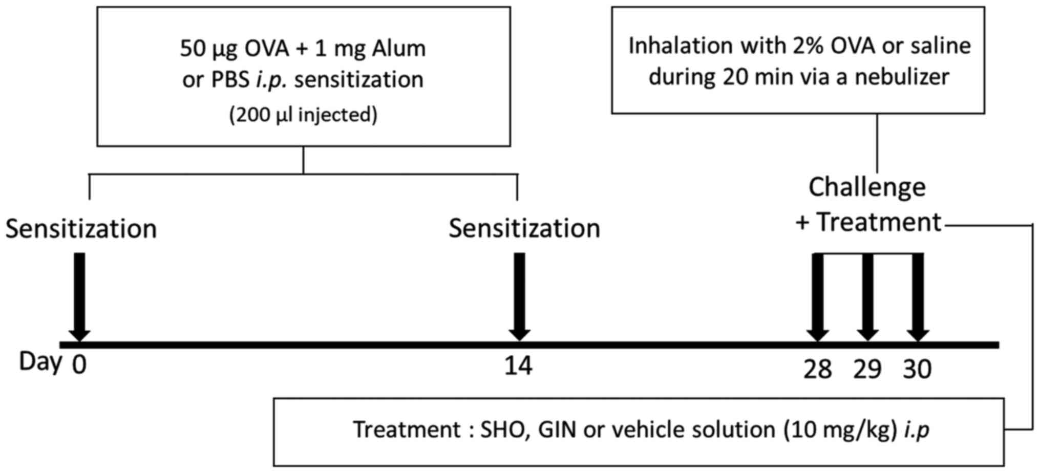 | Figure 1Protocol for the OVA/alum-induced
asthma mouse model. The OVA, OVA + SHO and OVA + GIN groups were
sensitized with 50 µg OVA and 1 mg alum (i.p.) in 200 µl PBS, and
the control group was sensitized with 200 µl PBS on days 0 and 14
(n=6/group). The OVA, OVA + SHO and OVA + GIN groups were
challenged with 2% OVA, and the control group received saline
solution for 20 min through a nebulizer on days 28, 29 and 30. i.p.
injections of SHO or GIN were performed 2 h before each challenge
step. i.p., intraperitoneal; OVA, ovalbumin; SHO, 6-shogaol; GIN,
6-gingerol; alum, aluminum hydroxide. |
On days 28, 29 and 30, the mice received an i.p.
injection of SHO at 10 mg/kg or GIN at 10 mg/kg 2 h before they
were challenged with OVA. SHO and GIN were dissolved in 0.5 µl
DMSO, and 89.5 µl PBS with 10 µl 50% Tween-20 was added to the
100-µl final dose. The mice were then challenged with 2% OVA in
sterile saline through a whole-body exposure system with a
nebulizer on days 28, 29 and 30 for 20 min. The control group
inhaled sterile saline. The mice were sacrificed at 24 h after the
final OVA challenge by cervical dislocation.
Measurement of bronchoalveolar lavage
fluid (BALF)
BALF was obtained from the mice at 24 h after the
final OVA challenge by flushing 0.5 ml cold PBS through the lungs
three times with a tracheal catheter. The BALF was centrifuged at
318 x g for 10 min at 4˚C to collect the pellet, which was
resuspended with PBS and stored at 4˚C for a differential cell
count. The BALF cells in the pellet were stained with the Diff-Quik
stain kit (cat. no. 38721; Sysmex Corporation), and 5 µl BALF cells
were smeared onto one end of a glass slide according to the
manufacturer's instructions. The cells were stained with Diff quick
solution II and I for 30 sec at room temperature each, and rinsed
with tap water. Stained cells were counted with a hematocytometer
(HSU-0650030; Paul Marienfeld GmbH & Co. KG). The differential
cells in the BALFs were classified as lymphocytes, neutrophils,
macrophages or eosinophils. The distribution of the cells in the
BALFs was expressed as a percentage.
Lung histological analysis
The right lung lobes of the mice were fixed in 4%
formaldehyde at 4˚C for 48 h, embedded in paraffin and sliced to
4-µm sections (6 slides/mouse). The sections were stained with
hematoxylin at room temperature for 5 min and then with eosin at
room temperature for 3 min to analyze the degree of inflammation in
the lung tissues. The degree of inflammation was assigned an
arbitrary score of 0 (normal = no inflammation), 1 (minimal =
perivascular, peribronchial or patchy interstitial inflammation
involving <10% of lung volume), 2 (mild = perivascular,
peribronchial or patchy interstitial inflammation involving 10-20%
of lung volume), 3 (moderate = perivascular, peribronchial, patchy
interstitial or diffuse inflammation involving 20-50% of lung
volume) and 4 (severe = diffuse inflammation involving >50% of
lung volume). The average inflammatory score was analyzed using a
total of 18 slides (3 slides/mouse). In addition, the thickness of
the lung epithelial cells was measured, calculated as the average
value of the top, bottom, left and right of three random alveoli
per slide using a light microscope (Leica Microsystems GmbH) with
Leica AF6000 modular systems.
The sections also were stained with periodic
acid-Schiff (PAS) at room temperature for 15 min to visualize the
goblet cells to determine the extent of mucus production. A
point-counting method was performed to quantify the number of cells
stained positively in three random fields from each slide. The
average of the cells was analyzed with a total of 18 slides (3
slides/mouse). These lung sections were observed using light
microscope (magnification, x100) and analyzed with LAS AF Ink
(Leica Microsystems GmbH) and ImageJ software (v1.53e; National
Institutes of Health).
Reverse transcription-quantitative
(RT-q)PCR analysis
Total RNA was isolated from the mouse lung tissues
using the TRI-Solution (cat. no. TS200-001; Bio Science
Technology). The total RNA was synthesized to cDNA using a
PrimeScript™ 1st strand cDNA Synthesis kit (cat. no. 6110; Takara
Biotechnology Co., Ltd.) with oligo-dT primers. The qPCR reaction
mixture contained 8 µl cDNA, 10 µl Power SYBR®-Green PCR
Master mix (cat. no. 4367659; Applied Biosystems; Thermo Fisher
Scientific, Inc.), 1 µl 0.2-pmol forward primer and 1 µl 0.2-pmol
reverse primer. The forward and reverse primers for β-actin
(5'-GGCTCTTTTCCAGCCTTCCT-3' and 5'-GTCTTTACGG ATGTCAACGTCACA-3',
respectively), IL-4 (5'-CCACGGAT GCGACAAAAATC-3' and
5'-GACGTTTGGCACATCCAT CTC-3'), IL-5 (5'-GATGGACGCAGGAGGATCAC-3' and
5'-GTGTGGCATCCCTCAGCAA-3'), IL-13 (5'-GGCCAG CCCACAGTTCTACA-3' and
5'-ACCACCAAGGCAAGCAA GAG-3'), superoxide dismutase 1 (SOD1;
5'-GACTTGGGC AAAGGTGGAAA-3' and 5'-CAGGGAATGTTTACTGCGC AAT-3'),
SOD2 (5'-TGCTCTTGATTGAACATTTTCGTTA-3' and
5'-GCCCCCCAAAACAGAGATG-3'), catalase (5'-CGA CCAGGGCATCAAAAACT-3'
and 5'-ATTGGCGATGGCAT TGAAA-3') and glutathione peroxidase-1
(GPx-1; 5'-AGAAAG CGATGCCACGTGAT-3' and 5'-GGAGATGTTGGGACTC
AAACG-3') were used. The qPCR was run on the Applied Biosystems
real-time PCR program (StepOnePlus™ Real-Time PCR System) at 95˚C
for 10 min, followed by 40 cycles of a cycling stage at 95˚C for 15
sec and 60˚C for 1 min, and a melt curve stage at 95˚C for 15 sec,
60˚C for 1 min and 95˚C for 15 sec. All data were analyzed using
the 2-ΔΔCq method (23)
and expressed as fold change relative to controls. The relative
expression levels of IL-4, IL-5, IL-13, SOD1, SOD2, catalase and
GPx-1 were normalized to those of β-actin.
Western blotting assay
Lung tissues were lysed using the PRO-PREP for
Cell/Tissue Protein Extraction Solution kit (cat. no. 17081; Intron
Biotechnology, Inc.) with the Protease Inhibitor Cocktail (cat. no.
P3100; GenDEPOT, LLC). The protein concentrations of the tissue
lysates were determined using the BCA protein assay kit (cat. no.
23227; Thermo Fisher Scientific, Inc.). A total of 30 µg protein
from each sample was separated via 10-12% SDS-PAGE and transferred
to PVDF membranes. The membranes were blocked with 5% skim milk in
a 1X TBS-Tween-20 (TBST; containing 0.05% Tween-20) at room
temperature for 1 h and incubated with primary antibodies against
IL-4 (monoclonal; rat anti-mouse; 1:1,000; cat. no. ab11524;
Abcam), IL-5 (monoclonal; mouse anti-mouse; 1:1,000; cat. no.
sc-398334; Santa Cruz Biotechnology, Inc.), IL-13 (polyclonal;
rabbit anti-mouse; 1:1,000; cat. no. ab106732; Abcam), SOD1
(monoclonal; mouse anti-mouse; 1:1,000; cat. no. sc-101523; Santa
Cruz Biotechnology, Inc.), SOD2 (monoclonal; mouse anti-mouse;
1:1,000; cat. no. sc-133134; Santa Cruz Biotechnology, Inc.),
catalase (monoclonal; mouse anti-mouse; 1:1,000; cat. no.
sc-271803; Santa Cruz Biotechnology, Inc.), GPx-1/2 (monoclonal;
mouse anti-mouse; 1:1,000; cat. no. sc-133160; Santa Cruz
Biotechnology, Inc.) or β-actin (monoclonal; mouse anti-mouse;
1:1,000; cat. no. sc-47778; Santa Cruz Biotechnology, Inc.) diluted
with 3% skim milk in a 1X TBST buffer at 4˚C overnight. Then, the
blots were washed three times with 1X TBST buffer and incubated
with anti-mouse HRP-conjugated secondary antibodies (goat
anti-mouse IgG-HRP; cat. no. sc-2005; Santa Cruz Biotechnology,
Inc.) diluted at 1:5,000 with 3% skim milk in 1X TBST buffer at
room temperature for 1 h. Next, the membranes were washed three
times with 1X TBST buffer, and the protein bands were detected with
the ECL detection reagents (cat. no. 34580; Thermo Fisher
Scientific, Inc.) and semi-quantified with ImageQuant LAS 500
(Cytiva). The protein expression level of β-actin was used as the
loading control.
Cell lines and culture
Mast cell degranulation was evaluated by the release
of β-hexosaminidase from the mast cell line, RBL-2H3. Rat RBL-2H3
mast cells were obtained from the American Type Culture Collection
and maintained in MEM medium (cat. no. 61100061; Gibco; Thermo
Fisher Scientific, Inc.) supplemented with 10% FBS (cat. no.
A31604; Gibco; Thermo Fisher Scientific, Inc.) and 1%
penicillin/streptomycin (cat. no. 15140122; Gibco; Thermo Fisher
Scientific, Inc.) in a 5% CO2 incubator at 37˚C. RBL-2H3
cells were sensitized via an incubation with 0.2 µg/ml monoclonal
anti-dinitrophenyl mouse IgE (cat. no. D8406; Sigma-Aldrich; Merck
KGaA) diluted medium overnight at 37˚C. The cells were washed twice
with a piperazine-N, N'bis (2-ethanesulfonic acid) (PIPES) buffer
(pH 7.2) containing 25 mM PIPES, 0.05 mM NaOH, 110 mM DNP-IgE and
0.1% BSA (cat. no. A0100-010; GenDEPOT LLC). The cells were then
incubated in PIPES buffer containing different concentrations of
SHO and GIN (0, 10, 25, 50 and 100 nM) at 37˚C for 30 min. Next,
the cells were incubated with 1 µg/ml human dinitrophenyl albumin
(cat. no. A6661; Sigma-Aldrich; Merck KGaA) to induce degranulation
for 15 min at 37˚C. After centrifugation at 125 x g for 5 min at
4˚C, 25 µl supernatant from each reaction was transferred to a
96-well microplate and incubated for 110 min with 5 mM
4-nitrophenyl N-acetyl-β-D-glucosaminide in a 0.1-M citrate buffer
(pH 4.5; cat. no. N9376; Sigma-Aldrich; Merck KGaA). The reaction
was terminated by adding 0.05 M sodium carbonate buffer (pH 10).
The optical density of each reaction was measured at the absorbance
wavelength of 405 nm using a microplate reader.
The concentrations of SHO and GIN were selected
using a cell viability assay. Cell viability was evaluated to
determine cytotoxicity of SHO and GIN using a Cell Counting Kit-8
(CCK-8; Dojindo Laboratories, Inc.) assay, following the
manufacturer's instructions. Jurkat cells (human T cell line;
1x10⁴ cells/well) were obtained from American Type
Culture Collection and maintained in RPMI-1640 medium (cat. no.
31800022; Gibco; Thermo Fisher Scientific, Inc.) supplemented with
10% FBS and 1% penicillin/streptomycin in a 5% CO2
incubator at 37˚C. Jurkat cells were incubated at 37 ˚C in 96-well
plates with SHO and GIN at 0, 10, 50 and 100 nM for 0, 24, 48 and
72 h. RBL-2H3 cells (5x10³ cells/well) were incubated at
37˚C in 96-well plates with SHO (0, 1, 2.5 and 5 µM) and GIN (0,
0.5, 1 and 2 µM) for 0, 24, 48 and 72 h. Next, 10 µl CCK-8 reagent
was added to each well, and cells were incubated at 37˚C for an
additional 2 h. The absorbance was measured at 450 nm using a
microplate reader.
Statistical analysis
Data were collected from ≥3 independent experiments.
All results were expressed as the mean ± SD. All analyses were
performed using SPSS software (version 20; IBM Corp.). Statistical
significance between experimental groups was determined using
one-way ANOVA for pair-wise comparisons with Bonferroni's multiple
comparisons test. Ordinal data were analyzed using a Kruskal-Wallis
test followed by Dunn's test. P<0.05 was considered to indicate
a statistically significant difference.
Results
SHO and GIN inhibit eosinophil
recruitment in BALFs
In the OVA group, the eosinophil level was increased
significantly compared with that of the control group. In the OVA +
GIN group, this was decreased significantly compared with that of
the OVA group (Fig. 2). The
current study found no significant difference between the OVA + SHO
and OVA groups, but did identify a decreasing trend. These data
suggest that GIN strongly inhibited the recruitment of eosinophils
in asthma; SHO also may have such a potential. Therefore, GIN and
SHO likely have anti-inflammatory effects on asthma in a mouse
model. Thus, additional experiments were conducted to confirm the
anti-allergic effects of both using histological examination.
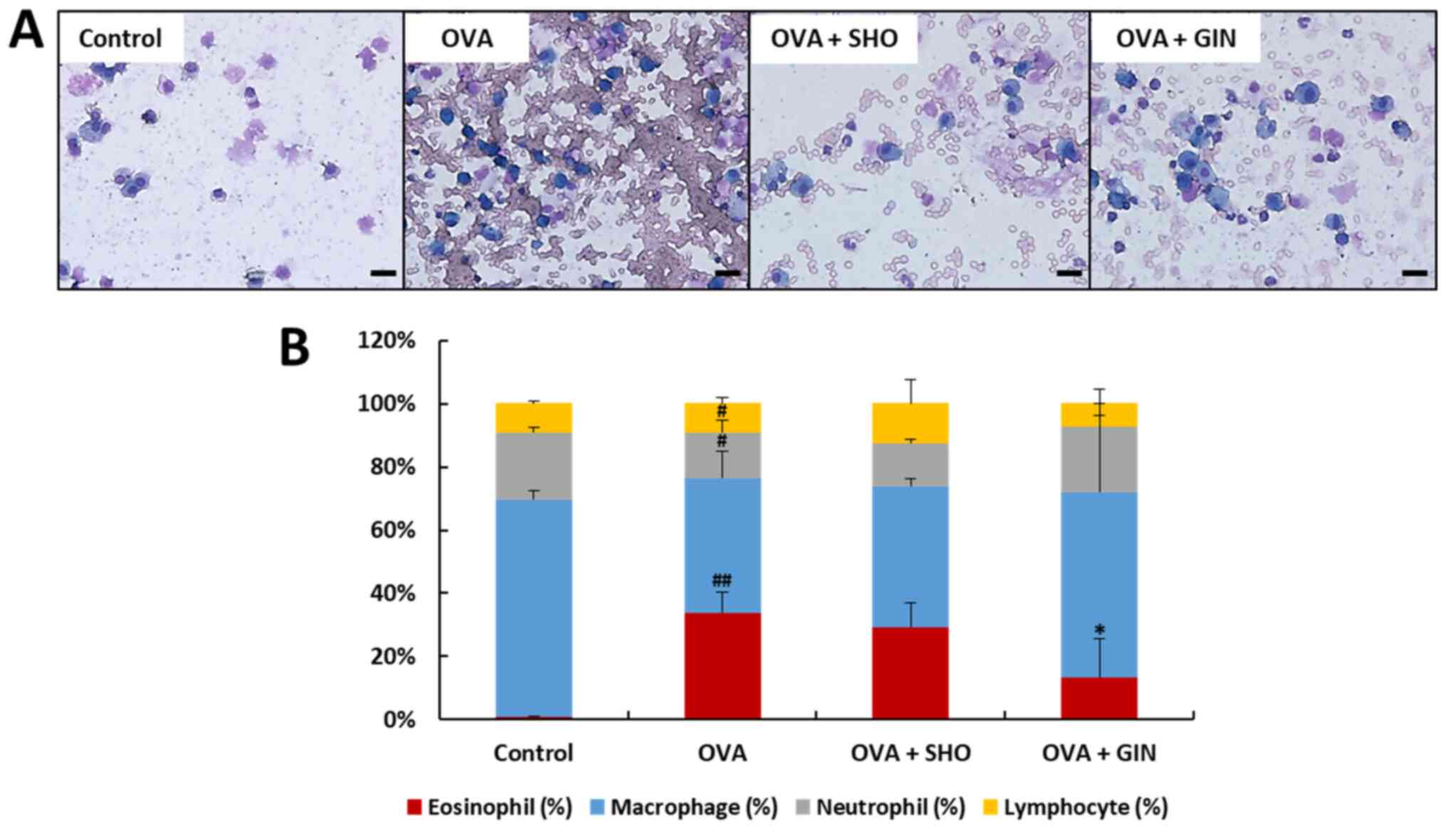 | Figure 2Effects of SHO and GIN on
inflammatory cell recruitment in BALF. BALF was obtained from the
control, OVA, OVA + SHO and OVA + GIN groups. (A) BALF was stained
with H&E (magnification, x400; scale bars, 20 µm), and cells
were (B) counted with a hematocytometer. Differential cells were
classified as lymphocytes (yellow), neutrophils (gray), macrophages
(blue) and eosinophils (red). The distribution of inflammatory
cells (white blood cells) is expressed as a percentage (mean ± SD,
n=6). #P<0.05, ##P<0.01 vs. control
group; *P<0.05 vs. OVA group. OVA, ovalbumin; SHO,
6-shogaol; GIN, 6-gingerol. |
SHO and GIN suppress the airway
inflammatory response and inflammatory cell infiltration in lung
tissues
The lung sections from the mice in different
treatment groups were stained with H&E to confirm the
inflammatory cell-counting results (Fig. 2). In chronic inflammation,
inflammatory cells infiltrate the ASM cell layer, inducing
eosinophilia in the lung tissues (24). The ASM cell thickness was increased
in the OVA group compared with that in the control group,
suggesting that a chronic inflammatory response, such as asthma,
was induced in the OVA group (Fig.
3A and B). In the OVA + SHO
and OVA + GIN groups, ASM thickness was significantly decreased
compared to that of the OVA group (Fig. 3A and B). Moreover, the inflammatory score was
significantly higher in the OVA group and was significantly lower
in the OVA + SHO and OVA + GIN groups (Fig. 3C).
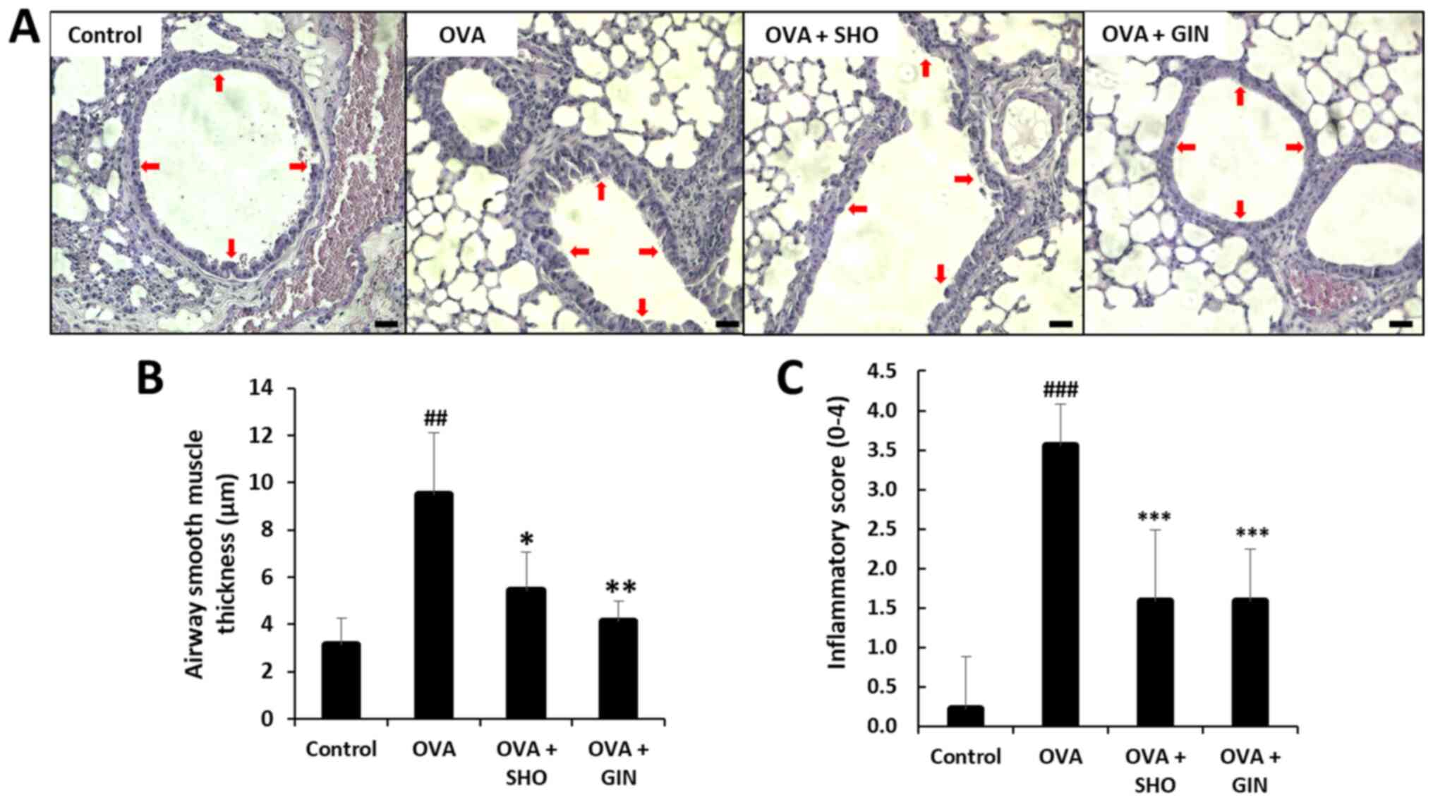 | Figure 3Effects of SHO and GIN on
inflammatory cell infiltration and airway inflammation in lung
tissues. The right lung lobes were isolated at 24 h after the final
OVA challenge. Next, 4-µm lung sections were stained with H&E
to analyze inflammatory cell infiltration and inflammatory score.
(A) Panels show H&E-stained lung sections obtained from the
control (first), OVA (second), OVA + SHO (third) and OVA + GIN
(fourth) groups. Magnification, x200; scale bar, 75 µm. (B) ASM
thickness was evaluated with LAS AF Ink. (C) Total inflammatory
score expressed as an average. Red arrow indicates measured
thickness site of epithelial cells. Values are presented as mean ±
SD (n=6). ##P<0.01, ###P<0.001 vs.
control group; *P<0.05, **P<0.01,
***P<0.001 vs. OVA group. OVA, ovalbumin; SHO,
6-shogaol; GIN, 6-gingerol. |
SHO and GIN decrease goblet cell
hyperplasia and mucus production in lung tissues
The mucins produced by the goblet cells were stained
using PAS (red arrows; Fig. 4A).
Mucus production was significantly increased in the OVA group
compared with that in the control group; however, it was
significantly reduced in the OVA + SHO and OVA + GIN groups as
compared with the OVA group (Fig.
4B).
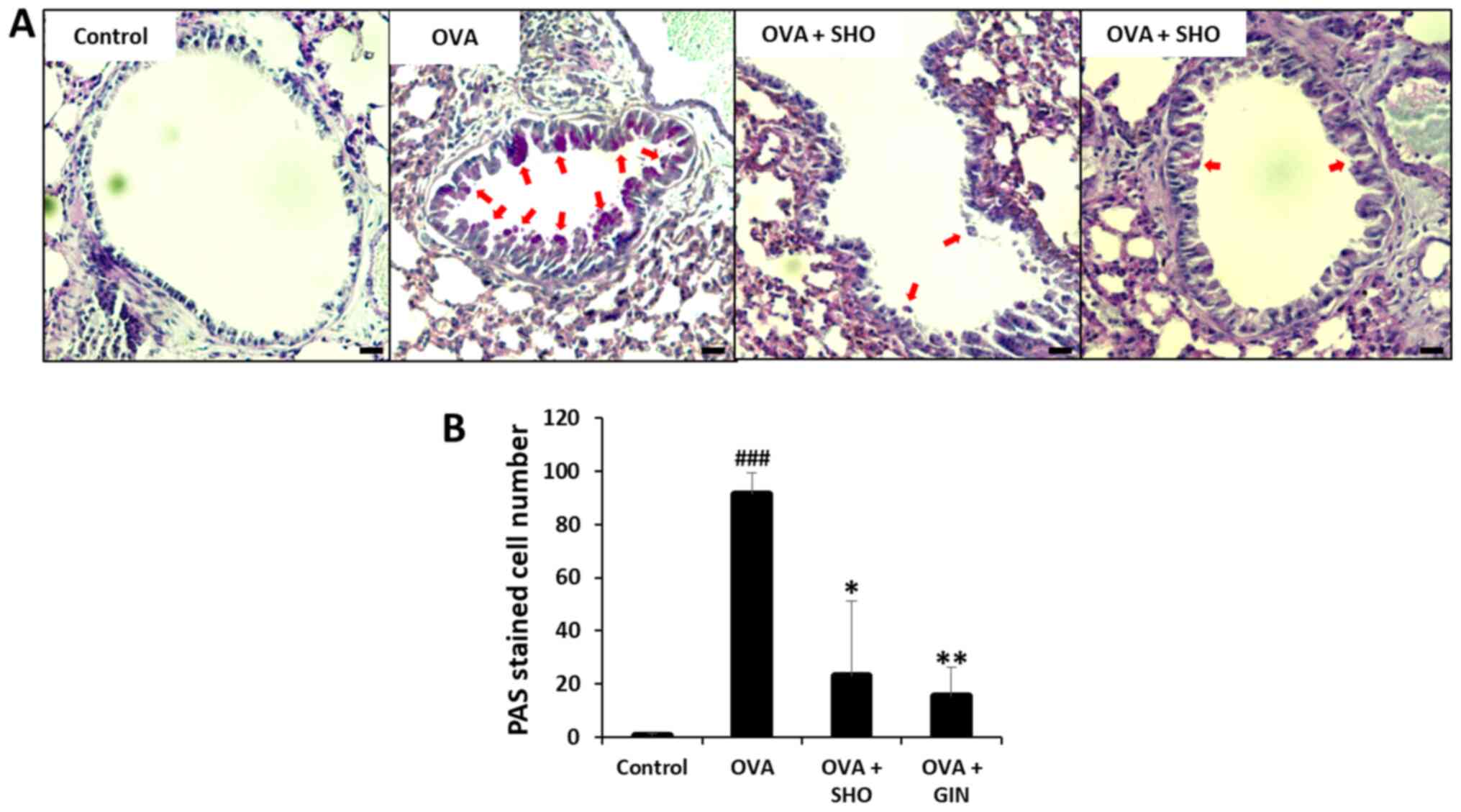 | Figure 4Effects of SHO and GIN on mucus
production in lung tissues. The right lung lobes were isolated 24 h
after the final OVA challenge. Then, 4 µm-cut-lung sections were
stained with PAS to analyze mucus production. (A) Panels show
PAS-stained lung sections obtained from the control (first), OVA
(second), OVA + SHO (third) and OVA + GIN (fourth) groups.
Magnification, x200; scale bars, 75 µm. Fixed lung tissues were
stained with PAS for visualizing mucus production. (B) PAS-stained
cells are goblet cells presented with a red arrow. Values are
presented as mean ± SD (n=6). ###P<0.001 vs. control
group; *P<0.05, **P<0.01 vs. OVA group.
OVA, ovalbumin; SHO, 6-shogaol; GIN, 6-gingerol; PAS, periodic
acid-Schiff. |
SHO and GIN suppress inflammatory
cytokine levels in lung tissues
Th2 cytokines regulate the inflammatory response in
allergic diseases such as asthma (25,26).
The expression of genes encoding Th2 cell-mediated cytokines,
including IL-4, IL-5 and IL-13, in the lung tissues after the final
OVA challenge was examined using RT-qPCR (Fig. 5). The mRNA expression levels of
IL-4, IL-5 and IL-13 were significantly increased in the OVA group
compared with those in the control group (Fig. 5A-C). SHO and GIN significantly
decreased the mRNA expression levels of IL-4 and IL-5 (Fig. 5A and B). In addition, both significantly
reduced the expression level of IL-13 (Fig. 5C). The protein expression levels of
IL-4, IL-5 and IL-13 were determined using a western blotting assay
to confirm the mRNA expression data (Fig. 5D-G). The protein expression levels
of IL-4, IL-5 and IL-13 were significantly increased in the OVA
group. Furthermore, SHO and GIN decreased the protein expression
levels of IL-4, IL-5 and IL-13.
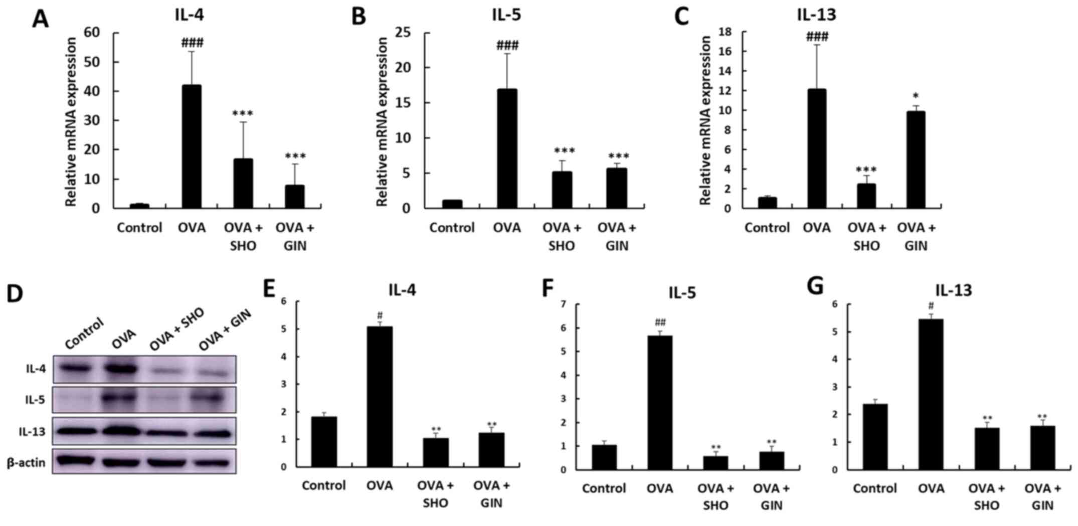 | Figure 5Effects of SHO and GIN on
inflammatory cytokines levels in lung tissues. Levels of Th2
cell-related inflammatory cytokines in lung tissues. Total RNA was
isolated from lung tissues. (A) IL-4, (B) IL-5 and (C) IL-6 mRNA
expression levels were detected using reverse
transcription-quantitative PCR with specific primers. The relative
mRNA expression levels were calculated based on β-actin mRNA
expression in lung tissues. (D) Protein expression levels of
cytokines were determined via western blotting, and (E) IL-4, (F)
IL-5 and (G) IL-6 protein expression levels were semi-quantified.
Values are presented as mean ± SD (n=6). #P<0.05,
##P<0.01, ###P<0.001 vs. control group;
*P<0.05, **P<0.01,
***P<0.001 vs. OVA group. OVA, ovalbumin; SHO,
6-shogaol; GIN, 6-gingerol. |
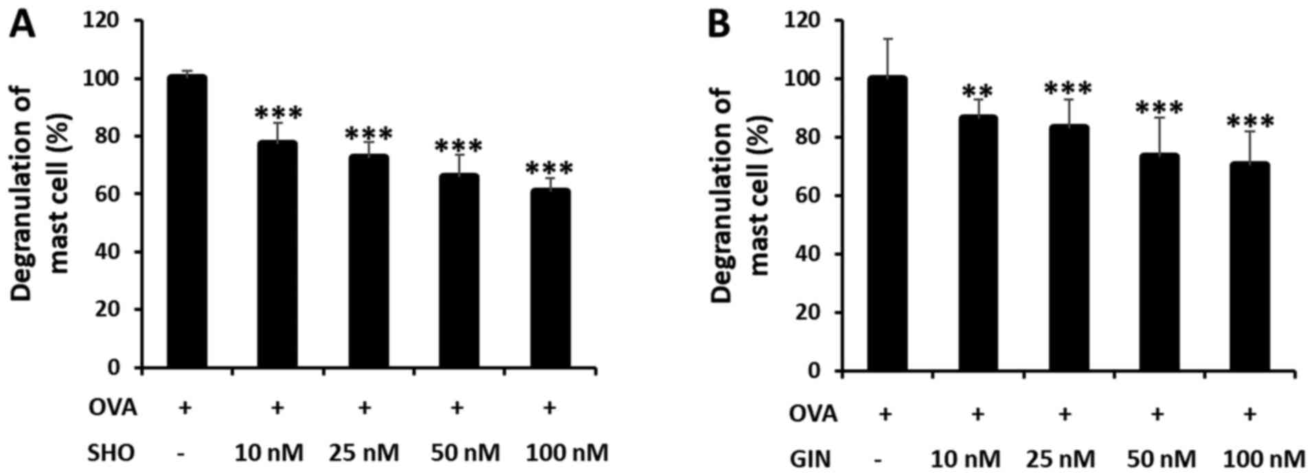
SHO and GIN inhibit mast cell
degranulation in RBL-2H3 cells
Cells were treated with SHO or GIN at 0, 10, 25, 50
and 100 nM to determine their dose-dependent effects on the mast
cells (Fig. 6). The OVA + SHO
group showed a significant dose-dependent decrease in mast cell
degranulation, to 61% in the group treated with 100 nM of SHO
(Fig. 6A). GIN had the same
tendency, decreasing degranulation to 70% in the group treated with
100 nM GIN (Fig. 6B).
Additionally, the cytotoxicity of SHO and GIN in the RBL-2H3 and
Jurkat cells were determined using CCK-8 assays, which demonstrated
that SHO and GIN were not toxic (Fig.
S1). The concentrations of SHO and GIN used for cell line
treatment were selected based on the results in Fig. S1. These data suggested that both
reagents effectively suppressed the allergic response in asthma by
inhibiting mast cell degranulation.
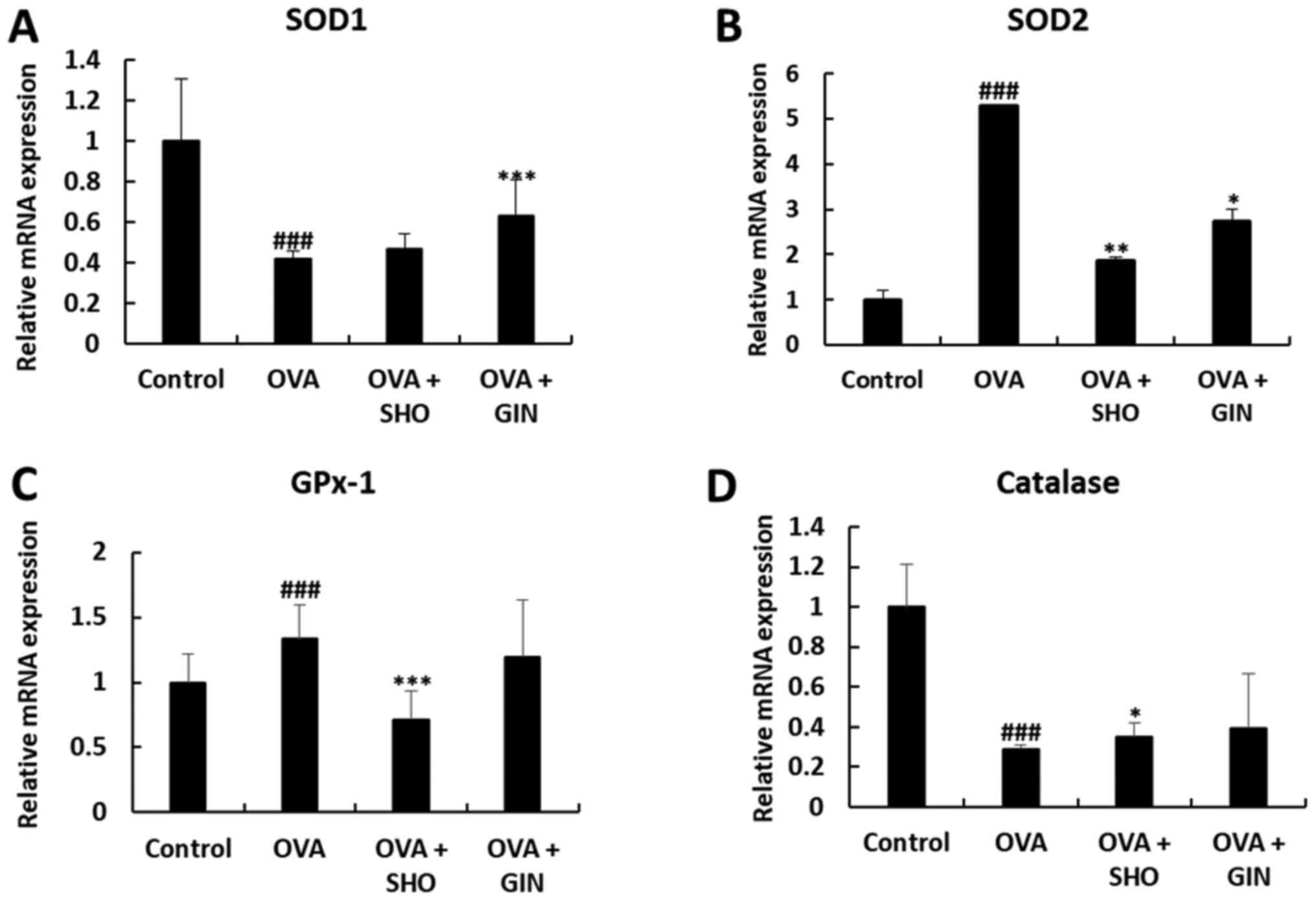
SHO and GIN restore the expression
levels of antioxidant factors
Oxidative stress is an important pathological
mechanism of asthma. SOD1, SOD2, catalase and GPx-1/2 are critical
antioxidant enzymes to reduce oxidative stress (27-29).
Total mRNA and proteins were isolated from the lung tissues from
different treatment groups to determine the effects of SHO and GIN
on the levels of various antioxidant enzymes. The mRNA expression
level of SOD1 was significantly decreased in the OVA group compared
with that in the control group, and the expression level of SOD1
was restored in the OVA + GIN group. In addition, the current study
found no difference between the OVA + SHO and OVA groups, but
observed an increasing tendency (Fig.
7A).
The mRNA expression levels of SOD2 and GPx-1 were
increased significantly in the OVA group compared with the control
group, but were decreased significantly in the OVA + SHO group as
compared with the OVA group (Fig.
7B and C). Similarly, SOD2
expression was significantly decreased in the OVA + GIN group
compared with the OVA group (Fig.
7B). However, no significant difference was detected in the
expression level of GPx-1 between the OVA and OVA + GIN groups
(Fig. 7C).
The mRNA expression levels of catalase were
significantly decreased in the OVA group compared with those in the
control group, and this expression was restored in the OVA + SHO
group. However, no difference was observed in catalase expression
between the OVA and OVA + GIN groups (Fig. 7D).
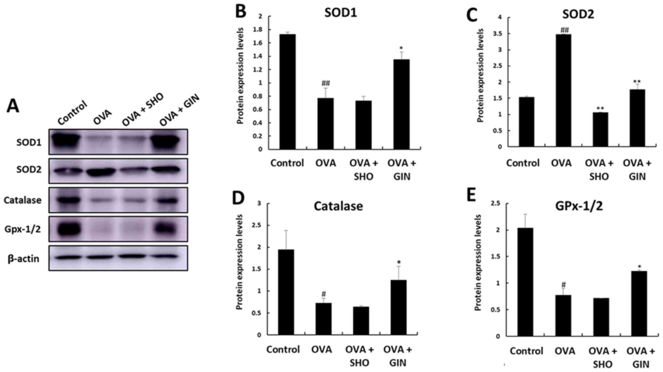
The expression patterns of the antioxidant genes
were confirmed by examining the corresponding protein expression
levels using western blot analysis (Fig. 8A). The protein expression levels of
SOD1, SOD2 and catalase exhibited the same trend as those of the
mRNA expression of the corresponding genes (Fig. 8B-D). However, GPx-1/2 expression
showed the opposite tendency to the mRNA expression levels of GPx-1
(Fig. 8D). GPx-1/2 was decreased
in the OVA group compared with the control group, was increased in
the OVA + GIN group, but showed no change in the OVA + SHO group
(Fig. 8D).
Discussion
The present study demonstrated the in vitro
and in vivo anti-inflammatory and anti-allergic effects of
SHO and GIN. In the OVA-induced asthma mouse model, allergic
response and chronic inflammation are mediated via the Th2
cell-signaling pathway (30). The
present study administrated 10 mg/kg SHO and GIN via i.p. injection
in mice. The dose for i.p. injection was selected based on previous
studies (31,32). The current results revealed the
anti-asthmatic effects of SHO and GIN on the cells, as determined
using inflammatory cell counting and histological examination in
BALF samples and lung tissue. The present study also examined the
compounds' effects on the balance of antioxidant enzymes in lung
tissues and their anti-allergic effects on mast cells RBL-2H3.
The accumulation of inflammatory cells is associated
with an allergic response in asthma (33). Among the inflammatory cells,
eosinophils are most important in chronic asthma. High levels of
eosinophils have been found in asthmatic sputum, blood and BALF
(34). Therefore, the current
study investigated the effects of SHO and GIN on the recruitment of
inflammatory cells, such as lymphocytes, neutrophils, macrophages
and eosinophils, in the BALF from different treatment groups. SHO
and GIN had a significant impact on the accumulation of
inflammatory cells, including eosinophils, in BALF. A significant
decrease occurred in the eosinophils in the OVA + SHO and OVA + GIN
groups compared with the OVA group. Furthermore, the histological
results indicated that the overall inflammation and eosinophilia
were substantially inhibited. Eosinophils produce inflammatory
cytokines, lipid mediators, eosinophil extracellular traps and ROS
that can cause inflammation and asthma symptoms (35,36).
SHO and GIN also significantly reduced mucus
production in lung tissues. Mucus production plays a vital role in
airway narrowing, obstruction and hyperresponsiveness in asthma
(37). Therefore, SHO and GIN
could treat allergic asthma by suppressing eosinophil infiltration
in the lung tissues and inhibiting eosinophil accumulation.
However, it is not sufficient to judge whether SHO and GIN can
inhibit eosinophilic asthma. Therefore, the current study also
confirmed the levels of inflammatory cytokines, namely IL-4, IL-5
and IL-13, which are associated with allergic responses and
eosinophilia (38-40).
The present results supported those reported by Tavernier et
al (41). It was found that
SHO and GIN significantly decreased IL-4, IL-5 and IL-13 levels,
according to the inflammatory cell counting results.
Th2 cell cytokines, including IL-4, IL-5 and IL-13,
are typical in asthma (42). The
main characteristic of the OVA-induced asthma mouse model is the
allergic response through IL-4(43). In the present study, the gene
expression analysis verified that OVA induced allergic asthma via
high levels of IL-4. IL-4 broadly influences the pathogenesis of
allergic asthma, including airway inflammation, eosinophilia and
bronchial hyperresponsiveness, through Th2 cell proliferation
(44,45). Furthermore, IL-4 and IL-13 are
cytokines that induce allergic reactions in asthma and worsen lung
inflammation (46,47). IL-5 affects the development of
eosinophilic inflammation in asthma. Moreover, IL-5 is involved in
the production, differentiation, maturation and activation of
eosinophils (48). Finally, these
reactions can lead to airway inflammation in lung epithelial cells
by increasing mucus production and bronchial hyperresponsiveness.
Therefore, SHO and GIN likely suppressed the allergic response and
eosinophilic inflammation by inhibiting the production of
inflammatory cytokines in the OVA-induced asthma mouse model.
However, the present study did not provide any evidence regarding
the Th2 pathway, such as the changes of Th cells in different
groups. Thus, the effect of SHO and GIN on Th cells using the
Jurkat cell line should be investigated to confirm this
association.
SHO and GIN also caused a significant dose-dependent
decrease in mast cell degranulation in the RBL-2H3 cells. Mast
cells, which secrete various allergy mediators such as histamines
and β-hexosaminidase, have emerged as primary cells in the
pathogenesis of allergic asthma (49). IgE-bound mast cells release various
allergic mediators, such as histamines, serotonin and
β-hexosaminidase, which can induce inflammation. Among the
mediators of mast cell degranulation, β-hexosaminidase is central
in airway remodeling and inflammation (50). Thus, the current study measured the
release of β-hexosaminidase to verify mast cell degranulation.
RBL-2H3 cells were sensitized with IgE to induce degranulation.
Treatment with SHO or GIN reduced the degranulation of these cells,
thereby confirming them in vitro anti-allergic effects and
supporting previous results.
The current study also examined the cytotoxicity of
SHO and GIN in the RBL-2H3 and Jurkat cells using CCK-8 assays,
which revealed that SHO and GIN were not toxic.
In asthma, oxidative stress causes inflammation of
the epithelial cells (51-53).
The present study determined the expression levels of the
antioxidant genes in lung tissues to verify the mechanism
underlying SHO's and GIN's inhibition of inflammation. Both have
been considered to possess antioxidant and anti-inflammatory
properties, and SHO has exhibited more potency (54). SHO can attenuate inflammation and
oxidative stress by modulating nuclear factor-erythroid factor
2-related factor 2 signaling in human epidermal keratinocytes
(55), as well as has been shown
to ameliorate oxidative stress and inflammation in an induced
middle-cerebral-artery occlusion mouse model (56). Moreover, GIN has the potential to
protect against arsenic-induced oxidative stress in the pancreas by
increasing the levels of antioxidant proteins, such as GPx,
catalase and SOD (57).
However, GPx-1/2 was found to have different mRNA
and protein expression levels. It was predicted that GPx-1/2 mRNA
was produced to protect cells against ROS in the OVA group but was
not converted to protein. Numerous factors come into play at the
translation from mRNA to protein (58). Moreover, the difference in mRNA and
protein levels may be observed due to time differences. Protein and
mRNA cannot always present equally (59); therefore, future studies should
confirm the relationship and reason for the different expression
levels. The present study demonstrated that GPx-1 was upregulated
by GIN, as the protein is the last form of the gene that performs
the final function. SHO and GIN were found to reduce oxidative
stress by regulating SOD1, SOD2, catalase and GPx-1. SOD1, GPx-1
and catalase expression levels were increased in the OVA + GIN
group and may regulate the balance of antioxidant proteins in lung
epithelial cells. However, SOD2 expression was decreased in the OVA
+ SHO and OVA + GIN groups, but the levels of antioxidant proteins
in these groups were similar to those of the control group. These
data indicate that SHO and GIN can suppress oxidative stress.
In summary, the present study identified that SHO
and GIN effectively suppressed the allergic response in an
OVA-induced asthma mouse model by inhibiting inflammatory cell
infiltration, airway mucus production and Th2 cell-mediated
inflammatory cytokine production in lung tissues. In addition, both
reagents can regulate the oxidant/antioxidant proteins to suppress
oxidative stress. Therefore, the current findings support the
therapeutic application of SHO and GIN for patients with allergic
and eosinophilic asthma.
Supplementary Material
Cytotoxicity assay of SHO and GIN in
the Jurkat and RBL-2H3 cell lines. Jurkat and RBL-2H3 cells were
maintained with RPMI-1600 medium and MEM. Jurkat cells (1x10⁴) were
transferred and incubated in a 96-well plate with (A) SHO (0, 10,
50 and 100 nM) or (B) GIN (0, 10, 50 and 100 nM) for 0, 24, 48 and
72 h. RBL-2H3 cells (5x10³) were transferred and incubated in a
96-well plate with (C) SHO (0, 1, 2.5 and 5 μl) and (D) GIN (0,
0.5, 1 and 2 μM for 0, 24, 48 and 72 h. Cell viability assays (Cell
Counting Kit-8) were performed to evaluate the non-toxic
concentrations of SHO and GIN in Jurkat and RBM-2H3 cells. Values
are presented as the mean ± SD. *P<0.05,
***P<0.001 vs. OVA group. OVA, ovalbumin; SHO,
6-shogaol; GIN, 6-gingerol; OD, optical density.
Acknowledgements
The authors thank Professor Jong-Hwan Park
(Laboratory Animal Medicine, Chonnam National University, South
Korea) and Professor Dong-Soon Im (College of Pharmacy, Pusan
National University, South Korea) for technical assistance with
this research.
Funding
Funding: This research was supported by the Basic Science
Research Program through the National Research Foundation of Korea
funded by the Ministry of Education (grant nos. 2020R1I1A2075315,
2020R1I1A1A01074542 and 2020R1A4A1018280).
Availability of data and materials
The datasets used and/or analyzed during the current
study are available from the corresponding author on reasonable
request.
Authors' contributions
EK and SJ designed and performed the experiments.
EK, HH, HK, HJK, NEC, HI and HZ performed formal analysis,
including statistical analysis and other techniques to analyze or
synthesize study data. YSC and YS designed the methods and
contributed to animal experiments. SHK, JKY and SL contributed to
analysis and interpretation of data and drafted the manuscript. EK,
SJ and HJK confirmed the authenticity of raw data. MOK and ZYR
contributed to conception and design and supervised the experiments
and reviewed the manuscript. All authors read and approved the
final manuscript.
Ethics approval and consent to
participate
The Kyungpook National University Industry
Foundation approved this study for animal experiments (approval no.
2018-0140).
Patient consent for publication
Not applicable.
Competing interests
The authors declare that they have no competing
interests.
References
|
1
|
Okuyama K, Ohwada K, Sakurada S, Sato N,
Sora I, Tamura G, Takayanagi M and Ohno I: The distinctive effects
of acute and chronic psychological stress on airway inflammation in
a murine model of allergic asthma. Allergol Int. 56:29–35.
2007.PubMed/NCBI View Article : Google Scholar
|
|
2
|
Meyer EH, DeKruyff RH and Umetsu DT: T
cells and NKT cells in the pathogenesis of asthma. Annu Rev Med.
59:281–292. 2008.PubMed/NCBI View Article : Google Scholar
|
|
3
|
Zhang J, Li C and Guo S: Effects of
inhaled inactivated Mycobacterium phlei on airway inflammation in
mouse asthmatic models. J Aerosol Med Pulm Drug Deliv. 25:96–103.
2012.PubMed/NCBI View Article : Google Scholar
|
|
4
|
O'Garra A: Commitment factors for T helper
cells. Curr Biol. 10:R492–R494. 2000.PubMed/NCBI View Article : Google Scholar
|
|
5
|
Williams AS, Eynott PR, Leung SY, Nath P,
Jupp R, De Sanctis GT, Resnick R, Adcock IM and Chung KF: Role of
cathepsin S in ozone-induced airway hyperresponsiveness and
inflammation. Pulm Pharmacol Ther. 22:27–32. 2009.PubMed/NCBI View Article : Google Scholar
|
|
6
|
Walker JA and McKenzie AN: TH2 cell
development and function. Nat Rev Immunol. 18:121–133.
2018.PubMed/NCBI View Article : Google Scholar
|
|
7
|
Asano T, Kume H, Taki F, Ito S and
Hasegawa Y: Thalidomide attenuates airway hyperresponsiveness and
eosinophilic inflammation in a murine model of allergic asthma.
Biol Pharm Bull. 33:1028–1032. 2010.PubMed/NCBI View Article : Google Scholar
|
|
8
|
Heo JY and Im DS: Anti-allergic effects of
salvianolic acid A and tanshinone IIA from Salvia miltiorrhiza
determined using in vivo and in vitro experiments. Int
Immunopharmacol. 67:69–77. 2019.PubMed/NCBI View Article : Google Scholar
|
|
9
|
Ishii T, Niikura Y, Kurata K, Muroi M,
Tanamoto K, Nagase T, Sakaguchi M and Yamashita N: Time-dependent
distinct roles of Toll-like receptor 4 in a house dust mite-induced
asthma mouse model. Scand J Immunol. 87(e12641)2018.PubMed/NCBI View Article : Google Scholar
|
|
10
|
White MC, Etzel RA, Wilcox WD and Lloyd C:
Exacerbations of childhood asthma and ozone pollution in Atlanta.
Environ Res. 65:56–68. 1994.PubMed/NCBI View Article : Google Scholar
|
|
11
|
Perez L, Declercq C, Iñiguez C, Aguilera
I, Badaloni C, Ballester F, Bouland C, Chanel O, Cirarda FB,
Forastiere F, et al: Chronic burden of near-roadway traffic
pollution in 10 European cities (APHEKOM network). Eur Respir J.
42:594–605. 2013.PubMed/NCBI View Article : Google Scholar
|
|
12
|
Norris G, YoungPong SN, Koenig JQ, Larson
TV, Sheppard L and Stout JW: An association between fine particles
and asthma emergency department visits for children in Seattle.
Environ Health Perspect. 107:489–493. 1999.PubMed/NCBI View Article : Google Scholar
|
|
13
|
Barnett AG, Williams GM, Schwartz J,
Neller AH, Best TL, Petroeschevsky AL and Simpson RW: Air pollution
and child respiratory health: A case-crossover study in Australia
and New Zealand. Am J Respir Crit Care Med. 171:1272–1278.
2005.PubMed/NCBI View Article : Google Scholar
|
|
14
|
Lipworth BJ: Clinical pharmacology of
corticosteroids in bronchial asthma. Pharmacol Ther. 58:173–209.
1993.PubMed/NCBI View Article : Google Scholar
|
|
15
|
Bjermer L and Diamant Z: Current and
emerging nonsteroidal anti-inflammatory therapies targeting
specific mechanisms in asthma and allergy. Treat Respir Med.
3:235–246. 2004.PubMed/NCBI View Article : Google Scholar
|
|
16
|
Oray M, Abu Samra K, Ebrahimiadib N, Meese
H and Foster CS: Long-term side effects of glucocorticoids. Expert
Opin Drug Saf. 15:457–465. 2016.PubMed/NCBI View Article : Google Scholar
|
|
17
|
Lee BK, Park SJ, Nam SY, Kang S, Hwang J,
Lee SJ and Im DS: Anti-allergic effects of sesquiterpene lactones
from Saussurea costus (Falc.) Lipsch. determined using in
vivo and in vitro experiments. J Ethnopharmacol. 213:256–261.
2018.PubMed/NCBI View Article : Google Scholar
|
|
18
|
Liu YN, Zha WJ, Ma Y, Chen FF, Zhu W, Ge
A, Zeng XN and Huang M: Galangin attenuates airway remodelling by
inhibiting TGF-β1-mediated ROS generation and MAPK/Akt
phosphorylation in asthma. Sci Rep. 5(11758)2015.PubMed/NCBI View Article : Google Scholar
|
|
19
|
Grzanna R, Lindmark L and Frondoza CG:
Ginger - an herbal medicinal product with broad anti-inflammatory
actions. J Med Food. 8:125–132. 2005.PubMed/NCBI View Article : Google Scholar
|
|
20
|
Prasad S and Tyagi AK: Ginger and its
constituents: Role in prevention and treatment of gastrointestinal
cancer. Gastroenterol Res Pract. 2015(142979)2015.PubMed/NCBI View Article : Google Scholar
|
|
21
|
Kawamoto Y, Ueno Y, Nakahashi E, Obayashi
M, Sugihara K, Qiao S, Iida M, Kumasaka MY, Yajima I, Goto Y, et
al: Prevention of allergic rhinitis by ginger and the molecular
basis of immunosuppression by 6-gingerol through T cell
inactivation. J Nutr Biochem. 27:112–122. 2016.PubMed/NCBI View Article : Google Scholar
|
|
22
|
Yocum GT, Hwang JJ, Mikami M, Danielsson
J, Kuforiji AS and Emala CW: Ginger and its bioactive component
6-shogaol mitigate lung inflammation in a murine asthma model. Am J
Physiol Lung Cell Mol Physiol. 318:L296–L303. 2020.PubMed/NCBI View Article : Google Scholar
|
|
23
|
Livak KJ and Schmittgen TD: Analysis of
relative gene expression data using real-time quantitative PCR and
the 2(-Delta Delta C(T)) method. Methods. 25:402–408.
2001.PubMed/NCBI View Article : Google Scholar
|
|
24
|
Shi H, Qin S, Huang G, Chen Y, Xiao C, Xu
H, Liang G, Xie Z, Qin X, Wu J, et al: Infiltration of eosinophils
into the asthmatic airways caused by interleukin 5. Am J Respir
Cell Mol Biol. 16:220–224. 1997.PubMed/NCBI View Article : Google Scholar
|
|
25
|
Deo SS, Mistry KJ, Kakade AM and Niphadkar
PV: Role played by Th2 type cytokines in IgE mediated allergy and
asthma. Lung India. 27:66–71. 2010.PubMed/NCBI View Article : Google Scholar
|
|
26
|
Barnes PJ: Th2 cytokines and asthma: An
introduction. Respir Res. 2:64–65. 2001.PubMed/NCBI View
Article : Google Scholar
|
|
27
|
Sahiner UM, Birben E, Erzurum S, Sackesen
C and Kalayci Ö: Oxidative stress in asthma: Part of the puzzle.
Pediatr Allergy Immunol. 29:789–800. 2018.PubMed/NCBI View Article : Google Scholar
|
|
28
|
Powell CV, Nash AA, Powers HJ and Primhak
RA: Antioxidant status in asthma. Pediatr Pulmono. 18:34–38.
1994.PubMed/NCBI View Article : Google Scholar
|
|
29
|
Ahmad A, Shameem M and Husain Q: Relation
of oxidant-antioxidant imbalance with disease progression in
patients with asthma. Ann Thorac Med. 7:226–232. 2012.PubMed/NCBI View Article : Google Scholar
|
|
30
|
Kumar RK, Herbert C and Foster PS: The
‘classical’ ovalbumin challenge model of asthma in mice. Curr Drug
Targets. 9:485–494. 2008.PubMed/NCBI View Article : Google Scholar
|
|
31
|
Park G, Kim HG, Ju MS, Ha SK, Park Y, Kim
SY and Oh MS: 6-Shogaol, an active compound of ginger, protects
dopaminergic neurons in Parkinson's disease models via
anti-neuroinflammation. Acta Pharmacol Sin. 34:1131–1139.
2013.PubMed/NCBI View Article : Google Scholar
|
|
32
|
Kim MO, Lee MH, Oi N, Kim SH, Bae KB,
Huang Z, Kim DJ, Reddy K, Lee SY, Park SJ, et al: [6]-shogaol
inhibits growth and induces apoptosis of non-small cell lung cancer
cells by directly regulating Akt1/2. Carcinogenesis. 35:683–691.
2014.PubMed/NCBI View Article : Google Scholar
|
|
33
|
Bogaert P, Tournoy KG, Naessens T and
Grooten J: Where asthma and hypersensitivity pneumonitis meet and
differ: Noneosinophilic severe asthma. Am J Pathol. 174:3–13.
2009.PubMed/NCBI View Article : Google Scholar
|
|
34
|
Brussino L, Heffler E, Bucca C, Nicola S
and Rolla G: Eosinophils target therapy for severe asthma: Critical
points. Biomed Res Int: Oct 25, 2018 (Epub ahead of print). doi:
10.1155/2018/7582057.
|
|
35
|
Patadia MO, Murrill LL and Corey J:
Asthma: Symptoms and presentation. Otolaryngol Clin North Am.
47:23–32. 2014.PubMed/NCBI View Article : Google Scholar
|
|
36
|
Silveira JS, Antunes GL, Kaiber DB, da
Costa MS, Marques EP, Ferreira FS, Gassen RB, Breda RV, Wyse ATS,
Pitrez P, et al: Reactive oxygen species are involved in eosinophil
extracellular traps release and in airway inflammation in asthma. J
Cell Physiol. 234:23633–23646. 2019.PubMed/NCBI View Article : Google Scholar
|
|
37
|
Fahy JV and Dickey BF: Airway mucus
function and dysfunction. N Engl J Med. 363:2233–2247.
2010.PubMed/NCBI View Article : Google Scholar
|
|
38
|
Woodruff PG, Modrek B, Choy DF, Jia G,
Abbas AR, Ellwanger A, Koth LL, Arron JR and Fahy JV: T-helper type
2-driven inflammation defines major subphenotypes of asthma. Am J
Respir Crit Care Med. 180:388–395. 2009.PubMed/NCBI View Article : Google Scholar
|
|
39
|
Wills-Karp M, Luyimbazi J, Xu X, Schofield
B, Neben TY, Karp CL and Donaldson DD: Interleukin-13: Central
mediator of allergic asthma. Science. 282:2258–2261.
1998.PubMed/NCBI View Article : Google Scholar
|
|
40
|
Lambrecht BN, Hammad H and Fahy JV: The
cytokines of asthma. Immunity. 50:975–991. 2019.PubMed/NCBI View Article : Google Scholar
|
|
41
|
Tavernier J, Plaetinck G, Guisez Y, van
der Heyden J, Kips J, Peleman R and Devos R: The role of
interleukin 5 in the production and function of eosinophils. In:
Hematopoietic cell Growth Factors and their Receptors. Whetton AD
and Gordon J (eds). Plenum Press, New York, NY, pp321-361,
1996.
|
|
42
|
Kips JC: Cytokines in asthma. Eur Respir J
Suppl. 34:24s–33s. 2001.PubMed/NCBI View Article : Google Scholar
|
|
43
|
Debeuf N, Haspeslagh E, van Helden M,
Hammad H and Lambrecht BN: Mouse models of asthma. Curr Protoc
Mouse Biol. 6:169–184. 2016.PubMed/NCBI View
Article : Google Scholar
|
|
44
|
Dabbagh K, Takeyama K, Lee HM, Ueki IF,
Lausier JA and Nadel JA: IL-4 induces mucin gene expression and
goblet cell metaplasia in vitro and in vivo. J Immunol.
162:6233–6237. 1999.PubMed/NCBI
|
|
45
|
Trautmann A, Krohne G, Bröcker EB and
Klein CE: Human mast cells augment fibroblast proliferation by
heterotypic cell-cell adhesion and action of IL-4. J Immunol.
160:5053–5057. 1998.PubMed/NCBI
|
|
46
|
Wills-Karp M, Luyimbazi J, Xu X, Schofield
B, Neben TY, Karp CL and Donaldson DD: Interleukin-13: Central
mediator of allergic asthma. Science. 282:2258–2261.
1998.PubMed/NCBI View Article : Google Scholar
|
|
47
|
Gour N and Wills-Karp M: IL-4 and IL-13
signaling in allergic airway disease. Cytokine. 75:68–78.
2015.PubMed/NCBI View Article : Google Scholar
|
|
48
|
Matucci A, Maggi E and Vultaggio A:
Eosinophils, the IL-5/IL-5Rα axis, and the biologic effects of
benralizumab in severe asthma. Respir Med.
160(105819)2019.PubMed/NCBI View Article : Google Scholar
|
|
49
|
Bradding P, Walls AF and Holgate ST: The
role of the mast cell in the pathophysiology of asthma. J Allergy
Clin Immunol. 117:1277–1284. 2006.PubMed/NCBI View Article : Google Scholar
|
|
50
|
Tomasiak MM, Tomasiak M, Zietkowski Z,
Skiepko R and Bodzenta-Lukaszyk A: N-acetyl-beta-hexosaminidase
activity in asthma. Int Arch Allergy Immunol. 146:133–137.
2008.PubMed/NCBI View Article : Google Scholar
|
|
51
|
Mishra V, Banga J and Silveyra P:
Oxidative stress and cellular pathways of asthma and inflammation:
Therapeutic strategies and pharmacological targets. Pharmacol Ther.
181:169–182. 2018.PubMed/NCBI View Article : Google Scholar
|
|
52
|
Erzurum SC: New insights in oxidant
biology in asthma. Ann Am Thorac Soc. (Suppl 1): 13:S35–S9.
2016.PubMed/NCBI View Article : Google Scholar
|
|
53
|
Comhair SA and Erzurum SC: Redox control
of asthma: Molecular mechanisms and therapeutic opportunities.
Antioxid Redox Signal. 12:93–124. 2010.PubMed/NCBI View Article : Google Scholar
|
|
54
|
Dugasani S, Pichika MR, Nadarajah VD,
Balijepalli MK, Tandra S and Korlakunta JN: Comparative antioxidant
and anti-inflammatory effects of [6]-gingerol, [8]-gingerol,
[10]-gingerol and [6]-shogaol. J Ethnopharmacol. 127:515–520.
2010.PubMed/NCBI View Article : Google Scholar
|
|
55
|
Chen F, Tang Y, Sun Y, Veeraraghavan VP,
Mohan SK and Cui C: 6-shogaol, a active constiuents of ginger
prevents UVB radiation mediated inflammation and oxidative stress
through modulating NrF2 signaling in human epidermal keratinocytes
(HaCaT cells). J Photochem Photobiol B. 197(111518)2019.PubMed/NCBI View Article : Google Scholar
|
|
56
|
Na JY, Song K, Lee JW, Kim S and Kwon J:
Pretreatment of 6-shogaol attenuates oxidative stress and
inflammation in middle cerebral artery occlusion-induced mice. Eur
J Pharmacol. 788:241–247. 2016.PubMed/NCBI View Article : Google Scholar
|
|
57
|
Chakraborty D, Mukherjee A, Sikdar S, Paul
A, Ghosh S and Khuda-Bukhsh AR: [6]-Gingerol isolated from ginger
attenuates sodium arsenite induced oxidative stress and plays a
corrective role in improving insulin signaling in mice. Toxicol
Lett. 210:34–43. 2012.PubMed/NCBI View Article : Google Scholar
|
|
58
|
Koussounadis A, Langdon SP, Um IH,
Harrison DJ and Smith VA: Relationship between differentially
expressed mRNA and mRNA-protein correlations in a xenograft model
system. Sci Rep. 5(10775)2015.PubMed/NCBI View Article : Google Scholar
|
|
59
|
Fortelny N, Overall CM, Pavlidis P and
Freue GV: Can we predict protein from mRNA levels? Nature.
547:E19–E20. 2017.PubMed/NCBI View Article : Google Scholar
|