1. Introduction
Autoimmune bullous diseases are a significant cause
of morbidity and mortality, particularly in the elderly population.
Given the advancing age of the general population, the incidence of
these disorders, particularly that of bullous pemphigoid (BP), is
expected to increase. BP is one of the most frequent subepidermal
autoimmune blistering diseases that predominantly affects
individuals aged >60 years (1).
BP is a chronic condition that may resolve spontaneously, although
it may take years before remission. The mortality rate of BP is
mainly dependent on the age of onset and on the systemic therapies
prescribed, rather than on the extent and severity of BP
itself.
The typical clinical presentation consists of tense
bullae of variable dimensions with clear or hemorrhagic fluid
content, forming on erythematous or occasionally on apparently
healthy skin (2). The blisters of
BP have a high breaking strength and can persist for a few days
before rupturing, leaving eroded and crusted areas. These lesions
are commonly observed in the lower abdomen, in the anterior and
inner thighs and in the flexor region of the forearm, and they are
associated with intense itching (3). In certain cases, in which
erythematous lesions are predominant, the patient may present with
widespread urticarial lesions and no blistering, particularly in
the early stages of the disease. Mucous membranes are affected in
only a minority of the patients, ranging between 10 and 35%, and
the buccal mucosa is mainly involved, displaying blisters or
erosions which are generally non-scarring and limited to small
parts of mucosa (4).
BP is a complex multifactorial disorder that is
common in elderly patients (5). In
the present review, the disease mechanisms and related triggers and
comorbidities of BP have been explored, with the aim of
highlighting conditions that are typically found in elderly
patients. The alterations in skin barrier integrity and immune
system function associated with aging, which may increase
susceptibility to BP in the elderly, are also described.
BP is the most frequent autoimmune blistering
disorder, with a reported prevalence of >250 patients/million
individuals and an incidence rate of 43 cases/million annually for
the general population in the United Kingdom (6,7). It
is traditionally considered a disease of the elderly, with the
average age of onset ranging from 66-83 years. Furthermore, the
incidence rate of BP increases exponentially with age, reaching
~190-312 cases/million each year when considering only individuals
over the age of 80(8). A 300-fold
risk increase in developing BP has been estimated in patients aged
over 90 compared to individuals of 60 years or younger (9).
BP is a prototypical model of antibody-mediated
immune disease. Pathogenic IgG1 and IgG4 autoantibodies are
primarily directed against BP antigen 180 (BP180) and BP antigen
230 (BP230), two hemidesmosomal proteins that are components of the
dermoepidermal junction (10). The
main disease antigen is BP180, also known as type XVII collagen,
which is a type II transmembrane protein of the hemidesmosome.
However, BP230 is an intracellular cytoplasmic protein and there is
controversy as to whether anti-BP230 IgG alone can cause BP
(11).
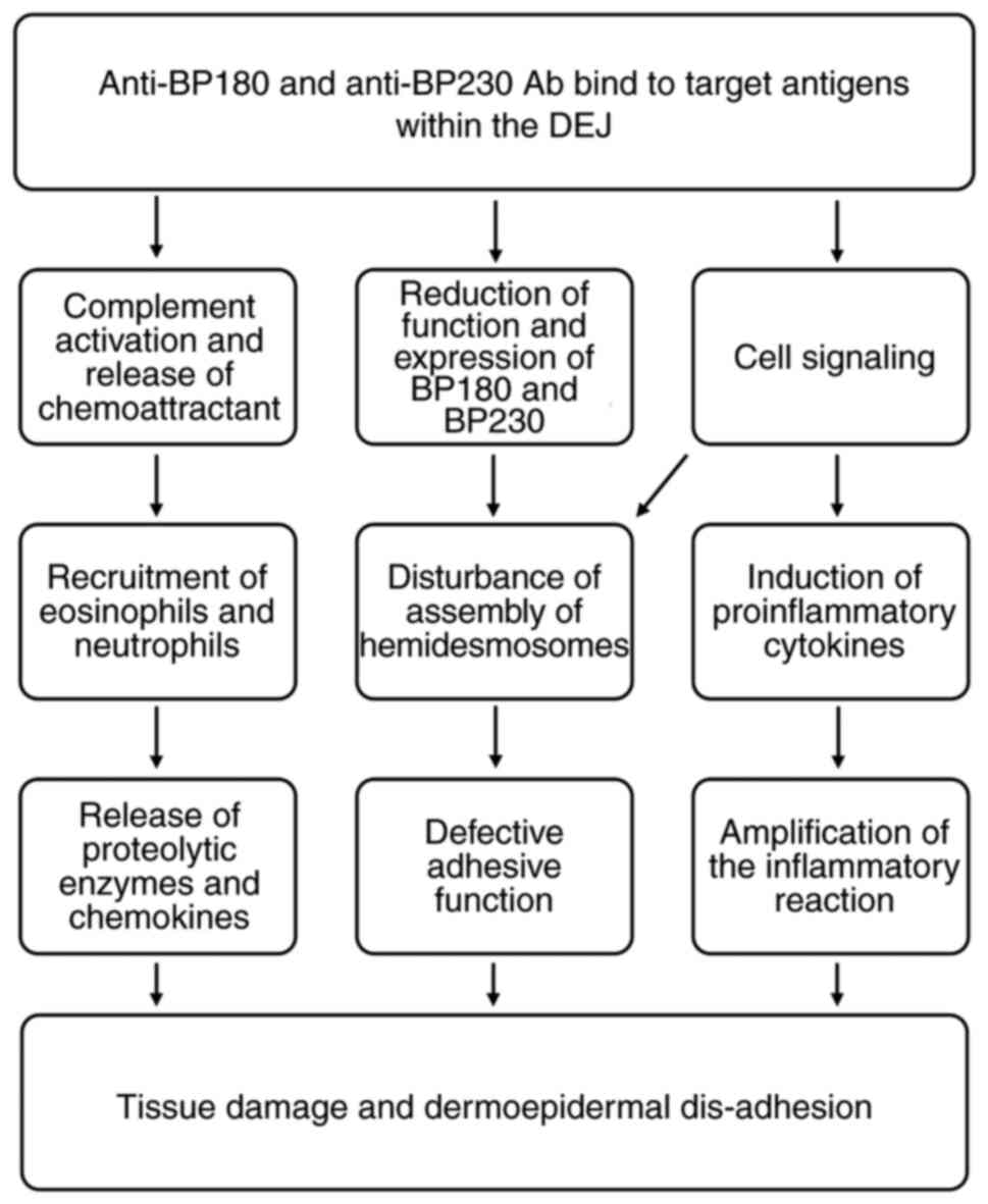
The binding of autoreactive IgG antibodies to BP230
and BP180 antigens initiates a cascade of immunological events,
which are detailed in Fig. 1.
Complement activation promotes inflammation by summoning
leukocytes, mast cells and other immune cell populations to
affected skin sites. These cells trigger the release of cytokines,
proteases and lysosomal enzymes, which leads to the destruction of
hemidesmosomes, exposure of neo-antigens and, ultimately, to the
formation of blisters (12).
Previous studies have reported that anti-BP180 IgE autoantibodies
are also observed in patients with BP (13,14).
Autoimmune IgEs are considered to synergize with self-reactive IgG1
and IgG4, leading to complement activation, mast cell degranulation
and the release of leukotrienes, platelet-activating factor, TNF
and other cytokines. Neutrophils and eosinophils recruited to skin
lesion sites release proteolytic enzymes, which impair adhesion
molecules and contribute to subepidermal blistering (15).
Although the underlying cause of BP resides in
autoimmunity, to the best of our knowledge, it is not known how
this loss of self-tolerance occurs and why the elderly population
is almost exclusively affected (9). An increase in autoantibody levels is
observed with increasing age and may be related to loss of
tolerance (16,17). However, with the exception of
specific disorders, including BP, the incidence of autoimmune
disease in the elderly population is not as high as could be
expected.
These observations support the hypothesis that BP is
not exclusively dependent on the autoantibodies produced against
skin structural proteins, but also requires the development of skin
barrier abnormalities. These changes associated with aging, on a
background of genetic predisposition, would make elderly subjects
susceptible to the action of triggering factors, leading to the
exposure of normally sequestered antigens (18).
2. Triggers of BP
Known triggers of BP include drugs, topically
applied products, such as wound dressings, vaccines, viral
infections, physical factors, such as UV radiation, radiotherapy,
surgery and neurological or cardiac disorders.
Drugs
BP has been frequently associated with systemic
therapy. The pathogenesis of drug-induced BP is poorly understood
and, in most elderly patients, particularly in the context of
polytherapy, it is difficult to establish the triggering role of a
specific drug. A complex and multifactorial pathogenesis involving
numerous immunological mechanisms has been hypothesized for
drug-induced BP. According to the ‘two-step’ theory, interactions
among the immune system and two different drugs displaying similar
molecular structures could represent the first and second hit,
triggering and enhancing an immune reaction leading to the onset of
BP (19). The literature provides
an example of ‘two-step’ induction following intake of captopril
and, subsequently, of enalapril. While both drugs are ACE
inhibitors, the former also contains a thiol group. The first onset
of blisters after captopril is attributed to acantholysis induced
by a thiol drug, while relapse after enalapril is associated with
the production of autoantibodies and activation of the kinin system
caused by the drug (20). Another
theory considers the interaction between sulfhydryl groups of
hemidesmosomes/components of the basement membrane and
sulfur-containing drugs, whereby the integrity of the
dermoepidermal junction is disrupted, which could lead to exposure
of hidden antigenic sites (21).
Furthermore, certain oncological treatments, such as monoclonal
antibodies against programmed death-1 and its corresponding ligand,
act by inactivating endogenous immunoregulatory checkpoints of
T-cell function and they are associated with the development of
immune-related adverse events, including cutaneous autoimmune
blistering disorders (22).
Finally, the emergence of BP following administration of vaccines
has been reported in the literature. It has previously been
hypothesized that, in predisposed subjects, the inflammatory
response following vaccination may promote a conformational change
of the dermoepidermal junction and expose hidden epitopes or
antigens triggering the production of specific anti-basement
membrane antibodies (23).
Infections
Viruses can interact with B and T lymphocytes,
stimulating the production of antibodies and the secretion of IL-4
and IL-10, which determines the switch from T helper type 1 cell
(Th1) to T helper type 2 cell (Th2) immunity. Pathogen-induced
antibodies may cross-react with shared epitopes on host cells.
Furthermore, viruses may induce autoimmunity by directly infecting
keratinocytes, exposing hidden epitopes or modifying the expression
of existing antigens (24). Host
cell fragments can also become inserted into viral envelopes and be
presented as novel antigens to the host immune system (24,25).
Moreover, cases of BP triggered by Sarcoptes scabiei
infestation have been reported. This suggests that parasitic
diseases may contribute to the onset of BP, which may be a result
of Koebner's phenomenon (26).
Physical factors
The onset of BP has been observed following exposure
to numerous physical triggers, including trauma, surgery, thermal
or electrical burns, UV exposure, radiation therapy and
photodynamic therapy. The association of BP with radiotherapy, as
well as that with thermal or electrical burns, has been documented
in a study (27). The irritant
mechanism of certain topical drugs on the skin, such as benzyl
benzoate, or allergic contact hypersensitivity, for example by
5-fluorouracil, have been considered as possible triggers for the
onset of BP (28). BP can also be
induced by UV radiation or photodynamic therapy, both of which are
established skin-directed treatment approaches in dermatology
(29-33).
This hypothesis has been investigated in animal models, which
demonstrate that UV irradiation leads to the development of severe
inflammation and blisters in subjects with circulating
autoantibodies against BP180 or BP230 (34,35).
Furthermore, underlying alterations of the basement membrane which
are associated with preexisting skin disorders such as psoriasis,
are hypothetical predisposing factors (36-38).
In patients with psoriasis treated using narrow-band UVB radiation
therapy, BP lesions have been reported to develop over pre-existing
psoriatic lesions, leading to the hypothesis that UVB- and psolaren
UVA-induced antigenic changes in psoriatic skin may trigger blister
formation (39,40). The pathogenetic mechanisms by which
other environmental physical agents could cause or exacerbate BP
are unclear. Previous studies have reported that trauma-induced
tissue rupture may lead to the exposure of previously masked
antigens and the development of autoantibodies directed towards the
basement membrane (29,41,42).
Following physical trauma, the damaged tissue may release a variety
of pro-inflammatory factors that contribute to the recruitment of
circulating inflammatory cells. This would result in the activation
of granulocytes and the complement pathway and may lead to the
production of antibodies and, finally, blistering (12).
3. Comorbidities of BP
An association has been highlighted between BP and a
number of conditions, including autoimmune, neurological (43-51)
and cardiovascular disorders (47,51-56),
as well as tumors (47,51,57-60).
These are listed in Table I. The
association between BP and certain neurological diseases could be
explained by the fact that damage to brain cells exposes neural
antigens. These molecules may lead to the production of
cross-reactive autoantibodies against antigens of the skin basement
membrane (18).
 | Table IClassification of comorbidities
reported in association with bullous pemphigoid. |
Table I
Classification of comorbidities
reported in association with bullous pemphigoid.
| Classification | Disease | (Refs.) |
|---|
| Autoimmune
diseases | Psoriasis | (61-66) |
| | Rheumatoid
arthritis | |
| | Lupus
erythematosus | |
| | Lichen planus | |
| | Polymyositis | |
| | Thyroiditis | |
| | Pernicious
anemia | |
| | Primary biliary
cirrhosis | |
| | Membranous
nephropathy | |
| Neurological
diseases | Parkinson's
disease | (43-51) |
| | Alzheimer's
disease | |
| | Stroke | |
| | Dementia | |
| | Multiple
sclerosis | |
| | Epilepsy | |
| | Schizophrenia | |
| Cardiovascular
diseases | Hypertension | (47,51-56) |
| | Stroke | |
| | Venous
thromboembolism | |
| | Pulmonary
embolism | |
| Neoplasms | Kidney cancer | (47,51,57-60) |
| | Laryngeal
cancer | |
| | Hematological
malignancies | |
BP has also been associated with other autoimmune
diseases, including lupus erythematosus, lichen planus, membranous
nephropathy, pernicious anemia, thyroiditis, primary biliary
cirrhosis, multiple sclerosis, psoriasis and polymyositis (61-65).
The onset of BP in patients with psoriasis may be a result of a
series of epigenetic events involved in the psoriatic inflammatory
cascade (66). Moreover, basement
membrane changes in psoriasis make the patient more susceptible to
autoantibody formation. Furthermore, an increase in chemokine C-C
motif ligand (CCL) 28 and T helper type 17 cell (Th17) serum levels
has been demonstrated in psoriasis as well as in BP (67). This is consistent with the concept
that IL-17-producing Th17 cells are involved in the secretion of
pro-inflammatory cytokines and MMPs and in the recruitment of
neutrophils and eosinophils, all of which are important factors in
the pathogenesis of BP (66).
4. Aging
The epidemiology of BP clearly shows that this is a
disease of the elderly. Its triggers and comorbidities have been
reviewed thus far, highlighting their association with advanced
age. The following sections focus on the immune mechanisms
associated with aging that contribute to the induction of BP. Aging
is accompanied by a gradual remodeling of the immune system called
immunosenescence. Immunosenescence is the process of immune system
dysregulation, which is characteristic of elderly patients and
gives rise to the following three events: i) A reduction in the
immune response; ii) an increase in autoantibody production; and
iii) chronic, sterile, low-grade inflammation (68).
In elderly individuals, low affinity immunoglobulin
production increases, as do the serum levels of IgG1 and IgG4.
Chronic production of cytokines and the associated low-grade
inflammation is typical of elderly patients and could possibly
serve as a stimulus for the onset of autoimmunity (69). Furthermore, persistent infections
commonly develop in old age. Infectious agents, in the context of a
dysregulated immune system, may provide foreign antigens that
cross-react with self-antigens displaying similar sequences. It has
also been observed that CD4+ CD25+ regulatory
T cells decrease with age and, therefore, their protective
mechanism against the development of autoimmunity may become
gradually compromised (70).
Considering its anatomical characteristics and
defensive role against the external environment, the skin serves as
a barrier organ. It is not merely a mechanical barrier, but a
complex immune organ, hosting both innate and adaptive immune
functions, with the physiological role of protecting the organism
from infectious or inflammatory diseases. The process of aging is
particularly evident in the cutaneous compartment, which is
vulnerable to intrinsic alterations and to lifelong environmental
exposure (71). Intrinsic factors
mainly include hormonal changes, such as those related to
menopause, ethnicity, which affects pigmentation and lipid
proportions, and stress, which may exaggerate degenerative
processes (72-74).
The influence of the environment on skin aging is mainly dependent
on air pollutants and sunlight (75,76).
5. Skin changes associated with aging
Skin aging is a slow, complex process resulting from
the interplay of cellular, molecular and genetic factors. Different
cutaneous layers undergo progressive morphological and functional
changes that alter the appearance and mechanical behavior of the
skin (77). The principal changes
that make elderly individuals more susceptible to mechanical
injury, alter the skin microbiome and impair skin barrier immune
function, contributing to the onset of chronic low-grade
inflammation, are summarized in Table
II.
| Table IIChanges in cutaneous barrier function
and local skin immunity associated with aging. |
Table II
Changes in cutaneous barrier function
and local skin immunity associated with aging.
| Skin function | Associated
change |
|---|
| Inflammation | ↑ Senescent
cells |
| | ↑ Cytokines of
inflammaging |
| | ↑ MMP production
(with ECM degradation) |
| Phagocytosis | ↓ TNF
production |
| | ↓ LC number |
| | ↓ LC migration |
| T cell
function | ↓ Response to
antigens |
| | ↑ FOXP3 regulatory
T cells (inhibit antigen-specific immunity) |
| | ↑ Inhibitory
receptors |
| Stromal
support | ↓ ECM production by
fibroblasts |
| | ↓ Ability of
fibroblasts to differentiate |
| | ↓ Dermal white
adipose tissue |
Senescence of the epidermis is accompanied by a dull
appearance, pigmentation and dehydration (77). The epidermis of aged skin exhibits
an overall decreased thickness, whereas that of the basement
membrane may increase up to 50% with aging. However, collagen IV
content is decreased in the basement membrane, reflecting a
reduction in tissue turnover (78). Keratinocytes acquire degenerative
morphological changes due to a loss of epidermal cell stemness, as
well as reduced support and nutrition from the underlying dermis
(79). A thinning of the
epidermis, with atrophy of the keratinocytes, consequently leads to
increases in trans-epidermal water loss and skin dryness (80,81).
Melanocytes are responsible for cellular hyperpigmentation of
senescent skin due to an enhanced dihydroxyphenylalanine reaction
following chronic irradiation (82).
Dermal alterations are a result of UVA-induced
photoaging, mediated by an increase in cellular reactive oxygen
species and a decrease in the thickness of the dermis, elastosis
and fibroblast apoptosis. These aforementioned factors are
responsible for wrinkles, dehydration, scar formation and skin
susceptibility to trauma (83,84).
A decrease in the number and size of dermal fibroblasts has been
demonstrated to be associated with skin thinning and the reduced
production of pro-collagen (85).
Moreover, an accumulation of senescent dermal fibroblasts is known
to occur with aging and is classically defined by the expression of
cyclin-dependent kinase inhibitor 2A in the skin of elderly
individuals and mice (86-88).
Senescent fibroblasts are characterized by irreversible cell growth
arrest and changes in the levels of secretory proteins involved in
inflammation and matrix degradation. Secretion of proinflammatory
mediators, such as IL-8, IL-6, TNF and CCL2, is upregulated and
sustains chronic inflammation (89). Furthermore, increased expression
levels of MMPs, including MMP-1, -3 and -9, in senescent skin
decreases the total amount of collagen and is also responsible for
the fragmentation of elastin, which results in reduced skin
elasticity and classic signs of skin aging, such as wrinkles
(90). Elastin is an inert protein
that forms during early development and is not replenished, so any
loss in elastin that occurs throughout life tends to be
irreversible (91). In photoaged
skin, collagen and elastin content are both reduced by ~50%, which
means that hydration, tensile strength and elasticity are
compromised (92). The dermis also
hosts sweat glands that, although they may show atrophy, generally
continue to function in elderly individuals.
With advancing age, wasting of subcutaneous adipose
tissue is observed due to a reduction in the number of white
adipocytes (93). This change
affects the mechanical protection as well as antimicrobial defense
that is physiologically provided from dermal fat in response to
infection (94). Dermal adipocytes
may promote innate immunity through production of antimicrobial
peptide cathelicidin, and may support adaptive immunity by
providing a hub for memory T cells (95). The enhancement of transforming
growth factor b pathway in the elderly is largely responsible for
this age-dependent loss of immune protection from dermal fat
(94).
Immune compartments of the skin are impaired
following changes in Langerhans cell function, weakening of
antigen-specific responses and modification of regulatory T-cell
populations (96). These changes
occur in the context of chronic low-grade inflammation that is
typical of senescence and has been defined as ‘inflammaging’.
Potential markers of inflammaging include C-reactive protein, which
is characteristically elevated in the serum, and IL-1β, the
elevation of which is associated with increased morbidity and
mortality in elderly patients (69). Macrophages may also contribute to
the development of inflammaging due to structural changes in the
skin, such as thinning. These innate immune cells are more easily
exposed to pathogens leading to chronic activation and cytokine
production (97). Moreover, the
accumulation of senescent cells was also shown to contribute to
increased inflammation in the elderly (98).
In summary, changes impairing the physical, chemical
and immunological barrier function of the skin are all likely
culprits responsible for susceptibility to BP in genetically
predisposed elderly individuals who have been exposed to specific
triggers.
6. BP and aging
Aging significantly alters the anatomy and function
of the skin. A triggering factor on damaged skin easily destroys
keratinocytes via necrosis or necroptosis, releasing into the
extracellular space intracellular components that act as
autoantigens (99). BP180 and
BP230 are autoantigens taken up by specialized antigen presenting
cells (APC), which express the class II major histocompatibility
complex. APCs present to self-reactive T cells leading to an
adaptive immune response and the release of pro-inflammatory
cytokines from activated T cells (100). Subsequently, this leads to an
increase in co-stimulatory receptors on the surface of APCs and to
the activation of autoreactive autoantibody-producing B cells.
Furthermore, certain molecules released following cell damage may
behave as autoantigens and endogenous Toll-like receptor (TLR)
ligands. TLRs and B-cell receptors can be directly activated
triggering the secretion of autoantibodies. These antibodies must
then acquire pathogenicity by performing a class switch from IgM to
IgA or IgG. Damaged keratinocytes also release additional
self-antigens that contribute to the production of new
autoantibodies and further impairment of the skin barrier, which
establishes a perpetual cycle of tissue damage and autoantibody
production (10,14).
7. Conclusion
The pathology of BP results from the complex
interaction of multiple mechanisms, including genetic
predisposition, skin barrier changes, immunosenescence and
triggering factors. The epidemiology of BP displays a predilection
for elderly individuals. In the present review, the triggers and
comorbidities associated with BP have been described, highlighting
conditions that are typically found in advanced age. Moreover, the
effects of aging on skin barrier integrity and the role of immune
system function in BP susceptibility were investigated.
The present review has indicated that the management
of BP should be aimed at removing potential triggers, treating
concomitant disease conditions and counteracting the detrimental
effects of aging on the skin. The latter objective may be achieved
by improving skin barrier integrity and maintaining cutaneous
homeostasis, for example with systematic applications of topical
emollients and photoprotection. Furthermore, immunosuppressive
treatment for BP must be tailored according to the presence of
comorbidities, age and overall clinical conditions of each patient
(101).
In conclusion, restoring skin barrier integrity
should be a treatment goal for all patients with BP. This strategy
could prove particularly beneficial in elderly individuals, in whom
frequent comorbidities associated with age often limit the
availability of treatment options. Moreover, the safety of regimens
can markedly affect outcome and prognosis.
Acknowledgements
Not applicable.
Funding
Funding: No funding was received.
Availability of data and materials
Not applicable.
Authors' contributions
MLD and ASp were responsible for data collection and
the writing of the draft manuscript. ASe was responsible for
reviewing, editing and the presentation of data in the manuscript.
MA was responsible for the concept of the review and supervision
and for reviewing and editing the manuscript. All authors read and
approved the final manuscript. Data sharing is not applicable.
Ethics approval and consent to
participate
Not applicable.
Patient consent for publication
Not applicable.
Competing interests
The authors declare that they have no competing
interests.
References
|
1
|
Parker SR and MacKelfresh J: Autoimmune
blistering diseases in the elderly. Clin Dermatol. 29:69–79.
2011.PubMed/NCBI View Article : Google Scholar
|
|
2
|
Bernard P and Antonicelli F: Bullous
pemphigoid: A review of its Diagnosis, Associations and Treatment.
Am J Clin Dermatol. 18:513–528. 2017.PubMed/NCBI View Article : Google Scholar
|
|
3
|
Miyamoto D, Santi CG, Aoki V and Maruta
CW: Bullous pemphigoid. An Bras Dermatol. 94:133–146.
2019.PubMed/NCBI View Article : Google Scholar
|
|
4
|
Patel F, Wilken R, Patel FB, Sultani H,
Bustos I, Duong C, Zone JJ, Raychaudhuri SP and Maverakis E:
Pathophysiology of autoimmune bullous diseases: Nature versus
nurture. Indian J Dermatol. 62:262–267. 2017.PubMed/NCBI View Article : Google Scholar
|
|
5
|
Yang M, Wu H, Zhao M, Chang C and Lu Q:
The pathogenesis of bullous skin diseases. J Transl Autoimmun.
2(100014)2019.PubMed/NCBI View Article : Google Scholar
|
|
6
|
Marazza G, Pham HC, Schärer L, Pedrazzetti
PP, Hunziker T, Trüeb RM, Hohl D, Itin P, Lautenschlager S, Naldi
L, et al: Incidence of bullous pemphigoid and pemphigus in
Switzerland: A 2-year prospective study. Br J Dermatol.
161:861–868. 2009.PubMed/NCBI View Article : Google Scholar
|
|
7
|
Langan SM, Smeeth L, Hubbard R, Fleming
KM, Smith CJ and West J: Bullous pemphigoid and pemphigus
vulgaris-incidence and mortality in the UK: Population based cohort
study. BMJ. 337(a180)2008.PubMed/NCBI View
Article : Google Scholar
|
|
8
|
Gudi VS, White MI, Cruickshank N, Herriot
R, Edwards SL, Nimmo F and Ormerod AD: Annual incidence and
mortality of bullous pemphigoid in the Grampian Region of
North-east Scotland. Br J Dermatol. 153:424–427. 2005.PubMed/NCBI View Article : Google Scholar
|
|
9
|
Hübner F, Recke A, Zillikens D, Linder R
and Schmidt E: Prevalence and age distribution of pemphigus and
pemphigoid diseases in Germany. J Invest Dermatol. 136:2495–2498.
2016.PubMed/NCBI View Article : Google Scholar
|
|
10
|
Feliciani C, Caldarola G, Kneisel A,
Podstawa E, Pfütze M, Pfützner W and Hertl M: IgG autoantibody
reactivity against bullous pemphigoid (BP) 180 and BP230 in elderly
patients with pruritic dermatoses. Br J Dermatol. 161:306–312.
2009.PubMed/NCBI View Article : Google Scholar
|
|
11
|
Hopkinson SB and Jones JC: The N terminus
of the transmembrane protein BP180 interacts with the N-terminal
domain of BP230, thereby mediating keratin cytoskeleton anchorage
to the cell surface at the site of the hemidesmosome. Mol Biol
Cell. 11:277–286. 2000.PubMed/NCBI View Article : Google Scholar
|
|
12
|
Giang J, Seelen MAJ, van Doorn MBA,
Rissmann R, Prens EP and Damman J: Complement activation in
inflammatory skin diseases. Front Immunol. 9(639)2018.PubMed/NCBI View Article : Google Scholar
|
|
13
|
Fania L, Caldarola G, Müller R, Brandt O,
Pellicano R, Feliciani C and Hertl M: IgE recognition of bullous
pemphigoid (BP)180 and BP230 in BP patients and elderly individuals
with pruritic dermatoses. Clin Immunol. 143:236–245.
2012.PubMed/NCBI View Article : Google Scholar
|
|
14
|
Di Zenzo G, Thoma-Uszynski S, Fontao L,
Calabresi V, Hofmann SC, Hellmark T, Sebbag N, Pedicelli C, Sera F,
Lacour JP, et al: Multicenter prospective study of the humoral
autoimmune response in bullous pemphigoid. Clin Immunol.
128:415–426. 2008.PubMed/NCBI View Article : Google Scholar
|
|
15
|
Park SH, Lee SH, Kim JH and Kim SC:
Circulating eosinophil and neutrophil counts correlate with disease
severity in bullous pemphigoid. Ann Dermatol. 30:544–549.
2018.PubMed/NCBI View Article : Google Scholar
|
|
16
|
Budamagunta V, Foster TC and Zhou D:
Cellular senescence in lymphoid organs and immunosenescence. Aging
(Albany NY). 13:19920–19941. 2021.PubMed/NCBI View Article : Google Scholar
|
|
17
|
Liu YD, Wang YH, Ye YC, Zhao WL and Li L:
Prognostic factors for mortality in patients with bullous
pemphigoid: a meta-analysis. Arch Dermatol Res. 309:335–347.
2017.PubMed/NCBI View Article : Google Scholar
|
|
18
|
Lai YC, Yew YW and Lambert WC: Bullous
pemphigoid and its association with neurological diseases: A
systematic review and meta-analysis. J Eur Acad Dermatology
Venereol. 30:2007–2015. 2016.PubMed/NCBI View Article : Google Scholar
|
|
19
|
Adam J, Pichler WJ and Yerly D: Delayed
drug hypersensitivity: Models of T-cell stimulation. Br J Clin
Pharmacol. 71:701–707. 2011.PubMed/NCBI View Article : Google Scholar
|
|
20
|
Ruocco V, Satriano RA and Guerrera V:
‘Two-step’ pemphigus induction by ACE-inhibitors. Int J Dermatol.
31:33–36. 1992.PubMed/NCBI View Article : Google Scholar
|
|
21
|
Ruocco V and Sacerdoti G: Pemphigus and
Bullous pemphigoid due to drugs. Int J Dermatol. 30:307–312.
1991.PubMed/NCBI View Article : Google Scholar
|
|
22
|
Faina V, Sernicola A, Russo I, Michelotto
A, Szathvary V, Frigo AC and Alaibac M: Programmed cell death-1
rs2227981 polymorphism in patients with autoimmune skin blistering
disorders: A pilot study. Meta Gene. 26(100793)2020.
|
|
23
|
Stavropoulos PG, Soura E and Antoniou C:
Drug-induced pemphigoid: A review of the literature. J Eur Acad
Dermatology Venereol. 28:1133–1140. 2014.PubMed/NCBI View Article : Google Scholar
|
|
24
|
Lo Schiavo A, Ruocco E, Brancaccio G,
Caccavale S, Ruocco V and Wolf R: Bullous pemphigoid: Etiology,
pathogenesis, and inducing factors: Facts and controversies. Clin
Dermatol. 31:391–399. 2013.PubMed/NCBI View Article : Google Scholar
|
|
25
|
Drago F, Nozza P, Casazza S, Brusati C,
Bandelloni R and Rebora A: Human herpesviruses in bullous
pemphigoid lesions. Br J Dermatol. 152:375–376. 2005.PubMed/NCBI View Article : Google Scholar
|
|
26
|
Iriki H, Adachi T, Matsuda H, Chinen K,
Arakawa H, Yamagami J, Nishie W and Yokouchi M: Case of dipeptidyl
peptidase 4 inhibitor-associated bullous pemphigoid that developed
after a scabies infestation. J Dermatol. 47:e258–e260.
2020.PubMed/NCBI View Article : Google Scholar
|
|
27
|
Mai Y, Nishie W, Sato K, Hotta M, Izumi K,
Ito K, Hosokawa K and Shimizu H: Bullous pemphigoid triggered by
thermal burn under medication with a dipeptidyl peptidase-IV
inhibitor: A case report and review of the literature. Front
Immunol. 9(542)2018.PubMed/NCBI View Article : Google Scholar
|
|
28
|
Vassileva S: Drug-induced pemphigoid:
Bullous and cicatricial. Clin Dermatol. 16:379–387. 1998.PubMed/NCBI View Article : Google Scholar
|
|
29
|
Dănescu S, Chiorean R, Macovei V, Sitaru C
and Baican A: Role of physical factors in the pathogenesis of
bullous pemphigoid: Case report series and a comprehensive review
of the published work. J Dermatol. 43:134–140. 2016.PubMed/NCBI View Article : Google Scholar
|
|
30
|
Caca-Biljanovska N, Arsovska-Bezhoska I
and V'lckova-Laskoska M: PUVA-induced bullous pemphigoid in
psoriasis. Acta Dermatovenerol Croat. 24:214–217. 2016.PubMed/NCBI
|
|
31
|
Perl S, Rappersberger K, Födinger D, Anegg
B, Hönigsmann H and Ortel B: Bullous pemphigoid induced by PUVA
therapy. Dermatology. 193:245–247. 1996.PubMed/NCBI View Article : Google Scholar
|
|
32
|
Preesman AH, Toonstra J, Van der Putte SC,
De Geer DB, Van Weelden H and Van Vloten WA: UV-B-induced bullous
pemphigoid restricted to mycosis fungoides plaques. Clin Exp
Dermatol. 15:363–366. 1990.PubMed/NCBI View Article : Google Scholar
|
|
33
|
Korekawa A, Kaneko T, Nakajima K, Rokunohe
D, Akasaka E, Nakano H, Sawamura D, Fukui T, Takiyoshi N, Kitamura
H and Harada K: Mycosis fungoides bullosa associated with bullous
pemphigoid. Int J Dermatol. 54:e366–e368. 2015.PubMed/NCBI View Article : Google Scholar
|
|
34
|
Mitsuhashi Y, Nakano H, Murai T, Ohta T,
Sawamura D, Hanada K and Hashimoto I: Bullous pemphigoid sera
induce bullous-pemphigoid-like lesions in neonatal mice pretreated
with a limited dose of ultraviolet B irradiation. Dermatology. 189
(Suppl 1):S76–S81. 1994.PubMed/NCBI View Article : Google Scholar
|
|
35
|
Hall RP III, Murray JC, McCord MM, Rico MJ
and Streilein RD: Rabbits Immunized with a Peptide Encoded for by
the 230-kD bullous pemphigoid antigen cDNA develop an enhanced
inflammatory response to UVB irradiation: A potential animal model
for bullous pemphigoid. J Invest Dermatol. 101:9–14.
1993.PubMed/NCBI View Article : Google Scholar
|
|
36
|
Ho YH, Hu HY, Chang YT, Li CP and Wu CY:
Psoriasis is associated with increased risk of bullous pemphigoid:
A nationwide population-based cohort study in Taiwan. J Dermatol.
46:604–609. 2019.PubMed/NCBI View Article : Google Scholar
|
|
37
|
Barnadas MA, Gilaberte M, Pujol R, Agustí
M, Gelpí C and Alomar A: Bullous pemphigoid in a patient with
psoriasis during the course of PUVA therapy: Study by ELISA test.
Int J Dermatol. 45:1089–1092. 2006.PubMed/NCBI View Article : Google Scholar
|
|
38
|
Wilczek A and Sticherling M: Concomitant
psoriasis and bullous pemphigoid: Coincidence or pathogenic
relationship? Int J Dermatol. 45:1353–1357. 2006.PubMed/NCBI View Article : Google Scholar
|
|
39
|
Danno K, Takigawa M and Horio T:
Alterations in lectin binding to the epidermis following treatment
with 8-methoxypsoralen plus long-wave ultraviolet radiation. J
Invest Dermatol. 82:176–179. 1984.PubMed/NCBI View Article : Google Scholar
|
|
40
|
George PM: Bullous pemphigoid possibly
induced by psoralen plus ultraviolet a therapy. Photodermatol
Photoimmunol Photomed. 11:185–187. 1996.PubMed/NCBI View Article : Google Scholar
|
|
41
|
Yesudian PD, Dobson CM, Ahmad R and
Azurdia RM: Trauma-induced bullous pemphigoid around venous access
site in a haemodialysis patient. Clin Exp Dermatol. 27:70–72.
2002.PubMed/NCBI View Article : Google Scholar
|
|
42
|
Neville JA and Yosipovitch G: Flare of
bullous pemphigoid in surgically treated skin. Cutis. 75:169–170.
2005.PubMed/NCBI
|
|
43
|
Taghipour K, Chi CC, Vincent A, Groves RW,
Venning V and Wojnarowska F: The association of bullous pemphigoid
with cerebrovascular disease and dementia: A case-control study.
Arch Dermatol. 146:1251–1254. 2010.PubMed/NCBI View Article : Google Scholar
|
|
44
|
Langan SM, Groves RW and West J: The
relationship between neurological disease and bullous pemphigoid: A
population-based case-control study. J Invest Dermatol.
131:631–636. 2011.PubMed/NCBI View Article : Google Scholar
|
|
45
|
Brick KE, Weaver CH, Savica R, Lohse CM,
Pittelkow MR, Boeve BF, Gibson LE, Camilleri MJ and Wieland CN: A
population-based study of the association between bullous
pemphigoid and neurologic disorders. J Am Acad Dermatol.
71:1191–1197. 2014.PubMed/NCBI View Article : Google Scholar
|
|
46
|
Gambichler T, Segert H, Höxtermann S,
Schmitz L, Altmeyer P and Teegen B: Neurological disorders in
patients with bullous pemphigoid: clinical and experimental
investigations. J Eur Acad Dermatology Venereol. 29:1758–1762.
2015.PubMed/NCBI View Article : Google Scholar
|
|
47
|
Kibsgaard L, Bay B, Deleuran M and
Vestergaard C: A retrospective consecutive case-series study on the
effect of systemic treatment, length of admission time, and
co-morbidities in 98 bullous pemphigoid patients admitted to a
tertiary centre. Acta Derm Venereol. 95:307–311. 2015.PubMed/NCBI View Article : Google Scholar
|
|
48
|
Försti AK, Jokelainen J, Ansakorpi H,
Seppänen A, Majamaa K, Timonen M and Tasanen K: Psychiatric and
neurological disorders are associated with bullous pemphigoid-a
nationwide finnish care register study. Sci Rep.
6(37125)2016.PubMed/NCBI View Article : Google Scholar
|
|
49
|
Khosravani S, Handjani F, Alimohammadi R
and Saki N: Frequency of neurological disorders in bullous
pemphigoid patients: A cross-sectional study. Int Sch Res Notices.
2017(6053267)2017.PubMed/NCBI View Article : Google Scholar
|
|
50
|
Ravn AH, Thyssen JP and Egeberg A: Skin
disorders in Parkinson's disease: Potential biomarkers and risk
factors. Clin Cosmet Investig Dermatol. 10:87–92. 2017.PubMed/NCBI View Article : Google Scholar
|
|
51
|
Kalińska-Bienias A, Kowalczyk E, Jagielski
P, Bienias P, Kowalewski C and Wozniak K: The association between
neurological diseases, malignancies and cardiovascular
comorbidities among patients with bullous pemphigoid: Case-control
study in a specialized Polish center. Adv Clin Exp Med. 28:637–642.
2019.PubMed/NCBI View Article : Google Scholar
|
|
52
|
Langan SM, Hubbard R, Fleming K and West
J: A population-based study of acute medical conditions associated
with bullous pemphigoid. Br J Dermatol. 161:1149–1152.
2009.PubMed/NCBI View Article : Google Scholar
|
|
53
|
Heelan K, Mahar AL, Walsh S and Shear NH:
Pemphigus and associated comorbidities: A cross-sectional study.
Clin Exp Dermatol. 40:593–599. 2015.PubMed/NCBI View Article : Google Scholar
|
|
54
|
Kremer N, Zeeli T, Sprecher E and Geller
S: Failure of initial disease control in bullous pemphigoid: A
retrospective study of hospitalized patients in a single tertiary
center. Int J Dermatol. 56:1010–1016. 2017.PubMed/NCBI View Article : Google Scholar
|
|
55
|
Hsu DY, Brieva J, Nardone B, West D and
Silverberg JI: Association of pemphigus and systemic corticosteroid
use with comorbid health disorders: A case-control study. Dermatol
Online J. 23(13030/qt1vk2m30m)2017.PubMed/NCBI
|
|
56
|
Ungprasert P, Wijarnpreecha K and
Thongprayoon C: Risk of venous thromboembolism in patients with
bullous pemphigoid: A systematic review and meta-analysis. Indian J
Dermatol Venereol Leprol. 84:22–26. 2018.PubMed/NCBI View Article : Google Scholar
|
|
57
|
Venning VA and Wojnarowska F: The
association of bullous pemphigoid and malignant disease: A case
control study. Br J Dermatol. 123:439–445. 1990.PubMed/NCBI View Article : Google Scholar
|
|
58
|
Bastuji-Garin S, Joly P, Lemordant P,
Sparsa A, Bedane C, Delaporte E, Roujeau JC, Bernard P, Guillaume
JC, Ingen-Housz-Oro S, et al: Risk factors for bullous pemphigoid
in the elderly: A prospective case-control study. J Invest
Dermatol. 131:637–643. 2011.PubMed/NCBI View Article : Google Scholar
|
|
59
|
Schulze F, Neumann K, Recke A, Zillikens
D, Linder R and Schmidt E: Malignancies in Pemphigus and Pemphigoid
Diseases. J Invest Dermatol. 135:1445–1447. 2015.PubMed/NCBI View Article : Google Scholar
|
|
60
|
Balestri R, Magnano M, La Placa M, Patrizi
A, Angileri L, Tengattini V and Bardazzi F: Malignancies in bullous
pemphigoid: A controversial association. J Dermatol. 43:125–133.
2016.PubMed/NCBI View Article : Google Scholar
|
|
61
|
Callen JP: Internal disorders associated
with bullous disease of the skin. A critical review. J Am Acad
Dermatol. 3:107–119. 1980.PubMed/NCBI View Article : Google Scholar
|
|
62
|
Dahl MV: Bullous pemphigoid: Associated
diseases. Clin Dermatol. 5:64–70. 1987.PubMed/NCBI View Article : Google Scholar
|
|
63
|
Chen YJ, Wu CY, Lin MW, Chen TJ, Liao KK,
Chen YC, Hwang CY, Chu SY, Chen CC, Lee DD, et al: Comorbidity
profiles among patients with bullous pemphigoid: A nationwide
population-based study. Br J Dermatol. 165:593–599. 2011.PubMed/NCBI View Article : Google Scholar
|
|
64
|
Ljubojevic S and Lipozenčić J: Autoimmune
bullous diseases associations. Clin Dermatol. 30:17–33.
2012.PubMed/NCBI View Article : Google Scholar
|
|
65
|
Akarsu S, Özbağçivan Ö, Dolaş N and Aktan
Ş: Possible triggering factors and comorbidities in newly diagnosed
autoimmune bullous diseases. Turkish J Med Sci. 47:832–840.
2017.PubMed/NCBI View Article : Google Scholar
|
|
66
|
Dainichi T and Kabashima K: Interaction of
psoriasis and bullous diseases. Front Med. 5(222)2018.PubMed/NCBI View Article : Google Scholar
|
|
67
|
Harper EG, Guo C, Rizzo H, Lillis JV,
Kurtz SE, Skorcheva I, Purdy D, Fitch E, Iordanov M and Blauvelt A:
Th17 cytokines stimulate CCL20 expression in keratinocytes in vitro
and in vivo: Implications for psoriasis pathogenesis. J Invest
Dermatol. 129:2175–2183. 2009.PubMed/NCBI View Article : Google Scholar
|
|
68
|
Watad A, Bragazzi NL, Adawi M, Amital H,
Toubi E, Porat BS and Shoenfeld Y: Autoimmunity in the elderly:
Insights from basic science and clinics-a mini-review. Gerontology.
63:515–523. 2017.PubMed/NCBI View Article : Google Scholar
|
|
69
|
Lee YI, Choi S, Roh WS, Lee JH and Kim TG:
Cellular senescence and inflammaging in the skin microenvironment.
Int J Mol Sci. 22(3849)2021.PubMed/NCBI View Article : Google Scholar
|
|
70
|
Scott MM and Liang SY: Infections in older
adults. Emerg Med Clin North Am. 39:379–394. 2021.PubMed/NCBI View Article : Google Scholar
|
|
71
|
Ganceviciene R, Liakou AI, Theodoridis A,
Makrantonaki E and Zouboulis CC: Skin anti-aging strategies.
Dermatoendocrinol. 4:308–319. 2012.PubMed/NCBI View Article : Google Scholar
|
|
72
|
Raine-Fenning NJ, Brincat MP and
Muscat-Baron Y: Skin aging and menopause: Implication for
treatment. Am J Clin Dermatol. 4:371–378. 2003.PubMed/NCBI View Article : Google Scholar
|
|
73
|
Farage MA, Miller KW, Elsner P and Maibach
HI: Intrinsic and extrinsic factors in skin ageing: A review. Int J
Cosmet Sci. 30:87–95. 2008.PubMed/NCBI View Article : Google Scholar
|
|
74
|
Chen Y and Lyga J: Brain-skin connection:
Stress, inflammation and skin aging. Inflamm Allergy Drug Targets.
13:177–190. 2014.PubMed/NCBI View Article : Google Scholar
|
|
75
|
Schikowski T and Hüls A: Air pollution and
skin aging. Curr Environ Health Rep. 7:58–64. 2020.PubMed/NCBI View Article : Google Scholar
|
|
76
|
Nakyai W, Saraphanchotiwitthaya A, Viennet
C, Humbert P and Viyoch J: An in vitro model for fibroblast
photoaging comparing single and repeated UVA irradiations.
Photochem Photobiol. 93:1462–1471. 2017.PubMed/NCBI View Article : Google Scholar
|
|
77
|
Bhatia E, Kumari D, Sharma S, Ahamad N and
Banerjee R: Nanoparticle platforms for dermal antiaging
technologies: Insights in cellular and molecular mechanisms. Wiley
Interdiscip Rev Nanomed Nanobiotechnol: Aug 22, 2021 (Epub ahead of
print).
|
|
78
|
Xi YP, Nette EG, King DW and Rosen M:
Age-related changes in normal human basement membrane. Mech Ageing
Dev. 19:315–324. 1982.PubMed/NCBI View Article : Google Scholar
|
|
79
|
Bonté F, Girard D, Archambault JC and
Desmoulière A: Skin changes during ageing. Subcell Biochem.
91:249–280. 2019.PubMed/NCBI View Article : Google Scholar
|
|
80
|
Waller JM and Maibach HI: Age and skin
structure and function, a quantitative approach (I): Blood flow,
pH, thickness, and ultrasound echogenicity. Ski Res Technol.
11:221–235. 2005.PubMed/NCBI View Article : Google Scholar
|
|
81
|
Wilhelm KP, Cua AB and Maibach HI: Skin
aging. Effect on transepidermal water loss, stratum corneum
hydration, skin surface pH, and casual sebum content. Arch
Dermatol. 127:1806–1809. 1991.PubMed/NCBI View Article : Google Scholar
|
|
82
|
Gilchrest BA, Blog FB and Szabo G: Effects
of aging and chronic sun exposure on melanocytes in human skin. J
Invest Dermatol. 73:141–143. 1979.PubMed/NCBI View Article : Google Scholar
|
|
83
|
Trivisonno A, Rossi A, Monti M, Di Nunno
D, Desouches C, Cannistra C and Toietta G: Facial skin rejuvenation
by autologous dermal microfat transfer in photoaged patients:
Clinical evaluation and skin surface digital profilometry analysis.
J Plast Reconstr Aesthetic Surg. 70:1118–1128. 2017.PubMed/NCBI View Article : Google Scholar
|
|
84
|
Buranasirin P, Pongpirul K and Meephansan
J: Development of a global subjective skin aging assessment score
from the perspective of dermatologists. BMC Res Notes.
12(364)2019.PubMed/NCBI View Article : Google Scholar
|
|
85
|
Mine S, Fortunel NO, Pageon H and
Asselineau D: Aging alters functionally human dermal papillary
fibroblasts but not reticular fibroblasts: A new view of skin
morphogenesis and aging. PLoS One. 3(e4066)2008.PubMed/NCBI View Article : Google Scholar
|
|
86
|
Hall BM, Balan V, Gleiberman AS, Strom E,
Krasnov P, Virtuoso LP, Rydkina E, Vujcic S, Balan K, Gitlin I, et
al: Aging of mice is associated with p16(Ink4a)- and
β-galactosidase-positive macrophage accumulation that can be
induced in young mice by senescent cells. Aging (Albany NY).
8:1294–1315. 2016.PubMed/NCBI View Article : Google Scholar
|
|
87
|
Ressler S, Bartkova J, Niederegger H,
Bartek J, Scharffetter-Kochanek K, Jansen-Durr P and Wlaschek M:
p16 INK4A is a robust in vivo biomarker of cellular aging in human
skin. Aging Cell. 5:379–389. 2006.PubMed/NCBI View Article : Google Scholar
|
|
88
|
Pereira BI, Devine OP, Vukmanovic-Stejic
M, Chambers ES, Subramanian P, Patel N, Virasami A, Sebire NJ,
Kinsler V, Valdovinos A, et al: Senescent cells evade immune
clearance via HLA-E-mediated NK and CD8+ T cell
inhibition. Nat Commun. 10(2387)2019.PubMed/NCBI View Article : Google Scholar
|
|
89
|
Waldera Lupa DM, Kalfalah F, Safferling K,
Boukamp P, Poschmann G, Volpi E, Götz-Rösch C, Bernerd F, Haag L,
Huebenthal U, et al: Characterization of skin aging-associated
secreted proteins (SAASP) produced by dermal fibroblasts isolated
from intrinsically aged human skin. J Invest Dermatol.
135:1954–1968. 2015.PubMed/NCBI View Article : Google Scholar
|
|
90
|
Fligiel SE, Varani J, Datta SC, Kang S,
Fisher GJ and Voorhees JJ: Collagen degradation in
aged/photodamaged skin in vivo and after exposure to matrix
metalloproteinase-1 in vitro. J Invest Dermatol. 120:842–848.
2003.PubMed/NCBI View Article : Google Scholar
|
|
91
|
Le Page A, Khalil A, Vermette P, Frost EH,
Larbi A, Witkowski JM and Fulop T: The role of elastin-derived
peptides in human physiology and diseases. Matrix Biol. 84:81–96.
2019.PubMed/NCBI View Article : Google Scholar
|
|
92
|
Zhang Z, Zhu H, Zheng Y, Zhang L, Wang X,
Luo Z, Tang J, Lin L, Du Z and Dong C: The effects and mechanism of
collagen peptide and elastin peptide on skin aging induced by
D-galactose combined with ultraviolet radiation. J Photochem
Photobiol B. 210(111964)2020.PubMed/NCBI View Article : Google Scholar
|
|
93
|
Farage MA, Miller KW, Elsner P and Maibach
HI: Functional and physiological characteristics of the aging skin.
Aging Clin Exp Res. 20:195–200. 2008.PubMed/NCBI View Article : Google Scholar
|
|
94
|
Zhang LJ, Chen SX, Guerrero-Juarez CF, Li
F, Tong Y, Liang Y, Liggins M, Chen X, Chen H, Li M, et al:
Age-related loss of innate immune antimicrobial function of dermal
fat is mediated by transforming growth factor beta. Immunity.
50:121–136.e5. 2019.PubMed/NCBI View Article : Google Scholar
|
|
95
|
Zhang L, Guerrero-Juarez CF, Hata T, Bapat
SP, Ramos R, Plikus MV and Gallo RL: Innate immunity. Dermal
adipocytes protect against invasive Staphylococcus aureus skin
infection. Science. 347:67–71. 2015.PubMed/NCBI View Article : Google Scholar
|
|
96
|
Chambers ES and Vukmanovic-Stejic M: Skin
barrier immunity and ageing. Immunology. 160:116–125.
2020.PubMed/NCBI View Article : Google Scholar
|
|
97
|
Guimarães GR, Almeida PP, de Oliveira
Santos L, Rodrigues LP, de Carvalho JL and Boroni M: Hallmarks of
aging in macrophages: Consequences to skin inflammaging. Cells.
10(1323)2021.PubMed/NCBI View Article : Google Scholar
|
|
98
|
Low E, Alimohammadiha G, Smith LA,
Costello LF, Przyborski SA, von Zglinicki T and Miwa S: How good is
the evidence that cellular senescence causes skin ageing? Ageing
Res Rev. 71(101456)2021.PubMed/NCBI View Article : Google Scholar
|
|
99
|
Stevens NE, Cowin AJ and Kopecki Z: Skin
barrier and autoimmunity-mechanisms and novel therapeutic
approaches for autoimmune blistering diseases of the skin. Front
Immunol. 10(1089)2019.PubMed/NCBI View Article : Google Scholar
|
|
100
|
Liu Y, Li L and Xia Y: BP180 is critical
in the autoimmunity of bullous pemphigoid. Front Immunol.
8(1752)2017.PubMed/NCBI View Article : Google Scholar
|
|
101
|
Hooten J, Hall RP III and Cardones A:
Updates on the management of autoimmune blistering diseases. Skin
Therapy Lett. 19:1–6. 2014.PubMed/NCBI
|