Introduction
Peripheral nerve injury commonly occurs in the form
of laceration due to trauma. Where possible, primary end-to-end
repair is the best repair option for lacerated nerves and includes
the techniques of epineural and fascicular repair (1-3).
It has been increasingly recognized that nerve repair is not simply
a mechanical problem and that surgery is not the only key to
recovery (4). Several factors may
influence nerve regeneration. While contact with Schwann cell basal
laminae and Schwann cell development are important at the cellular
level, inflammatory and neurotrophic factors are important at the
extracellular level (5-7).
Numerous treatments that may improve nerve recovery at the cellular
and extracellular level are still at an experimental stage and have
yet to progress to the clinic. In addition to immunosuppressive
agents such as tacrolimus (8),
healing accelerating agents, including stem cells and cytokines
(9-11),
and antioxidative agents, including trimetazidine (12), have been used in experimentally and
their effectiveness has been demonstrated. The quality of nerve
regeneration and reinnervation time are crucial for an ideal
functional outcome following nerve injury, but traditional nerve
repair alone cannot effectively shorten reinnervation time to
ensure a satisfactory recovery (13,14).
The inflammatory process that occurs following
peripheral nerve injury increases fibrosis and oxidative stress
levels. Excess scar tissue can subsequently develop and exert a
negative influence on signal transmission within neural tubes. It
is thought that agents with anti-inflammatory properties may
decrease the oxidative stress load and contribute to healing of
nerve injuries. Dexpanthenol is an alcohol analogue of pantothenic
acid (PA). Also known as provitamin B5, PA specifically exerts
anti-inflammatory and antioxidant effects. Dexpanthenol is known to
increase the reduced levels of glutathione, coenzyme A (CoA;
especially mitochondrial CoA) and adenosine-5'-triphosphate
synthesis within a cell. This provides support to anti-inflammatory
and antioxidant activities that themselves serve major roles in
cellular defense and repair systems against oxidative stress and
the inflammatory response (15).
The aim of the present study was to use an
experimental sciatic nerve injury model to investigate the
potential effect of dexpanthenol on nerve conduction and the
healing of peripheral nerve injuries following laceration and
treatment with epineurial neurorrhaphy.
Materials and methods
Animals
In the present study, 30 mature (5-6 months), male
Sprague-Dawley albino rats each weighing 200-240 g were used. The
animals were fed and given water ad libitum and housed in
pairs in metal cages in a temperature and humidity-controlled
environment (22±2˚C, 40-70% relative humidity) under a 12 h
light/dark cycle. The Committee for Animal Research of Demiroglu
Science University (approval no. 20172805-3) approved the
experimental procedures used in the present study and the research
strictly conformed to the American Psychological Association
Committee on Animal Research and Ethics Guidelines for Ethical
Conduct in the Care and Use of Nonhuman Animals in Research
(13).
Experimental protocol
A total of 20 rats were designated as the
experimental group, and surgical sciatic nerve dissection and
repair surgery was performed on each. The remaining 10 rats served
as the normal control group (n=10); no surgical operation was
performed on these animals and no medications were administered.
The experimental group of 20 was divided into 2 subgroups. The
surgery + saline rats (SSLE; n=10) were a placebo group and were
given 1 ml/kg 0.9% sodium chloride saline intraperitoneally. The
surgery + dexpanthenol group (SDPL; n=10) rats were given 500
mg/kg/day dexpanthenol (Bepanthen; 500 mg ampoule; Bayer AG)
intraperitoneally. All of the injections were administered for 12
weeks. At the conclusion of the treatment period, a motor function
test and electromyography (EMG) recordings were performed. Blood
samples were collected via tail vein puncture (1 ml) using an
insulin syringe and placed into tubes containing heparin for
biochemical analysis. The animals were sedated with a
ketamine/xylazine mixture (75 mg/kg ketamine and 10 g/kg xylazine)
and euthanized via decapitation, after which sciatic nerve samples
were collected for immunohistochemistry analysis.
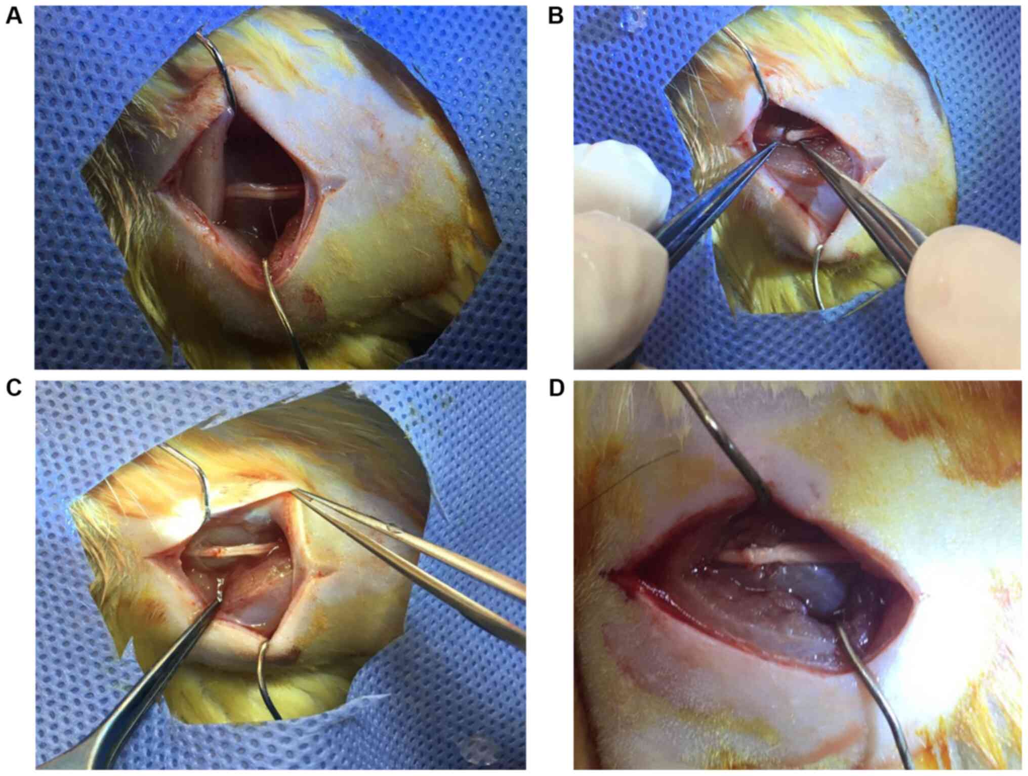
Surgical procedure
Following the induction of general anesthesia (75
mg/kg ketamine and 10 g/kg xylazine, via intraperitoneal
injection), the rats were fixed to the operating surface in the
prone position. Both sciatic nerves were exposed from 1 cm distal
of the sciatic notch to 1 cm distal to the trifurcation of the
nerve using an aseptic technique. Nerve segments above the
trifurcation, each 3-3.5 cm in length, were carefully dissected to
isolate the sciatic nerve from the surrounding soft tissue. The
nerves were then transected using micro scissors at 1.5 cm above
the trifurcation (starting point of the tibial nerve, common
peroneal nerve and caudal sural cutaneous nerve). The nerves were
repaired with 3 epineurial sutures (Ethilon 9-0; Ethicon Inc.) by
the same surgeon (Fig. 1). The
wound was closed with 3-0 Vicryl (Ethicon, Inc.) and the rats were
allowed to heal. Upon recovery from anesthesia, the rats were
returned to their cages and allowed to freely consume food and
water. Lipid peroxidation was determined in the tissue samples by
measuring malondialdehyde (MDA) levels as a thiobarbituric acid
reactive substance (TBARS), as previously described (16). Briefly, trichloroacetic acid and
TBARS (Sigma-Alrich; Merck KGaA) reagents were added to the tissue
samples, then mixed and incubated at 100˚C for 60 min. After
cooling on ice, the samples were centrifuged at 1,000 x g for 20
min at 4˚C and the absorbance of the supernatant was
read at 535 nm using a microplate reader immediately. MDA levels
were calculated from the standard calibration curve using
tetraethoxypropane and expressed as nmol/µg protein.
Electrophysiological recordings
The rats were anesthetized with a combination of
ketamine hydrochloride at a dose of 80 mg/kg (Alfamine; Alfasan
International B.V.) and xylazine hydrochloride at a dose of 10
mg/kg (Alfazyne, Alfasan International B.V.). Electrophysiological
recordings (EMG studies) were performed in all of the groups. EMG
results were obtained 3 times from the sciatic nerve stimulated
supramaximally (intensity, 10 V; duration, 0.05 msec; frequency, 1
Hz; range, 0.5-5000 Hz; sampling rates, 40 kHz/s) using a bipolar
subcutaneous needle stimulation electrode (BIOPAC Systems, Inc.;
model no. EL451; 25 mm x 30 g concentric bipolar electrode) placed
at the sciatic notch. The compound muscle action potential (CMAP)
was recorded from 2-3 interosseous muscles using unipolar platinum
electrodes. The data were evaluated using Biopac Student Lab Pro
version 3.6.7 software (BIOPAC Systems, Inc.) with distal latency
and amplitude of the CMAP as parameters. During the EMG recordings,
the rectal temperature of the rats was monitored with a rectal
probe (model no. HP Viridia 24C; Hewlett-Packard Company) and the
temperature of each rat was maintained at ~36-37˚C with a heating
pad. All of the tests were performed between 10:00 a.m. and 2:00
p.m.
Assessment of motor function
The motor performance of the rats was evaluated
using an inclined plane test as described by Rivlin and Tator
(17). Briefly, rats were placed in
an oblique position to the long axis of an inclined plane. The
initial angle of the inclined plane was 10˚. The incline angle was
slowly increased and the maximum angle of the plane at which the
rat maintained its position for 5 sec without falling was recorded
as the motor score. The angle was measured 3 times in each rat to
determine an average value.
Histology and quantitative
immunohistochemistry
Intracardiac 4% formaldehyde perfusion (at room
temperature for 4 min) was performed for histology and quantitative
immunohistochemistry analysis. Briefly, the sciatic nerves were
embedded in paraffin, sectioned at 5 µm thickness with a microtome
(model no. Leica RM 2145; Leica Microsystems GmbH) and stained with
hematoxylin and eosin. The thickness of the sciatic epineurium
nerve was measured, and the stained tissue sections were then
examined with an Olympus C-5050 digital camera mounted on an
Olympus BX51 microscope (Olympus Corporation). The Image-Pro
Express 4.5 program (Media Cybernetics, Inc.) was used to measure
the total number of axons, the thickness of the perineural layers
in the middle regions of the grafts, and the level of fibrosis
covering these layers in the histological specimens of each group.
These data were used for statistical analyses.
For the immunohistochemical examination, sections
were incubated with hydrogen peroxide (10%) for 30 min at room
temperature to eliminate endogenous peroxidase activity and then
blocked with 10% normal goat serum (Invitrogen; Thermo Fisher
Scientific, Inc.) for 1 h at room temperature. Subsequently,
sections were incubated with a primary antibody for nerve growth
factor (NGF; 1:100; Santa Cruz Biotechnology Inc.; cat. no.
sc-365944) for 24 h at 4˚C. Antibody detection was conducted using
the Histostain-Plus Bulk kit (Invitrogen; Thermo Fisher Scientific,
Inc.; cat. no. 85-8943) according to the manufacturer's protocol
for rabbit immunoglobulin G, and 3,3' diaminobenzidine was used to
visualize the final product. All sections were washed with PBS and
examined under an Olympus BX51 microscope and photographed with the
Olympus C-5050 digital camera. A total of 6 sections from each
study animal and 10 different fields from each of these sections
were used for quantitative immunohistochemistry analysis. Two
blinded observers counted the total number of immune-positive
Schwann cells and the number of axons under a light microscope at
x10 and x20 magnification. The data are presented as the mean ±
SD.
Statistical analysis
SPSS version 20.0 (IBM Corp.) was used to perform
statistical analysis. All experiments were performed in triplicate
and the data for each group are presented as the mean ± SD.
Statistical analysis was performed using one-way ANOVA with a Tukey
post hoc test to determine statistical significance. P<0.05 was
considered to indicate a statistically significant difference.
Results
Functional evaluation
Analysis of the inclined plane test revealed that
the results in the SDPL group (79.1±6.93˚) were more similar to
that of the control group (88.7±5.8˚) than those in the SSLE group
(39.6±5.5˚) but there is no statistically significance between
these groups. Statistical evaluation demonstrated a significant
difference between the control and SSLE groups (P<0.001) and the
SSLE and SDPL groups (P<0.001; Table
I).
 | Table IComparison of EMG CMAP latency, EMG
CMAP amplitude and inclined plane score between groups. |
Table I
Comparison of EMG CMAP latency, EMG
CMAP amplitude and inclined plane score between groups.
| Parameter | Normal control | Surgery + saline
group | Surgery +
dexpanthenol group |
|---|
| EMG CMAP latency
(ms) | 2,31±0,19 |
3,74±0,15a | 2,9±0,19c |
| EMG CMAP amplitude
(mV) | 12,65±1,1 |
2,36±0,42b |
7,02±0,97d |
| Inclined plane score
(˚) | 88,7±5,8 | 39,6±5,5b |
79,1±6,93d |
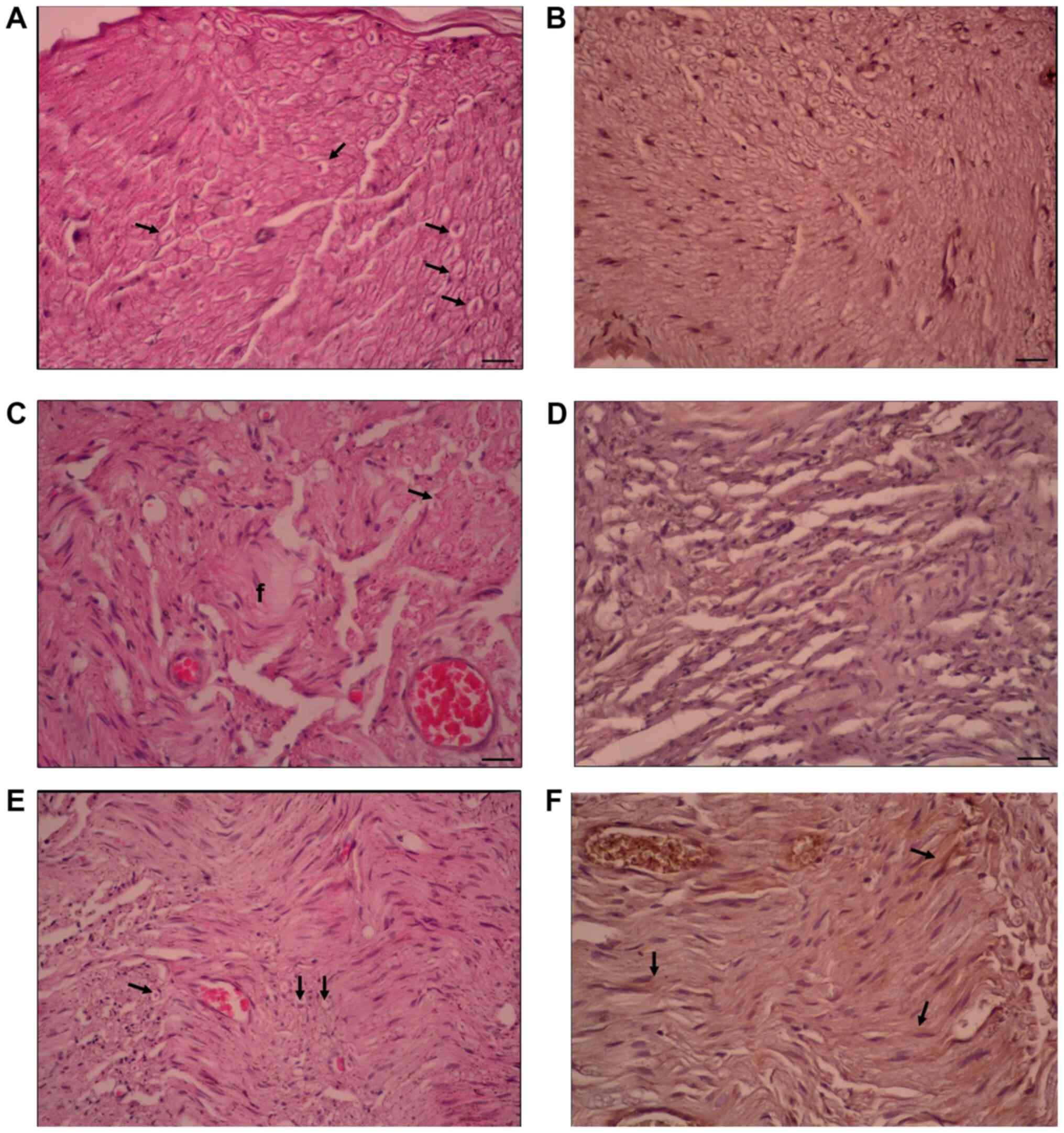
Results of histopathological
evaluation
Histological evaluation of sciatic nerve tissue
revealed that the fibrosis score was significantly lower in the
SDPL group when compared with the SSLE group (P<0.001).
Increased NGF expression and increased Schwann cell numbers were
seen in the SDPL group in comparison with the SSLE group
(P<0.001). The histopathological evaluation of the number of
axons in the sciatic nerve demonstrated that the results in the
SDPL group were significantly higher than those in the SSLE group
(P<0.001) (Table II; Fig. 2).
| Table IIComparison of NGF expression in
Schwann cells, total axon number and fibrosis score between
groups. |
Table II
Comparison of NGF expression in
Schwann cells, total axon number and fibrosis score between
groups.
| Parameter | Normal control | Surgery+saline
group | Surgery +
dexpanthenol group |
|---|
| NGF expression on
Schwann cells (%) | 41,8±9,6 | 4,6±1,1a | 12,5±4,2c |
| Total axon
number | 1450,2±85,6 |
315,8±45,9b |
514,2±30,06c |
| Fibrosis score
(%) | 0,9±0,08 |
68,5±6,2b |
25,4±5,65d |
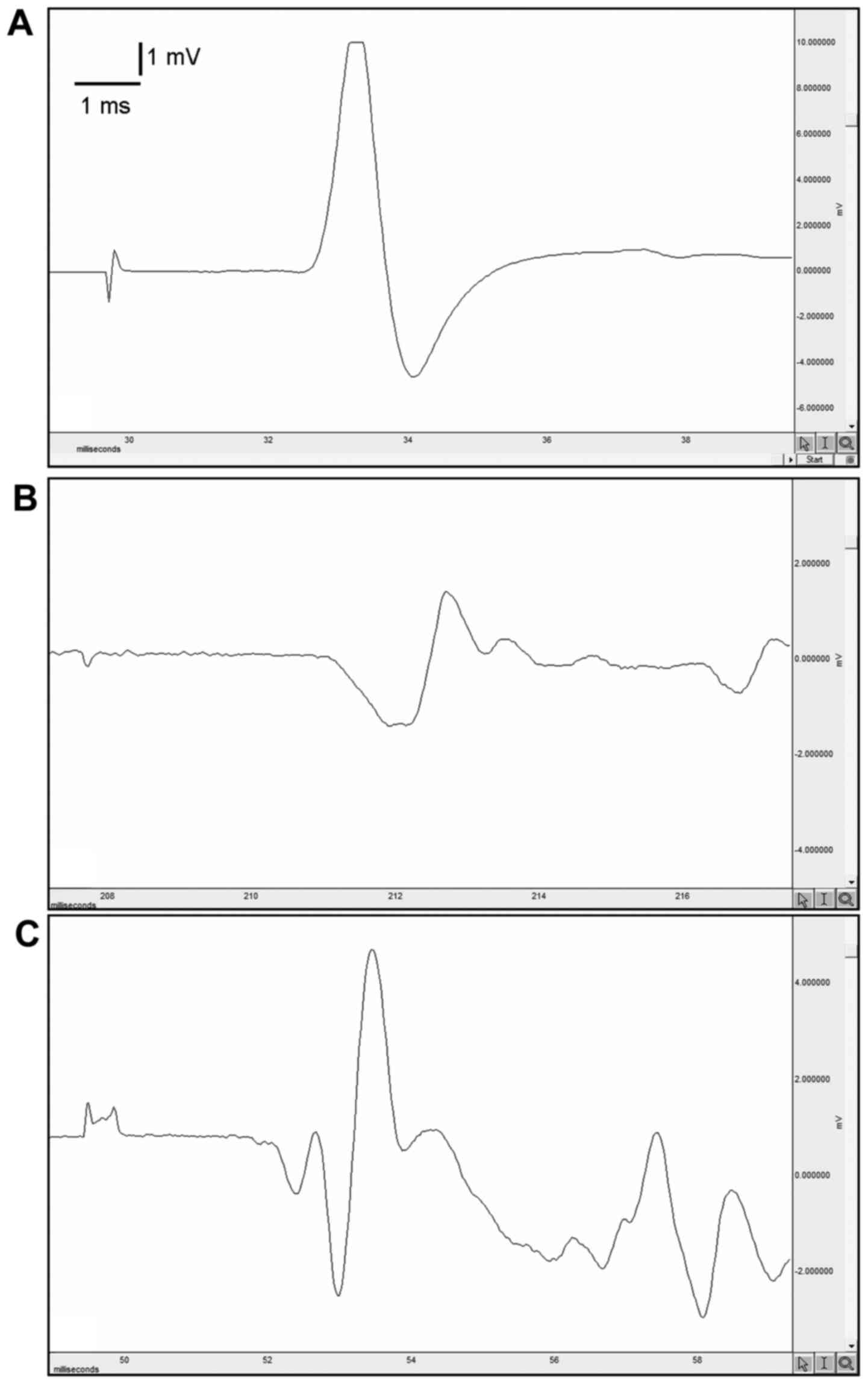
Results of electrophysiological
studies
EMG results indicated that the CMAP level was
significantly lower in the SSLE group when compared with the
control group (P<0.001). The CMAP level of the SDPL group was
significantly higher than that of the SSLE group (P<0.001). The
CMAP latency period was lower in the SDPL group in comparison with
the SSLE group (P<0.001; Table
II; Fig. 3).
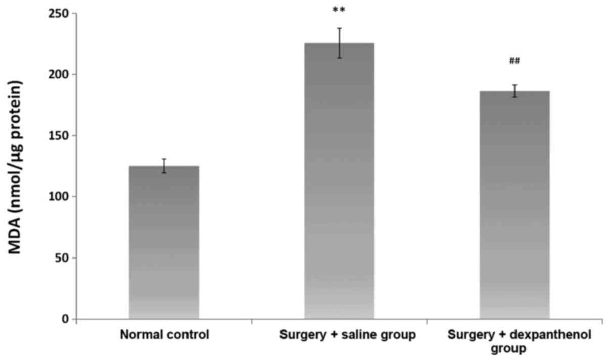
Evaluation of oxidative stress
The level of MDA, which was used as a marker of
oxidative stress, was higher in the SSLE group when compared with
the control group (P<0.0001). When the SDPL group was compared
with the SSLE group, MDA levels were significantly lower
(P<0.001; Fig. 4).
Discussion
Peripheral nerve injuries are a common clinical
problem and often result in long-term functional impairment.
Despite advances in microsurgical techniques, the functional
outcome of peripheral nerve trauma is rarely completely
satisfactory and has resulted in extensive experimental research to
develop methods to improve regeneration (13). The rat sciatic nerve model has been
widely used in these studies, and various regeneration assessment
methods have been described (18).
The results of the present study revealed that
dexpanthenol had a statistically significant effect on nerve
recovery compared with the control group post epineurial
neurosurgery in the repair of a laceration-type peripheral nerve
injury. Previous research has demonstrated that fibrosis occurring
during nerve healing adversely affected functional results
(19). A number of studies have
revealed that dexpanthenol decreases fibrosis (20,21).
Ermis et al (21)
investigated intraurethral fibrosis with histopathological
evaluation that used fibrosis and inflammation scar scoring and
demonstrated that dexpanthenol decreased fibrosis of the epithelial
tissue. In the present study, the fibrosis tissue at the field of
the nerve anastomosis was scored and evaluated. The influence of
the decrease in fibrous tissue on axonal advancement was assessed
histopathologically. The results indicated that the fibrosis score
was greater in the SSLE group than in the control and significantly
lower in the SDPL than the SSLE group. This influence was observed
histopathologically as an increase in the number of axons and
Schwann cells in the distal region of the anastomosis.
The most commonly referenced evaluation method in
peripheral nerve injury models is the electrophysiological method
(13,22). Ogden et al (15) examined the effect of dexpanthenol on
compression type injuries of the sciatic nerve and found no
significant influence when used alone in treatment; however,
electrophysiological tests were not performed. Laceration-type
peripheral nerve injuries are frequently encountered in the clinic,
and in the present study, the assessment of the influence of
dexpanthenol on this type of injury included electrophysiological
methods, which demonstrated significant healing when compared with
the control group.
One of the main factors contributing to neural
damage following injury is oxidative stress (23). Studies have shown that decreasing
oxidative stress after a peripheral nerve injury may stimulate the
repair process and improve functional recovery (24,25).
MDA is a product of lipid oxidation and it is a commonly used index
for oxidative stress (26). Tutun
et al (27) investigated the
effect of dexpanthenol on tissue damage and lipid oxidation in a
rat testis torsion model and found a significant decrease in the
serum MDA level (28). In the
present study, oxidative stress was evaluated using the level of
serum MDA and it was observed that the MDA level was significantly
lower in the SDPL group when compared with the SSLE group.
Functional results commonly used in experimental
animal nerve studies include histopathological evaluation,
electrophysiological studies and measurements of biomarkers, such
as lipid peroxidation (13,22,24).
Many procedures and evaluation scales have been developed for the
proper measurement of functional outcome. Some of the most common
assessments used are the Basso, Beattie and Bresnahan (BBB) scale
(29), the Tarlov open field test
(30) and the inclined plane test.
In the present study, the inclined plane test, as defined by Rivlin
and Tator, was used to evaluate nerve healing and its influence on
motor function (17). It was
observed that the inclined plane test results of the group that
were administered dexpanthenol were increased in comparison to
those of the SSLE group. In another experimental study
investigating the effect of dexpanthenol on the healing of
compression-type nerve injuries, the walking track analysis method
(sciatic static index) was used for functional evaluation. The
results revealed that dexpanthenol had a positive effect on
functional recovery (31). The
Tarlov and inclined plane tests help evaluate general locomotor
abilities, but they do not reflect specific changes in motor or
sensory functions (32). Although
the BBB scale is more advanced than the Tarlov test, it does not
provide a detailed motor function evaluation. The full measurement
of fine motor and sensory function in rats is very challenging, and
is a shortcoming of previous studies.
The lack of a positive control group was an
important limitation of the current study. This was due to only
using a small number of animals for ethical reasons (ethical
committee limited the number of animals used). Another limitation
of this study is that anti-oxidative and inflammatory factors were
not detected directly. Instead, measurement of MDA levels, an
oxidative stress marker, was used. Strengths of this study include
the fact that the injury model was prepared to simulate a type of
injury commonly encountered in the clinic. Peripheral nerve
injuries seen in the clinic resulting in functional loss are mostly
in the form of a laceration. To the best of our knowledge, in the
current literature, there are no studies evaluating the effect of
dexpanthenol on lacerations with a peripheral nerve injury model
repaired with neurorrhaphy. In this regard, the present study may
enhance clinical trials and influence the surgical treatment of
nerve injuries.
Dexpanthenol was observed to have a positive effect
on nerve tissue repaired with neurorrhaphy in this rat sciatic
model of laceration-type injuries of the sort frequently seen in
the clinic. The influence of the antioxidant and fibrosis-reducing
effect on axonal healing is promising for peripheral nerve
surgery.
Acknowledgements
Not applicable.
Funding
No funding was received.
Availability of data and materials
The datasets used and/or analyzed during the current
study are available from the corresponding author on reasonable
request.
Authors' contributions
GK and MAE conducted the experiments and wrote the
manuscript. RSE and HK collected the data and performed the
experiments. OE and GY designed the study. The final version of the
manuscript was read and approved by all authors, and each author
believes that the manuscript represents honest work.
Ethics approval and consent to
participate
The Committee for Animal Research of Demiroglu
Science University (approval no. 20172805-3) approved the
experimental procedures. All animal studies are strictly conformed
to the American Psychological Association Committee on Animal
Research and Ethics Guidelines for Ethical Conduct in the Care and
Use of Nonhuman Animals in Research.
Patient consent for publication
Not applicable.
Competing interests
The authors declare that they have no competing
interests.
References
|
1
|
Brandon WS, Sarada S, David AS, Jacob RJ,
Lynda JS and Thomas JW: An update on the management of adult
traumatic nerve injuries-replacing old paradigms: A review. J
Trauma Acute Care Surg. 86:299–306. 2019.PubMed/NCBI View Article : Google Scholar
|
|
2
|
Rasulić L, Simić V, Savić A, Lepić M,
Kovačević V, Puzović V, Vitošević F, Novaković N, Samardžić M and
Rotim K: Management of brachial plexus missile injuries. Acta Clin
Croat. 57:487–496. 2018.PubMed/NCBI View Article : Google Scholar
|
|
3
|
Kline DG, Kim D, Midha R, Harsh C and Tiel
R: Management and results of sciatic nerve injuries: A 24-year
experience. J Neurosurg. 89:13–23. 1998.PubMed/NCBI View Article : Google Scholar
|
|
4
|
Mark LW, Michael R, Jack GG and Pedro KB:
Peripheral nerve injury, scarring, and recovery. Connect Tissue
Res. 60:3–9. 2019.PubMed/NCBI View Article : Google Scholar
|
|
5
|
Cho HH, Jang S, Lee SC, Jeong HS, Park JS,
Han JY, Lee KH and Cho YB: Effect of neural-induced mesenchymal
stem cells and platelet-rich plasma on facial nerve regeneration in
an acute nerve injury model. Laryngoscope. 120:907–913.
2010.PubMed/NCBI View Article : Google Scholar
|
|
6
|
Yin ZS, Zhang H, Bo W and Gao W:
Erythropoietin promotes functional recovery and enhances nerve
regeneration after peripheral nerve injury in rats. AJNR Am J
Neuroradiol. 31:509–515. 2010.PubMed/NCBI View Article : Google Scholar
|
|
7
|
Mahanthappa NK, Anton ES and Matthew WD:
Glial growth factor 2, a soluble neuregulin, directly increases
Schwann cell motility and indirectly promotes neurite outgrowth. J
Neurosci. 16:4673–683. 1996.PubMed/NCBI View Article : Google Scholar
|
|
8
|
Ghoraba SM, Mahmoud WH, Elsergany MA and
Ayad HM: Ulnar nerve injuries (Sunderland Grade V): A simplified
classification system and treatment algorithm. Plast Reconstr Surg
Glob Open. 7(e2474)2019.PubMed/NCBI View Article : Google Scholar
|
|
9
|
Tulaci KG, Tuzuner A, Karadas Emir H,
Tatar İ, Sargon MF, Tulaci T, Karadavut Y and Samim EE: The effect
of tacrolimus on facial nerve injury: Histopathological findings in
a rabbit model. Am J Otolaryngol. 37:393–397. 2016.PubMed/NCBI View Article : Google Scholar
|
|
10
|
Lopatina T, Kalinina N, Karagyaur M,
Stambolsky D, Rubina K, Revischin A, Pavlova G, Parfyonova Y and
Tkachuk V: Correction: Adipose-derived stem cells stimulate
regeneration of peripheral nerves: BDNF secreted by these cells
promotes nerve healing and axon growth de novo. PLoS One.
14(e0219946)2019.PubMed/NCBI View Article : Google Scholar
|
|
11
|
Klein SM, Vykoukal J, Li DP, Pan HL,
Zeitler K, Alt E, Geis S, Felthaus O and Prantl L: Peripheral motor
and sensory nerve conduction following transplantation of
undifferentiated autologous adipose tissue-derived stem cells in a
biodegradable U.S. food and drug administration-approved nerve
conduit. Plast Reconstr Surg. 138:132–139. 2016.PubMed/NCBI View Article : Google Scholar
|
|
12
|
Lars K, Barbara H and Doychin NA: The pros
and cons of growth factors and cytokines in peripheral axon
regeneration. Int Rev Neurobiol. 108:137–171. 2013.PubMed/NCBI View Article : Google Scholar
|
|
13
|
Karahan G, Kaya H, Erdogan MA, Yigitturk
G, Gokyayla E and Erbas O: Effects of trimetazidine on nerve
regeneration in a rat sciatic nerve injury model. Bratisl Lek
Listy. 120:777–782. 2019.PubMed/NCBI View Article : Google Scholar
|
|
14
|
Lundborg G: A 25-year perspective of
peripheral nerve surgery: Evolving neuroscientific concepts and
clinical significance. J Hand Surg Am. 25:391–414. 2000.PubMed/NCBI View Article : Google Scholar
|
|
15
|
Ogden M, Karaca SB, Aydin G, Yuksel U,
Dagli AT, Akkaya S and Bakar B: The healing effects of thymoquinone
and dexpanthenol in sciatic nerve compression injury in rats. J
Invest Surg. 1–9. 2019.PubMed/NCBI View Article : Google Scholar
|
|
16
|
Demougeot C, Marie C and Beley A:
Importance of iron location in iron-induced hydroxyl radical
production by brain slices. Life Sci. 67:399–410. 2000.PubMed/NCBI View Article : Google Scholar
|
|
17
|
Rivlin AS and Tator CH: Objective clinical
assessment of motor function after experimental spinal cord injury
in the rat. J Neurosurg. 47:577–581. 1977.PubMed/NCBI View Article : Google Scholar
|
|
18
|
Geuna S: The sciatic nerve injury model in
pre-clinical research. J Neurosci Methods. 243:39–46.
2015.PubMed/NCBI View Article : Google Scholar
|
|
19
|
Sunderland S, McArthur RA and Nam DA:
Repair of a transected sciatic nerve. A study of nerve regeneration
and functional recovery: Report of a case. J Bone Joint Surg Am.
75:911–914. 1993.PubMed/NCBI View Article : Google Scholar
|
|
20
|
Yardimci I, Karakan T, Resorlu B, Doluoglu
OG, Ozcan S, Aydın A, Demirbas A, Unverdi H and Eroglu M: The
effect of intraurethral dexpanthenol on healing and fibrosis in
rats with experimentally induced urethral trauma. Urology.
85:274.e9–13. 2015.PubMed/NCBI View Article : Google Scholar
|
|
21
|
Ermis H, Parlakpinar H, Gulbas G, Vardi N,
Polat A, Cetin A, Kilic T and Aytemur ZA: Protective effect of
dexpanthenol on bleomycin-induced pulmonary fibrosis in rats.
Naunyn Schmiedebergs Arch Pharmacol. 386:1103–1110. 2013.PubMed/NCBI View Article : Google Scholar
|
|
22
|
Dellon AL and Mackinnon SE: Selection of
the appropriate parameter to measure neural regeneration. Ann Plast
Surg. 23:197–202. 1989.PubMed/NCBI View Article : Google Scholar
|
|
23
|
Siqueira Mietto B, Kroner A, Girolami EI,
Santos-Nogueira E, Zhang J and David S: Role of IL-10 in resolution
of inflammation and functional recovery after peripheral nerve
injury. J Neurosci. 35:16431–442. 2015.PubMed/NCBI View Article : Google Scholar
|
|
24
|
Renno WM, Benov L and Khan KM: Possible
role of antioxidative capacity of (-)-epigallocatechin-3-gallate
treatment in morphological and neurobehavioral recovery after
sciatic nerve crush injury. J Neurosurg Spine. 27:593–613.
2017.PubMed/NCBI View Article : Google Scholar
|
|
25
|
Zhang L, Johnson D and Johnson JA:
Deletion of Nrf2 impairs functional recovery, reduces clearance of
myelin debris and decreases axonal remyelination after peripheral
nerve injury. Neurobiol Dis. 54:329–338. 2013.PubMed/NCBI View Article : Google Scholar
|
|
26
|
Draper HH and Hadley M: Malondialdehyde
determination as index of lipid Peroxidation. Methods Enzymol.
186:421–431. 1990.PubMed/NCBI View Article : Google Scholar
|
|
27
|
Tutun B, Elbe H, Vardi N, Parlakpinar H,
Polat A, Gunaltili M, Guclu MM and Yasar EN: Dexpanthenol reduces
diabetic nephropathy and renal oxidative stress in rats. Biotech
Histochem. 94:84–91. 2019.PubMed/NCBI View Article : Google Scholar
|
|
28
|
Etensel B, Ozkisacik S, Ozkara E, Karul A,
Oztan O, Yazici M and Gürsoy H: Dexpanthenol attenuates lipid
peroxidation and testicular damage at experimental ischemia and
reperfusion injury. Pediatr Surg Int. 23:177–181. 2007.PubMed/NCBI View Article : Google Scholar
|
|
29
|
Basso DM, Beattie MS and Bresnahan JC: A
sensitive and reliable locomotor rating scale for open field
testing in rats. J Neurotrauma. 12:1–21. 1995.PubMed/NCBI View Article : Google Scholar
|
|
30
|
Tarlov IM and Klinger H: Spinal cord
compression studies: II Time limits for recovery after acute
compression in dogs. AMA Arch Neurol Psychiatry. 71:271–290.
1954.PubMed/NCBI
|
|
31
|
Korkmaz MF, Parlakpinar H, Erdem MN,
Ceylan MF, Ediz L, Samdanci E and Kekilli E: The therapeutic
efficacy of dexpanthenol on sciatic nerve injury in a rat model. Br
J Neurosurg. 34:397–401. 2020.PubMed/NCBI View Article : Google Scholar
|
|
32
|
Kunkel-Bagden E, Dai HN and Bregman BS:
Methods to assess the development and recovery of locomotor
function after spinal cord injury in rats. Exp Neurol. 119:153–164.
1993.PubMed/NCBI View Article : Google Scholar
|