Introduction
The prevalence of premature delivery is on the rise
worldwide, and in 2019 it was estimated that premature birth
accounted for 10.6% of all live births (1). With economic development and an
increase in medical knowledge, the survival rate of premature
infants is increasing, reaching >90%, and the survival rate of
infants with an extremely low birth weight (ELBW) with gestational
age <28 weeks increased from 0 to 35-70% in high-income
countries during the previous two decades (1-4).
However, premature infants that survive may develop detrimental
conditions, such as neurodevelopmental disabilities and
bronchopulmonary dysplasia (BPD) (5).
The pulmonary cellular structure, thoracic structure
and muscle strength mature in the last trimester (6), and metabolic substances that are
important for effective breathing, such as pulmonary surfactant and
antioxidant enzyme, are highly reserved in the last trimester
(7). Therefore, pulmonary disease
is a major cause of mortality in premature infants, especially in
extremely premature infants (8).
These infants require respiratory assistance to clear lung fluid,
aerate the lungs and establish a consistent functional residual
capacity to ensure gas exchange (9). Prior to the 1970s, neonatal
respiratory distress syndrome (NRDS) was the most common cause of
mortality in premature infants until the emergence of assisted
ventilation, which resulted in a significant increase in the
survival rate of patients with the disease (10). Respiratory assistance is also an
important treatment for other complications that are common in
premature infants, including BPD and respiratory failure (8,11,12).
Respiratory assistance includes mechanical
ventilation, nasal continuous positive airway pressure (CPAP) and
nasal intermittent positive pressure ventilation. A previous study
has revealed that extremely preterm births require positive
pressure ventilation if infants have spontaneous breathing and the
use of nasal or mask CPAP is recommended (13). CPAP failure is defined as hypoxia,
severe respiratory distress or apnea when FiO2 >0.6
and pCO2 >65 mmHg (14-16),
and often requires mechanical ventilation. Due to the
underdeveloped body function and organ development of premature
infants who exhibit hypoimmunity, long-term respiratory assistance
may also result in a number of complications. For example, an
Australian study revealed that 24% of 500 pediatric patients who
used respiratory assistance developed complications, including
croup, epiglottitis, lung atelectasis, infection and others
(17). Despite the continuous
advancement of medical care, ventilator-associated pneumonia,
infection and retinopathy of prematurity occur occasionally,
resulting in prolonged hospital stay of newborns, of which severe
cases can be life-threatening (1,18,19).
Therefore, investigating the risk factors of respiratory assistance
in premature infants may reduce the use of ventilators and the
incidence of complications via targeted pregnancy and prenatal
education and can strengthen medical skills. Furthermore, using the
correct personalized respiratory management in a timely manner (by
determining whether premature infants require respiratory
assistance as early as possible) will be of great significance in
improving survival rates of premature infants. However, to the best
of our knowledge, there are few relevant studies at present.
The current study retrospectively analyzed the
factors associated with the use of ventilators in the treatment of
premature infants to investigate risk factors for respiratory
assistance. The results of the current study may provide evidence
for the clinical early judgment of respiratory assistance
contributing to an increase in survival rate of premature infants
by effectively managing risk factors of respiratory assistance.
Materials and methods
Subjects
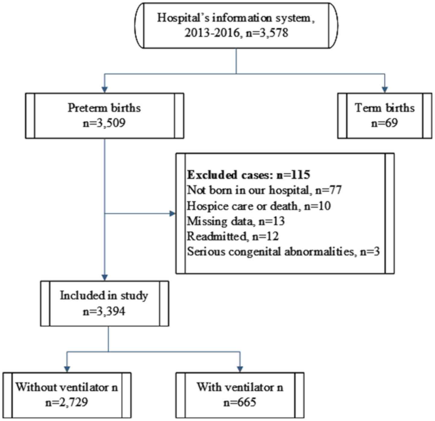
The subjects were selected from 3,578 premature
infants ranging from 24 to 36 week of gestational age (mean, 33.73;
SD, 2.29). They were born in Changzhou Maternal and Child Health
Care Hospital (Changzhou, China) between January 1, 2013 and
December 31, 2016 and were hospitalized in the neonatology
department. In total, 56.48% of participants were male, 43.46% were
female and 0.06% were unknown. The inclusion criteria were as
follows: i) Born in Changzhou Maternal and Child Health Care
hospital; ii) gestational week of delivery <37 weeks. The
exclusion criteria were as follows: i) Patients with hospice care;
ii) missing data on maternal pregnancy period or perinatal period;
iii) pediatrics who were readmitted for various reasons after
recovery from neonatology department; and iv) congenital
abnormalities including fatal fetal anomaly, such as Edwards
syndrome, anencephaly, Patau Syndrome, Renal Agenesis/Potter
Syndrome, triploidy, serious open spina bifida, severe
encephalocele, monocardian, severe fatal achondroplasia and severe
cleft lip and palate.
The included subjects were divided into two groups:
The respiratory assistance group, which had a history of ventilator
use during neonatal hospitalization, including invasive ventilator,
non-invasive ventilator and high-frequency ventilation; and the no
ventilator use group, of which patients had no history of
ventilator use during hospitalization.
The indications for non-invasive ventilation were:
i) Early prophylactic application in the delivery room for
premature infants with spontaneous breathing (gestational age 25-28
weeks); ii) preterm infants at high risk of respiratory distress
syndrome (RDS); iii) when the fraction of inspired oxygen
(FiO2) >0.3, arterial oxygen saturations
(PaO2) <50 mmHg (1 mmHg=0.133 kpa) or transcutaneous
oxygen saturations (TcSO2) <90%; iv) premature infant
apnea; v) the condition of children with RDS was stable following
pulmonary surfactant treatment, and the tracheal tube was removed;
and vi) after conventional mechanical ventilation or high-frequency
ventilation was removed, there were obvious tri-retraction signs
and/or respiratory distress (20-22).
The indications of conventional mechanical
ventilation (CMV) were as follows: i) Frequent apnea, with no
effect after medication or CPAP intervention; ii) children with RDS
who require PS treatment; iii) FiO2 >0.6-0.7,
PaO2 <50-60 mmHg or TcSO2 <85% (except
the cyanotic congenital heart disease); iv) PaCO2
>60-65 mmHg, accompanied by persistent acidosis (pH <7.20);
and v) newborns under general anesthesia (22).
High-frequency ventilation is often used for the
following situations (22-25):
i) Pulmonary air leak syndromes, including pneumothorax,
interstitial emphysema or bronchopleural fistula; ii) some
congenital diseases, such as diaphragmatic hernia, pulmonary
dysplasia and severe thoracic deformity; iii) persistent pulmonary
hypertension, especially for those requiring inhalation of NO; iv)
lung diseases with severe non-uniform changes, such as meconium
inhalation syndrome and severe pneumonia; and v) premature infant
RDS, and can be used as the first choice after CMV failure.
The current study protocol followed the ethical
guidelines of the Declaration of Helsinki (26) and was approved by the Ethics
Committee of Changzhou Women and Children Health Care Hospital.
Patient information collection
Using the hospital's information system, a
retrospective study was conducted to collect factors that may be
associated with the use of ventilators in 3,578 patients. The data
are anonymous, and the requirement for informed consent was
therefore waived. Data included the general information and
complication condition of mothers during the maternal pregnancy
period, including the mothers' hospital number, age, polyembryony
or not, gravidity and parity, the complications of pregnancy and
delivery (such as anemia, hypertension, diabetes, infection,
scarred uterus, cholestasis, hypoproteinemia, thrombocytopenia,
uterine fibroid and hypothyroidism), whether the fetal position,
placenta and umbilical cord were abnormal and whether the preterm
birth was in vitro fertilization and embryo transfer.
Furthermore, data regarding the condition of pediatric patients
during the perinatal period was collected, and included the
following: Whether there was premature rupture of membranes, the
mode of delivery, the gestational age (weeks), 1-min and 5-min
Apgar score (27), sex, birth
weight and whether a ventilator was used. After removing
unqualified cases, 3,394 subjects were included, of which 19.59%
(665/3,394) required a ventilator (Fig.
1).
Statistical analysis
A total of 3,394 patients were included in the
statistical analysis, 2,729 cases did not require ventilator use
and 665 cases used a ventilator. Continuous variables were tested
for normality using the Kolmogorov-Smirnov test. Data are presented
as the mean ± standard deviation or median (percentile 25 to
percentile 75). When appropriate, continuous variables, including
maternal age, birth weight and gestational age, were compared using
unpaired Student's t-test or Mann-Whitney U test. Pregnancy times,
1-min and 5-min Apgar scores were compared using the Wilcoxon
signed rank test. The remaining categorical variables were
summarized using frequency and percentage and compared using
χ2 test or Fisher's exact test. For all possible
determinants of treatment with a ventilator or not, univariate
logistic regression analysis was performed. All variables with a
P<0.1 (defined ‘a priori’) were considered relevant and
included into the multivariate logistic regression analysis.
Multivariate logistic regression analysis was used to define the
factors that were independently associated with using a ventilator.
Statistical analyses were performed with R version 3.4.1
(www.R-project.org). P<0.05 was considered to
indicate a statistically significant difference.
Results
Demographics and clinical
characteristics
The results identified that among the factors during
the maternal pregnancy period, there were significant differences
between the two groups in terms of maternal anemia, abnormal fetal
position, pregnancy with diabetes, placental abnormality, maternal
infection, abnormality of umbilical cord, cholestasis during the
maternal pregnancy period and neonates conceived by IVF (Table I). However, there were no
statistically significant differences between the two groups in
terms of maternal hypertension, scarred uterus, hypothyroidism and
thrombocytopenia (Table I). Among
the perinatal factors, the proportion of male infants who received
ventilator rescue was significantly higher compared with the number
of female infants. Furthermore, the patients who received
ventilator rescue exhibited a lower 1-min and 5-min Apgar score,
lower birth weight and smaller gestational age compared with those
who did not receive ventilator treatment. There were significant
differences between the groups in terms of whether the weight was
suitable for gestational age (SGA/AGA/LGA), the mole of delivery
(eutocia/caesarean section), pregnancy times, and premature rupture
of membranes.
 | Table IDemographics and clinical
characteristics in premature babies with or without ventilator
use. |
Table I
Demographics and clinical
characteristics in premature babies with or without ventilator
use.
| A, Maternal
pregnancy period |
|---|
| Factor | Without ventilator,
n=2,729 | With ventilator,
n=665 | P-value |
|---|
| Anemia | | | 0.001b |
|
Yes | 1,234 | 347 | |
|
No | 1,495 | 318 | |
| Hypertension | | | 0.118 |
|
Yes | 545 | 115 | |
|
No | 2,184 | 550 | |
| Abnormal fetal
position | | | 0.045a |
|
Yes | 441 | 129 | |
|
No | 2,288 | 536 | |
| Diabetes | | | 0.018a |
|
Yes | 368 | 67 | |
|
No | 2,361 | 598 | |
| Placental
abnormality | | |
<0.001b |
|
Yes | 544 | 234 | |
|
No | 2,185 | 431 | |
| Infection | | |
<0.001b |
|
Yes | 603 | 219 | |
|
No | 2,126 | 446 | |
| Abnormality of
umbilical cord | | | 0.004b |
|
Yes | 423 | 74 | |
|
No | 2,306 | 591 | |
| Scarred uterus | | | 0.243 |
|
Yes | 274 | 77 | |
|
No | 2,455 | 588 | |
| Cholestasis | | | 0.004b |
|
Yes | 305 | 49 | |
|
No | 2,424 | 616 | |
|
Hypoproteinemia | | | 0.776 |
|
Yes | 269 | 68 | |
|
No | 2,460 | 597 | |
| Conceived by
IVF | | | 0.013a |
|
Yes | 364 | 65 | |
|
No | 2,365 | 600 | |
|
Thrombocytopenia | | | 0.405 |
|
Yes | 73 | 14 | |
|
No | 2,656 | 651 | |
| Uterine
fibroid | | | 0.786 |
|
Yes | 53 | 14 | |
|
No | 2,676 | 651 | |
| Hypothyroidism | | | 0.573 |
|
Yes | 41 | 12 | |
|
No | 2,688 | 653 | |
| Maternal age
(years)c | 28.18±4.94 | 28.42±5.07 | 0.266 |
| B, Perinatal
period |
| Factor | Without ventilator,
n=2,729 | With ventilator,
n=665 | P-value |
| Age
(days)c | 1.12±0.86 | 1.10±0.77 | 0.571 |
| Sex,
female/male | | |
<0.001b |
|
Female | 1,231 | 250 | |
|
Male | 1,498 | 415 | |
| AGA or not | | | 0.007b |
|
SGA | 321 | 72 | |
|
AGA | 2,328 | 587 | |
|
LGA | 81 | 6 | |
| Mode of
delivery | | |
<0.001b |
|
Eutocia | 1,142 | 329 | |
|
Cesarean | 1,587 | 336 | |
| 1-min Apgar
scored | 8
(8-9)c | 7 (6-8) |
<0.001b |
| 5-min Apgar
scored | 8
(8-9)c | 8 (7-8) |
<0.001b |
| Monocyesis | | | 0.005b |
|
Yes | 1,949 | 511 | |
|
No | 780 | 154 | |
| Pregnancy
times | | | 0.010a |
|
1 | 1,208 | 262 | |
|
2 | 710 | 176 | |
|
3 | 403 | 108 | |
|
≥4 | 408 | 119 | |
| Parity | | | 0.767 |
|
Primiparous | 1,265 | 304 | |
|
Multiparous | 1,464 | 361 | |
| Birth weight
(g)c | 2339.83±473.06 | 1735.64±523.24 |
<0.001b |
| Gestational age
(weeks)c | 34.29±1.62 | 31.24±2.59 |
<0.001b |
| Premature rupture
of membrane | | | 0.001b |
|
Yes | 1124 | 229 | |
|
No | 1605 | 436 | |
Risk factors associated with preterm
infants who used ventilation
Univariate and multivariate logistic regression were
used to analyze the risk factors for respiratory assistance in
pediatric patients. The results demonstrated that placental
abnormality, being male, cesarean section, low 1-min Apgar score,
low birth weight and small gestational age were the independent
risk factors for using respiratory assistance in rescue of
premature infants (Table II).
| Table IIAnalysis of risk factors for using a
ventilator in preterm infants. |
Table II
Analysis of risk factors for using a
ventilator in preterm infants.
| A, Maternal
pregnancy period |
|---|
| | Univariate
regression analysis | Multivariate
regression analysis |
|---|
| Variables | OR | 95% CI | P-value | OR | 95% CI | P-value |
|---|
| Anemia, yes/no | 1.322 | 1.116-1.566 | 0.001b | 1.021 | 0.819-1.273 | 0.854 |
| Hypertension,
yes/no | 0.838 | 0.671-1.046 | 0.118 | | | |
| Abnormal fetal
position, yes/no | 1.249 | 1.005-1.552 | 0.045a | 0.778 | 0.581-1.042 | 0.092 |
| Diabetes,
yes/no | 0.719 | 0.546-0.947 | 0.019a | 0.864 | 0.613-1.217 | 0.402 |
| Placental
abnormality, yes/no | 2.181 | 1.813-2.623 |
<0.001b | 1.284 | 1.002-1.646 | 0.048a |
| Infection,
yes/no | 1.731 | 1.438-2.083 |
<0.001b | 1.151 | 0.901-1.471 | 0.259 |
| Abnormality of
umbilical cord, yes/no | 0.683 | 0.525-0.888 | 0.004b | 0.832 | 0.605-1.145 | 0.259 |
| Scarred uterus,
yes/no | 1.173 | 0.897-1.534 | 0.243 | | | |
| Cholestasis,
yes/no | 0.632 | 0.462-0.866 | 0.004b | 0.992 | 0.675-1.457 | 0.967 |
| Hypoproteinemia,
yes/no | 1.042 | 0.787-1.379 | 0.776 | | | |
| Conceived by IVF,
yes/no | 0.704 | 0.533-0.930 | 0.014a | 0.789 | 0.528-1.181 | 0.249 |
| Thrombocytopenia,
yes/no | 0.782 | 0.439-1.395 | 0.406 | | | |
| Uterine fibroid,
yes/no | 1.086 | 0.599-1.969 | 0.786 | | | |
| Hypothyroidism,
yes/no | 1.205 | 0.630-2.305 | 0.574 | | | |
| Maternal age
(years) | 1.010 | 0.993-1.027 | 0.258 | | | |
| B, Perinatal
period |
| | Univariate
regression analysis | Multivariate
regression analysis |
| Variables | OR | 95% CI | P-value | OR | 95% CI | P-value |
| Sex,
female/male | 0.733 | 0.616-0.872 |
<0.001b | 0.696 | 0.558-0.869 | 0.001b |
| Gestational age,
SGA/AGA | 0.892 | 0.680-1.170 | 0.41 | 0.887 | 0.579-1.356 | 0.579 |
| Gestational age,
LGA/AGA | 0.294 | 0.128-0.667 | 0.004b | 1.182 | 0.432-3.235 | 0.745 |
| Delivery method:
Cesarean section/eutocia | 0.735 | 0.620-0.871 |
<0.001b | 1.538 | 1.197-1.977 |
<0.001b |
| 1-min Apgar
score | 0.479 | 0.443-0.518 |
<0.001b | 0.727 | 0.638-0.828 |
<0.001b |
| 5-min Apgar
score | 0.300 | 0.261-0.343 |
<0.001b | 0.908 | 0.740-1.114 | 0.356 |
| Polyembryony,
yes/no | 0.753 | 0.618-0.918 | 0.005b | 0.763 | 0.571-1.018 | 0.066 |
| Pregnancy
times | 1.105 | 1.024-1.192 | 0.01a | 1.005 | 0.911-1.109 | 0.916 |
| Parity, ≥2/1 | 1.026 | 0.866-1.216 | 0.767 | | | |
| Birth weight
(g) | 0.997 | 0.997-0.998 |
<0.001b | 0.999 | 0.999-1.000 | 0.005b |
| Gestational age
(weeks) | 0.519 | 0.493-0.546 |
<0.001b | 0.616 | 0.560-0.678 |
<0.001b |
| Premature rupture
of membrane, yes/no | 0.750 | 0.628-0.895 | 0.001b | 0.966 | 0.769-1.214 | 0.769 |
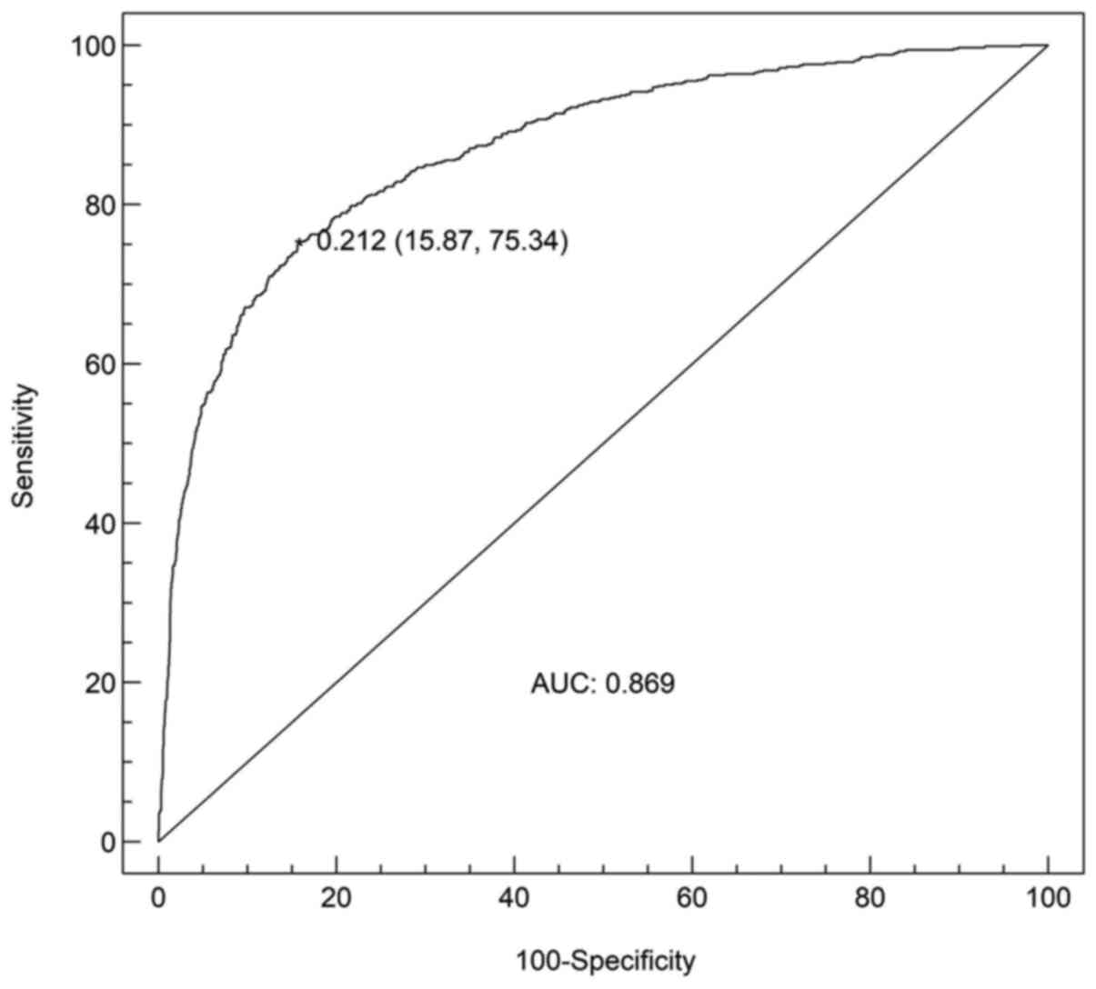
Analysis of receiver operating
characteristic (ROC) curve
The results from the multivariate regression
analyses, which included birth weight, gestational age, 1-min
Apgar, mode of delivery (cesarean section/vaginal delivery), sex
and placental abnormality, were used to construct the ROC curve
analysis that predicted the use of infant ventilator in preterm
infants. The area under the curve of the prediction model
constructed in the present study was 0.869 and the cut-off value
was 0.212, which indicated that the prediction ability and
discrimination of the model were generally effective (Fig. 2).
Discussion
Ventilator-assisted ventilation, which is an
important treatment tool used in the Neonatal Intensive Care Unit
(NICU), has developed rapidly and aided a number of premature
infants (13). However, if
respiratory assistance is delayed, the rescue outcomes for newborn
patients whose conditions are continuously changing and difficult
to predict may be unfavorable. The present study investigated the
risk factors for the use of ventilation in advance, and whether it
is possible to predict if preterm births will require ventilator
support or not, which will determine whether the relevant equipment
should be prepared and a pediatrician made available in order to
rescue infants quickly and improve their chances of survival. In
addition, the economic cost of treatment for premature infants is
high, and with the improvement of medicine, more premature infants
of decreased gestational ages survive, which increases this cost
(28-30).
Ventilator support involves a number of different pieces of
equipment, medical workers and expenses, which can be reduced if
the risk factors of ventilator support are managed effectively.
As an effective method to solve high-risk pregnancy,
dystocia and other obstetric critical illness, the use of a
cesarean section has been increasing each year, and in 2015, an
estimated 29.7 million (21.1%) births globally were by CS, which
represented almost a doubling in the proportion since 2000, when
16.0 million (12.1%) births were by CS (31). Although cesarean delivery can reduce
the incidence of neonatal asphyxia and meconium inhalation, the
risk of respiratory morbidity and the probability of admission into
the NICU are significantly higher (32,33).
Furthermore, previous studies have reported that cesarean section
is a risk factor for respiratory diseases, such as respiratory
distress syndrome (34-36),
transient shortness of breath (37)
and pulmonary hypertension (38),
but few of these studies examine the association between ventilator
use and cesarean section. In the present study, cesarean section
was demonstrated to be an independent risk factor for ventilator
use.
Fetuses need to overcome the viscosity resistance,
surface tension and tissue resistance of the lung fluid by
accelerating their breathing rate after delivery, but fetuses
delivered by cesarean section have significant restricted
ventilatory dysfunction within 12 h of birth due to the lower tidal
volume and minute ventilation volume compared with fetuses born via
vaginal delivery (39). It has been
previously suggested that the clearance of fetal lung fluid begins
before birth, and neonates born via cesarean section without
uterine contractions may have difficulty in removing fluid from
their lungs, which can result in obstruction of ventilation
(39). In addition, cesarean
delivery makes infants more susceptible to NRDS and its
accompanying increase in endothelin-1 levels, which may indirectly
result in the occurrence of Persistent Pulmonary Hypertension of
Newborn (38). Exposure of newborns
delivered via caesarean section with the combined effect of these
pathophysiological events may result in the occurrence of
respiratory distress, hypoxia, acidosis and transition-delay
(34). Therefore, the risk of
respiratory diseases in premature infants delivered by cesarean
section is higher, which suggests that the indications for elective
cesarean section should be strictly controlled when selecting this
method of delivery for premature infants in clinical practice, thus
reducing their risk of ventilator use.
A previous study which measured the association
between fetal sex and preterm birth in four original datasets found
that premature birth is more common in males, with ~55% of
premature births occurring in male infants (40). In addition, male infants exhibit a
higher mortality rate compared with female infants born in the same
gestation period (41). The results
of the present study suggested that the male sex is an independent
risk factor for ventilator use, which is consistent with previous
studies reporting that the incidence of certain respiratory-related
diseases in male newborns is significantly higher compared with
that of female infants. For example, the incidence of NRDS in
late-stage premature infants with different sexes is statistically
different (male infants have a higher incidence compared with
female infants) (42,43). Another study compared 130 premature
male infants with 106 premature female infants and revealed that
60.8% of male infants required machinery ventilation support
compared with only 46.2% of female infants, and the incidence of
Chronic Lung Disease in male infants was higher compared with that
of female infants (44). A previous
study reported that after birth, male infants express less
transforming growth factor (TGF)-β compared with female infants,
and low levels of TGF-β are important indicators for predicting
aerobic therapy, and at 36 weeks, male infants require a higher
oxygen supply compared with female infants, which may cause
premature male infants to lack oxygen, thus deepening and
accelerating the breathing and aggravating damage to the
respiratory system (45). Another
previous study indicated that the carbon dioxide partial pressure
of male infants is lower compared with female infants within 24 h
after birth (46), while the low
level of carbon dioxide partial pressure is associated with a low
level of pulmonary surfactant, which may account for the sex
difference in respiratory diseases. In addition, genetic factors
may be responsible for sex differences. For example, a previous
study revealed that allele 186 Asn and haploid 138 Asn-186 Asn are
independent risk factors for RDS (47), and the association between allele
186 Asn and RDS was observed among male infants (48).
In the present study, it was revealed that placental
abnormalities, such as placental abruption, placenta previa,
placental adhesions and abnormal invasion placenta (AIP), may also
increase the risk of ventilator use. Ahn et al (49) studied newborns born by 2,067 mothers
with placenta previa and demonstrated that newborns whose anterior
placenta previa in the second and third trimesters was correlated
with NRDS. Furthermore, a previous study reported that AIP
increased the risk of NRDS, and AIP cases required longer
respiratory assistance compared with the control group, which was
consistent with the results of the current study (50).
Apgar score includes heart rate, respiratory effort,
muscle tone, reflex irritability and color, which is the standard
evaluation method of checking the health condition of the infant
immediately after birth (27). This
score has been used as a predictive index for neonatal mortality
and morbidity, and for later neurologic or developmental disability
(51). Numerous factors can
influence Apgar score, such as maternal education and BMI,
gestational age, pathological obstetrics, longer duration of the
second stage of labor, neonatal weight and meconium-stained
amniotic fluid (52). The present
results suggested that 1-min Apgar score was a risk factor for
respiratory assistance in premature infants, indicating that weight
control in pregnant women, reduction of the preterm birth and other
measures may reduce the use of ventilators.
As aforementioned, lung-related structures and
metabolites mature in the last trimester, and thus premature
infants often require respiratory assistance (6,7,53). In
the current study, the ventilator utilization rates of gestational
age <28 weeks, 28-31 weeks and 32-36 weeks were 89.58% (43/48),
65.38% (321/491) and 10.54% (301/2,855), respectively (data not
shown); therefore, it was suggested that the younger the
gestational age, the more ventilator support is required. Birth
weight is also a risk factor for numerous respiratory diseases
(54,55). To avoid these diseases, the
pregnancy period should be extended as soon as possible, as well as
ensuring good nutrition, and timely and regular antenatal visits
during pregnancy. However, multiple sociodemographic, nutritional,
biological and environmental factors can increase the risk of
preterm birth and low birth weight (56,57).
Therefore, methods for appropriate interventions to reduce the
incidence of premature infants and low birth weight remain
challenging.
With the increasing incidence of premature infants,
how to reduce the use of ventilators, and use the ventilators
correctly and timely is of great concern (1). However, there is limited research on
the risk factors of ventilator use in premature infants worldwide,
and there are few predictive models constructed for its use. The
high-risk factors associated with respiratory assistance observed
in the present study, including placental abnormality, being male,
cesarean section, low 1-min Apgar score, low birth weight and small
gestational age, may reduce the use of respiratory assistance by
developing strategies to decrease these factors, and to clinically
guide medical staff to identify pediatric patients with high risk
factors, which will allow advanced preparation of the ventilator
and ensure the availability of a pediatrician. Collectively, these
actions could help to minimize the time of hypoxia within newborns,
which will increase their survival.
There are some limitations of the present study that
require discussion. Firstly, the current study used data from a
single-center that has not been validated in other hospitals.
Secondly, there was no in-depth discussion of the individual risk
factors, such as a stratified analysis based on gestational age,
which would provide more accurate risk factor analysis for the
respiratory assistance of premature infants in different
gestational age groups. This should be performed in future studies
in order to provide a basis for preparing for the respiratory
assistance of premature infants.
In conclusion, the present results suggested that
cesarean section, male neonates, placental abnormality, low 1-min
Apgar score, low birth weight and lower gestational age represented
independent risk factors for the use of ventilators in premature
infants. Thus, increased effort to reduce the use of ventilators
and provide early detection of those requiring assistance should be
performed for premature infants who require respiratory assistance
to improve their survival rate and quality of life. Firstly, the
indications for elective cesarean section should be strictly
controlled when selecting cesarean section for premature infants in
clinical practice. Secondly, ensuring nutrition and the timely and
regular antenatal visits during pregnancy are important, as well as
the pregnancy period should be extended as soon as possible
Finally, for premature infants who have risk factors for using the
ventilator, ventilator support such as the relevant equipment and
personnel should be prepared in time to improve their chances of
survival.
Acknowledgements
Not applicable.
Funding
No funding was received.
Availability of data and materials
The datasets used and/or analyzed during the current
study are available from the corresponding author on reasonable
request.
Authors' contributions
HXL, ZLM and HYW were study investigators and
participated in study design, patient recruitment, acquisition of
data, and/or analysis and interpretation of the findings. HXL, ZLM,
HYW, CJG and SC contributed to data analysis, drafting and revising
the article, gave final approval of the version to be published,
and agree to be accountable for all aspects of the work. All
authors read and approved the final version of the manuscript.
Ethics approval and consent to
participate
The present study protocol followed the ethical
guidelines of the Helsinki Declaration revised in 2008 and was
approved by the Ethics Committee of Changzhou Women and Children
Health Care Hospital (Changzhou, China).
Patient consent for publication
Not applicable.
Competing interests
The authors declare that they have no competing
interests.
References
|
1
|
Chawanpaiboon S, Vogel JP, Moller AB,
Lumbiganon P, Petzold M, Hogan D, Landoulsi S, Jampathong N,
Kongwattanakul K, Laopaiboon M, et al: Global, regional, and
national estimates of levels of preterm birth in 2014: A systematic
review and modelling analysis. Lancet Glob Health. 7:e37–e46.
2019.PubMed/NCBI View Article : Google Scholar
|
|
2
|
Ancel PY, Goffinet F, Kuhn P, Langer B,
Matis J, Hernandorena X, Chabanier P, Joly-Pedespan L, Lecomte B,
Vendittelli F, et al: EPIPAGE-2 Writing Group: Survival and
morbidity of preterm children born at 22 through 34 weeks'
gestation in France in 2011: Results of the EPIPAGE-2 cohort study.
JAMA Pediatr. 169:230–238. 2015.PubMed/NCBI View Article : Google Scholar
|
|
3
|
Kiechl-Kohlendorfer U, Simma B,
Urlesberger B, Maurer-Fellbaum U, Wald M, Wald M, Weissensteiner M,
Ehringer-Schetitska D, Berger A, Kurz H, et al: Austrian Preterm
Outcome Study Group: Low mortality and short-term morbidity in very
preterm infants in Austria 2011-2016. Acta Paediatr. 108:1419–1426.
2019.PubMed/NCBI View Article : Google Scholar
|
|
4
|
Fellman V, Hellström-Westas L, Norman M,
Westgren M, Källén K, Lagercrantz H, Marsál K, Serenius F and
Wennergren M: EXPRESS Group. One-year survival of extremely preterm
infants after active perinatal care in Sweden. JAMA. 301:2225–2233.
2009.PubMed/NCBI View Article : Google Scholar
|
|
5
|
Platt MJ: Outcomes in preterm infants.
Public Health. 128:399–403. 2014.PubMed/NCBI View Article : Google Scholar
|
|
6
|
Alphonse RS, Rajabali S and Thébaud B:
Lung injury in preterm neonates: The role and therapeutic potential
of stem cells. Antioxid Redox Signal. 17:1013–1040. 2012.PubMed/NCBI View Article : Google Scholar
|
|
7
|
Vento M, Aguar M, Escobar J, Arduini A,
Escrig R, Brugada M, Izquierdo I, Asensi MA, Sastre J, Saenz P, et
al: Antenatal steroids and antioxidant enzyme activity in preterm
infants: Influence of gender and timing. Antioxid Redox Signal.
11:2945–2955. 2009.PubMed/NCBI View Article : Google Scholar
|
|
8
|
Cools F, Offringa M and Askie LM: Elective
high frequency oscillatory ventilation versus conventional
ventilation for acute pulmonary dysfunction in preterm infants
Cochrane Database Syst Rev: Mar 19, 2015 (Epub ahead of print).
doi: 10.1002/14651858.CD000104.pub4.
|
|
9
|
Hillman NH, Kallapur SG and Jobe AH:
Physiology of transition from intrauterine to extrauterine life.
Clin Perinatol. 39:769–783. 2012.PubMed/NCBI View Article : Google Scholar
|
|
10
|
Owen LS, Manley BJ, Davis PG and Doyle LW:
The evolution of modern respiratory care for preterm infants.
Lancet. 389:1649–1659. 2017.PubMed/NCBI View Article : Google Scholar
|
|
11
|
Spotswood N, Orsini F, Dargaville P,
Marshall P, Schmidt P, Craven P, de Waal K, Simmer K, Gill A,
Pillow J, et al: Australian and New Zealand Neonatal Network:
Association of center-specific patient volumes and early
respiratory management practices with death and bronchopulmonary
dysplasia in preterm infants. J Pediatr. 210:63–68.e2.
2019.PubMed/NCBI View Article : Google Scholar
|
|
12
|
Yang X, Xu PF, Shan L, Lang LG, Du L and
Jia FY: Advances in respiratory assessment and treatment in
children undergoing invasive mechanical ventilation. Zhongguo Dang
Dai Er Ke Za Zhi. 21:94–99. 2019.PubMed/NCBI View Article : Google Scholar : (In Chinese).
|
|
13
|
Vento M and Lista G: Managing preterm
infants in the first minutes of life. Paediatr Respir Rev.
16:151–156. 2015.PubMed/NCBI View Article : Google Scholar
|
|
14
|
Dunn MS, Kaempf J, de Klerk A, de Klerk R,
Reilly M, Howard D, Ferrelli K, O'Conor J and Soll RF: Vermont
Oxford Network DRM Study Group. Randomized trial comparing 3
approaches to the initial respiratory management of preterm
neonates. Pediatrics. 128:e1069–e1076. 2011.PubMed/NCBI View Article : Google Scholar
|
|
15
|
Tooley J and Dyke M: Randomized study of
nasal continuous positive airway pressure in the preterm infant
with respiratory distress syndrome. Acta Paediatr. 92:1170–1174.
2003.PubMed/NCBI View Article : Google Scholar
|
|
16
|
Pfister RH and Soll RF: Initial
respiratory support of preterm infants: The role of CPAP, the
INSURE method, and noninvasive ventilation. Clin Perinatol.
39:459–481. 2012.PubMed/NCBI View Article : Google Scholar
|
|
17
|
Rivera R and Tibballs J: Complications of
endotracheal intubation and mechanical ventilation in infants and
children. Crit Care Med. 20:193–199. 1992.PubMed/NCBI View Article : Google Scholar
|
|
18
|
Iosifidis E, Pitsava G and Roilides E:
Ventilator-associated pneumonia in neonates and children: A
systematic analysis of diagnostic methods and prevention. Future
Microbiol. 13:1431–1446. 2018.PubMed/NCBI View Article : Google Scholar
|
|
19
|
Mao JB, Yu XT, Shen LJ, Wu MY, Lyu Z, Lao
JM, Li HX, Wu HF and Chen YQ: Risk factors of retinopathy of
prematurity in extremely low birth weight infants by strictly
controlling oxygen inhalation after birth. Zhonghua Yan Ke Za Zhi.
55:280–288. 2019.PubMed/NCBI View Article : Google Scholar : (In Chinese).
|
|
20
|
Sweet DG, Carnielli V, Greisen G, Hallman
M, Ozek E, Plavka R, Saugstad OD, Simeoni U, Speer CP, Vento M, et
al: European Association of Perinatal Medicine: European consensus
guidelines on the management of neonatal respiratory distress
syndrome in preterm infants - 2013 update. Neonatology.
103:353–368. 2013.PubMed/NCBI View Article : Google Scholar
|
|
21
|
Committee on Fetus and NewbornAmerican
Academy of Pediatrics. Respiratory support in preterm infants at
birth. Pediatrics. 133:171–174. 2014.PubMed/NCBI View Article : Google Scholar
|
|
22
|
Cloherty JP, Eichenwald EC, Hansen AR and
Stark AR (eds): Manual of Neonatal Care. 7th edition. Lippincott
Williams and Wilkins, London, 2012.
|
|
23
|
Rimensberger PC (ed): Pediatric and
Neonatal Mechanical Ventilation. Springer, New York, NY, 2015.
|
|
24
|
Donn SM and Sinha SK (eds): Manual of
Neonatal Respiratory Care. 3rd edition. Springer, New York, NY,
2012.
|
|
25
|
Goldsmith JP, Karotkin EH and Siede BL:
Assisted Ventilation of the Neonate. 5th edition. Elsevier, Louis,
2011.
|
|
26
|
World Medical Association. World Medical
Association Declaration of Helsinki: Ethical principles for medical
research involving human subjects. JAMA. 310:2191–2194.
2013.PubMed/NCBI View Article : Google Scholar
|
|
27
|
Apgar V: A proposal for a new method of
evaluation of the newborn infant. Curr Res Anest Anal. 32:260–267.
1953.PubMed/NCBI
|
|
28
|
Rocha G, Soares P, Gonçalves A, Silva AI,
Almeida D, Figueiredo S, Pissarra S, Costa S, Soares H,
Flôr-de-Lima F, et al: Respiratory care for the ventilated neonate.
Can Respir J: Aug 13, 2018 (Epub ahead of print). doi:
10.1155/2018/7472964.
|
|
29
|
Lancet T: The Lancet. The global burden of
preterm birth. Lancet. 374:1214. 2009.PubMed/NCBI View Article : Google Scholar
|
|
30
|
Donda K, Vijayakanthi N, Dapaah-Siakwan F,
Bhatt P, Rastogi D and Rastogi S: Trends in epidemiology and
outcomes of respiratory distress syndrome in the United States.
Pediatr Pulmonol. 54:405–414. 2019.PubMed/NCBI View Article : Google Scholar
|
|
31
|
Boerma T, Ronsmans C, Melesse DY, Barros
AJ, Barros FC, Juan L, Moller AB, Say L, Hosseinpoor AR, Yi M, et
al: Global epidemiology of use of and disparities in caesarean
sections. Lancet. 392:1341–1348. 2018.PubMed/NCBI View Article : Google Scholar
|
|
32
|
Ahimbisibwe A, Coughlin K and Eastabrook
G: Respiratory morbidity in late preterm and term babies born by
elective Caesarean sectio. J Obstet Gynaecol Can. 41:1144–1149.
2019.PubMed/NCBI View Article : Google Scholar : (Epub ahead of
print). doi: org/10.1016/j.jogc.2018.11.002.
|
|
33
|
Kamath BD, Todd JK, Glazner JE, Lezotte D
and Lynch AM: Neonatal outcomes after elective cesarean delivery.
Obstet Gynecol. 113:1231–1238. 2009.PubMed/NCBI View Article : Google Scholar
|
|
34
|
Altman M, Vanpée M, Cnattingius S and
Norman M: Risk factors for acute respiratory morbidity in
moderately preterm infants. Paediatr Perinat Epidemiol. 27:172–181.
2013.PubMed/NCBI View Article : Google Scholar
|
|
35
|
Gerten KA, Coonrod DV, Bay RC and
Chambliss LR: Cesarean delivery and respiratory distress syndrome:
Does labor make a difference? Am J Obstet Gynecol. 193:1061–1064.
2005.PubMed/NCBI View Article : Google Scholar
|
|
36
|
Berthelot-Ricou A, Lacroze V, Courbiere B,
Guidicelli B, Gamerre M and Simeoni U: Respiratory distress
syndrome after elective caesarean section in near term infants: A
5-year cohort study. J Matern Fetal Neonatal Med. 26:176–182.
2013.PubMed/NCBI View Article : Google Scholar
|
|
37
|
Rijal P and Shrestha M: Scenario of
neonatal respiratory distress in Tertiary Hospital. J Nepal Health
Res Counc. 16:131–135. 2018.PubMed/NCBI
|
|
38
|
Babooa N, Shi WJ and Chen C: Factors
relating caesarean section to persistent pulmonary hypertension of
the newborn. World J Pediatr. 13:517–527. 2017.PubMed/NCBI View Article : Google Scholar
|
|
39
|
Dileep A, Khan NB and Sheikh SS: Comparing
neonatal respiratory morbidity in neonates delivered at term by
elective Caesarean section with and without dexamethasone:
Retrospective cohort study. J Pak Med Assoc. 65:607–611.
2015.PubMed/NCBI
|
|
40
|
Zeitlin J, Saurel-Cubizolles MJ, De Mouzon
J, Rivera L, Ancel PY, Blondel B and Kaminski M: Fetal sex and
preterm birth: are males at greater risk? Hum Reprod. 17:2762–2768.
2002.PubMed/NCBI View Article : Google Scholar
|
|
41
|
Kent AL, Wright IM and Abdel-Latif ME: New
South Wales and Australian Capital Territory Neonatal Intensive
Care Units Audit Group. Mortality and adverse neurologic outcomes
are greater in preterm male infants. Pediatrics. 129:124–131.
2012.PubMed/NCBI View Article : Google Scholar
|
|
42
|
Anadkat JS, Kuzniewicz MW, Chaudhari BP,
Cole FS and Hamvas A: Increased risk for respiratory distress among
white, male, late preterm and term infants. J Perinatol.
32:780–785. 2012.PubMed/NCBI View Article : Google Scholar
|
|
43
|
Ye W, Zhang T, Shu Y, Fang C, Xie L, Peng
K and Liu C: The influence factors of neonatal respiratory distress
syndrome in Southern China: A case-control study. J Matern Fetal
Neonatal Med. 33:1678–1682. 2020.PubMed/NCBI View Article : Google Scholar
|
|
44
|
Elsmén E, Hansen Pupp I and
Hellström-Westas L: Preterm male infants need more initial
respiratory and circulatory support than female infants. Acta
Paediatr. 93:529–533. 2004.PubMed/NCBI View Article : Google Scholar
|
|
45
|
Lecart C, Cayabyab R, Buckley S, Morrison
J, Kwong KY, Warburton D, Ramanathan R, Jones CA and Minoo P:
Bioactive transforming growth factor-beta in the lungs of extremely
low birthweight neonates predicts the need for home oxygen
supplementation. Biol Neonate. 77:217–223. 2000.PubMed/NCBI View Article : Google Scholar
|
|
46
|
Dammann O, Allred EN, Kuban KC, van Marter
LJ, Stewart JE, Pagano M and Leviton A: Development Epidemiology
Network Investigators. Hypocarbia during the first 24 postnatal
hours and white matter echolucencies in newborns < or = 28 weeks
gestation. Pediatr Res. 49:388–393. 2001.PubMed/NCBI View Article : Google Scholar
|
|
47
|
Fatahi N, Dalili H, Kalani M, Niknafs N,
Shariat M, Tavakkoly-Bazzaz J, Amini E, Esmaeilnia Shirvani T,
Hardani AK, Taheritafti R, et al: Association of SP-C gene codon
186 polymorphism (rs1124) and risk of RDS. J Matern Fetal Neonatal
Med. 30:2585–2589. 2017.PubMed/NCBI View Article : Google Scholar
|
|
48
|
Lahti M, Marttila R and Hallman M:
Surfactant protein C gene variation in the Finnish population -
association with perinatal respiratory disease. Eur J Hum Genet.
12:312–320. 2004.PubMed/NCBI View Article : Google Scholar
|
|
49
|
Ahn KH, Lee EH, Cho GJ, Hong SC, Oh MJ and
Kim HJ: Anterior placenta previa in the mid-trimester of pregnancy
as a risk factor for neonatal respiratory distress syndrome. PLoS
One. 13(e0207061)2018.PubMed/NCBI View Article : Google Scholar
|
|
50
|
Spillane NT, Zamudio S, Alvarez-Perez J,
Andrews T, Nyirenda T, Alvarez M and Al-Khan A: Increased incidence
of respiratory distress syndrome in neonates of mothers with
abnormally invasive placentation. PLoS One.
13(e0201266)2018.PubMed/NCBI View Article : Google Scholar
|
|
51
|
Hegyi T, Carbone T, Anwar M, Ostfeld B,
Hiatt M, Koons A, Pinto-Martin J and Paneth N: The apgar score and
its components in the preterm infant. Pediatrics. 101:77–81.
1998.PubMed/NCBI View Article : Google Scholar
|
|
52
|
Yang C, Chen X, Zu S and He F:
Retrospective analysis of risk factors for low 1-minute Apgar
scores in term neonates. Braz J Med Biol Res.
52(e9093)2019.PubMed/NCBI View Article : Google Scholar
|
|
53
|
Dargaville PA and Tingay DG: Lung
protective ventilation in extremely preterm infants. J Paediatr
Child Health. 48:740–746. 2012.PubMed/NCBI View Article : Google Scholar
|
|
54
|
Jiangsu Multicenter Study Collaborative
Group for Breastmilk Feeding in Neonatal Intensive Care Units.
[Clinical characteristics and risk factors of very low birth weight
and extremely low birth weight infants with bronchopulmonary
dysplasia: Multicenter retrospective analysis]. Zhonghua Er Ke Za
Zhi. 57:33–39. 2019.PubMed/NCBI View Article : Google Scholar
|
|
55
|
Undela K, Mohammed BTS and Gurumurthy P:
Impact of preterm birth and low birth weight on medical conditions,
medication use and mortality among neonates: a prospective
observational cohort studyWorld journal of pediatrics :
WJP,2019,():.https://doi.org/10.1007/s12519-019-00239-1.
|
|
56
|
Goldenberg RL, Culhane JF, Iams JD and
Romero R: Epidemiology and causes of preterm birth. Lancet.
371:75–84. 2008.PubMed/NCBI View Article : Google Scholar
|
|
57
|
Negandhi PH, Negandhi HN, Zodpey SP,
Ughade SN and Biranjan JR: Risk factors for low birth weight in an
Indian urban setting: A nested case control study. Asia Pac J
Public Health. 26:461–469. 2014.PubMed/NCBI View Article : Google Scholar
|