Introduction
The external auditory canal (EAC) and the middle ear
ossicular chain (OC) couple the air-transmitted sound wave from an
external source to the aqueous fluids of the inner ear. The
coupling mechanism is both subtle and ingenious; the EAC collects
the pressure wave which moves the tympanic membrane (TM) into a
particular shape leading to motion of the ossicles (1). The stapes footplate moves within the
oval window stimulating the perilymph fluid in the cochlea, a
spirally coiled organ of the inner ear (2). Research into sound transmission began
as early as 1941 with the works of von Békésy (3) and have evolved during decades to
create a clearer image of acoustic transmission.
A mechanical modelling of the ear was also first
reported by von Békésy and indicated that the TM moves as a stiff
plate, and that the mallear and incudal ligaments act as a rotation
axis for the ossicular chain at low frequencies. The OC rotates
about the center of mass at high frequencies (3). Various methods were devised for the
study of the mechanics of the middle ear: Scaled replicas of the
outer ear canal (4), electrical
analogues of the middle ear (5,6),
finite-element modelling (FEM) (7,8).
The etiology of hearing loss originates from genetic
factors and includes several other causes including infections,
working or living environment, as well as several endocrine and
metabolic disorders; all of them associated with various degrees of
the loss of hearing. When we approach the neuroendocrine system, we
discover that thyroid-parathyroid, hypothalamic-pituitary, adrenal
and diabetes disorders (9) play an
important role in this pathology together with metabolic bone
diseases such as osteogenesis imperfect (10) or Paget's disease (11).
Hearing loss of variable etiology represents one of
the most serious public health issues confronting the world's
population. According to data reported in 2020 by the World Health
Organization (WHO), over 466 million people (5% of the world's
population) currently suffer from a form of hearing loss (12,13).
In addition, the integrity of the auditory system is one of the
prerequisites for the acquisition and the proper development of
oral language (14).
Implantable hearing-aids, initially developed
against sensorineural hearing loss are recently becoming more
important with the extension of indication towards mixed hearing
loss. This might also revive the application for pure sensorineural
hearing loss.
Advantages of implantable hearing aids against
conventional ones are as follows: No ear canal closure, higher
possible gain at the high frequency range and no visible parts (in
case of totally implantable systems) (15).
The Vibrant® SoundbridgeTM
(VSB) is an implantable middle ear hearing device to treat
sensorineural hearing loss which involves damage to the inner ear
(aging, prenatal and birth-related issues, viral and bacterial
infection, heredity, trauma, exposure to loud noise, fluid backup,
benign tumor of the inner ear) and represents 60% of all hearing
loss. In the case of the VSB, defining the optimal position for
transducer attachment during surgery could mean optimization of
functional results. The VSB directly drives the ossicular chain,
bypassing the ear canal and tympanic membrane.
Materials and methods
Investigations were performed with the help of a FEM
of the middle ear which consists of the ear canal (acoustic fluid
with matched impedance at the canal entrance to the surrounding
air), the eardrum (orthotrop-elastic shell with constant damping
ratio), the ossicles (rigid bodies with mass and inertia
properties), ligaments (elastic bars), joints (elastic bodies with
constant damping ratio) and a spring-mass-damper model of the
cochlea (15) (Figs.
1-3).
FEM has been developed for investigations on middle
ear reconstructions with focus on the acoustic transfer
characteristics and the frequency range of speech. Accordingly, the
model is limited to the linear region (sound pressure up to 120-130
dB) and to frequencies up to 6 kHz. The model has been validated
against experimental data from measurements on human temporal bone
specimen.
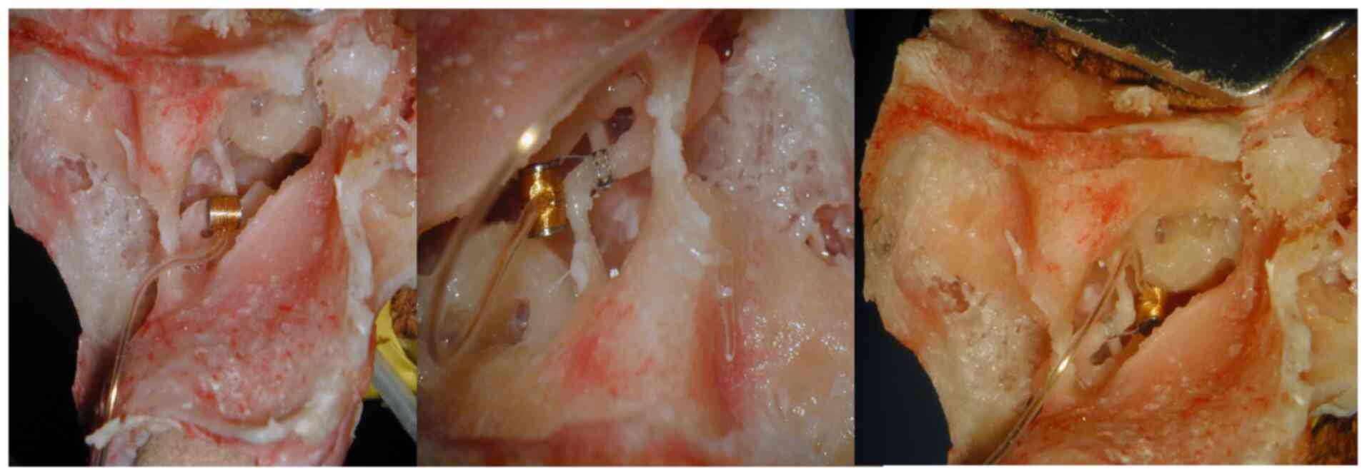
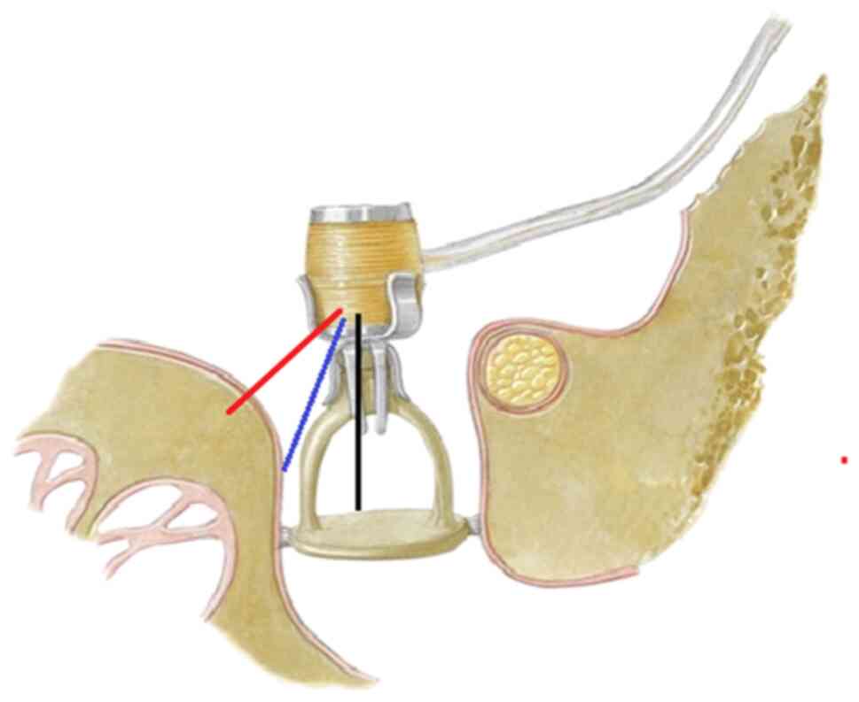
We investigated the VSB connected to the long
process of the incus in 3 different conditions: The floating mass
transducer (FMT) vibrating freely in the middle ear in the
direction of the longitudinal stapes axis, without contact to the
stapes; the FMT in contact to the stapes supra-structure, vibrating
in the direction of the longitudinal stapes axis; the FMT in
contact to the stapes supra-structure, vibrating in a direction of
45-60 degrees off the longitudinal stapes axis.
The three situations were investigated
experimentally on temporal bone specimens and theoretically by
means of a finite element simulation model of the middle ear. The
displacement of the stapes footplate was measured using laser
Doppler vibrometry (LDV) and calculated with the simulation model.
The Polytec LDV (CLV-700 sensor head, CLV-1000 vibrometer
controller; Polytec GmbH) was mounted onto a Zeiss surgical
microscope (Zeiss Co.). A micromanipulator was used to focus the
helium-neon laser on the target (squares of foil of 0.5
mm2 with reflective polystyrene microbeads). The sound
generator (insert earphone, eartone 3A) was inserted into the ear
canal and the probe microphone (ER7c; Ethymotic Research Inc.)
positioned through an extra opening in the external ear canal next
to the eardrum for reference measurements (16).
We compared the results obtained from these
measurements and simulations to determine the influence of
variations of coupling of the FMT.
The study was performed using unfixed human
cadaveric temporal bones (TBs). The temporal bones were harvested
within 48 h after death, using the classical techniques described
in literature (17) and stored in
isotonic NaCl saline until preparation for approximately 1 h.
All TBs were carefully inspected using an operating
microscope to exclude diseased middle ear or perforation of the
TM.
Results and discussion
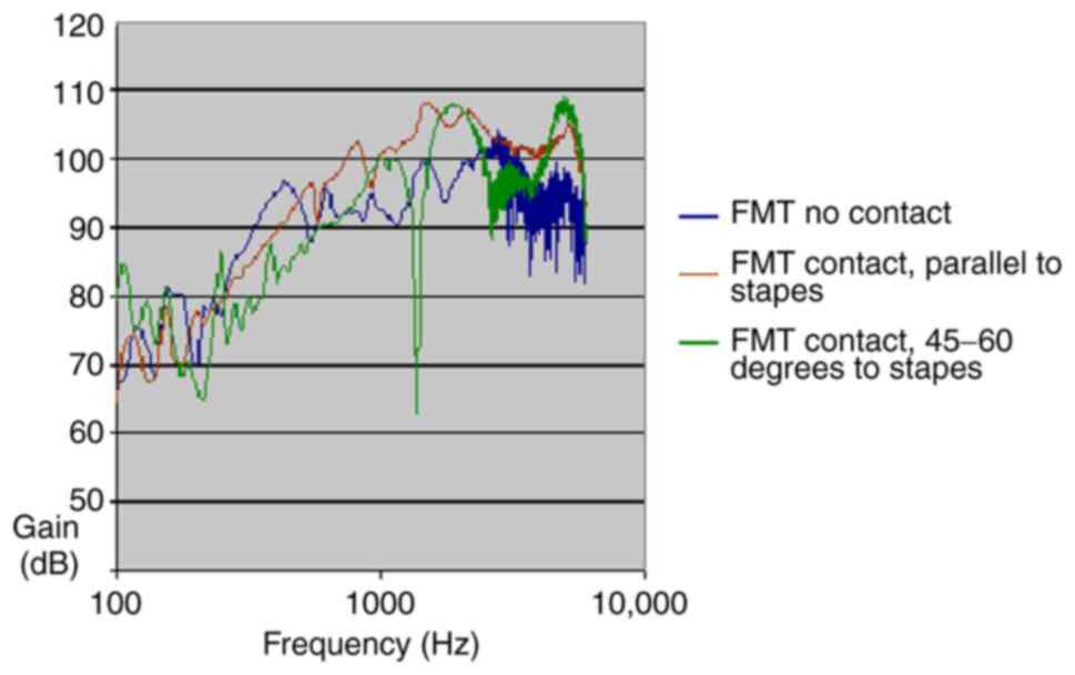
The measurements on human temporal bones, as shown
in Fig. 4 for each of the studied
positions, yielded a graphic representation depicted for comparison
(Fig. 5).
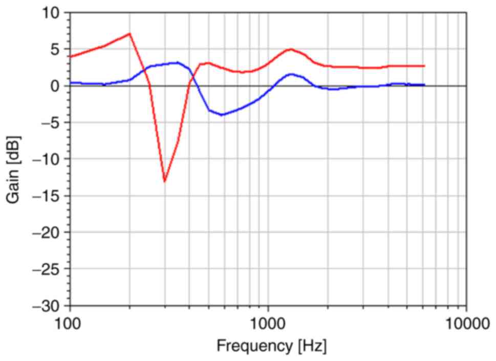
The FEM experiments for FMT coupling with contact to
the stapes parallel to its long axis and tilted at a 15,
respectively 45 angle showed similar changes in middle ear transfer
function (METF) to those obtained on temporal bone (Figs. 6 and 7).
While fresh frozen or thawed-frozen temporal bones
may not be a perfect model, due to loss of some elasticity of the
ossicular chain and the post-mortem effects caused by inner ear
pressure change, tissue oedema, temperature change, humidity, they
do represent an adequate model for relative performance gains due
to slight changes in prosthesis positions, provided that
comparisons are made within the same temporal bone.
LDV measurements of METF represents a suitable
method for monitoring of the VSB placement and functional results.
It is also reliable, easily applicable, not time consuming and
relatively cheap bearing in mind the possible long-term
benefits.
The FEM (a three-dimensional human outer and
middle-ear model, including muscles, ligaments and middle-ear
cavity) became popular after the massive development of computers
and allows the investigation of the motion of the entire ossicular
chain and precise definition of the outer ear canal and middle ear
space geometry (Table I).
 | Table IPreviously published finite element
models of the outer and middle earsa. |
Table I
Previously published finite element
models of the outer and middle earsa.
| Author(s) (year) | Description of FEM
experiments | (Refs.) |
|---|
| Funnell and Laszlo
(1978) | Three-dimensional
model of feline TM (cat), including curvature, isotropic
elasticity, static pressure load | (7) |
| Funnell and Laszlo
(1978) | Undamped natural
frequency analysis of previously presented three-dimensional
model | (7) |
| Williams and Lesser
(1990) | Three-dimensional
model of human TM; calculations of mode shapes for different
curvatures, thicknesses and stiffness | (8) |
| Lesser et al
(1991) | Two-dimensional plane
strain model of the ossicular chain under a static displacement;
stress contours in bones and joints reported | (21) |
| Wada et al
(1992) | Three-dimensional
human middle-ear model, including curved TM with peripheral sprung
restraints | (22) |
| Williams and Lesser
(1992) | Three-dimensional
model of the TM using shell elements and using beam elements for a
Fisch II spandral prosthesis; natural frequencies reported | (23) |
| Ladak and Funnell
(1996) | Three-dimensional
human middle-ear model, including curved TM with peripheral sprung
restraints; static displacement analysis | (24) |
| Koike et al
(1996) | Three-dimensional
human outer and middle-ear model, including muscles, ligaments and
middle-ear cavity | (25) |
| Beer et al
(1997) | Three-dimensional
model of TM and malleus, static and modal analyses | (26) |
| Williams et al
(1997) | Analyzing the mode
shapes of an intact and damaged TM by use of the finite element
model | (27) |
| Eiber (1997) | Three-dimensional
multibody analysis of the ossicular chain (no TM) for passive and
active prostheses | (28) |
| Eiber (1999) | Laser Doppler
Vibrometry and mechanical models used for simulations of the
dynamics of middle ear prosthesis | (29) |
| Zahnert et al
(1997) | Three-dimensional
model with a Dresden partial ossicular replacement prosthesis
(PORP) | (19) |
| Blayney et al
(1997) | Three-dimensional
model of a stapedectomy, with damping at the stapes footplate;
forced harmonic response | (30) |
| Bornitz (2010) | Evaluation of
implantable actuators by means of a middle ear simulation model
(finite element model) | (15) |
The disadvantage of FEM over experiments on
biological structures (LDV) is that the models can be difficult to
validate since the geometries differ between individuals, and
because material property data are usually not precisely known
(18). However, the finite-element
modelling approach may help explain the differences in the clinical
performance of ossicular replacement prostheses (19). It also has applications for the
feasibility of new passive and active middle ear implant design
concepts, especially since medical research can sometimes require a
high degree of abstraction (20).
In conclusion, experimental investigations and
simulations with the model yield the same main results. The first
fitting situation, with the FMT floating freely in the middle ear,
provided by far the worst possible results. Contact to the stapes
supra-structure of the FMT is necessary for optimal performance of
the FMT. Tilting the FMT off the longitudinal axis of the stapes
reduces the vibration of the stapes footplate. But this reduction
is less than for the first situation of the freely floating
FMT.
The mastoid specimen preserves its acoustic
properties that have been shown to be similar to those in the vital
human ear, under these conditions.
Properly coupling the electromagnetic transducer to
the ossicles can be difficult and it requires a certain degree of
experience.
A FEM is useful for functional evaluation of VSB
since it enables easy modelling of the complicated middle ear
structures and simulation of their dynamic behavior which makes it
easy to understand it in detail without experiments.
Acknowledgements
Not applicable.
Funding
FMTs were kindly provided by MED-EL (Innsbruck,
Austria). This research did not receive any specific grant from
funding agencies in the public, commercial, or not-for-profit
sectors. The authors have no conflicts of interest to declare.
Availability of data and materials
All data generated or analyzed during this study are
included in this published article.
Authors' contributions
HM contributed to all the stages of the article; he
designed the article and revised the manuscript for important
scientific content. MB, NL, TZ acquired the data and applied the
surgical procedure technic. HM also contributed to the conception
of the work and revised the language. All authors read and approved
the final manuscript.
Ethics approval and consent to
participate
Investigations did not involve studies in humans or
animals. Ethics approval for the use of human temporal bone
specimen was obtained by the Ethics Committee of the Technische
Universität Dresden (EK 59022014).
Patient consent for publication
Not applicable.
Competing interests
The authors declare that they have no competing
interests.
References
|
1
|
Prendergast PJ, Ferris P, Rice HJ and
Blayney AW: Vibro-acoustic modelling of the outer and middle ear
using the finite-element method. Audiol Neurotol. 4:185–191.
1999.PubMed/NCBI View Article : Google Scholar
|
|
2
|
Lighthill J: Biomechanics of hearing
sensitivity. J Vib Acoust. 113:1–13. 1991.
|
|
3
|
von Békésy G: Experiments in Hearing.
Mc-Graw-Hill, New York, NY, 1960.
|
|
4
|
Stinson MR: The spatial distribution of
sound pressure within scaled replicas of the human ear canal. J
Acoust Soc Am. 78:1596–1602. 1985.PubMed/NCBI View
Article : Google Scholar
|
|
5
|
Zwislocki JJ: Analysis of middle-ear
function. J Acoust Soc Am. 34:1514–1523. 1962.
|
|
6
|
Zwislocki JJ: Normal function of the
middle ear and its measurement. Audiology. 21:4–14. 1982.PubMed/NCBI View Article : Google Scholar
|
|
7
|
Funnell WR and Laszlo CA: Modeling of the
cat eardrum as a thin shell using the finite-element method. J
Acoust Soc Am. 63:1461–1467. 1978.PubMed/NCBI View
Article : Google Scholar
|
|
8
|
Williams KR and Lesser TH: A finite
element analysis of the natural frequencies of vibration of the
human tympanic membrane. Part I. Br J Audiol. 24:319–327.
1990.PubMed/NCBI View Article : Google Scholar
|
|
9
|
Trifu S: Neuroendocrine insights into
burnout syndrome. Acta Endocrinol (Bucur). 15:404–405.
2019.PubMed/NCBI View Article : Google Scholar
|
|
10
|
Trifu S, Vladuti A and Popescu A:
Neuroendocrine aspects of pregnancy and postpartum depression. Acta
Endocrinol (Bucur). 15:410–415. 2019.
|
|
11
|
Monsell EM: The mechanism of hearing loss
in Paget's disease of bone. Laryngoscope. 114:598–606.
2004.PubMed/NCBI View Article : Google Scholar
|
|
12
|
Mocanu H: The role of perinatal hearing
screening in the normal development of the Infant's language. In:
Debating Globalization. Identity, Nation and Dialogue. 4th edition.
Boldea I and Sigmirean C (eds). Arhipeleag XXI Press, Tirgu Mures,
pp562-569, 2017.
|
|
13
|
Mocanu H: The economic impact of early
diagnosis of congenital hearing loss. In: Debating Globalization.
Identity, Nation and Dialogue. 4th edition. Boldea I and Sigmirean
C (eds). Arhipeleag XXI Press, Tirgu Mures, pp556-561, 2017.
|
|
14
|
Mocanu H and Oncioiu I: The influence of
clinical and environmental risk factors in the etiology of
congenital sensorineural hearing loss in the Romanian population.
Iran J Public Health. 48:2301–2303. 2019.PubMed/NCBI
|
|
15
|
Bornitz M, Hardtke HJ and Zahnert T:
Evaluation of implantable actuators by means of a middle ear
simulation model. Hear Res. 263:145–151. 2010.PubMed/NCBI View Article : Google Scholar
|
|
16
|
Neudert M, Bornitz M, Mocanu H,
Lasurashvili N, Beleites T, Offergeld C and Zahnert T: Feasibility
study of a mechanical Real-time feedback system for optimizing the
sound transfer in the reconstructed middle Ear. Otol Neurotol.
39:e907–e920. 2018.PubMed/NCBI View Article : Google Scholar
|
|
17
|
Schuknecht HF: Pathology of the Ear
(Commonwealth Fund Publications). Harvard University Press,
Cambridge, 1974.
|
|
18
|
Prendergast PJ: Finite element models in
tissue mechanics and orthopaedic implant design. Clin Biomech
(Bristol Avon). 12:343–366. 1997.PubMed/NCBI View Article : Google Scholar
|
|
19
|
Zahnert T, Schmidt R, Hüttenbrink KB and
Hardtke HJ: F-E simulation of the Dresden middle-ear prosthesis.
In: Middle-Ear Mechanics in Research and Otosurgery. Hüttenbrink KB
(eds). University of Technology, Dresden, pp200-206, 1997.
|
|
20
|
Alecu I, Mocanu H and Călin IE:
Intellectual mobility in higher education system. Rom J Mil Med.
120:16–21. 2017.
|
|
21
|
Lesser TH, Williams KR and Blayney AW:
Mechanics and materials in middle ear reconstruction. Clin
Otolaryngol Allied Sci. 16:29–32. 1991.PubMed/NCBI View Article : Google Scholar
|
|
22
|
Wada H, Metoki T and Kobayashi T: Analysis
of dynamic behavior of human middle ear using a finite-element
method. J Acoust Soc Am. 92:3157–3168. 1992.PubMed/NCBI View
Article : Google Scholar
|
|
23
|
Williams KR and Lesser TH: A dynamic and
natural frequency analysis of the Fisch II spandrel using the
finite element method. Clin Otolaryngol Allied Sci. 17:261–270.
1992.PubMed/NCBI View Article : Google Scholar
|
|
24
|
Ladak HM and Funnell WR: Finite-element
modeling of the normal and surgically repaired cat middle ear. J
Acoust Soc Am. 100:933–944. 1996.PubMed/NCBI View
Article : Google Scholar
|
|
25
|
Koike T, Wada H and Kobayashi T: Modeling
of the human middle ear using the finite-element method. J Acoust
Soc Am. 111:1306–1317. 2002.PubMed/NCBI View Article : Google Scholar
|
|
26
|
Beer HJ, Bornitz M, Drescher J, Schmidt R,
Hardtke HJ, Hofmann G, Vogel U, Zahnert T and Hüttenbrink KB:
Finite element modelling of the human eardrum and applications. In:
Middle Ear Mechanics in Research and Otosurgery. Hüttenbrink KB
(ed). Proceedings of the International Workshop on Middle Ear
Mechanics, Dresden, pp40-47, 1997.
|
|
27
|
Williams KR, Blayney AW and Lesser TH:
Mode shapes of a damaged and repaired tympanic membrane as analysed
by the finite element method. Clin Otolaryngol Allied Sci.
22:126–131. 1997.PubMed/NCBI View Article : Google Scholar
|
|
28
|
Eiber A: Mechanical modeling and dynamical
investigation of middle ear. In: Middle-Ear Mechanics in Research
and Otosurgery. Hüttenbrink KB (ed). Proceedings of the
International Workshop on Middle Ear Mechanics, Dresden, pp61-66,
1997.
|
|
29
|
Eiber A: Mechanical modeling and dynamical
behavior of the human middle ear. Audiol Neurotol. 4:170–177.
1999.PubMed/NCBI View Article : Google Scholar
|
|
30
|
Blayney AW, Williams KR and Rice HJ: A
dynamic and harmonic damped finite element analysis model of
stapedotomy. Acta Otolaryngol. 117:269–273. 1997.PubMed/NCBI View Article : Google Scholar
|