Open Access

An original method of simulating the articular cartilage in the context of in vitro biomechanical studies investigating the proximal femur

  • Authors:
    • Teodor Stefan Gheorghevici
    • Eugen Carata
    • Paul-Dan Sirbu
    • Ovidiu Alexa
    • Manuela-Gabi Poroh
    • Alexandru Filip
    • Norin Forna
    • Bogdan Puha
  • View Affiliations

  • Published online on: January 5, 2022     https://doi.org/10.3892/etm.2022.11125
  • Article Number: 202
  • Copyright: © Gheorghevici et al. This is an open access article distributed under the terms of Creative Commons Attribution License.

Metrics: Total Views: 0 (Spandidos Publications: | PMC Statistics: )
Total PDF Downloads: 0 (Spandidos Publications: | PMC Statistics: )


Abstract

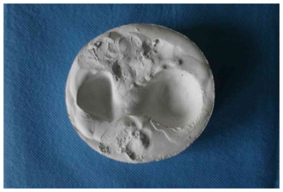
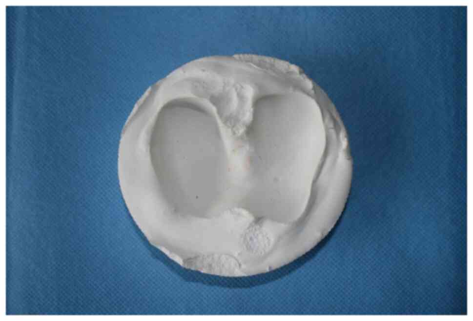
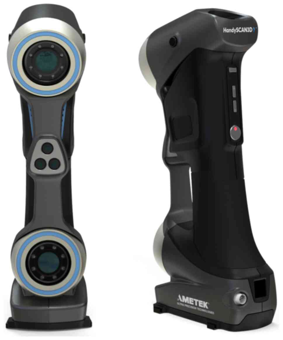
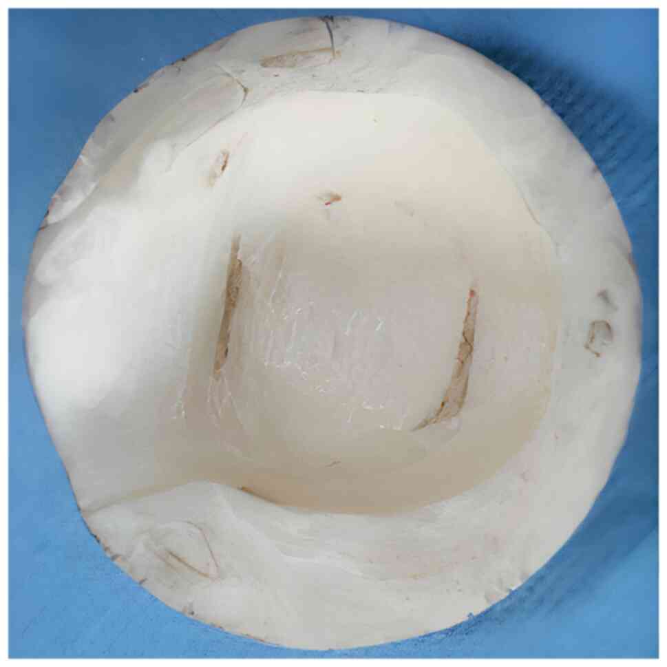
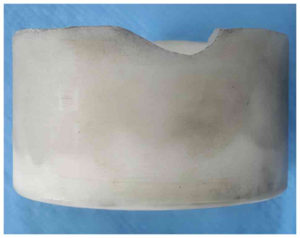
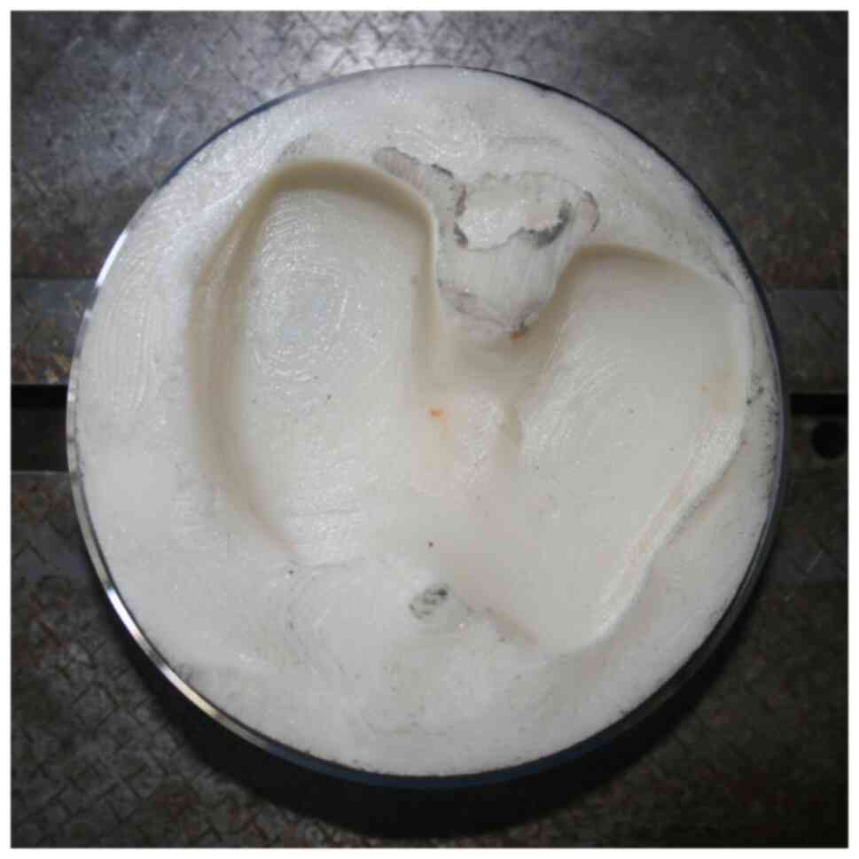
Biomechanical testing is a necessity given the development of novel implants used in the osteosynthesis of hip fractures. The purpose of biomechanical testing is to recreate realistic conditions similar to the in vivo conditions. Although biomechanical testing of hip arthroplasty has been standardized since the 1970s, there is no consensus at present on testing methodology for osteosynthesis of hip fractures. Most biomechanical studies examining the fractures of the proximal femur in order to optimize implants opt for loading the bone‑implant ensemble directly on the femoral head or using a metallic loading part. This loading technique fails to perform a mechanical stress distribution similar to in vivo conditions, which could alter the outcome. The present study aimed to design loading/unloading cups with mechanical properties that resemble those of the cartilage at the hip level. Through the impression and scanning of the cast models obtained, a digital 3D model was created in STL format and this was processed in order to obtain the computer numerical control (CNC) trajectories of the printing head. For prototyping using additive manufacturing technology, a thermoplastic polymer with biochemical properties, such as tensile strength, that resemble those of the adult hip and a Stratasys FORTUS 250 mc CNC machine were used. Loading/unloading cups with similar anatomy and biomechanical forces compared with those of the adult hip were created, which allowed the experimental simulation of the conditions during walking.
View Figures
View References

Related Articles

Journal Cover

March-2022
Volume 23 Issue 3

Print ISSN: 1792-0981
Online ISSN:1792-1015

Sign up for eToc alerts

Recommend to Library

Copy and paste a formatted citation
x
Spandidos Publications style
Gheorghevici TS, Carata E, Sirbu P, Alexa O, Poroh M, Filip A, Forna N and Puha B: An original method of simulating the articular cartilage in the context of <em>in vitro</em> biomechanical studies investigating the proximal femur. Exp Ther Med 23: 202, 2022
APA
Gheorghevici, T.S., Carata, E., Sirbu, P., Alexa, O., Poroh, M., Filip, A. ... Puha, B. (2022). An original method of simulating the articular cartilage in the context of <em>in vitro</em> biomechanical studies investigating the proximal femur. Experimental and Therapeutic Medicine, 23, 202. https://doi.org/10.3892/etm.2022.11125
MLA
Gheorghevici, T. S., Carata, E., Sirbu, P., Alexa, O., Poroh, M., Filip, A., Forna, N., Puha, B."An original method of simulating the articular cartilage in the context of <em>in vitro</em> biomechanical studies investigating the proximal femur". Experimental and Therapeutic Medicine 23.3 (2022): 202.
Chicago
Gheorghevici, T. S., Carata, E., Sirbu, P., Alexa, O., Poroh, M., Filip, A., Forna, N., Puha, B."An original method of simulating the articular cartilage in the context of <em>in vitro</em> biomechanical studies investigating the proximal femur". Experimental and Therapeutic Medicine 23, no. 3 (2022): 202. https://doi.org/10.3892/etm.2022.11125