Introduction
Ocular trauma, ocular tumors, or other diseases
often lead to enucleation and exenteration of the ocular contents
(1,2). After surgery, a local volume defect
resulting in orbital depression and orbital deformities may exist
even after hydroxyapatite implantation treatment with padding
materials (3). The thickness of
the orbital wall is 0.3 to 0.9 mm with a paucity of covered soft
tissue, which presents a challenge in reconstructive surgery
(4). The common complications of
hydroxyapatite implantation include exposure, prolapse, or
migration of the implants (5,6).
Additionally, it has been reported that the exposure risk of
hydroxyapatite implants is closely related to the vascularization
of implants in the orbit (7,8).
Vascularization in tissue engineering is a complex process. Some
investigators have argued that endothelial cells participate in
many physiological and pathological processes (9,10).
Endothelial cell sprouting from the parent vessels occurs with
migration, proliferation, alignment, tube formation, and
anastomosis to other vessels (11). Numerous studies have shown that the
formation of capillary-like tubes by endothelial cells creates a
foundation in in vitro assays (12) or in three-dimensional engineered
tissues (13). Coralline porous
hydroxyapatite (HA) was the material used in the first porous
orbital implant approved by the US Food and Drug Administration for
clinical operations in 1989(14).
Early studies reported that the architecture characteristics of
porous HA were important for cell perfusion seeding even within
large scaffolds (15). In
addition, cell proliferation and differentiation were enhanced in
the perfusion culture (16). It is
increasingly recognized that the angiogenesis process is not only
determined by endothelial cells, but also by growth factors and
scaffolds. Importantly, identification of the cytokines that are
expressed, such as proangiogenic factors and inflammatory factors,
have led to developments in vascularization. Several reports have
shown that vascular endothelial growth factor (VEGF) is upregulated
in most proangiogenic pathways (17). Recently, expression of various
pro-inflammatory factors, including interleukin (IL)-8, IL-1 and
IL-10, were found to be correlated with angiogenesis and effective
therapeutics in ophthalmology (18-20).
Previous studies have shown that ‘pre-vascularization’ provides the
basis for application of tissue engineering in orbital plastic
reconstruction. Preforming a microvasculature in vivo prior
to implantation (21) results in
acceleration of vascularization in the orbital implant complex.
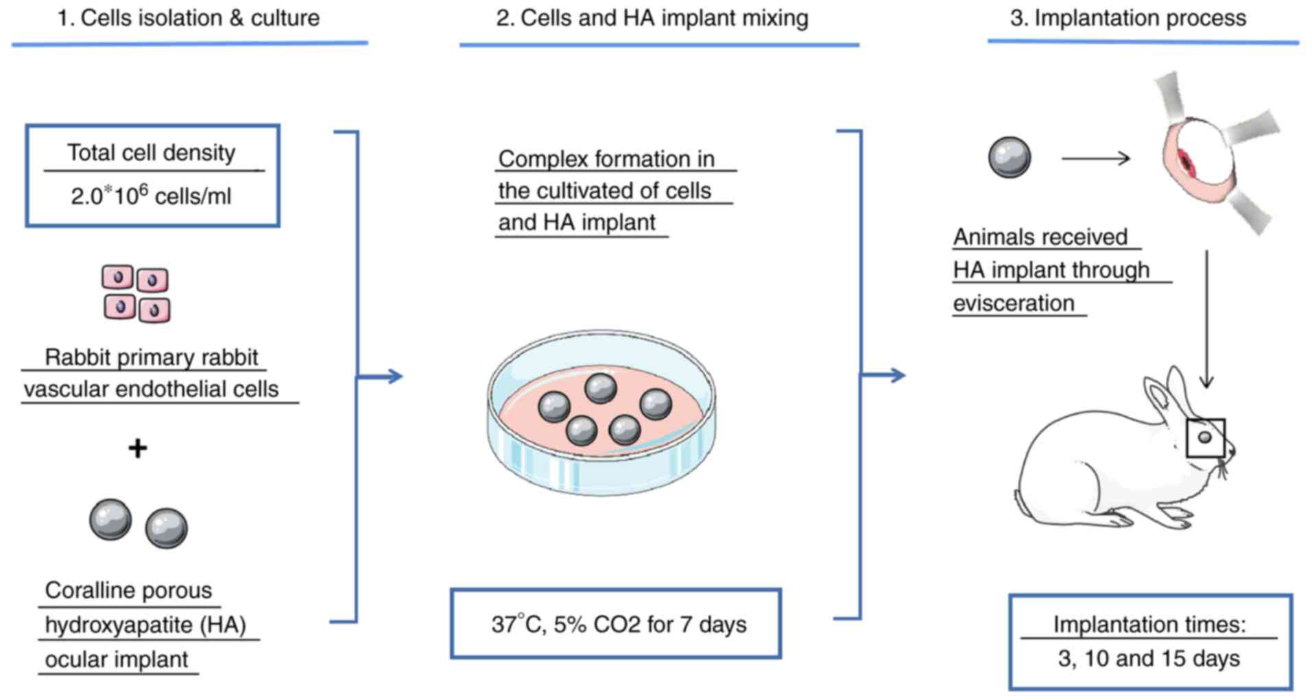
In the present study, a primary culture of a
vascular endothelial cell-hydroxyapatite orbital implant complex
was used. We investigated whether it reduced or prevented
postoperative infection. The results provide a theoretical basis
for the clinical application of tissue engineering techniques in
the future.
Materials and methods
Isolation and culture of rabbit
vascular endothelial cells
All animal experiments were carried out in
compliance with a protocol specifically approved by the
Experimental Animal Center of Nanchang University Animal Care and
Use Committee. Ten New Zealand white rabbits (1.5-2.0 kg) were
purchased from Tianqin Biology Technology Co., Ltd. (Hunan, China).
Ten rabbits were randomly divided into two groups, namely the
endothelial cell (EC) group and the blank group. Vascular
endothelial cells of the orbital microvascular tissue in the right
eye were extracted from rabbits in the ECs group under local
anesthesia of 0.5% proparacaine hydrochloride ophthalmic solution
and 1-2 drops per eye. The tissue samples were washed three times
with D-Hanks solution under aseptic conditions using an operating
microscope. These specimens were cut into 1-mm3 pieces
and digested with 0.1% collagenase I (Beijing Solarbio Science
& Technology Co., Ltd.) for 2 h at 37˚C with 5% CO2.
Rabbit vascular endothelial cells were then resuspended in
Dulbecco's modified Eagle's medium (DMEM) (Gibco; Thermo Fisher
Scientific, Inc.) containing 10% fetal bovine serum (FBS), 1%
penicillin/streptomycin (P/S) and 1% L-glutamine to terminate
digestion. When cell confluency reached 70-80%, the cells in
passage three were used in the experiments (22).
Implant preparation
An HA ocular implant was purchased from Beijing
Yihuajian Science and Trade Co., Ltd. The average HA ocular implant
diameter was approximately 18 mm and the maximum aperture was 300
µm. All of the above processes were performed under a sterile
environment. Cell seeding into the HA was performed as previously
reported (23). The HA ocular
implant in the EC group was treated with endothelial cells. The
numbers of transplanted cells were 2.0x10⁶. The cell-HA
implant was cultivated in DMEM containing 10% FBS, 1% P/S and 1%
L-glutamine at 37˚C with 5% CO2 for 7 days. The HA wells
did not receive any treatment and served as the blank HA implant
group. Culture conditions were the same as that in the EC
group.
Evisceration and hydroxyapatite
implants
Animal experiments were carried out in accordance
with the ARVO Statement for the Use of Animals in Ophthalmic and
Vision Research. The study obtained ethics approval from the Ethics
Committee of the Affiliated Eye Hospital of Nanchang University
(approval no. YLP200810001). The surgical approach for the
hydroxyapatite implant was similar to that reported in a previous
study (24). All 10 rabbits were
anesthetized with intramuscular injections of chlorpromazine (25
mg/kg body wt) and ketamine (50 mg/kg body wt). The fur was removed
at the surgical site in ~3 cm to prevent contamination. The eyeball
of the surgical eyes underwent iodophor disinfectant (250 mg/l) and
0.9% sodium chloride injection for douching repeatedly. All rabbits
received evisceration of the right eye in a sterile environment,
which was finished with oculis. The rabbits of the EC group
received implants of the cell-HA complex into the orbit, which was
then sutured and fixed to the temporalis muscle and frontal muscle
flap. The same operations were conducted in the blank group except
the blank-HA complex was implanted. Benzylpenicillin (400,000 U)
was injected over the following 3 days to prevent inflammation.
Fifteen days after surgery, the implants in the two groups were
extracted from the orbits of the experimental animals. Rabbits were
euthanized by i.v. injection of pentobarbital with 100 mg/kg.
Immunofluorescent assay
Immunofluorescent assay processes were similar to
those reported in a previous study (25). Rabbit vascular endothelial cells
were seeded on sterile coverslips in a 24-well plate in DMEM with
10% FBS. Twenty-four hours later, the cells were washed three times
with PBS and fixed with 4% paraformaldehyde for 30 min at 37˚C. The
cells were rinsed with PBS for 3x5 min, penetrated with 0.5% Triton
X-100 in PBS for 20 min, and then blocked in 10% FBS for 2 h. They
were then incubated with a 1:100 von Willebrand factor (vWF)
primary antibody (cat. no. 66682-1-lg; ProteinTech Group, Inc.) in
a wet box at 4˚C in the dark. Then, the cells were incubated with a
1:150 dilution of goat anti-mouse lgG (H+L) Cross-Adsorbed
secondary antibody, Alexa Fluor 594 (cat. no. SA00006-3;
ProteinTech Group, Inc.) in the dark for 1 h and stained with
4',6-diamidino-2-phenylindole (DAPI). Fluorescent images were
acquired with a fluorescence inverted microscope (IX73; Olympus
Corporation).
Cell tube formation assay
Cell tube formation assays were conducted as
previously reported (25).
In brief, all products that were exposed to the Matrigel matrix
(Corning, Inc.) adhesive were placed in a refrigerator at -20˚C for
6 h. Matrigel was precooled to a basal medium at 4˚C for 24 h.
Matrigel was added per well into 96-well plates with 60 µl and then
placed at 37˚C for 2 h. Rabbit vascular endothelial cells were
seeded onto the Matrigel bed (1.5x10⁴ cells/well) and
cultured for 4 h. Tube formation was imaged and assessed with
ImageJ software (version 1.52; National Institutes of Health).
Transwell cell migration assay
Endothelial cells were seeded into the upper level
of a Transwell chamber at a density of 1.0x 10⁵
cells/chamber, and the lower level was filled with 600 µl medium
containing 10% FBS in the EC group and 600 µl in serum-free medium
in the blank group. Cells were cultured at 37˚C with 5%
CO2 for 16 h. Subsequently, the cells that had invaded
the lower surface of the membrane were fixed for 25 min with 4%
paraformaldehyde, stained with 0.25% crystal violet dye for 5 min,
and then rinsed with PBS several times. Cells that had not migrated
were gently removed using a cotton swab once the chambers had
dried. The number of migrated cells were counted and imaged with a
microscope as previously described (25). The Transwell migration assays were
performed in triplicate.
Implant histopathological
examination
Implant samples were embedded in paraffin in 5-µm
serial sections. H&E was used to stain the implants. The slides
were examined under a light microscope with a photomicrographic
attachment as previously described (26). Pathologists examined all
H&E-stained slides from both the blank and EC groups, and
diagnosed various inflammatory cell infiltration (such as
neutrophils and eosinophils) according to the cellular
morphological features.
ELISA
ELISAs were performed according to the
manufacturer's guidelines (Boster Biological Technology). The
rabbits received intramuscular injections of chlorpromazine (25
mg/kg body wt) and ketamine (50 mg/kg body wt) for anesthesia, and
the right eye was exposed with a lid speculum at 3, 10, 15 days
after implantation. The conjunctiva flap at fornix was collected
from each group and fully broken down fully in liquid nitrogen.
Then, the samples were diluted five times in PBS and centrifuged at
4˚C for 10 min at 12,000 x g. Additionally, the supernatants were
collected for secretion measurement. Prior to the experiment
(27), the detection reagent and
each group of samples were pipetted onto pre-coated plates that had
been coated with rabbit interleukin-8 (IL-8) and vascular
endothelial growth factor (VEGF) antibodies. The optical density
value was determined at 450 nm by enzyme-labeled analyzer
immediately.
Statistical analysis
All data are reported as the mean ± SD, and each
experiment was repeated more than three times. The statistical
significance of differences was determined using Student's t-test
for the in vitro study. P<0.05 was considered to indicate
a statistically significant difference.
Results
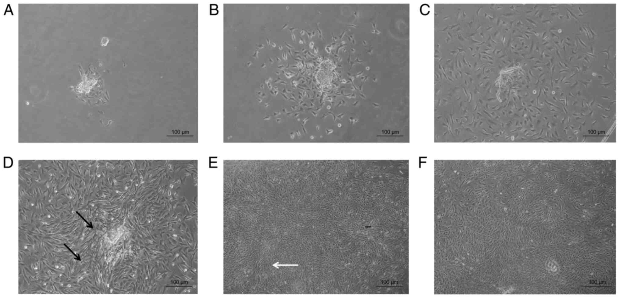
Cell isolation and
characterization
Primary rabbit vascular endothelial cells were
observed, and images were captured under an inverted microscope at
x100 magnification. At 13 days after cell culture, fusiforms were
observed, as shown in Fig. 1.
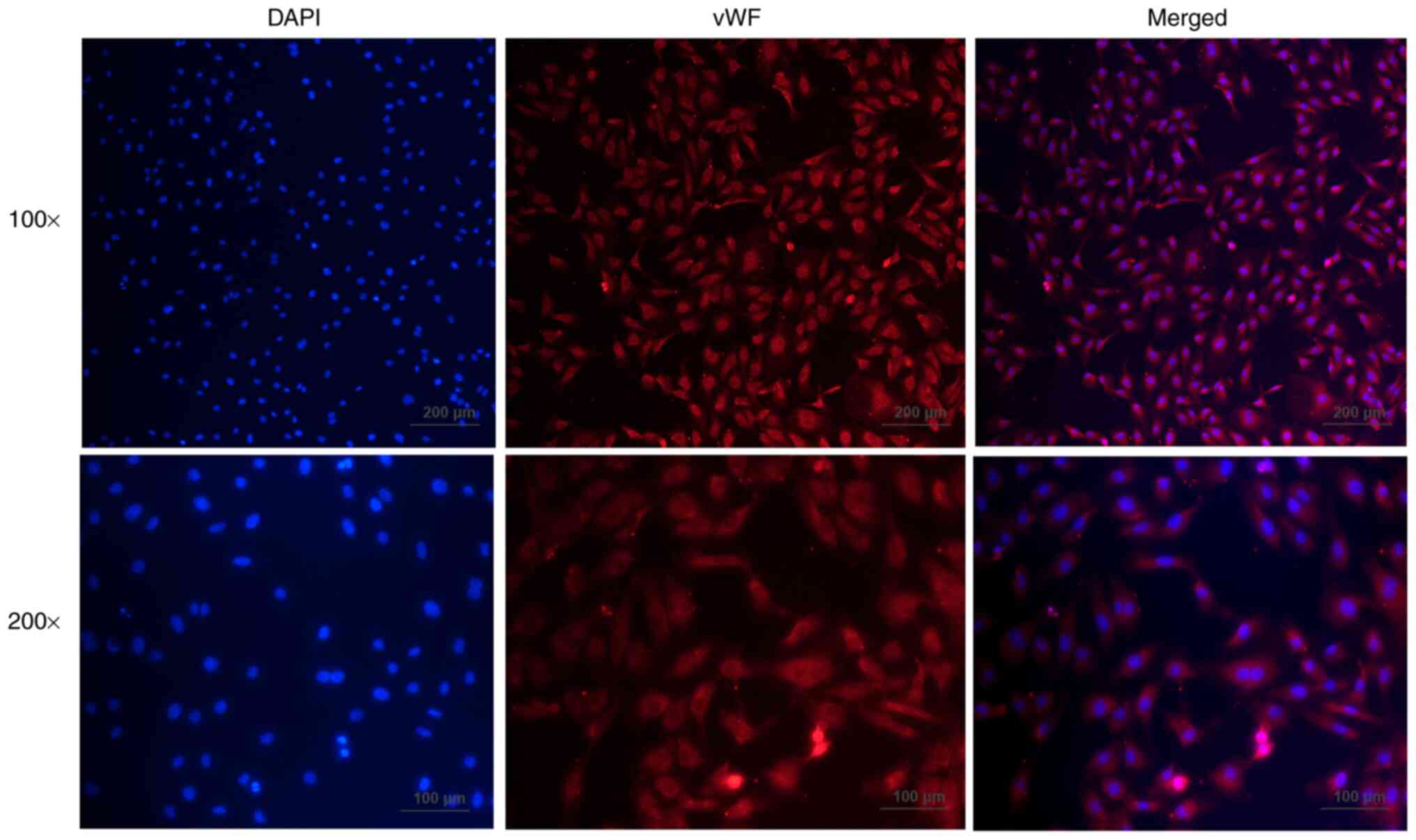
Immunofluorescence staining showed the distribution of vWF in
primary rabbit vascular endothelial cells with >90% of
vWF-positive cells (Fig. 2).
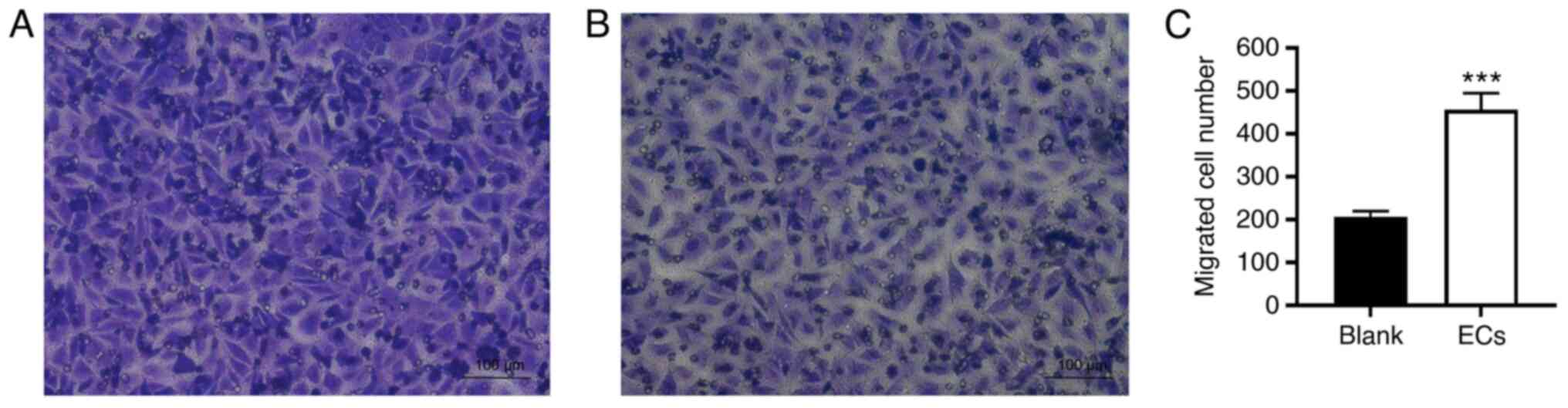
Migration of the primary vascular endothelial cells was assessed
using a Transwell assay. The number of cells in the EC group was
456±38.74, which was significantly increased when compared to the
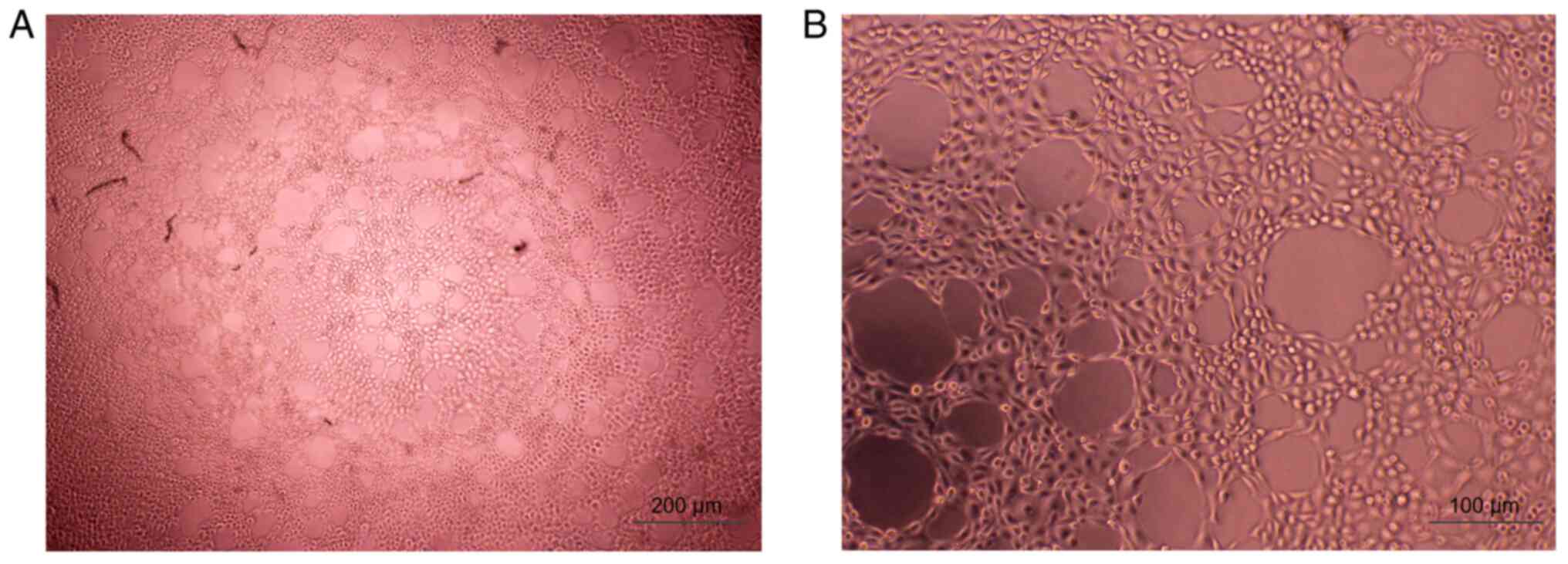
blank group (206.33±13.32) (P<0.001; Fig. 3). Tube formation occurred in the
primary cells and was observed under a microscope after culturing
for 4 h (Fig. 4).
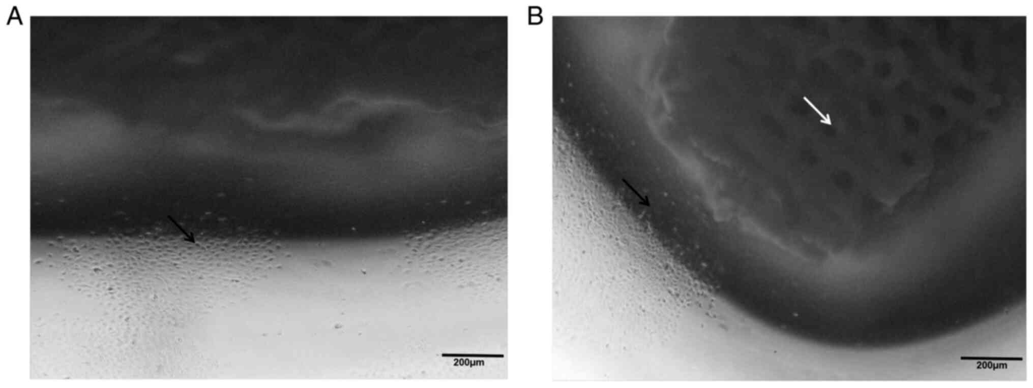
Primary vascular endothelial cells
grown in the HA implant
Primary rabbit vascular endothelial cells were
inoculated to the HA ocular implant (2.0x10⁶ cells/ml)
and cultured for 7 days before evisceration. As for the particular
structure of the HA implant, cells grew well inside the interiors
of the pore walls (Fig. 5). This
process was used to create the vascular endothelial cell-HA orbital
implant complex shown in Fig.
6.
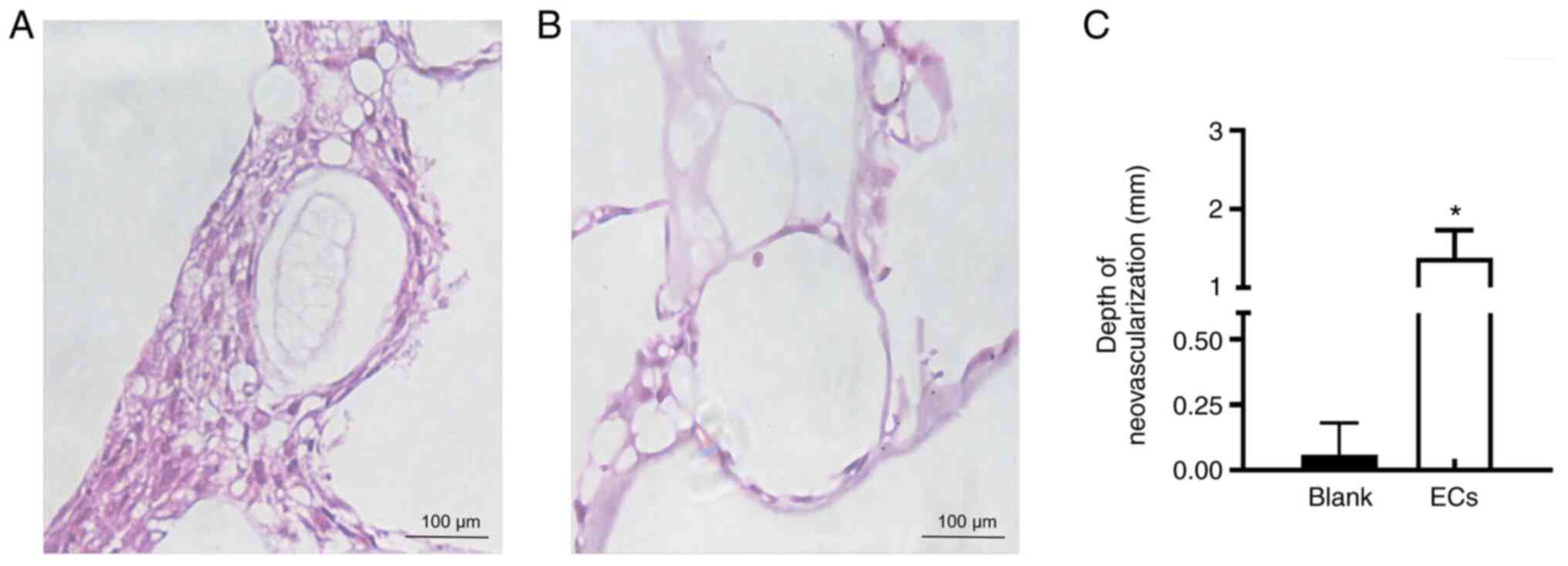
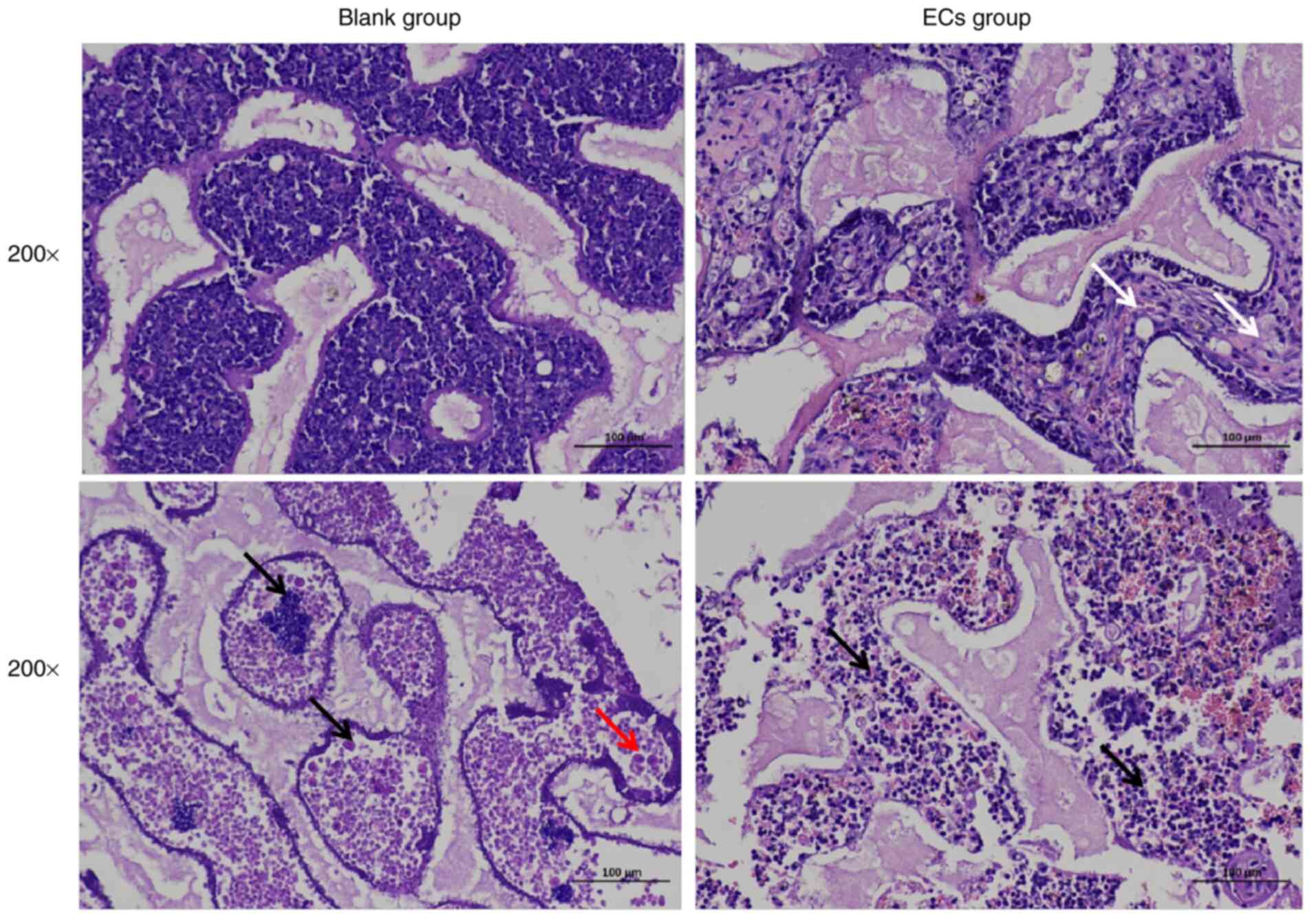
Comparison of fibrovascularization and
inflammatory cell infiltration
All rabbits underwent evisceration in the right eye
and the EC group received a cell-HA orbital complex implant. The
blank-HA complex was implanted in the blank group. At 15 days after
surgery, the implants in the EC group and blank group were
extracted from the orbits of the experimental animals. H&E
staining showed that fibrovascular ingrowth in the implants was
clearly observed in the EC group, but inapparent in the blank group
(Fig. 7A and B). The insertion depth (mm) of
neovascularization was 1.38±0.35 mm in the EC group, which was
significantly higher when compared with that in the blank group
(0.06±0.12 mm) (P<0.05; Fig.
7C). At day 15, both connective tissues and blood vessels were
clearly observed in the EC group, but not in the blank group. The
histopathological images of implantation in the blank group showed
evidence of inflammatory cell infiltration which indicated by
polymorphonuclear neutrophils and eosinophils. Numerous round cells
with obscure margins were presented in the edge zones with
occasional macrophage in the blank group. The neutrophils and
eosinophils were interspersed in the EC group, which indicated that
inflammatory cell infiltration in the blank group was higher than
that in the EC group (Fig. 8).
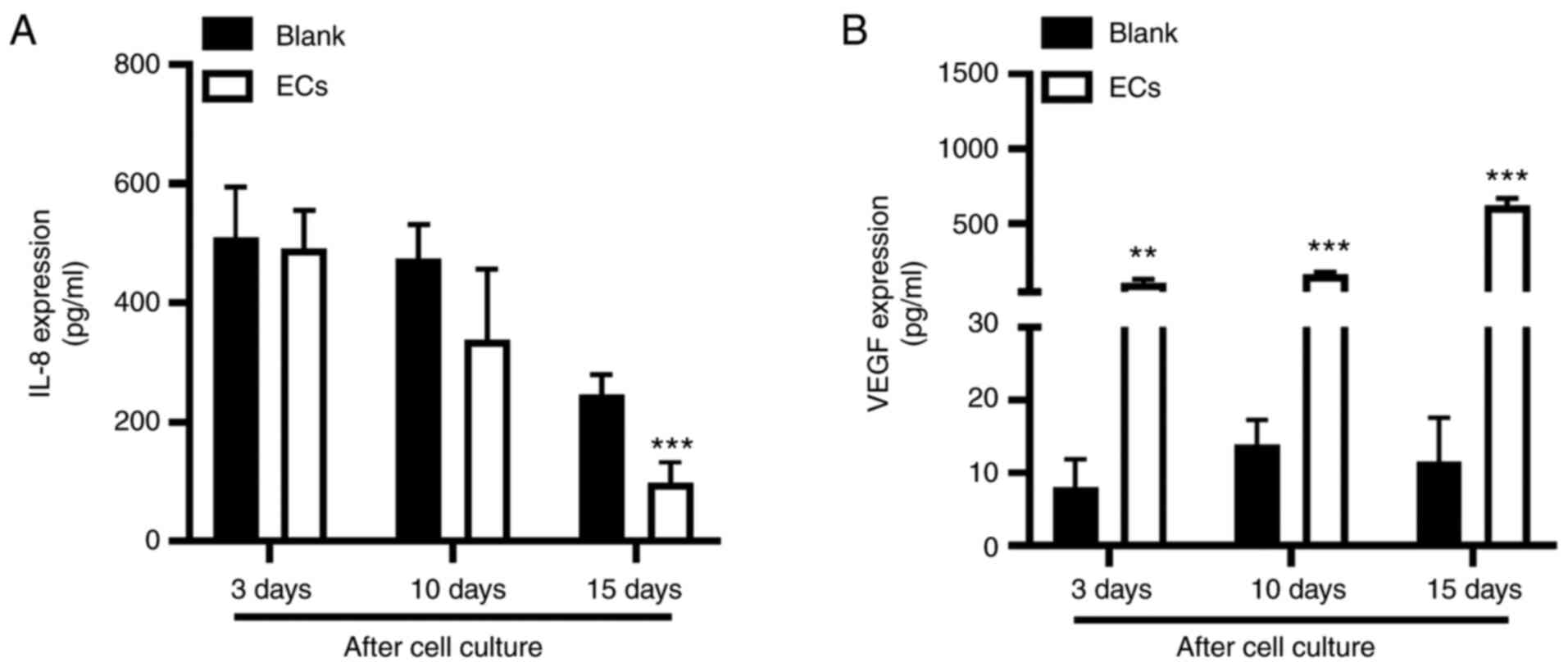
Levels of IL-8 and VEGF
An ELISA was used to measure the levels of rabbit
inflammatory factor IL-8 in the conjunctiva homogenate of the EC
group and blank group at 3, 10, and 15 days after implantation. At
day 3, the two groups showed similar levels of IL-8, without
statistical significance, which then decreased over time in the
culture as cells proliferated. At day 15, the differences reached
statistical significance when compared to the preoperative levels
with 84.72±38.88 pg/ml in the EC group and 230.78±41.62 pg/ml in
the blank group (P<0.001; Fig.
9A). The levels of rabbit angiogenesis factor VEGF in the EC
group at days 3, 10, and 15 after implantation were 101.32±23.62,
160.28±13.22, and 619.83±48.65 pg/ml, respectively, which were
significantly higher than those in the blank group, (8.32±3.86,
13.92±3.37, and 11.53±6.08 pg/ml; P<0.01 vs. day 3, P<0.001
vs. days 10 and 15; Fig. 9B).
Discussion
Tissue engineering technology has been employed
successfully in the management of orbital diseases (28). Endothelial cells, as the seeded
cells, play an important role in the in vitro formation of
the perivascular network (29).
Previous endothelial cell-related research has mostly focused on
human umbilical vein endothelial cells and their angiogenic
responses (30). Early reports
(31) suggested that rabbit bone
marrow mononuclear cell-derived endothelial cells can improve the
vascularization of bone replacements. Tan et al (32) showed that isolation of vascular
endothelial stem cells from liver tissue possess high proliferative
and revascularization potential. In our study, isolated endothelial
cells from microvascular tissue were conveniently obtained with
less trauma and they had high self-replication compared to other
tissue-derived endothelial cells. There have also been reports on
an anti-inflammatory role for endothelial cells (33,34)
with prevention of infection and postoperative complications. These
results suggest that endothelial cells are involved in dynamic
equilibrium with inflammatory mediators and angiogenic factors. HA
has good biocompatibility and provides a reticular structure for
fibrovascular ingrowth into the implant. It is widely used in the
repair of bone defects (35,36).
In our study, the HA implant was seeded with primary vascular
endothelial cells that were extracted from microvascular
endothelial tissue to avoid rejection. It is important to determine
the extraction rate and proliferation of vascular endothelial cells
in vitro.
In the present study, ELISA experiments and
pathological sections were used to demonstrate the activity of the
complex in vivo. IL-8, as one of the key proinflammatory
cytokines, plays an important role in modulating the inflammatory
response. The levels of IL-8 are associated with the progress and
severity of a disease state, including diabetes, atherosclerosis,
and various forms of liver injury (37,38).
The present study found that IL-8 expression was significantly
lower than in the blank group, suggesting that local
revascularization in the EC group might be enhanced when compared
with that of the blank group. Pathological sections showed that
fibrovascular ingrowth in the EC group was markedly higher than in
the blank group, which indicated a severe inflammatory reaction
with aggregation of mycelial pellets and scattered macrophages in
the blank group. This study demonstrated that the degree of
vascularization and the inflammatory reaction were approximately
inverse in proportion.
Vascular endothelial growth factor (VEGF) is an
essential growth factor that can promote angiogenesis and vascular
permeability (39). Due to the
large individual variations, gross changes in the local tissues,
healing of the conjunctival incision, and molding of the HA were
investigated after the operation with no significant difference
between the two groups. Specifically, it was demonstrated that the
isolation of orbital microvascular tissue and scaffold material of
HA provided an experimental basis both in vitro and in
vivo. These data suggest that the cell-hydroxyapatite orbital
implant complex had satisfactory biological activity in
experimental animals.
Postoperative complications of hydroxyapatite
orbital implantation, such as implant exposure, implant migration
conjunctival infection, conjunctival chronic discharge, and
conjunctival cysts, are inevitable (40). Previous reports have shown that
infection is a serious problem for orbital implants and it requires
antibiotic injection or surgical interventions (41). It was documented that infection
occurred in 5.19% in a series of 212 patients with HA implants over
a period of 12 years (42). Catalu
et al (43) reported that
radical surgery (evisceration or enucleation) can reduce the
occurrence of ocular infections. Thus, we investigated the
biological activity of a vascular endothelial cell-hydroxyapatite
orbital implant complex intraoperatively, which can promote and
improve the clinical treatment of orbital implantation. However,
there are some limitations to the present study. The efficacy of HA
transplantation was not thoroughly evaluated and whether or not
there are other factors in the process of blood vessels was not
assessed. We speculate that IL-8 is related to postoperative
anti-inflammatory effects, but more experiments, including
expression of other inflammatory factors, are needed to confirm
this speculation. In the future, it is necessary to provide
reference data for clinical operations and explore the possible
mechanisms of vascularization.
Acknowledgements
Not applicable.
Funding
Funding: This study was supported by the Natural Science
Foundation of China (no. 82060181) and Innovation Fund Designated
for Graduate Students of Jiangxi Province (no. YC2019-S040).
Availability of data and materials
The datasets used and/or analyzed during the current
study are available from the corresponding author on reasonable
request.
Authors' contributions
WW and HLu contributed to the conception and design
of the study and manuscript preparation. DW, MS and JY performed
the data collection, interpretation and statistical analysis. WW
and JY confirmed the authenticity of all the raw data. HLi designed
the study, analyzed the data and wrote the paper. All authors have
read and approved the final manuscript for publication.
Ethics approval and consent to
participate
This study was approved by the Ethics Committee of
the Affiliated Eye Hospital of Nanchang University (no.
YLP200810001). All animal experiments were carried out in
compliance with a protocol specifically approved by the
Experimental Animal Center of Nanchang University Animal Care and
Use Committee.
Patient consent for publication
Not applicable.
Competing interests
The authors declare that they have no competing
interests.
References
|
1
|
Sahraravand A, Haavisto AK, Holopainen JM
and Leivo T: Ocular trauma in the finnish elderly-helsinki ocular
trauma study. Acta Ophthalmol. 96:616–622. 2018.PubMed/NCBI View Article : Google Scholar
|
|
2
|
Zhao Y, Zhao H, Lin JY, Pan Y, Zhai WJ and
Wang YC: Clinical and pathological analysis of ocular tumors in 504
children cases. Zhonghua Yan Ke Za Zhi. 52:764–768. 2016.PubMed/NCBI View Article : Google Scholar : (In Chinese).
|
|
3
|
Pförtner R, Mohr C, Daamen J and Metz A:
Orbital tumors: Operative and therapeutic strategies. Facial Plast
Surg. 30:570–577. 2014.PubMed/NCBI View Article : Google Scholar
|
|
4
|
Kasper FK, Melville J, Shum J, Wong M and
Young S: Tissue engineered prevascularized bone and soft tissue
flaps. Oral Maxillofac Surg Clin North Am. 29:63–73.
2017.PubMed/NCBI View Article : Google Scholar
|
|
5
|
Bee YS, Lin MC, Sheu SJ and Ng JD:
Elevated white blood cell count may predict risk of orbital implant
exposure. Can J Ophthalmol. 49:45–49. 2014.PubMed/NCBI View Article : Google Scholar
|
|
6
|
Lu L, Shi W, Luo M, Sun Y and Fan X:
Repair of exposed hydroxyapatite orbital implants by
subconjunctival tissue flaps. J Craniofac Surg. 22:1452–1456.
2011.PubMed/NCBI View Article : Google Scholar
|
|
7
|
Buettner H and Bartley GB: Tissue
breakdown and exposure associated with orbital hydroxyapatite
implants. Am J Ophthalmol. 113:669–673. 1992.PubMed/NCBI View Article : Google Scholar
|
|
8
|
Zhao Y, Zhang MN, Gao YX, Gao XW and Ren
B: Comparative study of modified and conventional secondary
hydroxyapatite orbital implantations. Int J Ophthalmol. 6:646–649.
2013.PubMed/NCBI View Article : Google Scholar
|
|
9
|
Rioja AY, Annamalai RT, Paris S, Putnam AJ
and Stegemann JP: Endothelial sprouting and network formation in
collagen- and fibrin-based modular microbeads. Acta Biomater.
29:33–41. 2016.PubMed/NCBI View Article : Google Scholar
|
|
10
|
Rouwkema J and Khademhosseini A:
Vascularization and angiogenesis in tissue engineering: Beyond
creating static networks. Trends Biotechnol. 34:733–745.
2016.PubMed/NCBI View Article : Google Scholar
|
|
11
|
Dai B, Zhang Y, Zhan Y, Zhang D, Wang N
and He L: A novel tissue model for angiogenesis: Evaluation of
inhibitors or promoters in tissue level. Sci Rep.
4(3693)2014.PubMed/NCBI View Article : Google Scholar
|
|
12
|
Ayata RE, Chabaud S, Auger M and Pouliot
R: Behaviour of endothelial cells in a tridimensional in vitro
environment. Biomed Res Int. 2015(630461)2015.PubMed/NCBI View Article : Google Scholar
|
|
13
|
Caneparo C, Baratange C, Chabaud S and
Bolduc S: Conditioned medium produced by fibroblasts cultured in
low oxygen pressure allows the formation of highly structured
capillary-like networks in fibrin gels. Sci Rep.
10(9291)2020.PubMed/NCBI View Article : Google Scholar
|
|
14
|
Ye J, Gao Q, He JJ, Gao T, Ning QY and Xie
JJ: Exposure rate of unwrapped hydroxyapatite orbital implants in
enucleation surgery. Br J Ophthalmol. 100:860–865. 2016.PubMed/NCBI View Article : Google Scholar
|
|
15
|
Li J, Zhi W, Xu T, Shi F, Duan K, Wang J,
Mu Y and Weng J: Ectopic osteogenesis and angiogenesis regulated by
porous architecture of hydroxyapatite scaffolds with similar
interconnecting structure in vivo. Regen Biomater. 3:285–297.
2016.PubMed/NCBI View Article : Google Scholar
|
|
16
|
Wang L, Ma XY, Zhang Y, Feng YF, Li X, Hu
YY, Wang Z, Ma ZS and Lei W: Repair of segmental bone defect using
Totally Vitalized tissue engineered bone graft by a combined
perfusion seeding and culture system. PLoS One.
9(e94276)2014.PubMed/NCBI View Article : Google Scholar
|
|
17
|
Brauer MJ, Zhuang G, Schmidt M, Yao J, Wu
X, Kaminker JS, Jurinka SS, Kolumam G, Chung AS, Jubb A, et al:
Identification and analysis of in vivo VEGF downstream markers link
VEGF pathway activity with efficacy of anti-VEGF therapies. Clin
Cancer Res. 19:3681–3692. 2013.PubMed/NCBI View Article : Google Scholar
|
|
18
|
Paulitti A, Andreuzzi E, Bizzotto D,
Pellicani R, Tarticchio G, Marastoni S, Pastrello C, Jurisica I,
Ligresti G, Bucciotti F, et al: The ablation of the matricellular
protein EMILIN2 causes defective vascularization due to impaired
EGFR-dependent IL-8 production affecting tumor growth. Oncogene.
37:3399–3414. 2018.PubMed/NCBI View Article : Google Scholar
|
|
19
|
Protopsaltis NJ, Liang W, Nudleman E and
Ferrara N: Interleukin-22 promotes tumor angiogenesis.
Angiogenesis. 22:311–323. 2019.PubMed/NCBI View Article : Google Scholar
|
|
20
|
Wooff Y, Man SM, Aggio-Bruce R, Natoli R
and Fernando N: IL-1 family members mediate cell death,
inflammation and angiogenesis in retinal degenerative diseases.
Front Immunol. 10(1618)2019.PubMed/NCBI View Article : Google Scholar
|
|
21
|
Laschke MW and Menger MD:
Prevascularization in tissue engineering: Current concepts and
future directions. Biotechnol Adv. 34:112–121. 2016.PubMed/NCBI View Article : Google Scholar
|
|
22
|
Lei M, Wang K, Li S, Zhao K, Hua W, Wu X
and Yang C: The c-Jun signaling pathway has a protective effect on
nucleus pulposus cells in patients with intervertebral disc
degeneration. Exp Ther Med. 20(123)2020.PubMed/NCBI View Article : Google Scholar
|
|
23
|
Jin K, Ye X, Li S, Li B, Zhang C, Gao C
and Ye J: A biomimetic collagen/heparin multi-layered porous
hydroxyapatite orbital implant for in vivo vascularization studies
on the chicken chorioallantoic membrane. Graefes Arch Clin Exp
Ophthalmol. 254:83–89. 2016.PubMed/NCBI View Article : Google Scholar
|
|
24
|
Lee H and Baek S: Comparison of early
fibrovascular proliferation according to orbital implant in orbital
floor fracture reconstruction. J Craniofac Surg. 23:1518–1523.
2012.PubMed/NCBI View Article : Google Scholar
|
|
25
|
Li Z, Liang J, Wu WK, Yu X, Yu J, Weng X
and Shen J: Leptin activates RhoA/ROCK pathway to induce
cytoskeleton remodeling in nucleus pulposus cells. Int J Mol Sci.
15:1176–1188. 2014.PubMed/NCBI View Article : Google Scholar
|
|
26
|
Yang WJ, Yan JB, Zhang L, Zhao F, Mei ZM,
Yang YN, Xiang Y and Xing YQ: Paxillin promotes the migration and
angiogenesis of HUVECs and affects angiogenesis in the mouse
cornea. Exp Ther Med. 20:901–909. 2020.PubMed/NCBI View Article : Google Scholar
|
|
27
|
Jiang Z, Wang J, Li X and Zhang X:
Echinacoside and Cistanche tubulosa (Schenk) R. wight ameliorate
bisphenol A-induced testicular and sperm damage in rats through
gonad axis regulated steroidogenic enzymes. J Ethnopharmacol.
193:321–328. 2016.PubMed/NCBI View Article : Google Scholar
|
|
28
|
Fathi E, Farahzadi R, Vietor I and
Javanmardi S: Cardiac differentiation of bone-marrow-resident
c-kit+ stem cells by L-carnitine increases through
secretion of VEGF, IL6, IGF-1, and TGF-β as clinical agents in
cardiac regeneration. J Biosci. 45(92)2020.PubMed/NCBI
|
|
29
|
Zhou H and Lee J: Nanoscale hydroxyapatite
particles for bone tissue engineering. Acta Biomater. 7:2769–2781.
2011.PubMed/NCBI View Article : Google Scholar
|
|
30
|
Zhang X, Yang J, Li Y, Liu S, Long K, Zhao
Q, Zhang Y, Deng Z and Jin Y: Functional neovascularization in
tissue engineering with porcine acellular dermal matrix and human
umbilical vein endothelial cells. Tissue Eng Part C Methods.
17:423–433. 2011.PubMed/NCBI View Article : Google Scholar
|
|
31
|
Du P, Subbiah R, Park JH and Park K:
Vascular morphogenesis of human umbilical vein endothelial cells on
cell-derived macromolecular matrix microenvironment. Tissue Eng
Part A. 20:2365–2377. 2014.PubMed/NCBI View Article : Google Scholar
|
|
32
|
Tan H, Yang B, Duan X, Wang F, Zhang Y,
Jin X, Dai G and Yang L: The promotion of the vascularization of
decalcified bone matrix in vivo by rabbit bone marrow mononuclear
cell-derived endothelial cells. Biomaterials. 30:3560–3566.
2009.PubMed/NCBI View Article : Google Scholar
|
|
33
|
Naito H, Wakabayashi T, Ishida M, Gil CH,
Iba T, Rahmawati FN, Shimizu S, Yoder MC and Takakura N: Isolation
of tissue-resident vascular endothelial stem cells from mouse
liver. Nat Protoc. 15:1066–1081. 2020.PubMed/NCBI View Article : Google Scholar
|
|
34
|
Iba T and Levy JH: Inflammation and
thrombosis: Roles of neutrophils, platelets and endothelial cells
and their interactions in thrombus formation during sepsis. J
Thromb Haemost. 16:231–241. 2018.PubMed/NCBI View Article : Google Scholar
|
|
35
|
Okamoto T, Akita N, Terasawa M, Hayashi T
and Suzuki K: Rhamnan sulfate extracted from Monostroma nitidum
attenuates blood coagulation and inflammation of vascular
endothelial cells. J Nat Med. 73:614–619. 2019.PubMed/NCBI View Article : Google Scholar
|
|
36
|
Swetha M, Sahithi K, Moorthi A, Srinivasan
N, Ramasamy K and Selvamurugan N: Biocomposites containing natural
polymers and hydroxyapatite for bone tissue engineering. Int J Biol
Macromol. 47:1–4. 2010.PubMed/NCBI View Article : Google Scholar
|
|
37
|
Gradinaru S, Popescu V, Leasu C, Pricopie
S, Yasin S, Ciuluvica R and Ungureanu E: Hydroxyapatite ocular
implant and non-integrated implants in eviscerated patients. J Med
Life. 8:90–93. 2015.PubMed/NCBI
|
|
38
|
Joshi-Barve S, Barve SS, Amancherla K,
Gobejishvili L, Hill D, Cave M, Hote P and McClain CJ: Palmitic
acid induces production of proinflammatory cytokine interleukin-8
from hepatocytes. Hepatology. 46:823–830. 2007.PubMed/NCBI View Article : Google Scholar
|
|
39
|
Jaeschke H: Inflammation in response to
hepatocellular apoptosis. Hepatology. 35:964–966. 2002.PubMed/NCBI View Article : Google Scholar
|
|
40
|
Alkharsah KR: VEGF upregulation in viral
infections and its possible therapeutic implications. Int J Mol
Sci. 19(19)2018.PubMed/NCBI View Article : Google Scholar
|
|
41
|
Sobti MM, Shams F, Jawaheer L, Cauchi P
and Chadha V: Unwrapped hydroxyapatite orbital implants: Our
experience in 347 cases. Eye (Lond). 34:675–682. 2020.PubMed/NCBI View Article : Google Scholar
|
|
42
|
Karsloğlu S, Serin D, Simşek I and Ziylan
S: Implant infection in porous orbital implants. Ophthal Plast
Reconstr Surg. 22:461–466. 2006.PubMed/NCBI View Article : Google Scholar
|
|
43
|
Catalu CT, Istrate SL, Voinea LM,
Mitulescu C, Popescu V and Radu C: Ocular implants-methods of
ocular reconstruction following radical surgical interventions. Rom
J Ophthalmol. 62:15–23. 2018.PubMed/NCBI
|