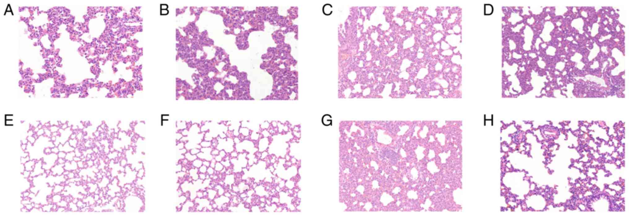
|
1
|
Ameno K, Fuke C, Shirakawa Y, Ogura S,
Ameno S, Kiriu T, Kinoshita H and Ijiri I: Different distribution
of paraquat and diquat in human poisoning cases after ingestion of
a combined herbicide. Arch Toxicol. 68:134–137. 1994.PubMed/NCBI View Article : Google Scholar
|
|
2
|
Van Vleet TR and Schnellmann RG: Toxic
nephropathy: Environmental chemicals. Semin Nephrol. 23:500–508.
2000.PubMed/NCBI View Article : Google Scholar
|
|
3
|
Jones GM and Vale JA: Mechanisms of
toxicity, clinical features, and management of diquat poisoning: A
review. J Toxicol Clin Toxicol. 38:123–128. 2000.PubMed/NCBI View Article : Google Scholar
|
|
4
|
Fortenberry GZ, Beckman J, Schwartz A,
Prado JB, Graham LS, Higgins S, Lackovic M, Mulay P, Bojes H, Waltz
J, et al: Magnitude and characteristics of acute paraquat- and
diquat-related illnesses in the US: 1998-2013. Environ Res.
146:191–199. 2016.PubMed/NCBI View Article : Google Scholar
|
|
5
|
Magalhães N, Carvalho F and Dinis-Oliveira
RJ: Human and experimental toxicology of diquat poisoning:
Toxicokinetics, mechanisms of toxicity, clinical features, and
treatment. Hum Exp Toxicol. 37:1131–1160. 2018.PubMed/NCBI View Article : Google Scholar
|
|
6
|
Dinis-Oliveira RJ, Duarte JA,
Sánchez-Navarro A, Remião F, Bastos ML and Carvalho F: Paraquat
poisonings: Mechanisms of lung toxicity, clinical features, and
treatment. Crit Rev Toxicol. 38:13–71. 2008.PubMed/NCBI View Article : Google Scholar
|
|
7
|
Fussell KC, Udasin RG, Gray JP, Mishin V,
Smith PJ, Heck DE and Laskin JD: Redox cycling and increased oxygen
utilization contribute to diquat-induced oxidative stress and
cytotoxicity in Chinese hamster ovary cells overexpressing
NADPH-cytochrome P450 reductase. Free Radic Biol Med. 50:874–882.
2011.PubMed/NCBI View Article : Google Scholar
|
|
8
|
Vanholder R, Colardyn F, De Reuck J, Praet
M, Lameire N and Ringoir S: Diquat intoxication: Report of two
cases and review of the literature. Am J Med Jun. 70:1267–1271.
1981.PubMed/NCBI View Article : Google Scholar
|
|
9
|
Clark DG and Hurst EW: The toxicity of
diquat. Br J Ind Med. 27:51–55. 1970.PubMed/NCBI View Article : Google Scholar
|
|
10
|
Rose MS, Crabtree HC, Fletcher K and Wyatt
I: Biochemical effects of diquat and paraquat. Disturbance of the
control of corticosteroid synthesis in rat adrenal and subsequent
effects on the control of liver glycogen utilization. Biochem J.
138:437–443. 1974.PubMed/NCBI View Article : Google Scholar
|
|
11
|
Lock EA: The effect of paraquat and diquat
on renal function in the rat. Toxicol Appl Pharmacol. 48:327–336.
1979.PubMed/NCBI View Article : Google Scholar
|
|
12
|
Zhang J, Zhao Y, Bai Y, Lv G, Wu J and
Chen Y: The significance of serum uric acid level in humans with
acute paraquat poisoning. Sci Rep. 5(9168)2015.PubMed/NCBI View Article : Google Scholar
|
|
13
|
Djukic M, Jovanovic MD, Ninkovic M,
Stevanovic I, Curcic M, Topic A, Vujanovic D and Djurdjevic D:
Intrastriatal pre-treatment with L-NAME protects rats from diquat
neurotoxcity. Ann Agric Environ Med. 19:666–672. 2012.PubMed/NCBI
|
|
14
|
Rogers LK, Bates CM, Welty SE and Smith
CV: Diquat induces renal proximal tubule injury in glutathione
reductase-deficient mice. Toxicol Appl Pharmacol. 217:289–298.
2006.PubMed/NCBI View Article : Google Scholar
|
|
15
|
Kashani K, Cheungpasitporn W and Ronco C:
Biomarkers of acute kidney injury: The pathway from discovery to
clinical adoption. Clin Chem Lab Med. 55:1074–1089. 2017.PubMed/NCBI View Article : Google Scholar
|
|
16
|
Calvier L, Chouvarine P, Legchenko E,
Hoffmann N, Geldner J, Borchert P, Jonigk D, Mozes MM and Hansmann
G: PPARγ Links BMP2 and TGF-β1 pathways in vascular smooth muscle
cells, regulating cell proliferation and glucose metabolism. Cell
Metab. 25:1118–1134.e7. 2017.PubMed/NCBI View Article : Google Scholar
|
|
17
|
Kim KK, Sheppard D and Chapman HA: TGF-β1
signaling and tissue fibrosis. Cold Spring Harb Perspect Biol.
10(a022293)2018.PubMed/NCBI View Article : Google Scholar
|
|
18
|
Wu Z, Hou Y, Dai Z, Hu CA and Wu G:
Metabolism, nutrition, and redox signaling of hydroxyproline.
Antioxid Redox Signal. 30:674–682. 2019.PubMed/NCBI View Article : Google Scholar
|