Introduction
Adenoid cystic carcinoma (ACC), also known as
cylindrical tumor or cylindrical adenocarcinoma, is one of the most
common types of malignant tumor found in the salivary glands. ACC
tumors are round or nodular in shape, with unclear boundaries
between the mass and surrounding tissue (1). Salivary ACC (SACC) accounts for 5-10%
of salivary gland tumors, 24% of salivary gland malignancies and is
usually located within the salivary glands (2). Although SACC may occur at any age, it
predominately occurs in middle-aged and elderly individuals (aged
40-60 years), with no discrepancies in the incidence rates between
males and females and this is more common in 40-60-year olds
compared with individuals older than 60. SACC is a slow-growing
salivary epithelial tumor but has a high malignant potential. Due
to the resistance of SACC to traditional treatment regimens, SACC
is characterized by a strong local invasive potential, high rates
of hematogenous metastasis and a poor long-term prognosis (3).
A previous study revealed that hsa_circular RNA
(circRNA/circ)_0011946 promoted the migration and invasion of
hepatocellular carcinoma by inducing epithelial-mesenchymal
transition (EMT) (4).
hsa_circ_0011946 also promoted the growth, migration and invasion
of oral squamous cell carcinoma cells by upregulating proliferating
cell nuclear antigen expression (5). The knockdown of hsa_circ_0011946 also
inhibited the migration and invasion of MCF-7 breast cancer cells
by targeting replication factor C subunit 3(6). However, to the best of our knowledge,
the role of has_circ_0011946 in SACC has not been reported.
miR-1205 has previously been reported to play an
anticancer role in ovarian cancer (7), gastric cancer (8), laryngeal squamous cell carcinoma
(9), colorectal cancer (10), thyroid papillary carcinoma (11). However, to the best of our
knowledge, the role of miR-1205 in SACC has not been reported.
Therefore, the present study aimed to analyze the expression levels
of hsa_circ_0011946 and miR-1205 in SACC and to determine the
specific underlying mechanisms in order to provide potential novel
targets for the treatment of SACC.
Materials and methods
Clinical samples
The samples of patients (two male and three female;
aged 45±0.26 years) with SACC selected in this paper were from The
People's Hospital of Guangxi Zhuang Autonomous Region (Nanning,
China) from January 2019 to January 2020. There were five cancerous
and five paracancerous tissues 5 cm away from tumor tissue in total
from five patients. The experiments were approved by the Ethics
Committee of The People's Hospital of Guangxi Zhuang Autonomous
Region, and informed consent was signed by all participants. The
inclusion criteria used were as follows: i) Patients that met
pathological diagnostic criteria of WHO and the diagnosis was
clear; and ii) patients who had the first onset. The exclusion
criteria used were as follows: i) Patients with malignant tumor of
other organs; ii) patients who received prereoperative radiotherapy
or chemotherapy; and iii) patients who disagreed to be
observed.
Cell lines and culture
The SACC cell line SACC-LM was obtained from The
Cell Bank of Type Culture Collection of The Chinese Academy of
Sciences. Cells were cultured in DMEM (Gibco; Thermo Fisher
Scientific, Inc.) supplemented with 10% FBS (Gibco; Thermo Fisher
Scientific, Inc.), and maintained at 37˚C with 5% CO2.
After digestion with trypsin at 37˚C for 2 min (Gibco; Thermo
Fisher Scientific, Inc.), the cells were separated in a low-speed
centrifuge at 4˚C for 10 min (300 x g, Hennuo Instrument Equipment
Co., Ltd.) and then the subsequent experiments were performed.
Reverse transcription-quantitative PCR
(RT-qPCR)
Cells were collected 48 h after transfection with
shRNA-hsa_circ_0011946 and by reference to the guidance of the
manufacturer (Ambion; Thermo Fisher Scientific, Inc.), cells were
segregated by means of a PARIS™ kit. Concisely, cells underwent
lysis in 1 ml cell segregation buffer and 15 min of centrifugation
at 500 x g at 4˚C. RNA was then extracted from the nuclear pellet
and cell supernatant using TRIzol LS and and TRIzol®
reagent (Invitrogen; Thermo Fisher Scientific, Inc.). RNA
concentration and quantification were assessed using a NanoDrop
spectrophotometer (Thermo Fisher Scientific, Inc.). Total RNA was
reverse transcribed into cDNA using an Invitrogen SuperScript™ III
Reverse Transcriptase kit (Thermo Fisher Scientific, Inc.)
according to the manufacturer's protocols. qPCR was subsequently
performed using SYBR Green qPCR Master mix (Takara Biotechnology
Co., Ltd.) on a StepOnePlus Real-Time PCR System (Thermo Fisher
Scientific, Inc.). hsa_circ_0011946 and miR-1205 expression levels
were analyzed using qPCR and normalized to U6 small nuclear RNA
expression as the internal control. The following thermocycling
conditions were used for qPCR: 95˚C for 10 min; followed by 40
cycles of 95˚C for 10 sec and 60˚C for 60 sec. The following primer
sequences were used for the qPCR: hsa_circ_0011946 forward,
5'-GCTGGTGTTCCTTGACTGGA-3' and reverse,
5'-CACTGTAGCAAACCAGCATTTCT-3'; miR-1205 forward,
5'-CACGCATCTGCAGGGTTT-3' and reverse, 5'-CCAGTGCAGGGTCCGAGGTA-3';
and U6 forward, 5'-GCGCGTCGTGAAGCGTTC-3' and reverse,
5'-GTGCAGGGTCCGAGGT-3'. hsa_circ_0011946 and miR-1205 expression
levels were normalized to U6 small nuclear RNA. Fold-changes in
circRNA expression and miR-1205 were calculated using the
2-∆∆Cq method (12).
Cell transfection
miR-1205 mimics and negative control (NC) mimic,
miR-1205 inhibitor and NC inhibitor, short hairpin RNA
(shRNA)-hsa_circ_0011946-1, shRNA-hsa_circ_0011946-2 and shRNA-NC
were purchased from Shanghai GenePharma Co., Ltd. The expression of
hsa_circ_0011946-1 gene was knocked down by shRNA using lentiviral
expression vector GV 493 (Shanghai GenePharma Co., Ltd.). Cells
were transfected with shRNA-hsa_circ_0011946, miR-1205 mimics and
NC mimic, miR-1205 inhibitors and NC inhibitor all at a
concentration of 20 nM using Lipofectamine® 2000 reagent
(Invitrogen; Thermo Fisher Scientific, Inc.) at 37˚C for 48 h
according to the manufacturer's protocol. The sequences used were
miR-1205 mimic (5'-UCUGCAGGGUUUGCUUUGAG-3') and NC mimic
(5'-UGACGUCGGUUUGCUUUGAG-3'); miR-1205 inhibitor
(5'-CUCAAAGCAAACCCUGCAGA-3') and NC inhibitor
(5'-CAGUACUUUUGUGUAGUACAAA-3'). RT-qPCR was used to detect the
transfection efficiency 48 h after transfection.
Cell Counting Kit-8 (CCK-8) assay
Transfected cells (4x10³ cells/well) were
seeded into 96-well plates for 24, 48 or 72 h. Following the
incubation, cells were incubated with 10 µl CCK-8 for a further 4 h
at 37˚C. The absorbance at a wavelength of 450 nm was measured
using a microplate reader (VersaMax; Beijing Yuechangxing
Technology Co., Ltd.).
Wound healing assay
Cells were seeded into six-well plates and cultured
in DMEM with 10% FBS to 80% confluence, before the cell monolayers
were scratched with sterile plastic pipette tips (width, 2 mm) and
washed with PBS to remove cell debris. Cells were incubated in
serum-free DMEM at 37˚C and then imaged at 0 and 24 h using a light
microscope (magnification, x100) in five randomly selected fields
of view. The cell migratory distance into the wound area was
calculated using ImageJ 1.48 software (National Institutes of
Health). The percentage of wound closure was determined according
to the following equation: [(Ai-At)/Ai] x100, where Ai represents
the initial area of the wound at 0 h and At represents the area of
the wound after 24 h.
Transwell assay
Cell suspensions were plated into the upper chambers
of 24-well Transwell chambers (8-µm pore size; Corning, Inc.) at a
density of 1-1.5x10⁶ cells/ml. The lower chambers were
filled with 0.5 ml DMEM supplemented with 10% FBS. The upper
chambers were precoated with Matrigel (1:8, BD Biosciences) for the
invasion assay at 37˚C for 24 h, according to the manufacturer's
protocol. Following incubation for 24 h at 37˚C, cells remaining in
the upper chamber of the membrane were removed using cotton swabs
cells, while cells in the lower chamber were fixed with 10%
paraformaldehyde at room temperature for 15 min and stained with
0.1% crystal violet for 30 min at room temperature. Stained cells
were counted under a light microscope (magnification, x100). Cells
on the bottom surface were quantified by counting five random
fields per well using ImageJ software version 146 (National
Institutes of Health), and the mean number of cells passing through
the chamber were calculated. Three independent experiments were
performed, and the data are presented as the average ± SD.
Western blotting
Total protein was extracted from each group of cells
using RIPA lysis buffer (Sigma-Aldrich; Merck KGaA) at 48 h after
cell transfection. Total protein was quantified using a
bicinchoninic acid (BCA) protein assay kit (Bio-Rad Laboratories,
Inc.). Protein (30 µg/lane) was separated via 10% SDS-PAGE, then
transferred onto polyvinylidene difluoride membranes (Bio-Rad
Laboratories, Inc.) that were blocked with 5% non-fat milk for 1 h
at room temperature. The membranes were subsequently incubated with
the following primary antibodies overnight at 4˚C: Anti-E-cadherin
(1:1,000; cat. no. ab40772; Abcam), anti-N-cadherin (1:1,000; cat.
no. ab76011; Abcam), anti-vimentin (1:1,000; cat. no. ab92547;
Abcam) and anti-GAPDH (1:1,000; cat. no. ab205718; Abcam).
Following the primary antibody incubation, the membranes were
washed with PBS and incubated with HRP-conjugated anti-rabbit
secondary (1:5,000; cat. no. ab205718; Abcam) for 1 h at room
temperature. Protein bands were visualized using an enhanced
chemiluminescence detection system (EMD Millipore). Densitometric
analysis was performed using ImageJ software version 146 (National
Institutes of Health). The ratio of the gray value of the target
protein band to that of GAPDH was regarded as the relative protein
expression.
Target prediction
circRNA interactome (https://circinteractome.irp.nia.nih.gov) was used to
predict the binding sites between hsa_circ_0011946 and
miR-1205.
Dual luciferase reporter assay
miR-1205 mimic or NC mimic were co-transfected with
wild-type or mutant hsa_circ_0011946-Luc plasmids all at a
concentration of 20 nM into SACC-LM cells for 48 h using
Lipofectamine 2000® (Invitrogen; Thermo Fisher
Scientific, Inc.). The cells were subsequently washed with PBS and
lysed using Passive Lysis Buffer (Promega Corporation). After
transfection for 48 h, the relative luciferase activity was
measured using a microplate reader (BD Biosciences) and normalized
to Renilla luciferase activity, which was measured using a
Renilla luciferase activity kit (pRL-TK; Invitrogen; Thermo
Fisher Scientific Inc.). The procedure was performed according to
the manufacturer's protocols. All plasmids were constructed by
Thermo Fisher Scientific, Inc.
Statistical analysis
All data are presented as the mean ± SD. All
experiments were repeated three times. A one-way ANOVA followed by
Tukey's post hoc test was used to determine the statistical
differences between multiple groups. P<0.05 was considered to
indicate a statistically significant difference.
Results
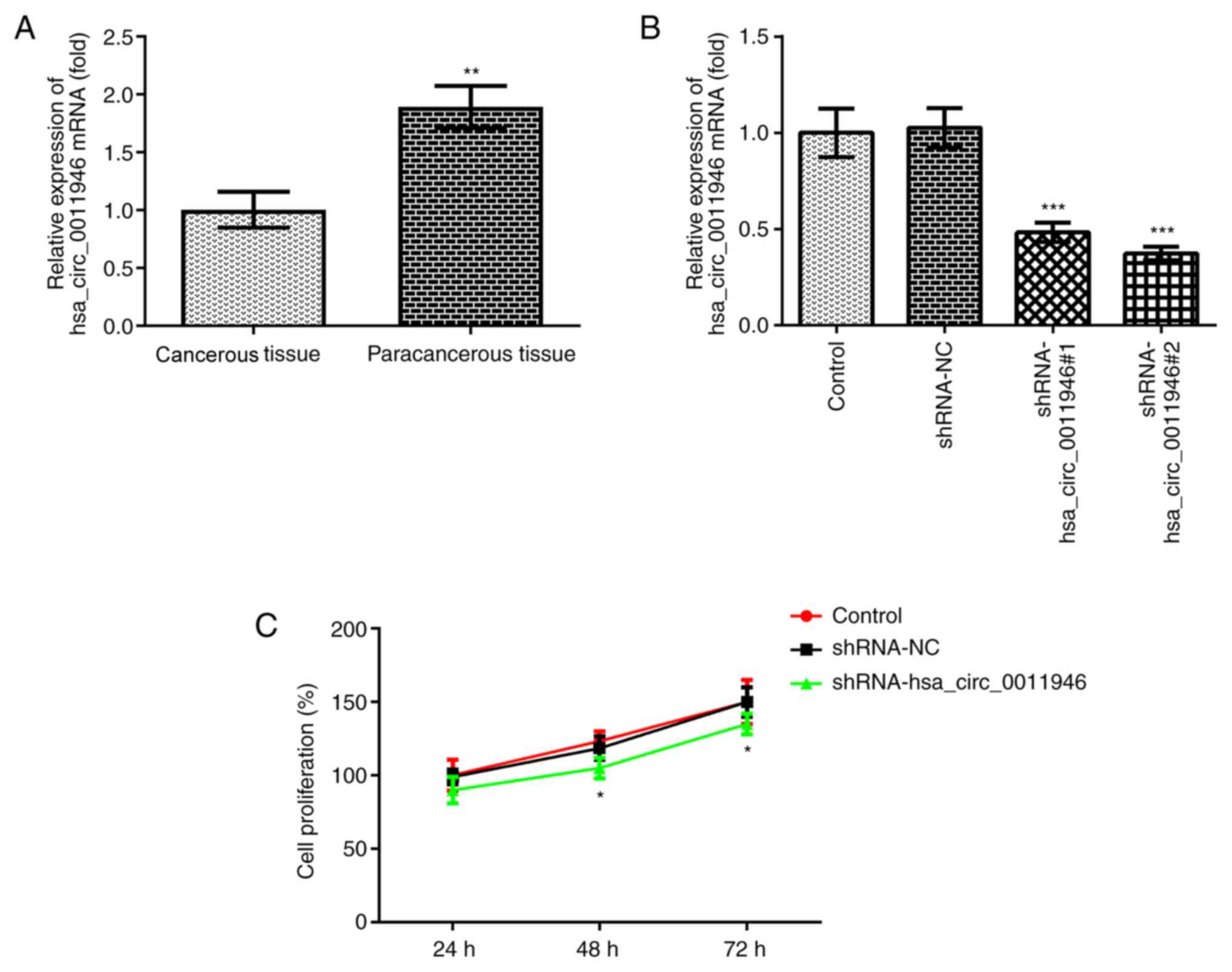
hsa_circ_0011946 expression levels are
upregulated in cancer tissues of patients with SACC
RT-qPCR was used to analyze the expression levels of
hsa_circ_0011946 in cancerous tissues of patients with SACC. The
results revealed that, compared with paracancerous tissue, the
expression levels of hsa_circ_0011944 were significantly
upregulated in SACC tissue (Fig.
1A). Subsequently, SACC-LM cells were selected for use in
subsequent experiments.
Knockdown of hsa_circ_0011946 inhibits
the proliferation of SACC cells
Cell transfection experiments were used to interfere
with the expression levels of hsa_circ_0011946 in SACC-LM cells,
and RT-qPCR was used to verify the transfection efficiency
(Fig. 1B). The results demonstrated
that the expression levels of hsa_circ_0011946 were significantly
downregulated in shRNA-hsa_circ_0011946-1- and
shRNA-hsa_circ_0011946-2-transfected cells compared with the
shRNA-NC group, and this was more notable in the
shRNA-hsa_circ_0011946-2 group. Therefore, shRNA-hsa_circ_0011946-2
was selected for use in subsequent experiments. The cells were then
divided into the following three groups: i) Control group; ii)
shRNA-NC group; and iii) shRNA-hsa_circ_0011946 group. CCK-8 assays
were used to determine the cell proliferation rate. Compared with
the shRNA-NC group, no statistically significant differences were
observed in the cell proliferation rate following 24 h of
transfection with shRNA-hsa_circ_0011946. However, following 48 and
72 h of interference, the cell proliferation ability was
significantly decreased compared with the shRNA-NC group (Fig. 1C). These results suggested that the
knockdown of hsa_circ_0011946 expression may inhibit the
proliferation of SACC cells.
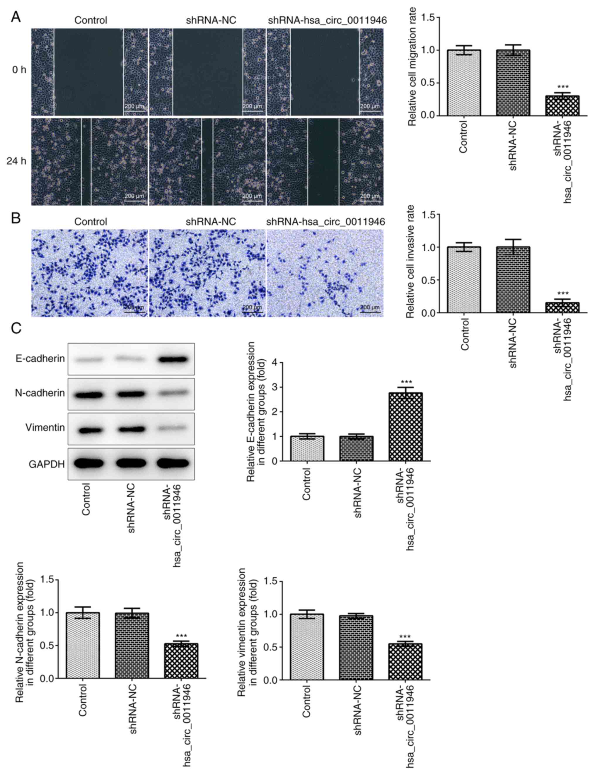
Knockdown of hsa_circ_0011946 inhibits
invasion, migration and EMT processes in SACC cells
The invasive and migratory abilities of cells
following the knockdown of hsa_circ_0011946 were analyzed, and the
results revealed that the invasive (Fig. 2A) and migratory (Fig. 2B) abilities of cells in the
shRNA-hsa_circ_0011946 group were significantly decreased compared
with the shRNA-NC group. The expression levels of the following
EMT-related proteins were analyzed using western blotting:
E-cadherin, N-cadherin and vimentin (Fig. 2C). Compared with the shRNA-NC group,
the expression levels of E-cadherin were significantly upregulated
in the shRNA-hsa_circ_0011946 group, while the expression levels of
N-cadherin and vimentin were significantly downregulated,
suggesting that hsa_circ_0011946 interfered with the EMT process in
SACC cells.
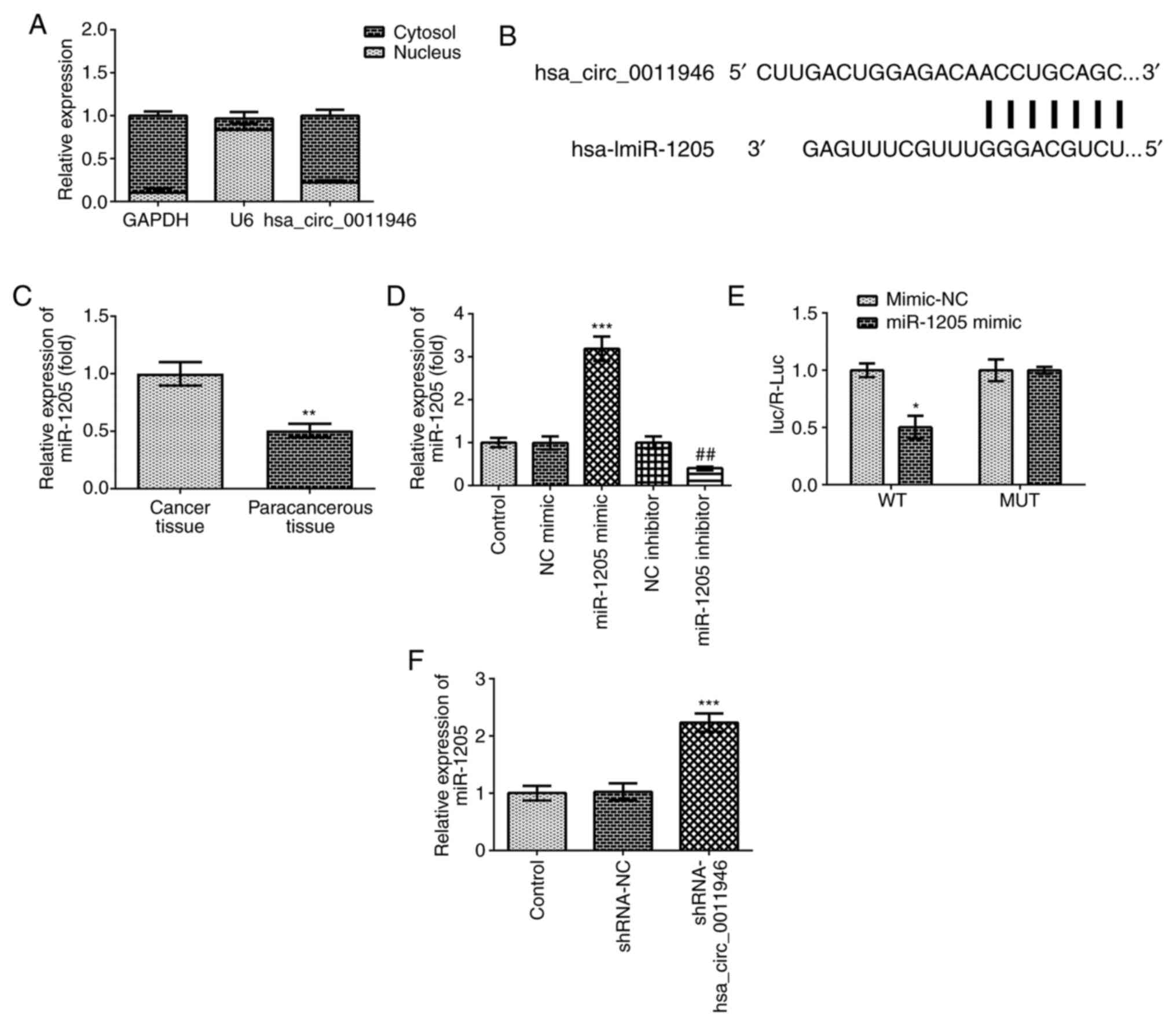
hsa_circ_0011946 negatively regulates
the expression levels of miR-1205 by acting as a sponge
The presence of hsa_circ_0011946 was confirmed by
RT-qPCR (Fig. 3A). The binding site
between hsa_circ_0011946 and miR-1205 was predicted using circRNA
interactome tools (Fig. 3B). The
expression levels of miR-1205 were subsequently found to be
significantly downregulated in SACC tissues compared with
paracancerous tissue (Fig. 3C).
RT-qPCR analysis revealed that cell transfection was successful
(Fig. 3D). A dual-luciferase
reporter assay was performed to validate the complementary binding
between hsa_circ_0011946 and miR-1205 (Fig. 3E). The expression levels of miR-1205
were significantly upregulated following the knockdown of
hsa_circ_0011946 (Fig. 3F). These
results suggested that hsa_circ_0011946 may negatively regulate the
expression levels of miR-1205 by acting as a sponge.
hsa_circ_0011946 promotes the
malignant process of SACC by downregulating miR-1205
expression
The cells were divided into control, shRNA-NC,
shRNA-hsa_circ_001194, shRNA-hsa_circ_0011946 + NC inhibitor and
shRNA-hsa_circ_0011946 + miR-1205 inhibitor groups. Cell viability
was detected using a CCK-8 assay and the results revealed that the
knockdown of miR-1205 could reverse the inhibition of cell
proliferation following the knockdown of hsa_circ_0011946 (Fig. 4A). Compared with
shRNA-hsa_circ_0011946 + NC inhibitor group, the cell invasive
(Fig. 4B) and migratory (Fig. 4C) abilities were significantly
increased in the shRNA-hsa_circ_0011946 + miR-1205 inhibitor group.
Subsequently, the expression levels of EMT-related proteins were
analyzed using western blotting. As shown in Fig. 4D, compared with the shRNA-NC group,
the expression levels of E-cadherin were significantly upregulated
in the shRNA-hsa_circ_0011946 group, while the expression levels of
N-cadherin and vimentin were significantly downregulated. The
expression levels of E-cadherin protein were downregulated in the
shRNA-hsa_circ_0011946 + miR-1205 inhibitor group, while the
expression levels of N-cadherin and vimentin were upregulated in
the shRNA-hsa_circ_0011946 + miR-1205 inhibitor group compared with
the shRNA-hsa_circ_0011946 + NC inhibitor group. Altogether, these
findings suggested that circRNA hsa_circ_0011946 may promote the
malignant progression of SACC by downregulating miR-1205
expression.
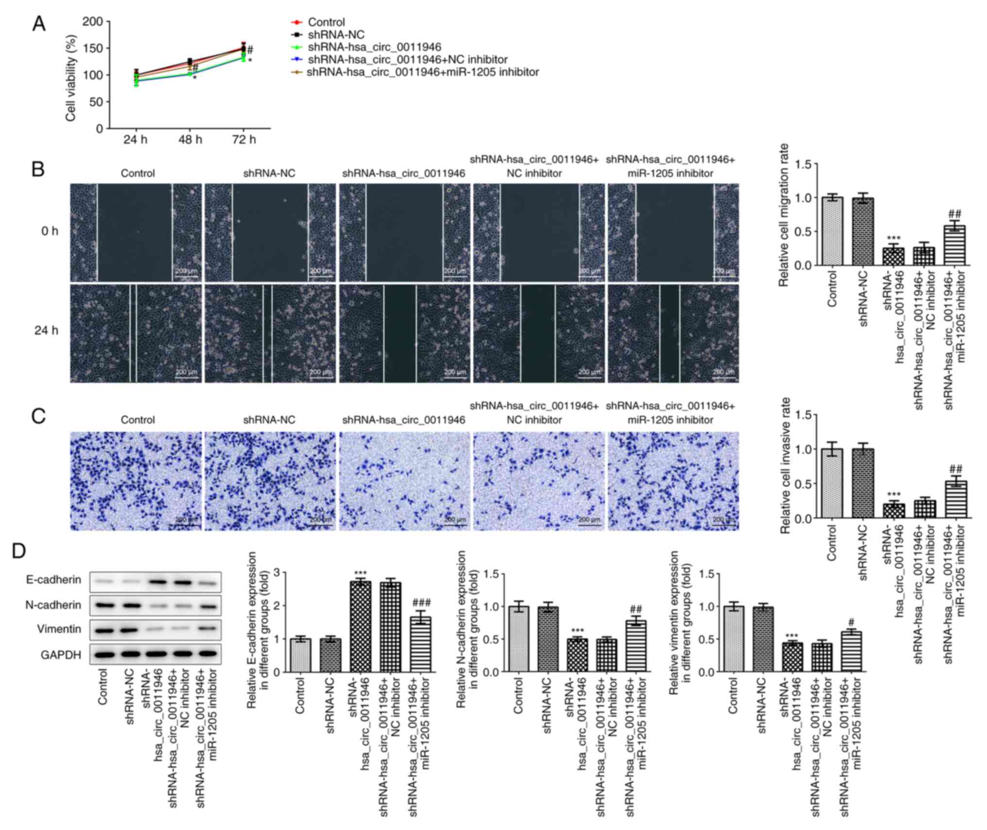 | Figure 4Hsa_circ_0011946 promotes the
malignant process of SACC by downregulating miR-1205. (A) Cell
Counting Kit-8 detected the cell viability. (B) Wound healing
detected the cell migration ability (magnification, x100). (C)
Transwell detected the cell invasion ability (magnification, x100).
(D) Western blot detected the expression of epithelial-mesenchymal
transition-related proteins. *P<0.05,
***P<0.001 vs. shRNA-NC. #P<0.05,
##P<0.001, ###P<0.001 vs.
shRNA-hsa_circ_0011946 + NC inhibitor. Circ, circular; NC, negative
control; SACC, salivary adenoid cystic carcinoma; miR, micoRNA; sh,
short hairpin. |
Discussion
SACC is a salivary gland malignancy with high
incidence rates and complex occurrence and progression mechanisms,
which remain poorly characterized. Following treatment with
surgical resection combined with chemoradiotherapy in the clinic,
the survival rate of patients with SACC can be improved (13); however, due to the strong invasive
nature of the tumor and the neurotropic growth characteristics,
recurrence and metastasis often occur following treatment, which
seriously affects the quality of life and survival time of patients
(14). Therefore, identifying the
underlying mechanisms of SACC progression and determining genes or
biomarkers associated with the invasion and metastasis of SACC may
be of vital importance for the early diagnosis and treatment of
patients, in addition to improving prognosis.
circRNAs, members of the non-coding RNA family, are
named due to their unique covalently closed circular structure
(15). circRNAs are characterized
by a strong stability, high abundance and species conservation, as
well as demonstrating cell and tissue specificity (16). circRNAs widely exist in the human
body and serve an important role in physiological and pathological
processes. Currently, circRNAs have been found to be differentially
expressed in a variety of cancer types, which suggests their
potential for use in tumor diagnosis and their potential ability to
predict the prognosis of patients (17). Thus, circRNAs are predicted to
become novel tumor biomarkers or prognostic indicators. circRNAs
regulate gene transcription and translation through various
different mechanisms, thereby promoting or inhibiting the
proliferation, apoptosis, invasion and metastasis of cancer cells,
and ultimately regulating tumor progression (18). The results of the present study
revealed that the expression levels of hsa_circ_0011946 were
upregulated in cancer tissues of SACC patients. In the present
study, no inhibitor of has_circ_0011946 was identified during the
literature review, so cell transfection techniques were used to
inhibit the expression of has_circ_0011946 in SACC-LM cells
(5,6). Following the knockdown of
hsa_circ_0011946, the proliferative, invasive and migratory
abilities of SACC cells were significantly decreased, and the EMT
process was inhibited. The present results also demonstrated that,
following the knockdown of hsa_circ_0011946, the proliferation of
SACC cells was not significantly inhibited, and the inhibitory
effect was only apparent following an extended period of knockdown
(48 and 72 h). The possible reasons for these results are that SACC
cells are locally aggressive (19),
thus the knockdown of hsa_circ_0011946 expression may exert little
effect on proliferation.
circRNAs have been found to serve a regulatory role
through a variety of mechanisms; for example, circRNAs can act as
sponges to absorb miRNAs by binding to their miRNA binding sites,
and the latter can influence the metabolic process of cells by
regulating the expression and activity of target proteins (20). In the present experiments, the
knockdown of hsa_circ_0011946 expression targeted and downregulated
miR-1205 expression to regulate the malignant progression of SACC
cells.
Notably, there were numerous limitations in the
current study. Hsa_circ_0011946 was not overexpressed in normal
human salivary gland cells, which may have represented an improved
study design. In future experiments, the functional differences of
the circRNA hsa_circ_0011946 should be investigated in normal HSG
and SACC tumor cells.
In conclusion, the results of the present study
suggested that the circRNA hsa_circ_0011946 may promote the
malignant process of SACC by downregulating miR-1205 expression,
which may provide a novel target for the treatment with SACC.
Acknowledgements
Not applicable.
Funding
Funding: No funding was received.
Availability of data and materials
The datasets used and/ot analyzed during the current
study are available from the corresponding author on reasonable
request.
Authors' contributions
HW and HD wrote the manuscript and analyzed the
data. JL and CX performed the experiments and supervised the study.
HD searched the literature and revised the manuscript for important
intellectual content. HD and HW confirmed the authenticity of all
the raw data. All authors read and approved the final
manuscript.
Ethics approval and consent to
participate
The experiments were approved by The Ethics
Committee of The People's Hospital of Guangxi Zhuang Autonomous
Region, and informed consent was signed by all participants.
Patients consent for publication
Informed consent was signed by all participants.
Competing interests
The authors declare that they have no competing
interests.
References
|
1
|
Bradley PJ: Adenoid cystic carcinoma
evaluation and management: Progress with optimism! Curr Opin
Otolaryngol Head Neck Surg. 25:147–53. 2017.PubMed/NCBI View Article : Google Scholar
|
|
2
|
Miller JA, An D, Shafique K, Song S, Rao
RA, Viswanathan K, Eykman E, Wiles A, Ali SZ, Field A, et al:
Mucoepidermoid carcinoma, acinic cell carcinoma, and adenoid cystic
carcinoma on fine-needle aspiration biopsy and The Milan System: An
international multi-institutional study. J Am Soc Cytopathol.
8:270–277. 2019.PubMed/NCBI View Article : Google Scholar
|
|
3
|
Šteiner P, Pavelka J, Vaneček T,
Miesbauerová M and Skálová A: Molecular methods for detection of
prognostic and predictive markers in diagnosis of adenoid cystic
carcinoma of the salivary gland origin. Cesk Patol. 54:132–136.
2018.PubMed/NCBI
|
|
4
|
Ren L, Zhai H, Wang XL, Li JZ and Xia YH:
Hsa_circ_0011946 promotes the migration and invasion of
hepatocellular carcinoma by inducing EMT process. Eur Rev Med
Pharmacol Sci. 24:1108–1115. 2020.PubMed/NCBI View Article : Google Scholar
|
|
5
|
Meng Y, Zhao EY, Zhou Y, Qiang DX, Wang S,
Shi L, Jiang LY and Bi LJ: Circular RNA hsa_circ_0011946 promotes
cell growth, migration, and invasion of oral squamous cell
carcinoma by upregulating PCNA. Eur Rev Med Pharmacol Sci.
24(7560)2020.PubMed/NCBI View Article : Google Scholar
|
|
6
|
Zhou J, Zhang WW, Peng F, Sun JY, He ZY
and Wu SG: Downregulation of hsa_circ_0011946 suppresses the
migration and invasion of the breast cancer cell line MCF-7 by
targeting RFC3. Cancer Manag Res. 10:535–544. 2018.PubMed/NCBI View Article : Google Scholar
|
|
7
|
Wang G, Zhang H and Li P: Upregulation of
hsa_circRNA_102958 indicates poor prognosis and promotes ovarian
cancer progression through miR-1205/SH2D3A axis. Cancer Manag Res.
12:4045–4053. 2020.PubMed/NCBI View Article : Google Scholar
|
|
8
|
Lin J, Liao S, Li E, Liu Z, Zheng R, Wu X
and Zeng W: circCYFIP2 acts as a sponge of miR-1205 and affects the
expression of its target gene E2F1 to regulate gastric cancer
metastasis. Mol Ther Nucleic Acids. 21:121–132. 2020.PubMed/NCBI View Article : Google Scholar
|
|
9
|
Li P, Lin XJ, Yang Y, Yang AK, Di JM,
Jiang QW, Huang JR, Yuan ML, Xing ZH, Wei MN, et al: Reciprocal
regulation of miR-1205 and E2F1 modulates progression of laryngeal
squamous cell carcinoma. Cell Death Dis. 10(916)2019.PubMed/NCBI View Article : Google Scholar
|
|
10
|
Jiang Y, Liu G, Ye W, Xie J, Shao C, Wang
X and Li X: ZEB2-AS1 accelerates epithelial/mesenchymal transition
through miR-1205/CRKL pathway in colorectal cancer. Cancer Biother
Radiopharm. 35:153–162. 2020.PubMed/NCBI View Article : Google Scholar
|
|
11
|
Yang Y, Ding L, Li Y and Xuan C:
Hsa_circ_0039411 promotes tumorigenesis and progression of
papillary thyroid cancer by miR-1179/ABCA9 and miR-1205/MTA1
signaling pathways. J Cell Physiol. 235:1321–1329. 2020.PubMed/NCBI View Article : Google Scholar
|
|
12
|
Livak KJ and Schmittgen TD: Analysis of
relative gene expression data using real-time quantitative PCR and
the 2(-Delta Delta C(T)) method. Methods. 25:402–408.
2001.PubMed/NCBI View Article : Google Scholar
|
|
13
|
Laurie SA, Ho AL, Fury MG, Sherman E and
Pfister DG: Systemic therapy in the management of metastatic or
locally recurrent adenoid cystic carcinoma of the salivary glands:
A systematic review. Lancet Oncol. 12:815–824. 2011.PubMed/NCBI View Article : Google Scholar
|
|
14
|
Garg M, Tudor-Green B and Bisase B:
Current thinking in the management of adenoid cystic carcinoma of
the head and neck. Br J Oral Maxillofac Surg. 57:716–721.
2019.PubMed/NCBI View Article : Google Scholar
|
|
15
|
Hsiao KY, Sun HS and Tsai SJ: Circular
RNA-new member of noncoding RNA with novel functions. Exp Biol Med
(Maywood). 242:1136–1341. 2017.PubMed/NCBI View Article : Google Scholar
|
|
16
|
Han B, Chao J and Yao H: Circular RNA and
its mechanisms in disease: From the bench to the clinic. Pharmacol
Ther. 187:31–44. 2018.PubMed/NCBI View Article : Google Scholar
|
|
17
|
Kristensen LS, Andersen MS, Stagsted LVW,
Ebbesen KK, Hansen TB and Kjems J: The biogenesis, biology and
characterization of circular RNAs. Nat Rev Genet. 20:675–691.
2019.PubMed/NCBI View Article : Google Scholar
|
|
18
|
Vo JN, Cieslik M, Zhang Y, Shukla S, Xiao
L, Zhang Y, Wu YM, Dhanasekaran SM, Engelke CG, Cao X, et al: The
landscape of circular RNA in cancer. Cell. 176:869–681.e13.
2019.PubMed/NCBI View Article : Google Scholar
|
|
19
|
Bouaichi A, Aimad-Eddine S, Mommers XA,
Ella B and Zwetyenga N: Intra-mandibular adenoid cystic carcinoma.
Rev Stomatol Chir Maxillofac Chir Orale. 115:100–104.
2014.PubMed/NCBI View Article : Google Scholar
|
|
20
|
Panda AC: Circular RNAs Act as miRNA
sponges. Adv Exp Med Biol. 1087:67–79. 2018.PubMed/NCBI View Article : Google Scholar
|