Non-small cell lung cancer (NSCLC) is the primary
histological type of lung cancer worldwide; it accounts for ~85% of
all lung cancer cases and is a leading cause of cancer-related
mortality. NSCLC is histologically divided into adenocarcinoma,
squamous cell carcinoma and large cell carcinoma (1). Epidemiology has revealed that lung
cancer accounts for ~18% of all cancer-related mortalities and
remains the major cause of cancer-related mortality worldwide. The
5-year survival rate ranges from 50 to 70% for stage I and from 1
to 5% for stage IV; however, only ~20% of patients are diagnosed
with stage I disease and the majority of patients with NSCLC are
diagnosed at an advanced stage (stage IV) with distant metastasis
and poor prognosis (2). Thus, it
is important to investigate the regulatory mechanisms of lung
cancer metastasis, and discover new predictive methods and
therapeutic targets. Findings from these investigations may aid in
the identification of new treatments for patients with NSCLC and
improve their prognosis.
Tumor metastasis occurs as a complex cascade
involving invasion, dissemination and distant colonization.
Specifically, primary tumor cells invade the surrounding tissues,
enter the circulatory system, survive during blood circulation,
enter the parenchyma of distant tissues and finally colonize
distant sites for subsequent proliferation (3). Postsurgical recurrence is the major
cause of mortality in the majority of patients with NSCLC (3). Brain and bone metastases can be
easily observed in patients with advanced NSCLC; for example, bone
metastasis occurs in 30-40% of patients with NSCLC (4). The present study summarized the
regulatory processes underlying metastasis of NSCLC, and analyzed
the diagnostic and prognostic factors related to tumor metastasis
of NSCLC to further clarify the molecular mechanisms underlying
metastasis in NSCLC.
Programmed cell death (PCD) refers to orderly and
autonomous cell death that is controlled by a series of endogenous
genes. PCD includes apoptosis, autophagy, pyroptosis, ferroptosis
and necroptosis (5). PCD serves a
key role in the treatment of various metastatic tumors. The present
study summarized the regulatory molecular mechanisms involved in
PCD and their corresponding regulatory factors, including
non-coding (nc)RNAs, proteins and compounds, during the process of
NSCLC bone metastasis. Simultaneously, it drew attention to future
research trends and identified several unsolved problems relevant
to NSCLC metastasis.
Aberrant apoptosis often leads to the progression
and metastasis of various types of human cancer. Cell apoptosis
inhibits metastasis at multiple stages ranging from invasion from
the primary site to distant colonization (3). During the progression and metastasis
of NSCLC, apoptosis is regulated by various factors, such as
proteins, ncRNAs and other compounds. Previously, research
examining the metastasis of NSCLC has primarily focused on the
regulatory mechanisms of novel proteins, microRNAs (miRNAs/miRs),
long ncRNAs (lncRNAs) and circular RNAs (circRNAs) (5,6).
Recently, a series of novel and important proteins
have been reported to be associated with apoptosis, metastasis and
prognosis of NSCLC by modulating epithelial-mesenchymal transition
(EMT) progression or chemotherapeutic drug resistance, and by
regulating various cancer-related signaling pathways in patients
with NSCLC. The proteins identified in the regulation of NSCLC
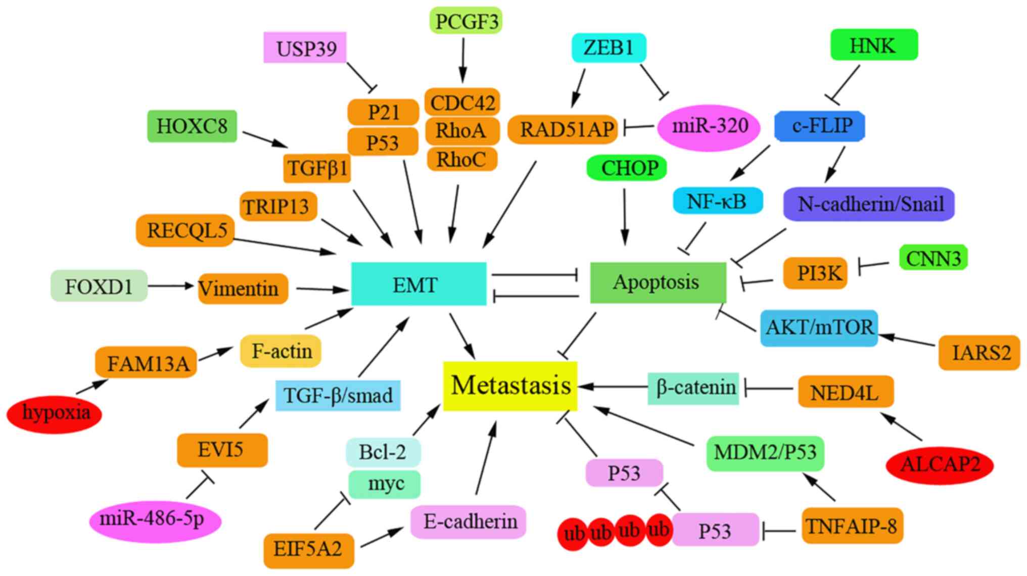
metastasis are summarized in Fig.
1. Interference with forkhead box D1 (FOXD1) has been shown to
inhibit proliferation and metastasis in vivo by increasing
the apoptotic rate of NSCLC cells (6). In addition, the oncogene ecotropic
viral integration site 5 (EVI5) has been reported to be upregulated
in NSCLC tissues. Knockdown of EVI5 may suppress cell invasion via
the TGF-β/Smad signaling pathway in NSCLC cells (7). The natural compound honokiol may also
inhibit the migration of NSCLC cells by targeting cellular
FLICE-inhibitory protein via the NF-κB pathway and the
N-cadherin/Snail pathway (8).
Knockdown of eukaryotic translation initiation factor 5A2 can
significantly inhibit cell proliferation and metastasis by inducing
the apoptosis of NSCLC cells (9).
Several regulators inhibit NSCLC metastasis by
inducing cell cycle arrest and apoptosis. Upregulation of thyroid
hormone receptor interacting protein 13 (TRIP13) has been reported
to be associated with tumor metastasis via EMT. TRIP13 knockdown
can promote cell apoptosis by inducing cell cycle arrest at the S
phase, and can inhibit the invasion and migration of NSCLC cells
(10). Calponin 3 overexpression
can also suppress the migration of NSCLC cells by inhibiting the
PI3K/AKT pathway and promoting G1-phase arrest (11). Furthermore, inhibition of
isoleucyl-tRNA synthetase 2, mitochondrial (IARS2) has been shown
to induce cell cycle arrest at the G0/G1
stage and mitochondrial apoptosis; thus, IARS2 may function as an
oncogene to promote NSCLC tumorigenesis by activating the AKT/mTOR
pathway (12). The
deubiquitinating enzyme ubiquitin-specific protease 39 (USP39) has
been reported to be overexpressed in NSCLC tissues. Notably,
interference with the expression of USP39 has been demonstrated to
induce cell cycle arrest at G2/M and to induce apoptosis
by activating the p53 and p21 pathways, thus resulting in
inhibition of metastasis (13).
Tumor necrosis factor-α-induced protein 8 may promote drug
resistance in NSCLC via the MDM2/p53 pathway (14). Chromatin assembly factor 1 subunit
B (CHAF1B) has been shown to be highly expressed in NSCLC tissues,
and high CHAF1B levels can lead to poor clinical outcomes and lymph
node metastasis (15). By
contrast, CHAF1B knockdown may contribute to cell cycle arrest and
the induction of apoptosis by activating the p53-dependent
apoptotic pathway.
Some newly identified regulators have been reported
to induce apoptosis and thus inhibit metastasis of NSCLC cells and
this has been demonstrated to be associated with the clinical
prognosis of patients with NSCLC. For example, homeobox (HOCX) 8
has been shown to be upregulated in NSCLC specimens and to function
as a transcriptional activator that induces the expression of
TGF-β1. High levels of HOXC8 have been reported to be associated
with tumor-node metastasis and poor relapse-free survival in
patients with NSCLC (16).
Polycomb group ring finger 3 has been demonstrated to be
overexpressed in tumors of patients with NSCLC and to be positively
associated with lymph node metastasis; it can promote migration via
the PI3K/AKT pathway (17).
Conversely, miR-320a expression levels have been shown to be
reduced in NSCLC tissues and cells. Zinc finger E-box binding
homeobox 1 can inhibit the expression of miR-320a that contributes
to the high expression level of RAD51AP1 to promote tumor growth
and metastasis in NSCLC (18).
Vitamin D may suppress metastasis of NSCLC cells by promoting
apoptosis via the PI3K/AKT/mTOR signaling pathway (19). In addition, C/EBP homologous
protein has been reported to suppress metastasis and to sensitize
NSCLC cells to cisplatin (DPP) by blocking the Bcl-2/JNK pathway
(20). RecQ-like helicase 5
(RECQL5), a member of the RecQ helicase family, may promote the
migration of NSCLC cells and has been shown to be strongly
associated with the poor prognosis of lung adenocarcinoma;
knockdown of RECQL5 can increase the level of apoptosis in
DPP-resistant A549 cells (21).
Elevated levels of FOXD1 have been revealed to be even higher in
NSLC tissues compared with normal controls and to be associated
with poor prognosis in patients with NSCLC (6).
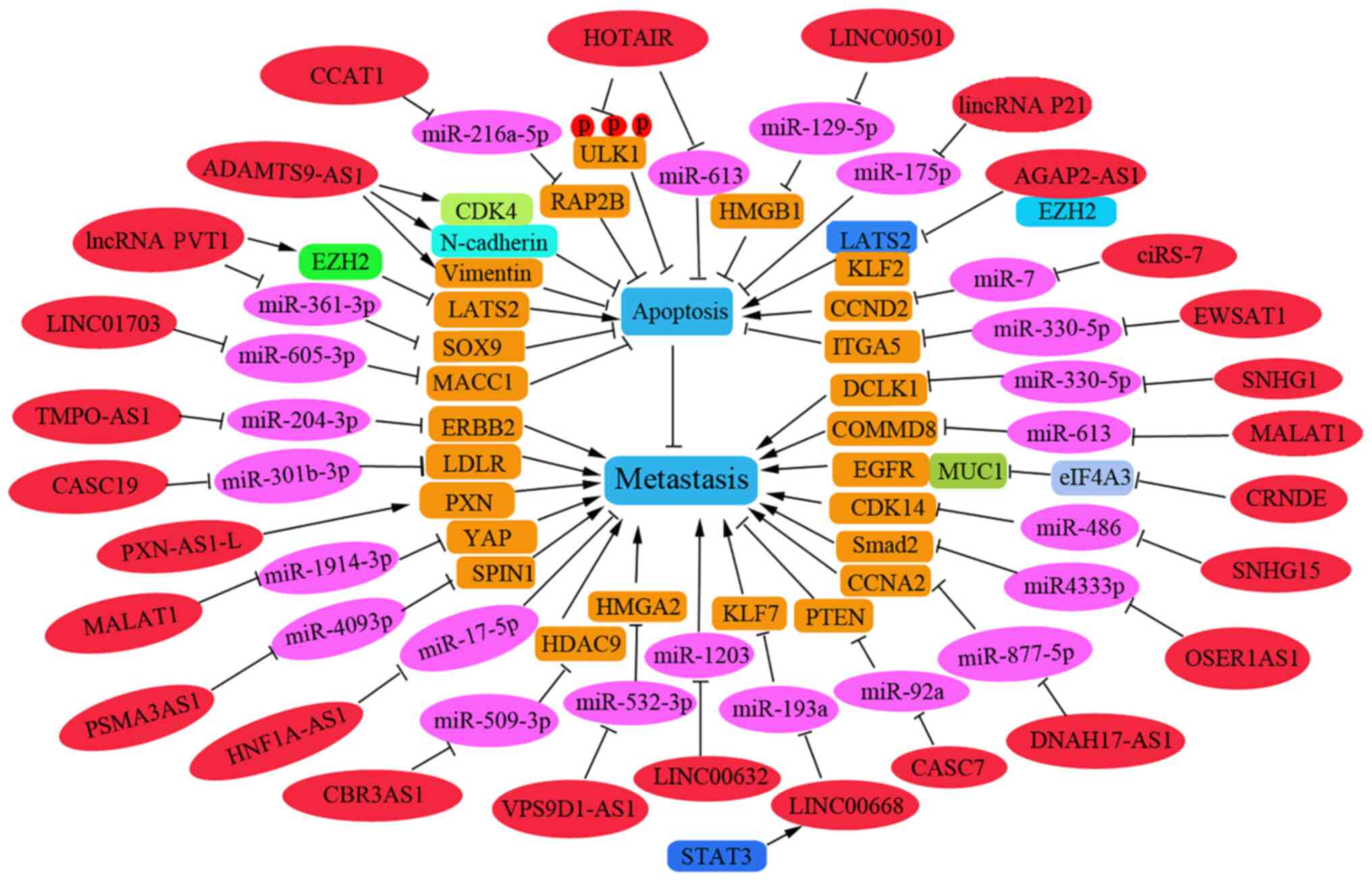
A number of lncRNAs have been reported to be
involved in NSCLC metastasis by regulating apoptosis-related
pathways (22-25).
As presented in Fig. 2, these
regulatory lncRNAs promote or inhibit the apoptosis process during
the metastasis of NSCLC cells. In addition, several lncRNAs and
associated miRNAs have been reported to be associated with the
prognosis of NSCLC, and thus could be used as effective therapeutic
and diagnostic targets in patients with NSCLC.
Certain lncRNAs have been reported to act as
suppressor genes in NSCLC metastasis by promoting apoptosis.
LINC00632 was shown to be downregulated in NSCLC tissues, and low
levels of LINC00632 can promote lymph node metastasis and distant
metastasis. Furthermore, LINC00632 was revealed to inhibit tumor
growth in nude mice with NSCLC by downregulating miR-1203(22). The lncRNA cancer susceptibility
candidate 7 may inhibit the growth and invasion of NSCLC cells and
promote apoptosis in vitro through phosphatase and tensin
homolog upregulation by sponging miR-92a (23). LINC00520 can inhibit cell
proliferation, metastasis and EMT, and increase the extent of
apoptosis in NSCLC cells. Mechanistically, LINC00520 has been shown
to serve as a competing endogenous (ce)RNA involved in the
regulation of miR-577 that affects the target gene CCNE2(24). Upregulated lncRNA-SNHG7 (SNHG7) has
been reported to be positively correlated with Fas apoptosis
inhibitory molecule 2 (FAIM2) in lung cancer cells and to function
as a molecular sponge to sequester endogenous miRNA (25). Knockdown of SNHG7 in vivo
has been reported to significantly inhibit tumor growth and
decrease tumor volume, which is accompanied by enhanced miR-193b
expression and reduced FAIM2 levels (25). Nuclear paraspeckle assembly
transcript (NEAT) 1 may act as a ceRNA against let-7a, and IGF-2 is
a direct target of let-7a; thus, knockdown of NEAT1 can inhibit
cell viability and reduce migration by increasing apoptosis of
NSCLC cells by regulating the NEAT1/let-7a/IGF2 pathway (26).
LncRNAs have recently been identified as oncogenes
that promote the invasion and migration of NSCLC cells. For
example, the ncRNA plasmacytoma variant translocation 1 (PVT1) was
shown to be highly expressed in 105 patients with NSCLC and to
promote tumor-to-node metastasis. Knockdown of PVT1 was revealed to
inhibit metastasis and proliferation by increasing LATS2
transcription via the Mdm2-p53 signaling pathway (27). In addition, PVT1 may act as a miRNA
sponge and could promote cell metastasis of NSCLC by regulating
miR-361-3p/SOX9 and activating the Wnt/β-catenin signaling pathway
(28). AGAP2-AS1 has been reported
to function as an oncogene to inhibit the transcription of the
tumor-suppressors LATS2 and KLF2; thus, interference with AGAP2-AS1
may suppress migration and induce apoptosis in NSCLC cells
(29). The lncRNA HOTAIR has been
reported to be overexpressed in NSCLC specimens and can negatively
regulate the levels of miR-613. Conversely, knockdown of HOTAIR
expression may promote cell apoptosis and suppress the migration of
NSCLC by down-regulating miR-613(30). Furthermore, interference with
lncRNA ADAMTS9-AS1 has been shown to suppress EMT in patients with
NSCLC, and cause cell cycle arrest at G0/G1
and the induction of cell apoptosis, which is associated with
downregulation of CDK4, N-cadherin and vimentin, and upregulation
of Bad and E-cadherin (31). The
lncRNA PXN-AS1-L has been shown to be upregulated in NSCLC tissues
compared with levels in noncancerous lung tissues and to be further
upregulated in NSCLC bone metastasis tissues, thus suggesting that
higher levels of PXN-AS1-L may be positively related to advanced
TNM stage and poor prognosis. This lncRNA can also promote NSCLC
progression and metastasis by upregulating PXN during NSCLC
progression (32).
The lncRNA HNF1A-AS1 may promote cell invasion by
targeting miR-17-5p in patients with NSCLC (33). By contrast, lincRNAp21 can
effectively inhibit migration and promote the apoptosis of NSCLC
cells by targeting miR-175p (34).
The lncRNA CBR3AS1 can promote metastasis of NSCLC by sponging
miR-509-3p and competitively upregulating histone deacetylase 9
expression (35). Overexpression
of SNHG15 has been reported to promote NSCLC tumorigenesis through
regulating the CDK14 protein and by sponging miR-486(36). An elevated expression level of
OSER1AS1 has been associated with lymph node metastasis in patients
with NSCLC. OSER1AS1 may promote the malignant properties of NSCLC
by sponging miR-4333p and upregulating Smad2 expression (37).
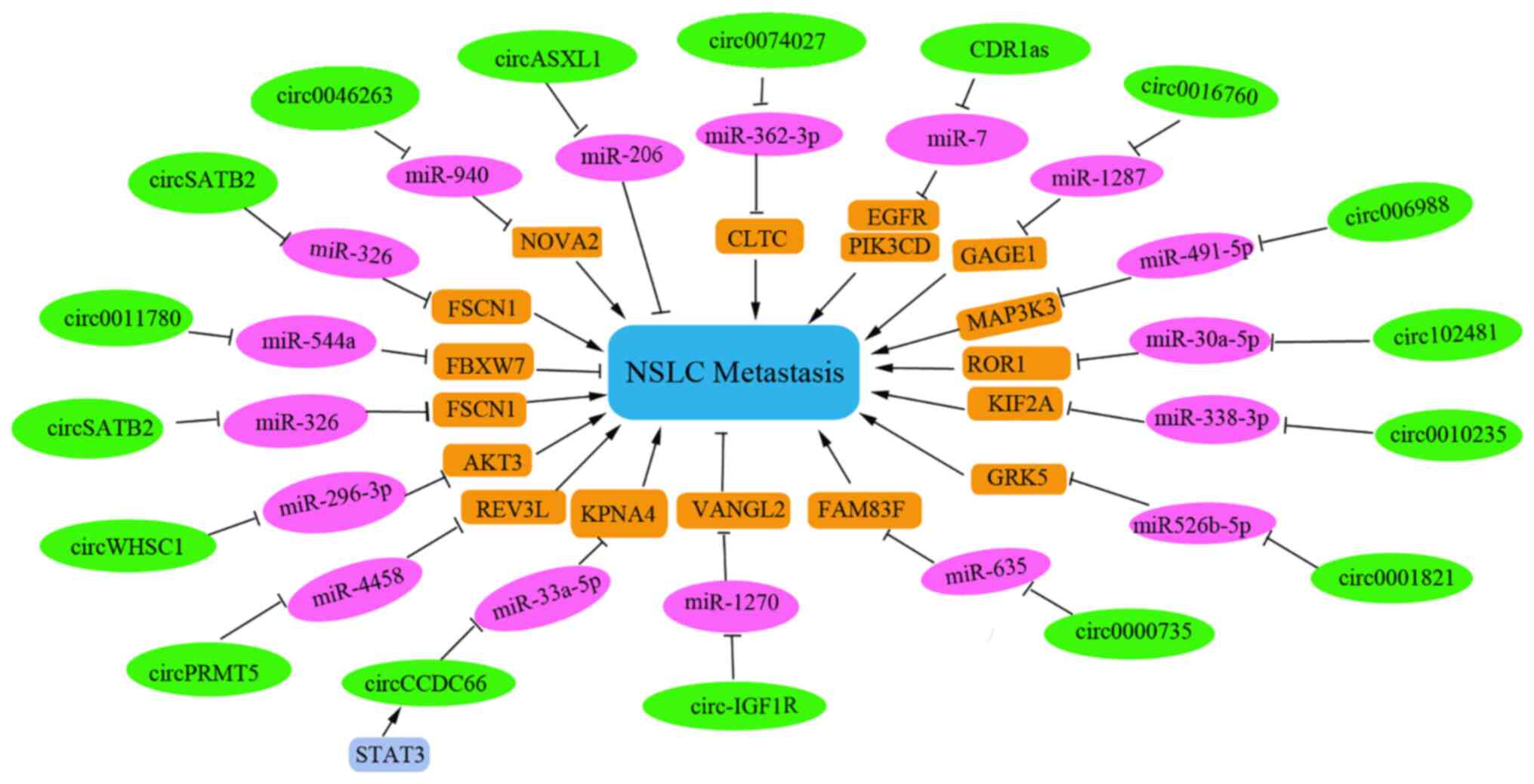
CircRNA is a new member of the ncRNA family that
functions as a miRNA sponge to influence gene transcription. These
RNAs are highly tissue-specific in mammals. An increasing number of
circRNAs have been identified that regulate apoptosis in NSCLC
metastasis. The specific and regulatory circRNAs involved in NSCLC
metastasis that function by inducing or suppressing apoptosis in
NSCLC cells have been summarized in the present study.
Certain circRNAs serve as tumor suppressor gene. For
instance, low hsa_circ_11780 expression levels have been reported
to be associated with distant metastasis and poor overall survival.
Overexpression of hsa_circ_11780 can significantly suppress the
metastasis of NSCLC cells in vitro and in vivo by
decreasing FBXW7 level to suppress the function of miR-544a
(46).
Several circRNAs have been reported to be involved
in chemotherapeutic drug resistance and the metastasis of NSCLC
cells. Knockdown of circ_0001821 may inhibit metastasis and
paclitaxel resistance in NSCLC cells via the miR-526b-5p/GRK5
pathway (47). Furthermore,
circASXL1 silencing can inhibit DDP resistance and tumorigenesis by
sponging miR-206 in NSCLC tissues and cells (48). Interference with circ-PRMT5 has
also been reported to induce apoptosis, reverse DDP resistance and
reduce metastasis in DDP-resistant NSCLC. Notably, circ-PRMT5 can
sponge miR-4458 to increase REV3L expression levels and to promote
DDP resistance in patients with NSCLC (49). Tumor-derived exosomal
circRNA_102481 has been reported to correlate with TNM stage, tumor
differentiation status, brain metastasis, progression-free survival
and overall survival duration. This RNA can promote cell
proliferation, inhibit cell apoptosis and induce EGFR-TKI
resistance by sponging miR-30a-5p to increase ROR1 levels in NSCLC
(50).
Furthermore, the expression of several circRNAs has
been shown to be associated with clinicopathological parameters and
prognostic value in patients with NSCLC. For example, CDR1as has
been considered an independent prognostic factor for patients with
NSCLC and knockdown of CDR1as can upregulate miR-7, a process that
leads to the induction of cell apoptosis and G1/S
arrest. Furthermore, high expression levels of CDR1as have been
shown to be correlated with advanced TNM stage, increased lymph
node metastasis and shorter overall survival time (51). Notably, circSATB2 has also been
reported to be highly expressed in NSCLC cells and tissues, and to
be associated with lung cancer metastasis. This circRNA can promote
the progression and invasion of NSCLC cells via upregulation of
fascin homolog 1 and actin-bundling protein 1, and downregulation
of miR-326; this can provide a potential biomarker for the
diagnosis of NSCLC (52).
Furthermore, upregulation of circ_0016760 can be indicative of
increased lymph node metastasis and poor prognosis in NSCLC, and
can also promote cell progression and metastasis and inhibit
apoptosis through the miR-1287/GAGE1 axis (53).
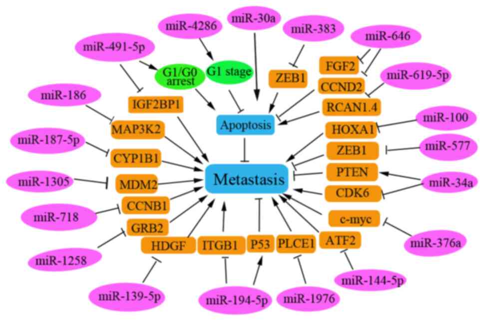
miRNAs are the most abundant endogenous small ncRNAs
and are abnormally expressed in various types of human cancer. The
miRNAs affecting NSCLC metastasis are summarized in Fig. 4. Several miRNAs function as
oncogenes that promote the invasion and migration of NSCLC cells.
Specifically, miR-4286 is considered an oncogenic miRNA in NSCLC
that is significantly enhanced in NSCLC tissues, and positively
related to lymph node metastasis and poor prognosis in patients
with NSCLC. Inhibition of miR-4826 has been reported to cause cell
cycle arrest at the G1 stage by regulating Runt-related
transcription factor 3 in NSCLC cells (54). miR-619-5p transported in
tumor-derived exosomes has been shown to promote angiogenesis and
metastasis of NSCLC by inhibiting RCAN1.4, which is a target of
miR-619-5p (55).
Furthermore, certain miRNAs have been reported to
function as tumor suppressors to promote apoptosis in NSCLC cells.
Tumor-suppressive miRNAs are significantly downregulated in NSCLC
tissues and cell lines. Downregulation of a number of
tumor-suppressive miRNAs has been reported to be correlated with
the poor prognosis of patients with NSCLC, including miR-187-5p
(56), miR-101(57), miR-718(58), miR-374b (59) and miR-30a (60). These miRNAs can exert anti-tumor
effects by inhibiting the invasion and migration of NSCLC cells by
targeting various key proteins to regulate their corresponding
signaling pathways. For example, miR-186 has been shown to inhibit
the metastasis of NSCLC cells by targeting MAP3K2(61). miR-491-5p can suppress NSCLC cell
proliferation by arresting cells in the G0/G1
phase and promoting cell apoptosis. In addition, miR-491-5p can
negatively regulate insulin-like growth factor 2 mRNA binding
protein 1 expression in malignant NSCLC (62) and miR-187-5p can negatively
regulate the target gene CYP1B1 to inhibit NSCLC cell metastasis
(56).
miR-718 has been demonstrated to serve a tumor
suppressive role by suppressing NSCLC progression in vivo by
directly targeting cyclin B1 mRNA (58). Overexpression of miR-374b may
inhibit metastasis of NSCLC by directly downregulating integrin β1
and upregulating p53(59).
Furthermore, overexpression of miR-139-5p in vitro can
induce apoptosis through a process that is partly mediated by
inhibiting hepatoma-derived growth factor (HDGF) expression in
NSCLC cells (63). In addition,
miR-139-5p may also induce apoptosis in NSCLC cells by targeting
and regulating YAF1 via the AKT/p38 MAPK signaling pathway
(64). The overexpression of
miR-34a has been shown to inhibit NSCLC growth and metastasis by
increasing PTEN and decreasing CDK6 expression levels, as reported
in tin miners in Gejiu County and farmers in Xuanwei (65). miR-30a may sensitize NSCLC cells by
promoting DDP/pemetrexed-induced apoptosis and by enhancing
autophagy in NSCLC cells (60).
miR-1258 has been reported to inhibit GRB2 expression and
inactivate the Ras/ERK pathway in NSCLC, thus inhibiting NSCLC
progression (66). Phospholipase C
ε1 has been demonstrated to promote metastasis of NSCLC cells, and
to be directly and negatively regulated by miR-1976 in NSCLC cells
(67). These identified miRNAs
function as tumor suppressors and could be used as therapeutic
targets for patients with NSCLC.
Autophagy is critical in cancer development and
metastasis, and serves a complex role in tumorigenesis in various
types of cancer (68).
Transfection with the autophagy-related gene ATG16L1-300T (vs.
300A) has been shown to promote the brain metastasis of NSCLC
cells, suggesting that autophagy may be closely associated with the
metastasis of NSCLC (68).
Autophagy is involved in cancer cell metastasis by regulating EMT.
The autophagy protein Beclin1 is a key regulator of tumorigenesis
and metastasis in NSCLC cells, and the expression levels of Beclin1
have been shown to be significantly reduced in NSCLC tissues
(69,70). Low Beclin1 expression levels have
also been observed in patients with NSCLC with more advanced stages
of cancer, increased lymph node metastasis and more poorly
differentiated tumors (70).
Reduced Beclin1 expression has also been associated with shorter
survival of patients with NSCLC, thus suggesting that autophagy,
which is an independent prognostic indicator for the survival of
patients with NSCLC, may be suppressed in these patients (69). Additionally, an increased level of
Nrf2 has been suggested as an independent prognostic factor in
patients with NSCLC (71). Thus,
the key autophagy-related proteins may be strongly associated with
the tumor stage and node metastasis of NSCLC, and this could help
to identify new independent prognostic biomarkers for patients with
NSCLC.
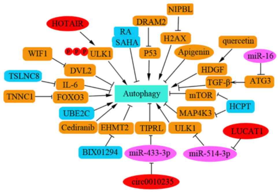
Previous research has suggested that autophagy
induction inhibits EMT or metastasis in NSCLC cells (72). Certain agents are involved in
regulating autophagy during the metastasis of NSCLC. For example,
the expression of transcription factor 21 (TCF21) has been reported
to be strongly associated with tumor stage, metastasis and invasion
of NSCLC, and to be significantly regulated by its methylation
level in patients with NSCLC. Promoter methylation of TCF21 may
suppress autophagy and increase cell apoptosis in lung cancer
metastasis (72). Furthermore,
knockdown of EHMT2 can decrease the proliferation of NSCLC cells by
inducing autophagy (73).
The combination of apigenin and gefitinib has been
shown to inhibit the activation of multiple oncogenes, including
c-Myc, HIF-1α and phosphorylated EGFR, and this combination can
also inhibit the AMPK signaling pathway in H1975 cells (74). These data revealed that apigenin
combined with gefitinib may promote apoptosis and inhibit autophagy
of EGFR L858R-T790M-mutated H1975 lung cancer cells and this could
be used as an alternative therapy for acquired resistance in
patients with NSCLC (74). The
drug 10-hydroxycamptothecin (HCPT) may promote autophagy in H1299
cells by suppressing MAP4K3 levels and downregulating the mTOR
pathway. HCPT in combination with Astragalus polysaccharide
(APS) has been reported to reduce the migration and invasion of
NSCLC cells compared with that observed in response to individual
treatments; this may be partly due to the induction of HCPT-induced
autophagy (75). Wnt inhibitory
factor 1-mediated autophagy may inhibit Wnt/βcatenin signaling by
downregulating dishevelled2 and suppressing activation of the
PI3K/AKT/mTOR pathway that contributes to the inhibition of
proliferation and promotion of apoptosis in NSCLC cells (76). In addition, treatment with miR-16
mimics can suppress TGF-β1-induced EMT by inducing autophagy in
NSCLC cells (76). Cell division
cycle-associated 4 can inhibit EMT and metastasis in NSCLC by
interacting with coactivator-associated arginine methyltransferase
1 to activate autophagy (77).
Consistently, previous studies have reported that
the induction of autophagy enhances the sensitivity of NSCLC cells
to chemotherapeutic drugs. For example, knockdown of NIPBL has been
shown to enhance the chemosensitivity of NSCLC cells via the DNA
damage response and autophagy pathways (78). Combined therapy with rapamycin and
suberoylanilide hydroxamic acid may also increase radiosensitivity
in these cells and inhibiting the levels of autophagy can markedly
weaken the radiotherapy sensitivity caused by this combined
treatment, thus suggesting that the inhibitory effect of
combination therapy may be partially due to the induction of
autophagy in NSCLC (79).
Cediranib has also been shown to exhibit effective anti-tumor
activities in NSCLC cells by inducing G1 phase cell
cycle arrest and autophagy via the MAPK/Erk1/2 and Akt/mTOR
pathways in A549 cells (80).
Notably, there is an opposing view that inducing
autophagy increases chemoresistance or radioresistance in NSCLC
cells. For example, troponin C1, slow skeletal and cardiac type can
promote gemcitabine chemoresistance in NSCLC by inducing autophagy
and this is negatively regulated by FOXO3(81). Inhibition of autophagy by
chloroquine has been shown to prevent the development of paclitaxel
resistance and to alleviate the metastatic potential of NSCLC cells
(82). Furthermore, downregulation
of CLDN1 can induce apoptosis in A549/CDDP cells, whereas
overexpression of CLDN1 can increase drug resistance and metastasis
of NSCLC by inducing autophagy through upregulating the
phosphorylation level of ULK1(83). Lung cancer-associated transcript 1
upregulation has also been shown to increase DPP resistance by
inducing autophagy, promoting metastasis and inhibiting the
apoptosis of NSCLC cells by regulating the miR-514a-3p/ULK1 axis in
human NSCLC (84).
Several studies have reported that autophagy
induction promotes metastasis of NSCLC cells. miR-21 has been shown
to be highly expressed in NSCLC tissues, and to be positively
associated with lymphatic metastasis and clinical staging. This
miRNA can promote the migration and invasion of A549 cells by
regulating autophagy activity via the AMPK/ULK1 signaling pathway
(85). In addition, UBE2C has been
demonstrated to selectively inhibit cell autophagy in NSCLC, which
is essentially associated with clonogenicity and invasion of NSCLC,
thus suggesting that UBE2C-mediated autophagy may contribute to
NSCLC progression (86).
Interference with PINK1 expression has been shown to inhibit cell
autophagy, whereas PINK1 overexpression can promote cell migration
by promoting autophagy and is associated with poor prognosis in
patients with lung cancer (87).
Hsa_circ_0010235 can sponge miR-433-3p to upregulate TOR signaling
pathway regulator-like expression, thus promoting proliferation and
autophagy but inhibiting apoptosis in NSCLC cells. Knockdown of
hsa_circ_0010235 or gain of miR-433-3p can block tumor growth in
vivo (88). Damage-regulated
autophagy modulator 2 (DRAM2) expression may contribute to
autophagy inhibition and has been shown to be highly expressed in
NSCLC tissues. Higher expression levels of DRAM2 in NSCLC are
associated with TNM stage and lymph node metastasis. As an
oncogene, interference with DRAM2 can induce upregulation of p53
and p21 expression in NSCLC cells (89). Quercetin, a chemical extracted from
a traditional Chinese drug, has been shown to promote HDGF
secretion and to induce autophagy. High serum levels of HDGF may
contribute to bone metastasis and poor prognosis in patients with
NSCLC (90) (Fig. 5).
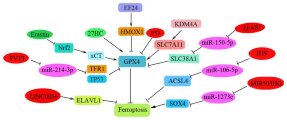
Ferroptosis is a newly reported iron-dependent PCD
process that is characterized by the accumulation of an
iron-dependent lethal lipid reactive oxygen species (ROS) response.
To date, the induction of cell ferroptosis has provided a new
insight into the design of anti-tumor drugs and is considered a
promising strategy to inhibit tumor metastasis or overcome
chemotherapeutic drug resistance (60,91).
Diphenyldifluoroketone (EF24) has been shown to exhibit
cytotoxicity against NSCLC via facilitating ROS accumulation
(91). In patients with
osteosarcoma, it has been determined that EF24 can induce
ferroptosis by suppressing GPX4 via the upregulation of HMOX1, thus
leading to increased MDA, ROS and intracellular ferric ion levels.
EF24 could therefore be considered a potential anti-cancer agent
for the clinical therapy of patients with NSCLC and osteosarcoma
(60). The light chain of the
cystine/glutamic acid reverse transporter System Xc (-) (xCT) has
been shown to be overexpressed in human cancer and to be associated
with tumor metastasis. Erastin/sorafenib can induce ferroptosis in
DPP-resistant NSCLC cells by inhibiting the Nrf2/xCT pathway
(92); thus, ferroptosis inducers,
such as erastin and sorafenib, may be used as novel therapies for
patients with NSCLC, particularly those with advanced NSCLC and DDP
failure. Metal-organic networks (MONs) encapsulated with a p53
plasmid (MON-p53) can evoke ferroptosis to prolong the survival
time of tumor-bearing mice (93).
In a study examining lung carcinoma, dihydroisotanshinone I,
extracted from Danshen (Salvia miltiorrhiza Bunge),
inhibited metastasis of lung carcinoma in vivo by triggering
apoptosis and ferroptosis (94).
The iron-sulfur cluster biosynthetic enzyme NFS1 has been reported
to be highly expressed in lung adenocarcinoma. Inhibition of NFS1
and of cysteine transport can induce ferroptosis in vitro to
suppress tumor growth in lung adenocarcinomas (95). GPX4 is a negative regulator of
ferroptosis and metastatic cells are resistant to ferroptosis.
Notably, high exposure to 27-hydroxycholesterol, an abundant
circulating cholesterol metabolite, can increase tumorigenic and
metastatic capacity that requires sustained expression of
GPX4(96). Fibroblast activation
protein α (FAP) has been reported to be overexpressed in >90% of
types of human cancer (97). A
newly discovered FAP gene-engineered tumor cell-derived
exosome-like nanovesicle (eNVs-FAP) vaccine can induce tumor cell
ferroptosis to overcome immunosuppression of the tumor
microenvironment (97). Thus,
ferroptosis exhibits promising potential to trigger an anti-cancer
immune response (98).
Recently, studies examining the regulation of
ferroptosis during cancer progression and metastasis have focused
primarily on ncRNAs. Regulatory ncRNAs that regulate NSCLC
progression and metastasis, including lncRNAs and circRNAs, have
been gradually and widely reported (99-106).
lncRNA MIR503HG serves as a sponge of miR-1273c and has been
reported to significantly increase NSCLC progression by regulating
SRY-box 4 expression and inhibiting ferroptosis (99). Furthermore, overexpression of
metallothionein 1D pseudogene can sensitize A549 and H1299 cells to
erastin-induced ferroptosis by downregulating Nrf2 in NSCLC cells
(100). LINC00336 can inhibit
ferroptosis by binding to ELAV-like RNA-binding protein 1 in human
lung cancer (101). Additionally,
H19 may promote cancer progression and suppress ferroptosis by
regulating the miR-106b-5p/ACSL4 axis (102). PVT1 can also regulate ferroptosis
via miR-214-mediated TFR1 and TP53 expression (104); notably, GPX4 has also been
demonstrated to be a target of miR-214-3p. Notably, ketamine has
been reported to inhibit the proliferation of liver cancer cells
in vitro and in vivo by inducing ferroptosis via the
lncRNA PVT1/miR-214-3p/GPX4 pathway (104). lncRNA ZFAS1 can also induce
ferroptosis by functioning as a ceRNA through the
miR-150-5p/SLC38A1 axis (105).
Additionally, LINC00618(106),
lncRNA GABPB1-AS1, lncRNA ZFAS1, lncRNA P53RRA and lncRNA MEG8 may
all be involved in regulating ferroptosis during cancer progression
(105). These RNAs function in
addition to the reported circRNAs, which include
circ_008035(107), circRNA cIARS
(108), circ_0067934(109), circ_0007142(110), circIL4R (111), circTTBK2(112) and circKIF4A (113), and the lncRNAs such as lncRNA
OIP5-AS1(114). However, the
relationship between these newly identified ncRNAs and ferroptosis
in NSCLC metastasis has not been clearly clarified and requires
further investigation (Fig. 6;
Table I).
Different types of PCD, such as apoptosis and
autophagy, apoptosis and necroptosis, and necroptosis and
ferroptosis, exist in close crosstalk (115). ROS-induced lipid peroxidation has
been shown to serve an important role in the crosstalk among
apoptosis, autophagy and ferroptosis (115). Lipid peroxidation products are
harmful as they destroy DNA, proteins and enzymes in various cell
types to inactivate PCD. For example, the products of lipid
peroxidation can induce cell apoptosis through the NF-κB, MAPK and
protein kinase C-related signaling pathways. Lipid peroxidation can
also interact with upstream regulators of autophagy-related
signaling pathways, such as AMP-activated protein kinase and
Akt-mTOR signaling to interfere with autophagy (115). In addition, GPX4 activity can
significantly affect lipid peroxidation and regulate ferroptosis
induction.
The association between lncRNA, circRNA and miRNAs
via miRNA recognition elements and negatively regulated the
expression of miRNAs. The ceRNA regulatory network can be
mRNA-miRNA-lncRNA, mRNA-miRNA-circRNA or mRNA-miRNA-lncRNA-circRNA.
Each miRNA can have multiple target genes and multiple miRNAs can
co-regulate the same gene. During the regulation of NSCLC
metastasis, lncRNAs, circRNAs and miRNAs may interact with each
other as ceRNAs, and can be involved in the progression and
metastasis of NSCLC (105,15).
The rapid progress in RNA sequencing and analysis
has revealed that a number of ncRNAs regulate tumor proliferation,
invasion and migration of NSCLC cells. The present study summarized
the regulatory role of PCD, including apoptosis, autophagy-related
cell death and ferroptosis, in NSCLC metastasis. It also discussed
the regulatory ncRNAs that promote PCD or inhibit the induction of
PCD by acting as oncogenes or tumor suppressor genes.
In addition, necroptosis and pyroptosis have been
newly identified as PCD mechanisms associated with various
inflammatory or autoimmune diseases. The functions of necroptosis
or pyroptosis in the context of cancer metastasis are complex; they
possess both pro- and anti-tumor effects according to different
pathological tissues, cell types or cancer stages during metastasis
(116). Additionally,
receptor-interacting kinase 3, the key regulator of necroptosis, is
normally silenced in types of cancer that are unable to induce
necroptosis. There are relatively few studies examining necroptosis
and pyroptosis in NSCLC metastasis and the present study did not
discuss the recent achievements in pyroptosis and necroptosis in
the context of NSCLC metastasis (117). Furthermore, several aspects
remain that must be addressed further: i) Novel and effective
biomarkers for identifying the progression and prognosis of
patients with NSCLC must be screened; ii) drug resistance remains a
major obstacle in the treatment of patients with NSCLC; therefore,
the effective induction of PCD, including apoptosis, ferroptosis
and autophagy, to overcome drug resistance in NSCLC cells must be
investigated in the future.
Not applicable.
Funding: The present study was supported by the Special
Scientific Research Project of Venous Thromboembolism Prevention
and Treatment (Heng Rui) of Sichuan Medical Association (grant no.
2019HR23).
Not applicable.
XH was responsible for article selection and
literature review. XH, LX, YL, WX and HZ wrote the manuscript. WX
constructed figures. Data authentication is not applicable. All
authors have read and approved the final manuscript.
Not applicable.
Not applicable.
The authors declare that they have no competing
interests.
|
1
|
Indini A, Rijavec E and Grossi F:
Circulating biomarkers of response and toxicity of immunotherapy in
advanced non-small cell lung cancer (NSCLC): A comprehensive
review. Cancers (Basel). 13(1794)2021.PubMed/NCBI View Article : Google Scholar
|
|
2
|
Chan MV, Huo YR, Cao C and Ridley L:
Survival outcomes for surgical resection versus CT-guided
percutaneous ablation for stage I non-small cell lung cancer
(NSCLC): A systematic review and meta-analysis. Eur Radiol.
31:5421–5433. 2021.PubMed/NCBI View Article : Google Scholar
|
|
3
|
Chen YS, Lin WH, Zhang AL and Lin Y:
Application of CT perfusion imaging in NSCLC and its correlation
with angiogenesis and lymph node metastasis. Eur Rev Med Pharmacol
Sci. 25:2511–2516. 2021.PubMed/NCBI View Article : Google Scholar
|
|
4
|
Lombardi M, D'Ascanio M, Scarpino S,
Scozzi D, Giordano M, Costarelli L, Raj ER, Mancini R, Cardillo G,
Cardaci V, et al: Full-length TrkB variant in NSCLC is associated
with brain metastasis. Biomed Res Int. 2020(4193541)2020.PubMed/NCBI View Article : Google Scholar
|
|
5
|
Su Z, Yang Z, Xu Y, Chen Y and Yu Q:
Apoptosis, autophagy, necroptosis, and cancer metastasis. Mol
Cancer. 14(48)2015.PubMed/NCBI View Article : Google Scholar
|
|
6
|
Li D, Fan S, Yu F, Zhu X, Song Y, Ye M,
Fan L and Lv Z: FOXD1 promotes cell growth and metastasis by
activation of vimentin in NSCLC. Cell Physiol Biochem.
51:2716–2731. 2018.PubMed/NCBI View Article : Google Scholar
|
|
7
|
Cai T, Zhou J, Zeng Y, Du W, Zhang Y, Liu
T, Fu Y, Huang JA, Qian Q, Zhu J, et al: EVI5 is an oncogene that
regulates the proliferation and metastasis of NSCLC cells. J Exp
Clin Cancer Res. 39(84)2020.PubMed/NCBI View Article : Google Scholar
|
|
8
|
Lv XQ, Qiao XR, Su L and Chen SZ: Honokiol
inhibits EMT-mediated motility and migration of human non-small
cell lung cancer cells in vitro by targeting c-FLIP. Acta Pharmacol
Sin. 37:1574–1586. 2016.PubMed/NCBI View Article : Google Scholar
|
|
9
|
Chen C, Zhang B, Wu S, Song Y and Li J:
Knockdown of EIF5A2 inhibits the malignant potential of non-small
cell lung cancer cells. Oncol Lett. 15:4541–4549. 2018.PubMed/NCBI View Article : Google Scholar
|
|
10
|
Lu R, Zhou Q, Ju L, Chen L, Wang F and
Shao J: Upregulation of TRIP13 promotes the malignant progression
of lung cancer via the EMT pathway. Oncol Rep.
46(172)2021.PubMed/NCBI View Article : Google Scholar
|
|
11
|
Yang C, Zhu S, Feng W and Chen X: Calponin
3 suppresses proliferation, migration and invasion of non-small
cell lung cancer cells. Oncol Lett. 22(634)2021.PubMed/NCBI View Article : Google Scholar
|
|
12
|
Di X, Jin X, Ma H, Wang R, Cong S, Tian C,
Liu J, Zhao M, Li R and Wang K: The oncogene IARS2 promotes
non-small cell lung cancer tumorigenesis by activating the AKT/MTOR
Pathway. Front Oncol. 9(393)2019.PubMed/NCBI View Article : Google Scholar
|
|
13
|
Yuan J, Zhang G, Li X, Ma Q, Cheng W, Wang
W, Zhang B, Hu T and Song G: Knocking down USP39 inhibits the
growth and metastasis of non-small-cell lung cancer cells through
activating the p53 pathway. Int J Mol Sci. 21(8949)2020.PubMed/NCBI View Article : Google Scholar
|
|
14
|
Xing Y, Liu Y, Liu T, Meng Q, Lu H, Liu W,
Hu J, Li C, Cao M, Yan S, et al: TNFAIP8 promotes the proliferation
and cisplatin chemoresistance of non-small cell lung cancer through
MDM2/p53 pathway. Cell Commun Signal. 16(43)2018.PubMed/NCBI View Article : Google Scholar
|
|
15
|
Duan Y, Liu T, Li S, Huang M, Li X, Zhao H
and Li J: CHAF1B promotes proliferation and reduces apoptosis in
95D lung cancer cells and predicts a poor prognosis in nonsmall
cell lung cancer. Oncol Rep. 41:2518–2528. 2019.PubMed/NCBI View Article : Google Scholar
|
|
16
|
Liu H, Zhang M, Xu S, Zhang J, Zou J, Yang
C, Zhang Y, Gong C, Kai Y and Li Y: HOXC8 promotes proliferation
and migration through transcriptional up-regulation of TGFbeta1 in
non-small cell lung cancer. Oncogenesis. 7(1)2018.PubMed/NCBI View Article : Google Scholar
|
|
17
|
Hu Y, Cheng Y, Jiang X, Zhang Y, Wang H,
Ren H, Xu Y, Jiang J, Wang Q, Su H, et al: PCGF3 promotes the
proliferation and migration of non-small cell lung cancer cells via
the PI3K/AKT signaling pathway. Exp Cell Res.
400(112496)2021.PubMed/NCBI View Article : Google Scholar
|
|
18
|
Wang H, Dong H, Qiao L, Wu Y, Wu B and Jin
X: ZEB1 induces non-small cell lung cancer development by targeting
microRNA-320a to increase the expression of RAD51AP1. Exp Cell Res.
405(112687)2021.PubMed/NCBI View Article : Google Scholar
|
|
19
|
Songyang Y, Song T, Shi Z, Li W, Yang S
and Li D: Effect of vitamin D on malignant behavior of non-small
cell lung cancer cells. Gene. 768(145309)2021.PubMed/NCBI View Article : Google Scholar
|
|
20
|
Wang LL, Hu RC, Dai AG, Tan SX, Xu M, Kong
CC, Chen YR and Fu DY: CHOP overexpression sensitizes human
non-small cell lung cancer cells to cisplatin treatment by
Bcl-2/JNK pathway. Am J Transl Res. 13:6279–6287. 2021.PubMed/NCBI
|
|
21
|
Xia HW, Zhang ZQ, Yuan J and Niu QL: Human
RECQL5 promotes metastasis and resistance to cisplatin in non-small
cell lung cancer. Life Sci. 265(118768)2021.PubMed/NCBI View Article : Google Scholar
|
|
22
|
Luo T, Yan L and Liu H: LINC00632 inhibits
the malignant development of non-small cell lung cancer by
downregulating miR-1203. J BUON. 25:1517–1524. 2020.PubMed/NCBI
|
|
23
|
Chen L, Li X, Lu C, Zhao Y, Zhu J and Yang
L: The long noncoding RNA CASC7 inhibits growth and invasion of
nonsmall cell lung cancer cells through phosphatase and tensin
homolog upregulation via sequestration of miR92a. Int J Oncol.
57:466–477. 2020.PubMed/NCBI View Article : Google Scholar
|
|
24
|
Wang JF, Xi ZN, Su HJ, Bao Z and Qiao YH:
SP1-induced overexpression of LINC00520 facilitates non-small cell
lung cancer progression through miR-577/CCNE2 pathway and predicts
poor prognosis. Hum Cell. 34:952–964. 2021.PubMed/NCBI View Article : Google Scholar
|
|
25
|
She K, Yan H, Huang J, Zhou H and He J:
miR-193b availability is antagonized by LncRNA-SNHG7 for
FAIM2-induced tumour progression in non-small cell lung cancer.
Cell Prolif. 51(e12406)2018.PubMed/NCBI View Article : Google Scholar
|
|
26
|
Qi L, Liu F, Zhang F, Zhang S, Lv L, Bi Y
and Yu Y: lncRNA NEAT1 competes against let-7a to contribute to
non-small cell lung cancer proliferation and metastasis. Biomed
Pharmacother. 103:1507–1515. 2018.PubMed/NCBI View Article : Google Scholar
|
|
27
|
Wan L, Sun M, Liu GJ, Wei CC, Zhang EB,
Kong R, Xu TP, Huang MD and Wang ZX: Long noncoding RNA PVT1
promotes non-small cell lung cancer cell proliferation through
epigenetically regulating LATS2 expression. Mol Cancer Ther.
15:1082–1094. 2016.PubMed/NCBI View Article : Google Scholar
|
|
28
|
Qi G and Li L: Long non-coding RNA PVT1
contributes to cell growth and metastasis in non-small-cell lung
cancer by regulating miR-361-3p/SOX9 axis and activating
Wnt/beta-catenin signaling pathway. Biomed Pharmacother.
126(110100)2020.PubMed/NCBI View Article : Google Scholar
|
|
29
|
Li W, Sun M, Zang C, Ma P, He J, Zhang M,
Huang Z, Ding Y and Shu Y: Upregulated long non-coding RNA
AGAP2-AS1 represses LATS2 and KLF2 expression through interacting
with EZH2 and LSD1 in non-small-cell lung cancer cells. Cell Death
Dis. 7(e2225)2016.PubMed/NCBI View Article : Google Scholar
|
|
30
|
Jiang C, Yang Y, Yang Y, Guo L, Huang J,
Liu X, Wu C and Zou J: Long noncoding RNA (lncRNA) HOTAIR affects
tumorigenesis and metastasis of non-small cell lung cancer by
upregulating miR-613. Oncol Res. 26:725–734. 2018.PubMed/NCBI View Article : Google Scholar
|
|
31
|
Li Z, Yue G, Zhang T, Wu J and Tian X:
LncRNA ADAMTS9-AS1 knockdown restricts cell proliferation and EMT
in non-small cell lung cancer. Histol Histopathol. 36:1063–1072.
2021.PubMed/NCBI View Article : Google Scholar
|
|
32
|
Zhang Z, Peng Z, Cao J, Wang J, Hao Y,
Song K, Wang Y, Hu W and Zhang X: Long noncoding RNA PXN-AS1-L
promotes non-small cell lung cancer progression via regulating PXN.
Cancer Cell Int. 19(20)2019.PubMed/NCBI View Article : Google Scholar
|
|
33
|
Zhang G, An X and Zhao H, Zhang Q and Zhao
H: Long non-coding RNA HNF1A-AS1 promotes cell proliferation and
invasion via regulating miR-17-5p in non-small cell lung cancer.
Biomed Pharmacother. 98:594–599. 2018.PubMed/NCBI View Article : Google Scholar
|
|
34
|
Ao X, Jiang M, Zhou J, Liang H, Xia H and
Chen G: lincRNAp21 inhibits the progression of nonsmall cell lung
cancer via targeting miR175p. Oncol Rep. 41:789–800.
2019.PubMed/NCBI View Article : Google Scholar
|
|
35
|
Guan Y, Yang J, Liu X and Chu L: Long
noncoding RNA CBR3 antisense RNA 1 promotes the aggressive
phenotypes of nonsmallcell lung cancer by sponging microRNA5093p
and competitively upregulating HDAC9 expression. Oncol Rep.
44:1403–1414. 2020.PubMed/NCBI View Article : Google Scholar
|
|
36
|
Jin B, Jin H, Wu HB, Xu JJ and Li B: Long
non-coding RNA SNHG15 promotes CDK14 expression via miR-486 to
accelerate non-small cell lung cancer cells progression and
metastasis. J Cell Physiol. 233:7164–7172. 2018.PubMed/NCBI View Article : Google Scholar
|
|
37
|
Liu X, Huang S, Guan Y and Zhang Q: Long
noncoding RNA OSER1AS1 promotes the malignant properties of
nonsmall cell lung cancer by sponging microRNA4333p and thereby
increasing Smad2 expression. Oncol Rep. 44:599–610. 2020.PubMed/NCBI View Article : Google Scholar
|
|
38
|
Li G, Zhao C, Zhang H, Yu J, Sun Y and
Zhang Y: Hsa_circ_0046263 drives the carcinogenesis and metastasis
of non-small cell lung cancer through the promotion of NOVA2 by
absorbing Mir-940 as a molecular sponge. Cancer Manag Res.
12:12779–12790. 2020.PubMed/NCBI View Article : Google Scholar
|
|
39
|
Xu Z, Xiang W, Chen W, Sun Y, Qin F, Wei
J, Yuan L, Zheng L and Li S: Circ-IGF1R inhibits cell invasion and
migration in non-small cell lung cancer. Thorac Cancer. 11:875–887.
2020.PubMed/NCBI View Article : Google Scholar
|
|
40
|
Wang Y, Zhao W and Zhang S: STAT3-induced
upregulation of circCCDC66 facilitates the progression of non-small
cell lung cancer by targeting miR-33a-5p/KPNA4 axis. Biomed
Pharmacother. 126(110019)2020.PubMed/NCBI View Article : Google Scholar
|
|
41
|
Shi F, Yang Q, Shen D and Chen J: CircRNA
WHSC1 promotes non-small cell lung cancer progression via sponging
microRNA-296-3p and up-regulating expression of AKT
serine/threonine kinase 3. J Clin Lab Anal.
35(e23865)2021.PubMed/NCBI View Article : Google Scholar
|
|
42
|
Tai G, Zhang M and Liu F: Circ_0000735
enhances the proliferation, metastasis and glycolysis of non-small
cell lung cancer by regulating the miR-635/FAM83F axis. Exp Lung
Res. 47:136–148. 2021.PubMed/NCBI View Article : Google Scholar
|
|
43
|
Wu Z, Jiang H, Fu H and Zhang Y: A
circGLIS3/miR-644a/PTBP1 positive feedback loop promotes the
malignant biological progressions of non-small cell lung cancer. Am
J Cancer Res. 11:108–122. 2021.PubMed/NCBI
|
|
44
|
Yang C, Shi J, Wang J, Hao D, An J and
Jiang J: Circ_0006988 promotes the proliferation, metastasis and
angiogenesis of non-small cell lung cancer cells by modulating
miR-491-5p/MAP3K3 axis. Cell Cycle. 20:1334–1346. 2021.PubMed/NCBI View Article : Google Scholar
|
|
45
|
Zhu Y, Ma C, Lv A and Kou C: Circular RNA
circ_0010235 sponges miR-338-3p to play oncogenic role in
proliferation, migration and invasion of non-small-cell lung cancer
cells through modulating KIF2A. Ann Med. 53:693–706.
2021.PubMed/NCBI View Article : Google Scholar
|
|
46
|
Liu Y, Yang C, Cao C, Li Q, Jin X and Shi
H: Hsa_circ_RNA_0011780 Represses the Proliferation and Metastasis
of Non-Small Cell Lung Cancer by Decreasing FBXW7 via Targeting
miR-544a. Onco Targets Ther. 13:745–755. 2020.PubMed/NCBI View Article : Google Scholar
|
|
47
|
Liu Y, Li C, Liu H and Wang J:
Circ_0001821 knockdown suppresses growth, metastasis, and TAX
resistance of non-small-cell lung cancer cells by regulating the
miR-526b-5p/GRK5 axis. Pharmacol Res Perspect.
9(e00812)2021.PubMed/NCBI View Article : Google Scholar
|
|
48
|
Yu L, Li J, Peng B, Cai P, Zhao B, Chen Y
and Zhu H: CircASXL1 knockdown restrains hypoxia-induced DDP
resistance and NSCLC progression by sponging miR-206. Cancer Manag
Res. 13:5077–5089. 2021.PubMed/NCBI View Article : Google Scholar
|
|
49
|
Pang J, Ye L, Zhao D, Zhao D and Chen Q:
Circular RNA PRMT5 confers cisplatin-resistance via miR-4458/REV3L
axis in non-small-cell lung cancer. Cell Biol Int. 44:2416–2426.
2020.PubMed/NCBI View Article : Google Scholar
|
|
50
|
Yang B, Teng F, Chang L, Wang J, Liu DL,
Cui YS and Li GH: Tumor-derived exosomal circRNA_102481 contributes
to EGFR-TKIs resistance via the miR-30a-5p/ROR1 axis in non-small
cell lung cancer. Aging (Albany NY). 13:13264–13286.
2021.PubMed/NCBI View Article : Google Scholar
|
|
51
|
Zhang X, Yang D and Wei Y: Overexpressed
CDR1as functions as an oncogene to promote the tumor progression
via miR-7 in non-small-cell lung cancer. Onco Targets Ther.
11:3979–3987. 2018.PubMed/NCBI View Article : Google Scholar
|
|
52
|
Zhang N, Nan A, Chen L, Li X, Jia Y, Qiu
M, Dai X, Zhou H, Zhu J, Zhang H and Jiang Y: Circular RNA
circSATB2 promotes progression of non-small cell lung cancer cells.
Mol Cancer. 19(101)2020.PubMed/NCBI View Article : Google Scholar
|
|
53
|
Li Y, Hu J, Li L, Cai S, Zhang H, Zhu X,
Guan G and Dong X: Upregulated circular RNA circ_0016760 indicates
unfavorable prognosis in NSCLC and promotes cell progression
through miR-1287/GAGE1 axis. Biochem Biophys Res Commun.
503:2089–2094. 2018.PubMed/NCBI View Article : Google Scholar
|
|
54
|
An X, Ge J, Guo H, Mi H, Zhou J, Liu Y,
Weiyue and Wu Z: Overexpression of miR-4286 is an
unfavorable prognostic marker in individuals with non-small cell
lung cancer. J Cell Biochem. 120:17573–17583. 2019.PubMed/NCBI View Article : Google Scholar
|
|
55
|
Kim DH, Park S, Kim H, Choi YJ, Kim SY,
Sung KJ, Sung YH, Choi CM, Yun M, Yi YS, et al: Tumor-derived
exosomal miR-619-5p promotes tumor angiogenesis and metastasis
through the inhibition of RCAN1.4. Cancer Lett. 475:2–13.
2020.PubMed/NCBI View Article : Google Scholar
|
|
56
|
Mao M, Wu Z and Chen J: MicroRNA-187-5p
suppresses cancer cell progression in non-small cell lung cancer
(NSCLC) through down-regulation of CYP1B1. Biochem Biophys Res
Commun. 478:649–655. 2016.PubMed/NCBI View Article : Google Scholar
|
|
57
|
Han L, Chen W, Xia Y, Song Y, Zhao Z,
Cheng H and Jiang T: MiR-101 inhibits the proliferation and
metastasis of lung cancer by targeting zinc finger E-box binding
homeobox 1. Am J Transl Res. 10:1172–1183. 2018.PubMed/NCBI
|
|
58
|
Wang S, Sun H, Zhan X and Wang Q:
MicroRNA718 serves a tumorsuppressive role in nonsmall cell lung
cancer by directly targeting CCNB1. Int J Mol Med. 45:33–44.
2020.PubMed/NCBI View Article : Google Scholar
|
|
59
|
Zhao M, Tong C, Hao Z, Zhao R and Wang L:
MicroRNA-374b mediates the initiation of non-small cell lung cancer
by regulating ITGB1 and p53 expressions. Thorac Cancer.
11:1670–1678. 2020.PubMed/NCBI View Article : Google Scholar
|
|
60
|
Lin X, Lai X, Feng W, Yu X, Gu Q and Zheng
X: MiR-30a sensitized lung cancer against neoadjuvant chemotherapy
by depressing autophagy. Jpn J Clin Oncol. 51:675–684.
2021.PubMed/NCBI View Article : Google Scholar
|
|
61
|
Huang T, She K, Peng G, Wang W, Huang J,
Li J, Wang Z and He J: MicroRNA-186 suppresses cell proliferation
and metastasis through targeting MAP3K2 in non-small cell lung
cancer. Int J Oncol. 49:1437–1444. 2016.PubMed/NCBI View Article : Google Scholar
|
|
62
|
Gong F, Ren P, Zhang Y, Jiang J and Zhang
H: MicroRNAs-491-5p suppresses cell proliferation and invasion by
inhibiting IGF2BP1 in non-small cell lung cancer. Am J Transl Res.
8:485–495. 2016.PubMed/NCBI
|
|
63
|
Zhang Z, Li W, Jiang D, Liu C and Lai Z:
MicroRNA-139-5p inhibits cell viability, migration and invasion and
suppresses tumor growth by targeting HDGF in non-small cell lung
cancer. Oncol Lett. 19:1806–1814. 2020.PubMed/NCBI View Article : Google Scholar
|
|
64
|
Yan Y, Jin X, Sun H, Pang S, Kong X, Bu J
and Xu S: MiR-139-5p targetedly regulates YAF2 and mediates the
AKT/P38 MAPK signaling pathway to alleviate the metastasis of
non-small cell lung cancer cells and their resistance against
cisplatin. Cancer Manag Res. 13:3639–3650. 2021.PubMed/NCBI View Article : Google Scholar
|
|
65
|
Chen Y, Hou C, Zhao LX, Cai QC, Zhang Y,
Li DL, Tang Y, Liu HY, Liu YY, Zhang YY, et al: The association of
microRNA-34a with high incidence and metastasis of lung cancer in
Gejiu and Xuanwei Yunnan. Front Oncol. 11(619346)2021.PubMed/NCBI View Article : Google Scholar
|
|
66
|
Jiang W, Wei K, Pan C, Li H, Cao J, Han X,
Tang Y, Zhu S, Yuan W, He Y, et al: MicroRNA-1258 suppresses tumour
progression via GRB2/Ras/Erk pathway in non-small-cell lung cancer.
Cell Prolif. 51(e12502)2018.PubMed/NCBI View Article : Google Scholar
|
|
67
|
Chen G, Hu J, Huang Z, Yang L and Chen M:
MicroRNA-1976 functions as a tumor suppressor and serves as a
prognostic indicator in non-small cell lung cancer by directly
targeting PLCE1. Biochem Biophys Res Commun. 473:1144–1151.
2016.PubMed/NCBI View Article : Google Scholar
|
|
68
|
Li QX, Zhou X, Huang TT, Tang Y, Liu B,
Peng P, Sun L, Wang YH and Yuan XL: The Thr300Ala variant of
ATG16L1 is associated with decreased risk of brain metastasis in
patients with non-small cell lung cancer. Autophagy. 13:1053–1063.
2017.PubMed/NCBI View Article : Google Scholar
|
|
69
|
Jiang L, Liang X, Liu M, Wang W, Ma J, Guo
Q, Han L, Yang C and Nan K: Reduced expression of liver kinase B1
and Beclin1 is associated with the poor survival of patients with
non-small cell lung cancer. Oncol Rep. 32:1931–1938.
2014.PubMed/NCBI View Article : Google Scholar
|
|
70
|
Du H, Chen L, Luo F, Chen X, Li Y and
Cheng Q: Beclin-1 expression is associated with prognosis in a
Bcl-2-dependent manner in non-small cell lung cancer. Oncol Lett.
20(9)2020.PubMed/NCBI View Article : Google Scholar
|
|
71
|
Yu S, Cheng C, Wang J, Wang J, Qu Z, Ren
H, Li Y, Ning Q, Chen M and Hu T: Loss of Beclin1 expression and
Nrf2 overexpression are associated with poor survival of patients
with non-small cell lung cancer. Anticancer Agents Med Chem.
18:1680–1687. 2018.PubMed/NCBI View Article : Google Scholar
|
|
72
|
Chen B, Zeng C, Ye Y, Wu D, Mu Z, Liu J,
Xie Y and Wu H: Promoter methylation of TCF21 may repress autophagy
in the progression of lung cancer. J Cell Commun Signal.
12:423–432. 2018.PubMed/NCBI View Article : Google Scholar
|
|
73
|
Kim H, Choi SY, Lim J, Lindroth AM and
Park YJ: EHMT2 inhibition induces cell death in human non-small
cell lung cancer by altering the cholesterol biosynthesis pathway.
Int J Mol Sci. 21(1002)2020.PubMed/NCBI View Article : Google Scholar
|
|
74
|
Chen Z, Tian D, Liao X, Zhang Y, Xiao J,
Chen W, Liu Q, Chen Y, Li D, Zhu L and Cai S: Apigenin combined
with gefitinib blocks autophagy flux and induces apoptotic cell
death through inhibition of HIF-1a, c-Myc, p-EGFR, and glucose
metabolism in EGFR L858R+T790M-mutated H1975 cells. Front
Pharmacol. 10(260)2019.PubMed/NCBI View Article : Google Scholar
|
|
75
|
Zhou Y, Hong T, Tong L, Liu W, Yang X, Luo
J, Wang F, Li J and Yan L: Astragalus polysaccharide combined with
10-hydroxycamptothecin inhibits metastasis in non-small cell lung
carcinoma cell lines via the MAP4K3/mTOR signaling pathway. Int J
Mol Med. 42:3093–3104. 2018.PubMed/NCBI View Article : Google Scholar
|
|
76
|
Wang H, Zhang Y, Wu Q, Wang YB and Wang W:
miR-16 mimics inhibit TGF-beta1-induced epithelial-to-mesenchymal
transition via activation of autophagy in non-small cell lung
carcinoma cells. Oncol Rep. 39:247–254. 2018.PubMed/NCBI View Article : Google Scholar
|
|
77
|
Xu C, Cao H, Sui Y, Zhang H, Shi C, Wu J,
Ma R and Feng J: CDCA4 suppresses epithelial-mesenchymal transtion
(EMT) and metastasis in Non-small cell lung cancer through
modulating autophagy. Cancer Cell Int. 21(48)2021.PubMed/NCBI View Article : Google Scholar
|
|
78
|
Zheng L, Zhou H, Guo L, Xu X, Zhang S, Xu
W and Mao W: Inhibition of NIPBL enhances the chemosensitivity of
non-small-cell lung cancer cells via the DNA damage response and
autophagy pathway. Onco Targets Ther. 11:1941–1948. 2018.PubMed/NCBI View Article : Google Scholar
|
|
79
|
Wang Y, Liu F, Fang C, Xu L, Chen L, Xu Z,
Chen J, Peng W, Fu B and Li Y: Combination of rapamycin and SAHA
enhanced radiosensitization by inducing autophagy and acetylation
in NSCLC. Aging (Albany NY). 13:18223–18237. 2021.PubMed/NCBI View Article : Google Scholar
|
|
80
|
Guo M, Liu Z, Si J, Zhang J, Zhao J, Guo
Z, Xie Y, Zhang H and Gan L: Cediranib induces apoptosis, G1 phase
cell cycle arrest, and autophagy in non-small-cell lung cancer cell
A549 in vitro. Biomed Res Int. 2021(5582648)2021.PubMed/NCBI View Article : Google Scholar
|
|
81
|
Ye X, Xie G, Liu Z and Tang J, Cui M, Wang
C, Guo C and Tang J: TNNC1 reduced gemcitabine sensitivity of
nonsmall-cell lung cancer by increasing autophagy. Med Sci Monit.
26(e922703)2020.PubMed/NCBI View Article : Google Scholar
|
|
82
|
Datta S, Choudhury D, Das A, Mukherjee DD,
Dasgupta M, Bandopadhyay S and Chakrabarti G: Autophagy inhibition
with chloroquine reverts paclitaxel resistance and attenuates
metastatic potential in human nonsmall lung adenocarcinoma A549
cells via ROS mediated modulation of beta-catenin pathway.
Apoptosis. 24:414–433. 2019.PubMed/NCBI View Article : Google Scholar
|
|
83
|
Zhao Z, Li J, Jiang Y, Xu W, Li X and Jing
W: CLDN1 increases drug resistance of non-small cell lung cancer by
activating autophagy via up-regulation of ULK1 phosphorylation. Med
Sci Monit. 23:2906–2916. 2017.PubMed/NCBI View Article : Google Scholar
|
|
84
|
Shen Q, Xu Z and Xu S: Long noncoding RNA
LUCAT1 contributes to cisplatin resistance by regulating the
miR514a3p/ULK1 axis in human nonsmall cell lung cancer. Int J
Oncol. 57:967–979. 2020.PubMed/NCBI View Article : Google Scholar
|
|
85
|
Li S, Zeng X, Ma R and Wang L: MicroRNA-21
promotes the proliferation, migration and invasion of non-small
cell lung cancer A549 cells by regulating autophagy activity via
AMPK/ULK1 signaling pathway. Exp Ther Med. 16:2038–2045.
2018.PubMed/NCBI View Article : Google Scholar
|
|
86
|
Guo J, Wu Y, Du J, Yang L, Chen W, Gong K,
Dai J, Miao S, Jin D and Xi S: Deregulation of UBE2C-mediated
autophagy repression aggravates NSCLC progression. Oncogenesis.
7(49)2018.PubMed/NCBI View Article : Google Scholar
|
|
87
|
Lu X, Liu QX, Zhang J, Zhou D, Yang GX, Li
MY, Qiu Y, Chen Q, Zheng H and Dai JG: PINK1 Overexpression
promotes cell migration and proliferation via regulation of
autophagy and predicts a poor prognosis in lung cancer cases.
Cancer Manag Res. 12:7703–7714. 2020.PubMed/NCBI View Article : Google Scholar
|
|
88
|
Zhang F, Cheng R, Li P, Lu C and Zhang G:
Hsa_circ_0010235 functions as an oncogenic drive in non-small cell
lung cancer by modulating miR-433-3p/TIPRL axis. Cancer Cell Int.
21(73)2021.PubMed/NCBI View Article : Google Scholar
|
|
89
|
Wudu M, Ren H, Hui L, Jiang J, Zhang S, Xu
Y, Wang Q, Su H, Jiang X, Dao R and Qiu X: DRAM2 acts as an
oncogene in non-small cell lung cancer and suppresses the
expression of p53. J Exp Clin Cancer Res. 38(72)2019.PubMed/NCBI View Article : Google Scholar
|
|
90
|
Zhang G, Liu Z, Chen Y and Zhang Y: High
serum HDGF levels are predictive of bone metastasis and unfavorable
prognosis in non-small cell lung cancer. Tohoku J Exp Med.
242:101–108. 2017.PubMed/NCBI View Article : Google Scholar
|
|
91
|
Chang M, Shang M, Yuan F, Guo W and Wang
C: EF24 exerts cytotoxicity against NSCLC via inducing ROS
accumulation. Cancer Cell Int. 21(531)2021.PubMed/NCBI View Article : Google Scholar
|
|
92
|
Lin H, Chen X, Zhang C, Yang T, Deng Z,
Song Y, Huang L, Li F, Li Q, Lin S and Jin D: EF24 induces
ferroptosis in osteosarcoma cells through HMOX1. Biomed
Pharmacother. 136(111202)2021.PubMed/NCBI View Article : Google Scholar
|
|
93
|
Li Y, Yan H, Xu X, Liu H, Wu C and Zhao L:
Erastin/sorafenib induces cisplatin-resistant non-small cell lung
cancer cell ferroptosis through inhibition of the Nrf2/xCT pathway.
Oncol Lett. 19:323–333. 2020.PubMed/NCBI View Article : Google Scholar
|
|
94
|
Wu CY, Yang YH, Lin YS, Chang GH, Tsai MS,
Hsu CM, Yeh RA, Shu LH, Cheng YC and Liu HT: Dihydroisotanshinone I
induced ferroptosis and apoptosis of lung cancer cells. Biomed
Pharmacother. 139(111585)2021.PubMed/NCBI View Article : Google Scholar
|
|
95
|
Alvarez SW, Sviderskiy VO, Terzi EM,
Papagiannakopoulos T, Moreira AL, Adams S, Sabatini DM, Birsoy K
and Possemato R: NFS1 undergoes positive selection in lung tumours
and protects cells from ferroptosis. Nature. 551:639–643.
2017.PubMed/NCBI View Article : Google Scholar
|
|
96
|
Liu W, Chakraborty B, Safi R, Kazmin D,
Chang CY and McDonnell DP: Dysregulated cholesterol homeostasis
results in resistance to ferroptosis increasing tumorigenicity and
metastasis in cancer. Nat Commun. 12(5103)2021.PubMed/NCBI View Article : Google Scholar
|
|
97
|
Hu S, Ma J, Su C, Chen Y, Shu Y, Qi Z,
Zhang B, Shi G, Zhang Y, Zhang Y, et al: Engineered exosome-like
nanovesicles suppress tumor growth by reprogramming tumor
microenvironment and promoting tumor ferroptosis. Acta Biomater.
135:567–581. 2021.PubMed/NCBI View Article : Google Scholar
|
|
98
|
Song R, Li T, Ye J, Sun F, Hou B, Saeed M,
Gao J, Wang Y, Zhu Q, Xu Z and Yu H: Acidity-activatable dynamic
nanoparticles boosting ferroptotic cell death for immunotherapy of
cancer. Adv Mater. 33(e2101155)2021.PubMed/NCBI View Article : Google Scholar
|
|
99
|
Yu H, Han Z, Xu Z, An C, Xu L and Xin H:
RNA sequencing uncovers the key long non-coding RNAs and potential
molecular mechanism contributing to XAV939-mediated inhibition of
non-small cell lung cancer. Oncol Lett. 17:4994–5004.
2019.PubMed/NCBI View Article : Google Scholar
|
|
100
|
Gai C, Liu C, Wu X, Yu M, Zheng J, Zhang
W, Lv S and Li W: MT1DP loaded by folate-modified liposomes
sensitizes erastin-induced ferroptosis via regulating
miR-365a-3p/NRF2 axis in non-small cell lung cancer cells. Cell
Death Dis. 11(751)2020.PubMed/NCBI View Article : Google Scholar
|
|
101
|
Wang M, Mao C, Ouyang L, Liu Y, Lai W, Liu
N, Shi Y, Chen L, Xiao D, Yu F, et al: Long noncoding RNA LINC00336
inhibits ferroptosis in lung cancer by functioning as a competing
endogenous RNA. Cell Death Differ. 26:2329–2343. 2019.PubMed/NCBI View Article : Google Scholar
|
|
102
|
Chen B, Wang H, Lv C, Mao C and Cui Y:
Long non-coding RNA H19 protects against intracerebral hemorrhage
injuries via regulating microRNA-106b-5p/acyl-CoA synthetase long
chain family member 4 axis. Bioengineered. 12:4004–4015.
2021.PubMed/NCBI View Article : Google Scholar
|
|
103
|
Lu J, Xu F and Lu H: LncRNA PVT1 regulates
ferroptosis through miR-214-mediated TFR1 and p53. Life Sci.
260(118305)2020.PubMed/NCBI View Article : Google Scholar
|
|
104
|
He GN, Bao NR, Wang S, Xi M, Zhang TH and
Chen FS: Ketamine induces ferroptosis of liver cancer cells by
targeting lncRNA PVT1/miR-214-3p/GPX4. Drug Des Devel Ther.
15:3965–3978. 2021.PubMed/NCBI View Article : Google Scholar
|
|
105
|
Yang Y, Tai W, Lu N, Li T, Liu Y, Wu W, Li
Z, Pu L, Zhao X, Zhang T and Dong Z: lncRNA ZFAS1 promotes lung
fibroblast-to-myofibroblast transition and ferroptosis via
functioning as a ceRNA through miR-150-5p/SLC38A1 axis. Aging
(Albany NY). 12:9085–9102. 2020.PubMed/NCBI View Article : Google Scholar
|
|
106
|
Wang Z, Chen X, Liu N, Shi Y, Liu Y,
Ouyang L, Tam S, Xiao D, Liu S, Wen F and Tao Y: A nuclear long
non-coding RNA LINC00618 accelerates ferroptosis in a manner
dependent upon apoptosis. Mol Ther. 29:263–274. 2021.PubMed/NCBI View Article : Google Scholar
|
|
107
|
Li C, Tian Y, Liang Y and Li Q:
Circ_0008035 contributes to cell proliferation and inhibits
apoptosis and ferroptosis in gastric cancer via miR-599/EIF4A1
axis. Cancer Cell Int. 20(84)2020.PubMed/NCBI View Article : Google Scholar
|
|
108
|
Liu Z, Wang Q, Wang X, Xu Z, Wei X and Li
J: Circular RNA cIARS regulates ferroptosis in HCC cells through
interacting with RNA binding protein ALKBH5. Cell Death Discov.
6(72)2020.PubMed/NCBI View Article : Google Scholar
|
|
109
|
Wang HH, Ma JN and Zhan XR: Circular RNA
Circ_0067934 attenuates ferroptosis of thyroid cancer cells by
miR-545-3p/SLC7A11 signaling. Front Endocrinol (Lausanne).
12(670031)2021.PubMed/NCBI View Article : Google Scholar
|
|
110
|
Wang Y, Chen H and Wei X: Circ_0007142
downregulates miR-874-3p-mediated GDPD5 on colorectal cancer cells.
Eur J Clin Invest. 51(e13541)2021.PubMed/NCBI View Article : Google Scholar
|
|
111
|
Xu Q, Zhou L, Yang G, Meng F, Wan Y, Wang
L and Zhang L: CircIL4R facilitates the tumorigenesis and inhibits
ferroptosis in hepatocellular carcinoma by regulating the
miR-541-3p/GPX4 axis. Cell Biol Int. 44:2344–2356. 2020.PubMed/NCBI View Article : Google Scholar
|
|
112
|
Zhang HY, Zhang BW, Zhang ZB and Deng QJ:
Circular RNA TTBK2 regulates cell proliferation, invasion and
ferroptosis via miR-761/ITGB8 axis in glioma. Eur Rev Med Pharmacol
Sci. 24:2585–2600. 2020.PubMed/NCBI View Article : Google Scholar
|
|
113
|
Chen W, Fu J, Chen Y, Li Y, Ning L, Huang
D, Yan S and Zhang Q: Circular RNA circKIF4A facilitates the
malignant progression and suppresses ferroptosis by sponging
miR-1231 and upregulating GPX4 in papillary thyroid cancer. Aging
(Albany NY). 13:16500–16512. 2021.PubMed/NCBI View Article : Google Scholar
|
|
114
|
Zhang Y, Guo S, Wang S, Li X, Hou D, Li H,
Wang L, Xu Y, Ma B, Wang H and Jiang X: LncRNA OIP5-AS1 inhibits
ferroptosis in prostate cancer with long-term cadmium exposure
through miR-128-3p/SLC7A11 signaling. Ecotoxicol Environ Saf.
220(112376)2021.PubMed/NCBI View Article : Google Scholar
|
|
115
|
Su LJ, Zhang JH, Gomez H, Murugan R, Hong
X, Xu D, Jiang F and Peng ZY: Reactive oxygen species-induced lipid
peroxidation in apoptosis, autophagy, and ferroptosis. Oxid Med
Cell Longev. 2019(5080843)2019.PubMed/NCBI View Article : Google Scholar
|
|
116
|
Liu Y, Chen Q, Zhu Y, Wang T, Ye L, Han L,
Yao Z and Yang Z: Non-coding RNAs in necroptosis, pyroptosis and
ferroptosis in cancer metastasis. Cell Death Discov.
7(210)2021.PubMed/NCBI View Article : Google Scholar
|
|
117
|
Tang R, Xu J, Zhang B, Liu J, Liang C, Hua
J, Meng Q, Yu X and Shi S: Ferroptosis, necroptosis, and pyroptosis
in anticancer immunity. J Hematol Oncol. 13(110)2020.PubMed/NCBI View Article : Google Scholar
|
|
118
|
Mei J, Liu G, Wang W, Xiao P, Yang D, Bai
H and Li R: OIP5-AS1 modulates epigenetic regulator HDAC7 to
enhance non-small cell lung cancer metastasis via miR-140-5p. Oncol
Lett. 20(7)2020.PubMed/NCBI View Article : Google Scholar
|
|
119
|
Dao R, Wudu M, Hui L, Jiang J, Xu Y, Ren H
and Qiu X: Knockdown of lncRNA MIR503HG suppresses proliferation
and promotes apoptosis of non-small cell lung cancer cells by
regulating miR-489-3p and miR-625-5p. Pathol Res Pract.
216(152823)2020.PubMed/NCBI View Article : Google Scholar
|
|
120
|
Shanshan W, Hongying M, Jingjing F, Yiming
Y, Yu R and Rui Y: CircDTL Functions as an Oncogene and Regulates
Both Apoptosis and Ferroptosis in Non-small Cell Lung Cancer Cells.
Front Genet. 12(743505)2021.PubMed/NCBI View Article : Google Scholar
|
|
121
|
Wu P, Li C, Ye DM, Yu K, Li Y, Tang H, Xu
G, Yi S and Zhang Z: Circular RNA circEPSTI1 accelerates cervical
cancer progression via miR-375/409-3P/515-5p-SLC7A11 axis. Aging
(Albany NY). 13:4663–4673. 2021.PubMed/NCBI View Article : Google Scholar
|
|
122
|
Ni H, Qin H, Sun C, Liu Y, Ruan G, Guo Q,
Xi T, Xing Y and Zheng L: MiR-375 reduces the stemness of gastric
cancer cells through triggering ferroptosis. Stem Cell Res Ther.
12(325)2021.PubMed/NCBI View Article : Google Scholar
|
|
123
|
Song Z, Jia G, Ma P and Cang S: Exosomal
miR-4443 promotes cisplatin resistance in non-small cell lung
carcinoma by regulating FSP1 m6A modification-mediated ferroptosis.
Life Sci. 276(119399)2021.PubMed/NCBI View Article : Google Scholar
|
|
124
|
Deng SH, Wu DM, Li L, Liu T, Zhang T, Li
J, Yu Y, He M, Zhao YY, Han R and Xu Y: miR-324-3p reverses
cisplatin resistance by inducing GPX4-mediated ferroptosis in lung
adenocarcinoma cell line A549. Biochem Biophys Res Commun.
549:54–60. 2021.PubMed/NCBI View Article : Google Scholar
|