Introduction
Sepsis is an intense immune response to infection
that contributes to 19.7% of all global deaths according to an
analysis published in 2020(1).
Currently, sepsis remains a leading cause of mortality in intensive
care units, as it may lead to multiple organ system dysfunction and
even multiorgan failure (2-4).
It was previously reported that the lung may be one of the organs
in the human body most vulnerable to sepsis, and sepsis contributes
to ~40% of the cases of acute lung injury (ALI) (2). Despite significant advances in the
overall treatment strategies, no specific treatment has yet been
developed for sepsis-induced ALI (5). Therefore, the development of novel
effective agents is crucial for optimizing the management,
prevention and treatment of patients with sepsis-mediated ALI.
Sepsis-induced lung damage promotes the
hypersecretion of inflammatory cytokines, leading to pathological
damage of the alveolar epithelium and vascular endothelial cells,
and to the development of ALI (6).
In addition, oxidative stress also serves a key role in the
development of ALI (7,8). Therefore, inhibition of inflammation
and oxidative stress may be a promising preventive and/or
therapeutic strategy for sepsis-induced ALI. Leonurine (LEO) is a
natural phenolic alkaloid extracted from Leonurus cardiaca
that has been shown to possess anti-inflammatory and antioxidant
properties in numerous studies (9-12).
It has been reported that LEO alleviates lipopolysaccharide
(LPS)-induced myocarditis through anti-inflammatory and antioxidant
mechanisms via the inactivation of the NF-кB signaling pathway
(13). In addition, LEO reduced
acute kidney injury and protected renal function against
LPS-induced inflammation (14).
However, the effects of LEO on ALI have not yet been determined, to
the best of our knowledge. Xu et al (15) reported that LEO exerted no
cytotoxic effects on human lung epithelial cells. Moreover, LEO was
reported to attenuate the aging process in mice through activation
of the nuclear factor erythroid 2-related factor 2 (Nrf2) signaling
pathway (16). A recent study
demonstrated that the activation of the Nrf2 signaling pathway
suppressed inflammation and oxidative stress in ALI (17). Therefore, it was hypothesized that
LEO may have a role in sepsis-induced ALI.
The aim of the present study was to explore the
effects of LEO on oxidative stress and on the inflammatory response
in LPS-induced BEAS-2B human lung epithelial cells. Notably, LEO
led to the activation of the Nrf2 signaling pathway. Therefore, LEO
may be an effective agent for the prevention of ALI, and the
findings of the present study may provide a theoretical basis for
the application of LEO in the prevention of ALI.
Materials and methods
Cell culture and treatment
Human lung epithelial cells (BEAS-2B; American Type
Culture Collection) were cultured in RPMI-1640 medium (Gibco;
Thermo Fisher Scientific, Inc.) supplemented with 10% fetal bovine
serum (Gibco; Thermo Fisher Scientific, Inc.) in a humidified
atmosphere with 5% CO2 at 37˚C. BEAS-2B cells were
pretreated with various doses of LEO (0-120 µg/ml) for 6 h and
subsequently stimulated with LPS (1 µg/ml) for 48 h. To block the
Nrf2 signaling pathway, ML385 (Abmole Bioscience, Inc.), an Nrf2
inhibitor, was used to treat the cells before the LEO
treatment.
MTT assay
The viability of treated BEAS-2B cells was measured
using an MTT assay, as described previously (18). Briefly, treated cells
(3x10³ cells/well) were cultured in a 96-well plate for
48 h and incubated for 4 h at 37˚C with 20 µl MTT solution (Beijing
Solarbio Science & Technology Co., Ltd.). Subsequently, the
formazan particles were dissolved in 200 µl DMSO and the absorbance
was measured at 580 nm using a microplate reader (Bio-Rad
Laboratories, Inc.).
Cell Counting Kit-8 (CCK-8) assay
BEAS-2B cells were transferred into 96-well plates
at a density of 3x10³ cells/well. Following 24 h of
incubation at 37˚C, 10 µl CCK-8 reagent (Beyotime Institute of
Biotechnology) was added to each well and cultured at 37˚C for 1 h.
The absorbance of each sample was measured at 450 nm using a
microplate reader (Bio-Rad Laboratories, Inc.).
Detection of reactive oxygen species
(ROS), lactate dehydrogenase (LDH), malondialdehyde (MDA) and
superoxide dismutase (SOD) levels
The levels of ROS (cat. no. CS-E64644; Shanghai
C-reagent Biotechnology Co., Ltd.), LDH (cat. no. A020-2-2; Nanjing
Jiancheng Bioengineering Institute), MDA (cat. no. A003-2-2;
Nanjing Jiancheng Bioengineering Institute) and the activity of SOD
(cat. no. A001-1-2; Nanjing Jiancheng Bioengineering Institute) in
cell culture medium were quantified using commercial assay kits,
following the manufacturers' instructions. The absorbance of each
sample was measured using a microplate reader (Bio-Rad
Laboratories, Inc.).
Western blot analysis
BEAS-2B cells were lysed using Lysis Buffer (Promega
Corporation) to extract the total protein. The concentration of the
protein samples were quantified using a Pierce BCA Protein Assay
Kit (Thermo Fisher Scientific, Inc.). The cell lysates (20 µg/lane)
were separated by 10% SDS-PAGE and subsequently transferred to PVDF
membranes. The membranes were blocked with 5% non-fat dried milk
diluted in PBS at room temperature for 1 h and further incubated
with primary antibodies overnight at 4˚C. The primary antibodies
used were as follows: Anti-TNF-α (cat. no. ab183218; 1:1,000),
anti-IL-6 (cat. no. ab233706; 1:1,000), anti-Nrf2 (cat. no.
ab62352; 1:1,000), anti-heme oxygenase (HO)-1 (cat. no. ab52947;
1:2,000) and anti-β-actin (cat. no. ab8227; 1:3,000) (all from
Abcam). The HRP-conjugated secondary antibody used was a goat
anti-rabbit IgG (cat. no. ab6721; 1:10,000, Abcam). Finally, the
protein bands were visualized using ECL Western Blotting Substrate
(cat. no. PE0010; Beijing Solarbio Science & Technology Co.,
Ltd.) and quantified using ImageJ software, version 1.x (National
Institutes of Health).
ELISA
The expression levels of the inflammatory cytokines,
including TNF-α (cat. no. 589201-480; AmyJet Scientific, Inc.) and
IL-6 (cat. no. ab178013; Abcam) were detected in the culture
supernatant of BEAS-2B cells using the corresponding ELISA kits,
according to the manufacturer's instructions.
Statistical analysis
The data are presented as the mean ± standard
deviation of three independent experiments and were analyzed using
GraphPad Prism version 5.01 (GraphPad Software, Inc.). Statistical
significance was determined using one-way ANOVA followed by Tukey's
post hoc test. P<0.05 was considered to indicate a statistically
significant difference.
Results
LEO increases the viability of
LPS-stimulated BEAS-2B cells
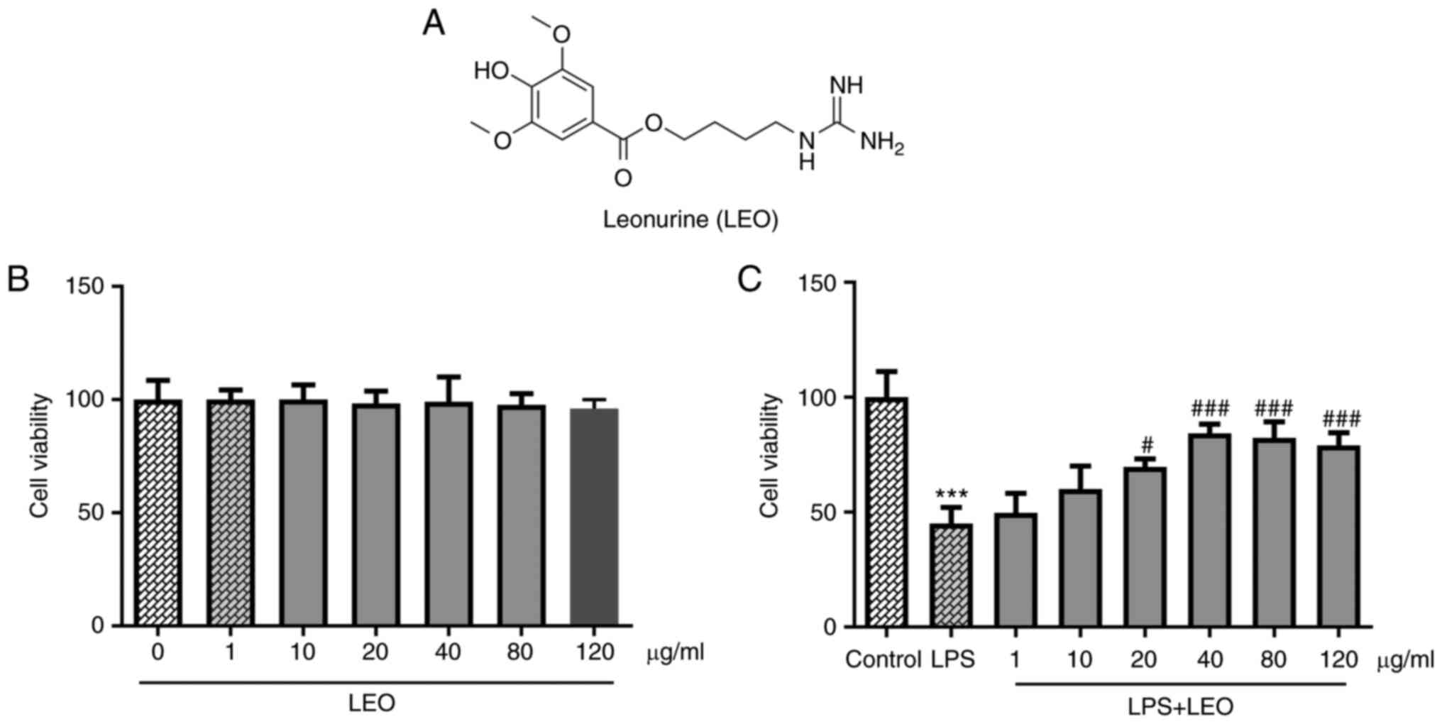
The chemical structure of LEO is presented in
Fig. 1A. To investigate the
potential cytotoxic effects of LEO, cell viability was measured
using an MTT assay. As shown in Fig.
1B, no significant changes were noted in the viability of
BEAS-2B cells treated with various doses of LEO (0, 1, 10, 20, 40,
80 and 120 µg/ml), suggesting that this compound was not cytotoxic
to BEAS-2B cells. In addition, the results of the CCK-8 assay
indicated that cell viability was reduced following LPS
stimulation, whereas it were increased by LEO treatment in a
dose-dependent manner in LPS-treated cells (Fig. 1C). Owing to the higher viability,
40 µg/ml LEO was selected for subsequent experiments. These results
suggested that LEO exerted potential therapeutic effects on
epithelial cells with LPS-induced injury.
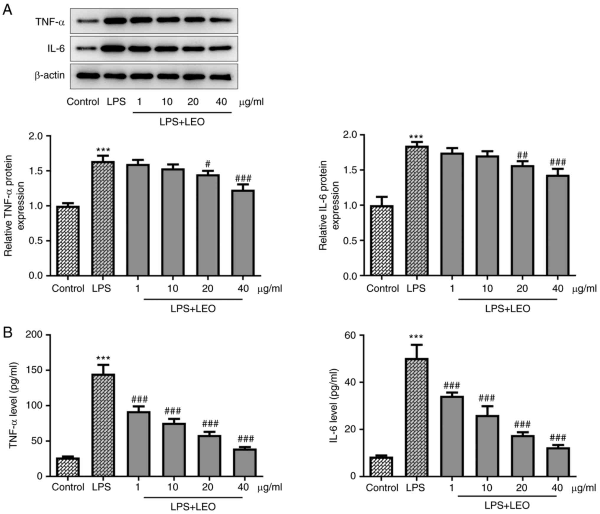
LEO suppresses LPS-induced oxidative
stress and inflammation
To investigate the mechanism underlying the function
of LEO, the induction of oxidative stress and inflammation were
analyzed in LPS-stimulated BEAS-2B cells. LPS stimulation increased
ROS, LDH and MDA levels, and decreased SOD activity in BEAS-2B
cells (Fig. 2); subsequent LEO
treatment abolished these effects. In addition, the results of the
western blot analysis and ELISA demonstrated that LPS induced the
upregulation of TNF-α and IL-6 in BEAS-2B cells, whereas LEO
treatment suppressed the expression levels of TNF-α and IL-6
(Fig. 3). These results indicated
that LEO treatment suppressed the LPS-induced oxidative stress and
inflammatory response in BEAS-2B cells.
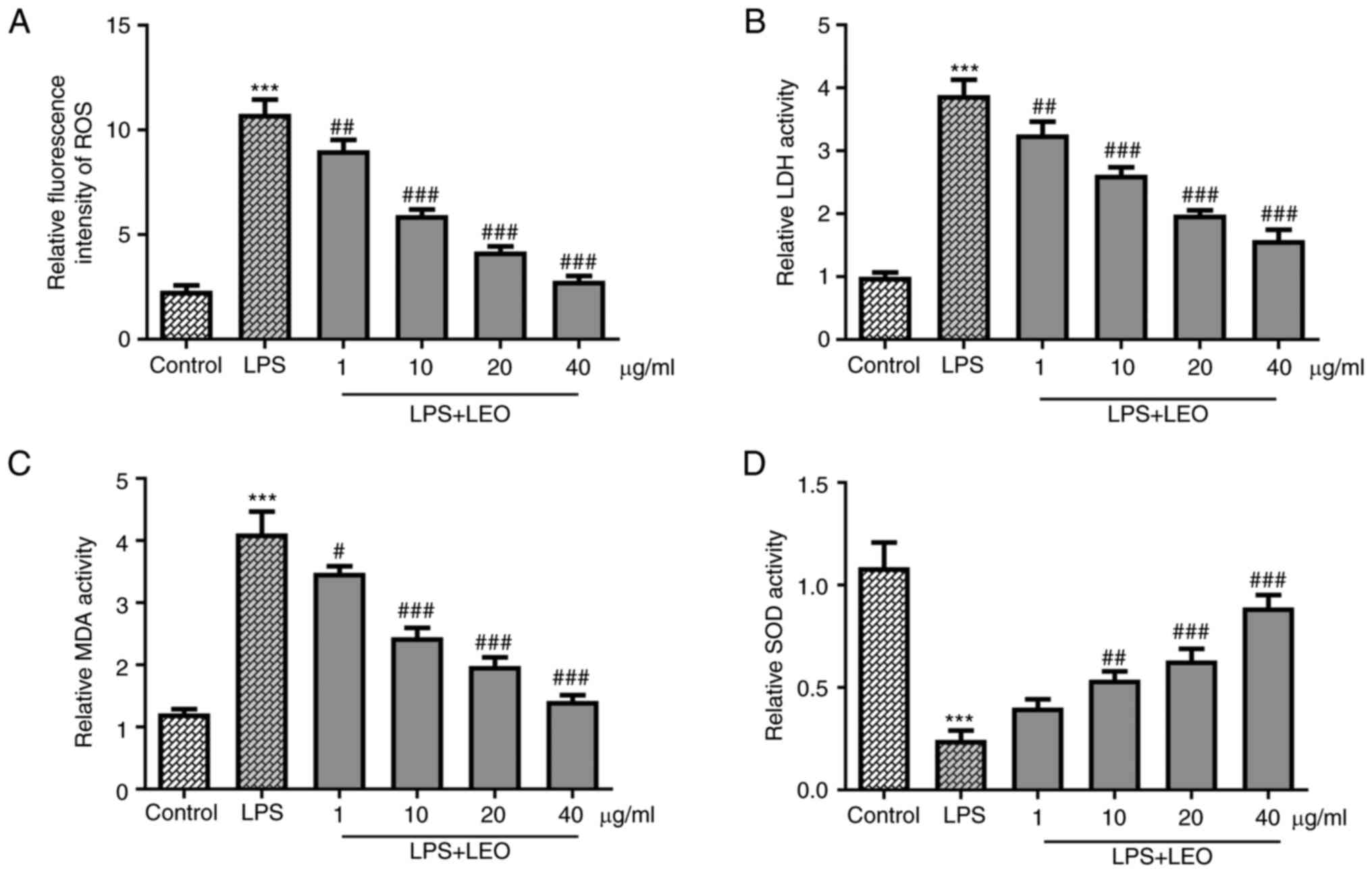 | Figure 2LEO suppresses LPS-induced oxidative
stress in BEAS-2B cells. (A) ROS, (B) LDH and (C) MDA levels, as
well as (D) SOD activity in BEAS-2B cells were measured using
specific assay kits. Data are presented as the mean ± standard
deviation of three independent experiments.
***P<0.001 vs. control; #P<0.05,
##P<0.01, ###P<0.001 vs. LPS. LDH,
lactate dehydrogenase; LEO, leonurine; LPS, lipopolysaccharide;
MDA, malondialdehyde; ROS, reactive oxygen species; SOD, superoxide
dismutase. |
Effect of LEO on the Nrf2 signaling
pathway
To investigate the mechanisms underlying the effects
of LEO, proteins involved in the Nrf2 signaling pathway were
examined by western blot analysis. LPS stimulation led to a
reduction in the expression levels of Nrf2 and HO-1 in BEAS-2B
cells, whereas LEO treatment exerted the opposite effects on the
expression levels of these markers (Fig. 4). These results suggested that LEO
treatment partially reversed the suppressive effects of LPS
stimulation on the activation of the Nrf2 signaling pathway.
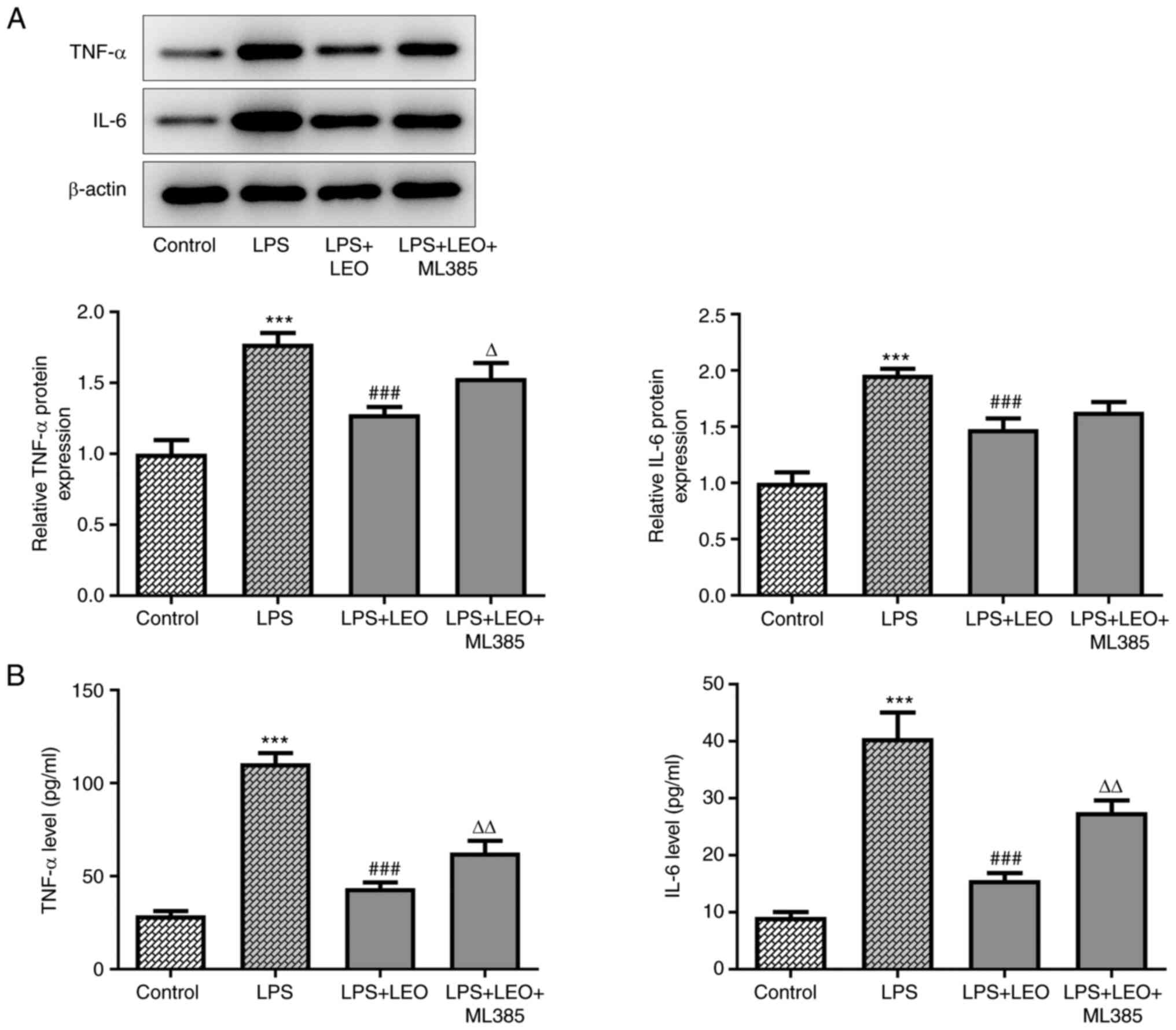
LEO enhances viability and inhibits
LPS-induced oxidative stress and inflammatory response in BEAS-2B
cells via activation of the Nrf2 signaling pathway
To confirm whether the Nrf2 signaling pathway
mediates the effects of LEO on LPS-induced epithelial cell injury,
ML385 (an Nrf2 inhibitor) was used. BEAS-2B cells were pretreated
with 10 µM ML385 to induce downregulation of Nrf2 expression. The
viability of BEAS-2B cells in the LPS + LEO + ML385 group was lower
compared with that observed in the LPS + LEO group (Fig. 5A). In addition, the inhibitory
effects of LEO treatment on ROS, LDH and MDA levels were reversed
by ML385 treatment (Fig. 5B-D,
respectively). Furthermore, treatment with ML385 significantly
reduced SOD activity in BEAS-2B cells co-treated with LPS and LEO
(Fig. 5E). Furthermore, the
expression level of TNF-α in BEAS-2B cells was significantly
enhanced by ML385 treatment, whereas that of IL-6 was only slightly
increased compared with those in the LPS + LEO group (Fig. 6A and B). Therefore, it was concluded that Nrf2
inhibition abrogated the effects of LEO treatment on the secretion
of oxidative markers and inflammatory cytokines.
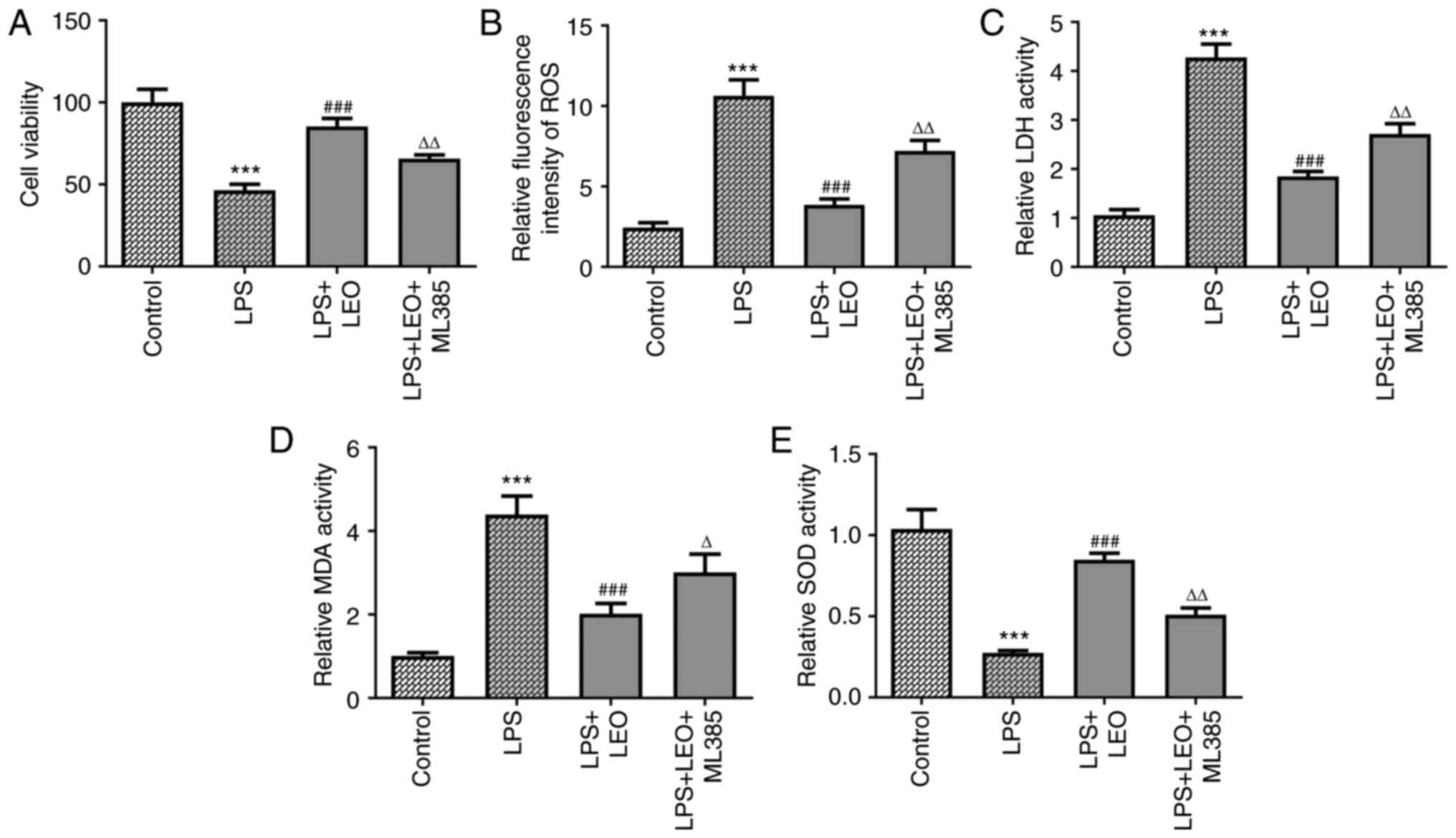 | Figure 5ML385 Nrf2 inhibitor treatment
ameliorates the effects of LEO on LPS-induced oxidative stress in
BEAS-2B cells. (A) BEAS-2B cell viability was quantified using a
Cell Counting Kit-8 assay. (B) ROS, (C) LDH and (D) MDA levels, as
well as (E) SOD activity in BEAS-2B cells were measured using
specific assay kits. Data are presented as the mean ± standard
deviation of three independent experiments.
***P<0.001 vs. control; ###P<0.001 vs.
LPS; ΔP<0.05, ΔΔP<0.01 vs. LPS + LEO.
LDH, lactate dehydrogenase; LEO, leonurine; LPS,
lipopolysaccharide; MDA, malondialdehyde; Nrf2, nuclear factor
erythroid 2-related factor 2; ROS, reactive oxygen species; SOD,
superoxide dismutase. |
Discussion
Sepsis is a common life-threatening organ
dysfunction that is triggered by a dysregulated host immune
response and remains one of the leading causes of mortality
worldwide (1). Previous studies
have demonstrated that sepsis-induced ALI is associated with high
morbidity and mortality rates (19,20).
Patients with ALI often present with hypoxemia, and oxygen therapy
can be administered to improve hypoxemia, such as nasal
catheterization. Oxygen inhalation by mask and mechanical
ventilation should be provided at an early stage when necessary
(2). Despite an improved
understanding of the pathophysiology of this disease, current
therapeutic strategies remain only partially effective (5). Therefore, the present study aimed to
develop a novel promising agent for the prevention of ALI.
The findings of the present study suggested that LEO
did not exert significant cytotoxic effects on BEAS-2B cell
viability. Subsequently, it was found that LEO treatment enhanced
the viability of LPS-stimulated BEAS-2B cells. BEAS-2B cells are
pulmonary epithelial cells, which are the major cells that form the
mechanical barrier in the lungs, and have a key protective function
against environmental aggressors (21). The results of the present study
suggested that LEO could protect against LPS-induced epithelial
cell injury and increase cell viability. LEO was first discovered
to have a protective effect on the survival of lung epithelial
cells. Subsequently, to investigate the underlying mechanism
related to the effects of LEO, the induction of oxidative stress
and inflammation were analyzed by additional experiments. In a
previous study, inflammation and oxidative stress were reported to
serve critical roles in the development of ALI. Moreover, the
inhibition of the induction of inflammation and oxidative stress
was shown to reverse the effects of ALI induced by LPS (22). Increased ROS production and
inflammatory cytokine hypersecretion have been implicated in the
development of lung injury (23).
It was reported that the levels of MDA and the expression levels of
TNF-α were significantly increased, whereas SOD levels were
significantly decreased in the lungs of ALI mice (24). Furthermore, IL-6 was found to be
elevated in the alveolar lavage fluid of patients with ALI
(25). In the present study, LEO
treatment reduced the levels of ROS, LDH and MDA, and increased SOD
activity and downregulated the expression levels of TNF-α and IL-6.
These findings demonstrated that LEO inhibited the LPS-induced
oxidative stress and inflammation in BEAS-2B cells. Subsequently,
the associated signaling pathway was assessed to explore the
molecular mechanism underlying the effects of LEO on oxidative
stress and inflammation.
Nrf2 is a transcription factor that improves
cytoprotective responses (26).
The Nrf2 signaling pathway has been reported to play an important
role in inhibiting multiple inflammatory and oxidative
stress-associated diseases, including ALI (27). More importantly, it has been
reported that LEO can induce activation of the Nrf2 signaling
pathway in vivo, which is consistent with the findings of
the current in vitro study (16). In the present study, LEO treatment
led to the activation of the Nrf2 signaling pathway in a
dose-dependent manner. Of note, the Nrf2 inhibitor (ML385)
abolished the effects of LEO treatment on cell viability,
inflammatory cytokine secretion and oxidative stress, suggesting
that LEO enhanced the viability and inhibited the LPS-induced
oxidative stress and inflammatory response in BEAS-2B cells via
activation of the Nrf2 signaling pathway. These findings suggested
that LEO specifically regulates the Nrf2 pathway to alleviate ALI,
further elucidating the pharmacological basis of its action.
However, the present study was limited to BEAS-2B cells, and
further research on additional cell lines, as well as in
vivo studies, are required. Our future plan is to explore the
effects of LEO on immune cells (such as macrophages) in
vitro and establish ALI mouse models to study the role of LEO
in vivo.
In summary, the present study demonstrated that LEO
suppressed oxidative stress and inflammation in LPS-induced BEAS-2B
cells via the Nrf2 signaling pathway, suggesting that LEO has the
potential to prevent sepsis-induced ALI.
Acknowledgements
Not applicable.
Funding
Funding: No funding was received.
Availability of data and materials
The datasets used and/or analyzed during the current
study are available from the corresponding author on reasonable
request.
Authors' contributions
LW wrote manuscript and participated in the
experimental design. GZ performed the experiments and analyzed the
results. LW and GZ confirm the authenticity of all the raw data.
Both authors have read and approved the final manuscript.
Ethics approval and consent to
participate
Not applicable.
Patient consent for publication
Not applicable.
Competing interests
The authors declare that they have no competing
interests.
References
|
1
|
Rudd KE, Johnson SC, Agesa KM, Shackelford
KA, Tsoi D, Kievlan DR, Colombara DV, Ikuta KS, Kissoon N, Finfer
S, et al: Global, regional, and national sepsis incidence and
mortality, 1990-2017: Analysis for the global burden of disease
study. Lancet. 395:200–211. 2020.PubMed/NCBI View Article : Google Scholar
|
|
2
|
Wang YM, Ji R, Chen WW, Huang SW, Zheng
YJ, Yang ZT, Qu HP, Chen H, Mao EQ, Chen Y and Chen EZ: Paclitaxel
alleviated sepsis-induced acute lung injury by activating MUC1 and
suppressing TLR-4/NF-κB pathway. Drug Des Devel Ther. 13:3391–3404.
2019.PubMed/NCBI View Article : Google Scholar
|
|
3
|
Zhang H, Wang W, Fang H, Yang Y, Li X, He
J, Jiang X, Wang W, Liu S, Hu J, et al: GSK-3β inhibition
attenuates CLP-induced liver injury by reducing inflammation and
hepatic cell apoptosis. Mediators Inflamm.
2014(629507)2014.PubMed/NCBI View Article : Google Scholar
|
|
4
|
Zhang Z, Han N and Shen Y: S100A12
promotes inflammation and cell apoptosis in sepsis-induced ARDS via
activation of NLRP3 inflammasome signaling. Mol Immunol. 122:38–48.
2020.PubMed/NCBI View Article : Google Scholar
|
|
5
|
Kim WY and Hong SB: Sepsis and acute
respiratory distress syndrome: Recent update. Tuberc Respir Dis
(Seoul). 79:53–57. 2016.PubMed/NCBI View Article : Google Scholar
|
|
6
|
Qiu N, Xu X and He Y: LncRNA TUG1
alleviates sepsis-induced acute lung injury by targeting
miR-34b-5p/GAB1. BMC Pulm Med. 20(49)2020.PubMed/NCBI View Article : Google Scholar
|
|
7
|
Fu H, Zhang J and Huang M: Topiroxostat
ameliorates oxidative stress and inflammation in sepsis-induced
lung injury. Z Naturforsch C J Biosci. 75:425–431. 2020.PubMed/NCBI View Article : Google Scholar
|
|
8
|
Hu Q, Wang Q, Han C and Yang Y: Sufentanil
attenuates inflammation and oxidative stress in sepsis-induced
acute lung injury by downregulating KNG1 expression. Mol Med Rep.
22:4298–4306. 2020.PubMed/NCBI View Article : Google Scholar
|
|
9
|
Xu W, Cui J, Zhou F, Bai M, Deng R and
Wang W: Leonurine protects against dexamethasone-induced
cytotoxicity in pancreatic β-cells via PI3K/Akt signaling pathway.
Biochem Biophys Res Commun. 529:652–658. 2020.PubMed/NCBI View Article : Google Scholar
|
|
10
|
Chen C, Zhu Z, Hu N, Liang X and Huang W:
Leonurine hydrochloride suppresses inflammatory responses and
ameliorates cartilage degradation in osteoarthritis via NF-κB
signaling pathway. Inflammation. 43:146–154. 2020.PubMed/NCBI View Article : Google Scholar
|
|
11
|
Ning K, Wang MJ, Lin G, Zhang YL, Li MY,
Yang BF, Chen Y, Huang Y, Li ZM, Huang YJ, et al: eNOS-nitric oxide
system contributes to a novel antiatherogenic effect of leonurine
via inflammation inhibition and plaque stabilization. J Pharmacol
Exp Ther. 373:463–475. 2020.PubMed/NCBI View Article : Google Scholar
|
|
12
|
Li YY, Lin YK, Liu XH, Wang L, Yu M, Li
DJ, Zhu YZ and Du MR: Leonurine: From gynecologic medicine to
pleiotropic agent. Chin J Integr Med. 26:152–160. 2020.PubMed/NCBI View Article : Google Scholar
|
|
13
|
Wang R, Li D, Ouyang J, Tian X, Zhao Y,
Peng X, Li S, Yu G and Yang J: Leonurine alleviates LPS-induced
myocarditis through suppressing the NF-кB signaling pathway.
Toxicology. 422:1–13. 2019.PubMed/NCBI View Article : Google Scholar
|
|
14
|
Xu D, Chen M, Ren X and Wu Y: Leonurine
ameliorates LPS-induced acute kidney injury via suppressing
ROS-mediated NF-κB signaling pathway. Fitoterapia. 97:148–155.
2014.PubMed/NCBI View Article : Google Scholar
|
|
15
|
Xu T, Li X, Leng T, Zhuang T, Sun Y, Tang
Y, Wang L, Yang M and Ji M: CYP2A13 acts as the main metabolic
CYP450s enzyme for activating leonurine in human bronchial
epithelial cells. Med Sci Monit. 26(e922149)2020.PubMed/NCBI View Article : Google Scholar
|
|
16
|
Chen P, Chen F and Zhou BH: Leonurine
ameliorates D-galactose-induced aging in mice through activation of
the Nrf2 signalling pathway. Aging (Albany NY). 11:7339–7356.
2019.PubMed/NCBI View Article : Google Scholar
|
|
17
|
Yuan CB, Tian L, Yang B and Zhou HY:
Isoalantolactone protects LPS-induced acute lung injury through
Nrf2 activation. Microb Pathog. 123:213–218. 2018.PubMed/NCBI View Article : Google Scholar
|
|
18
|
Kumar P, Nagarajan A and Uchil PD:
Analysis of cell viability by the MTT assay. Cold Spring Harb
Protoc. 2018:2018.PubMed/NCBI View Article : Google Scholar
|
|
19
|
Aziz M, Ode Y, Zhou M, Ochani M, Holodick
NE, Rothstein TL and Wang P: B-1a cells protect mice from
sepsis-induced acute lung injury. Mol Med. 24(26)2018.PubMed/NCBI View Article : Google Scholar
|
|
20
|
Mokra D and Kosutova P: Biomarkers in
acute lung injury. Respir Physiol Neurobiol. 209:52–58.
2015.PubMed/NCBI View Article : Google Scholar
|
|
21
|
Crystal RG, Randell SH, Engelhardt JF,
Voynow J and Sunday ME: Airway epithelial cells: Current concepts
and challenges. Proc Am Thorac Soc. 5:772–777. 2008.PubMed/NCBI View Article : Google Scholar
|
|
22
|
Lei J, Wei Y, Song P, Li Y, Zhang T, Feng
Q and Xu G: Cordycepin inhibits LPS-induced acute lung injury by
inhibiting inflammation and oxidative stress. Eur J Pharmacol.
818:110–114. 2018.PubMed/NCBI View Article : Google Scholar
|
|
23
|
Chen TH and Wang JJ: Niacin pretreatment
attenuates ischemia and reperfusion of pancreas-induced acute
pancreatitis and remote lung injury through suppressing oxidative
stress and inflammation and activation of SIRT1. Transplant Proc.
50:2860–2863. 2018.PubMed/NCBI View Article : Google Scholar
|
|
24
|
Wang X, An X, Wang X, Hu X, Bi J, Tong L,
Yang D, Song Y and Bai C: Peroxiredoxin 6 knockout aggravates cecal
ligation and puncture-induced acute lung injury. Int
Immunopharmacol. 68:252–258. 2019.PubMed/NCBI View Article : Google Scholar
|
|
25
|
Butt Y, Kurdowska A and Allen TC: Acute
lung injury: A clinical and molecular review. Arch Pathol Lab Med.
140:345–350. 2016.PubMed/NCBI View Article : Google Scholar
|
|
26
|
Matzinger M, Fischhuber K and Heiss EH:
Activation of Nrf2 signaling by natural products-can it alleviate
diabetes? Biotechnol Adv. 36:1738–1767. 2018.PubMed/NCBI View Article : Google Scholar
|
|
27
|
Pei X, Zhang XJ and Chen HM: Bardoxolone
treatment alleviates lipopolysaccharide (LPS)-induced acute lung
injury through suppressing inflammation and oxidative stress
regulated by Nrf2 signaling. Biochem Biophys Res Commun.
516:270–277. 2019.PubMed/NCBI View Article : Google Scholar
|