Open Access

Papillary mucinous cystadenoma of the kidney: Case report and review of literature

  • Authors:
    • Jingsheng Liu
    • Qian Zhao
    • Yuanyuan Zhang
    • Liang Xie
  • View Affiliations

  • Published online on: May 18, 2022     https://doi.org/10.3892/etm.2022.11378
  • Article Number: 451
  • Copyright: © Liu et al. This is an open access article distributed under the terms of Creative Commons Attribution License.

Metrics: Total Views: 0 (Spandidos Publications: | PMC Statistics: )
Total PDF Downloads: 0 (Spandidos Publications: | PMC Statistics: )


Abstract

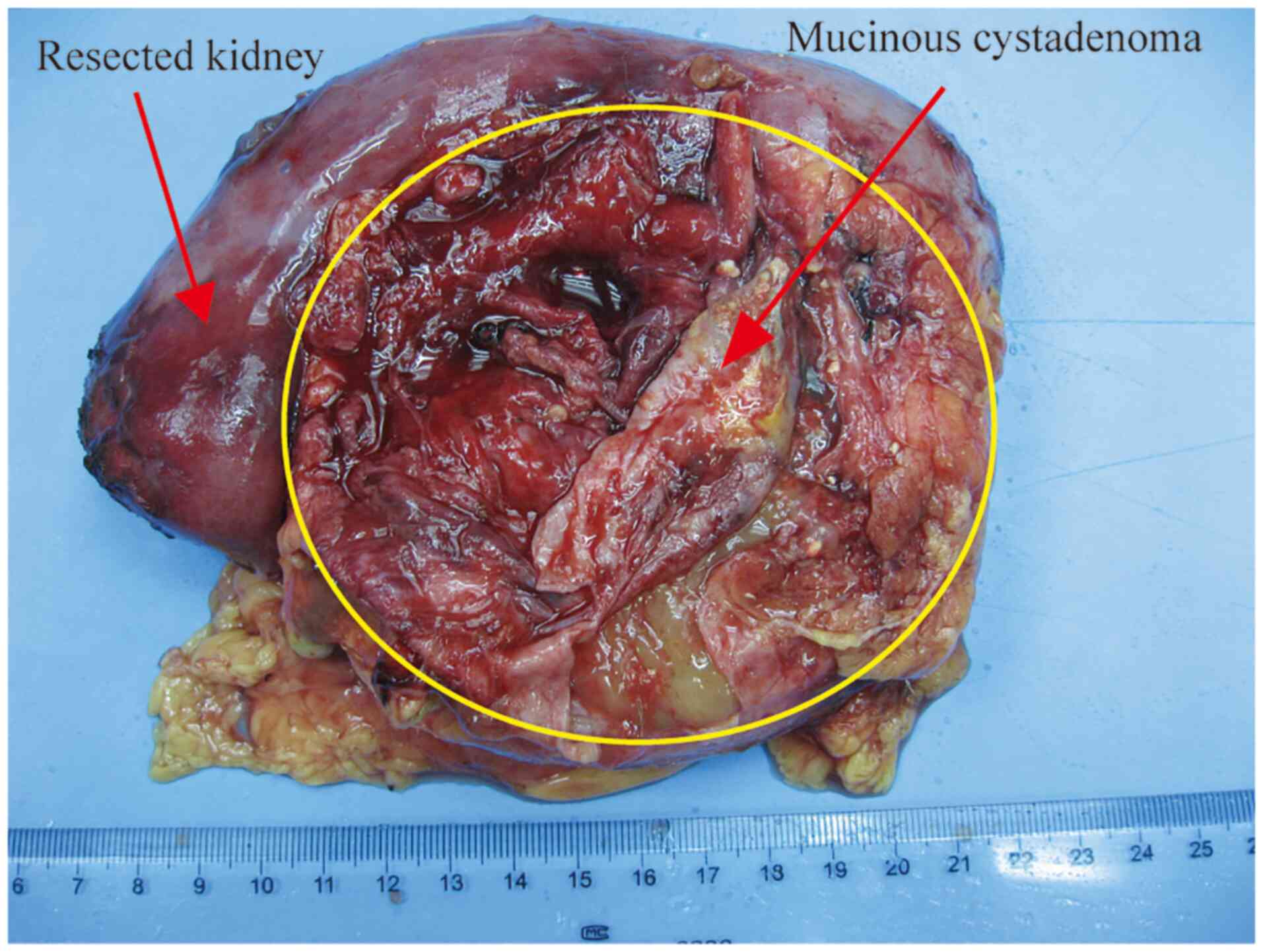
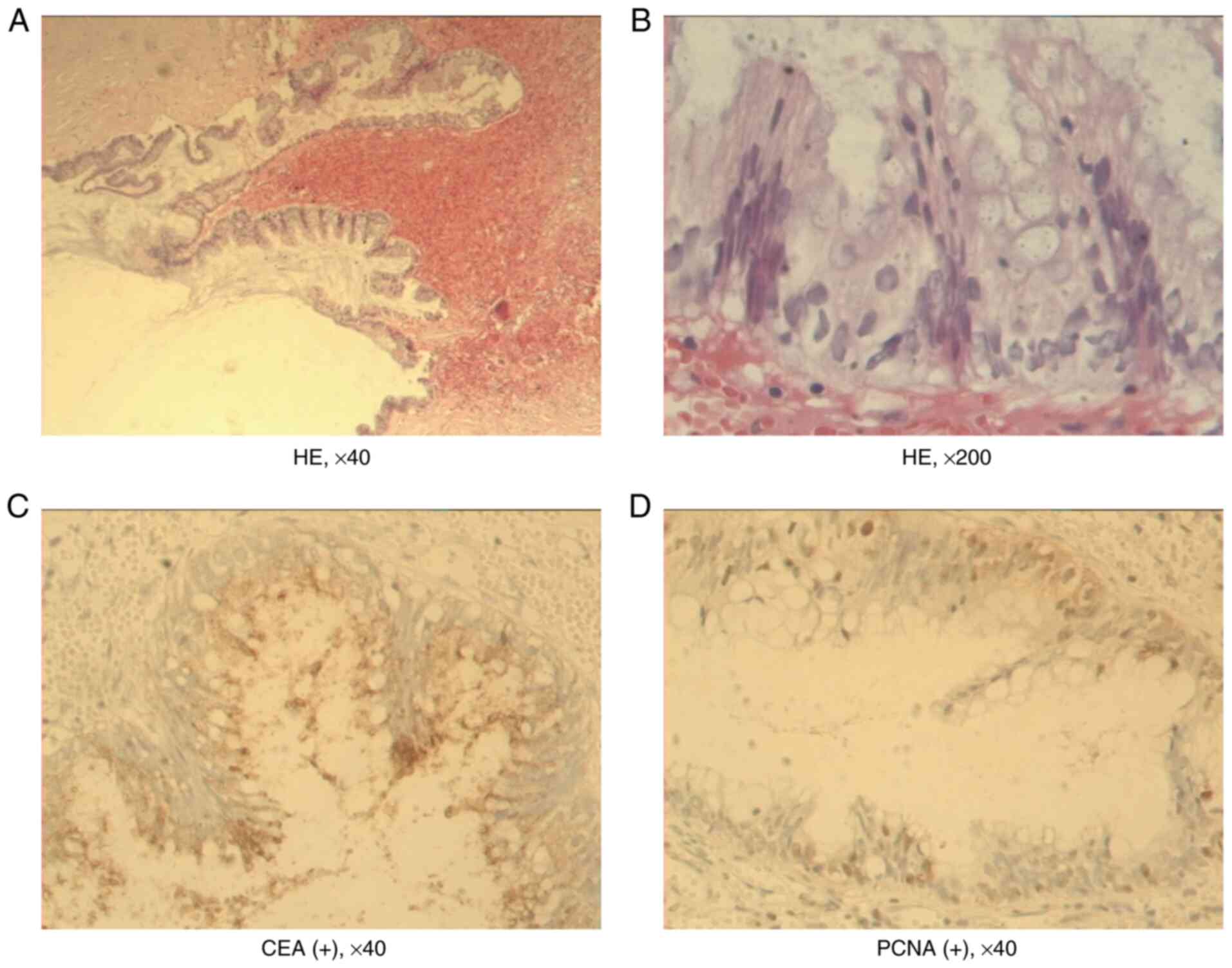
Epithelial tumors that originate from the kidney are uncommon. The majority of cases reported in the literature are malignant, with <1% of adenocarcinomas. Adenomas are even rarer. A case of mucinous cystadenoma of the kidney was diagnosed and treated in Cangzhou Hospital of Integrated TCM‑WM•HEBEI. The clinical feature of this disease was atypical. The principal symptom of mucinous cystadenoma of the kidney was a lump in the upper abdomen with intermittent pain in the abdomen. The tumor size ranged from 5‑12 cm. The patient was clinically diagnosed with multiple renal cysts with thickened walls and hemorrhage. The patient was admitted to the hospital five days later, and a left nephrectomy was performed. The tumor capsule was integrated and did not adhere to the adjacent tissue. One or multiple smooth‑walled dilated cystic areas were found on the cut section, some of which were filled with grey opaque gelatinous clots. Under light microscopy, the epithelial lining was characterized by a single layer of columnar with papillary proliferation and almost had no mitosis. Immunohistochemistry showed that the cyst was positive for carcinoembryonic antigen (CEA) and proliferating cell nuclear antigen (PCNA). Following nephrectomy, the patient was followed up for 8 years and no recurrence and metastasis were found. A total of five articles were retrieved, including six cases with mucinous cystadenoma of the kidney. The clinical feature of this disease was atypical, making clinical diagnosis difficult. Histopathological examination revealed that the cyst consisted of mucinous epithelium with supporting fibrous tissue and immunohistochemistry showed that the cyst was positive for CEA and PCNA. Patients with all resection of the affected kidney had an improved prognosis.
View Figures
View References

Related Articles

Journal Cover

July-2022
Volume 24 Issue 1

Print ISSN: 1792-0981
Online ISSN:1792-1015

Sign up for eToc alerts

Recommend to Library

Copy and paste a formatted citation
x
Spandidos Publications style
Liu J, Zhao Q, Zhang Y and Xie L: Papillary mucinous cystadenoma of the kidney: Case report and review of literature. Exp Ther Med 24: 451, 2022
APA
Liu, J., Zhao, Q., Zhang, Y., & Xie, L. (2022). Papillary mucinous cystadenoma of the kidney: Case report and review of literature. Experimental and Therapeutic Medicine, 24, 451. https://doi.org/10.3892/etm.2022.11378
MLA
Liu, J., Zhao, Q., Zhang, Y., Xie, L."Papillary mucinous cystadenoma of the kidney: Case report and review of literature". Experimental and Therapeutic Medicine 24.1 (2022): 451.
Chicago
Liu, J., Zhao, Q., Zhang, Y., Xie, L."Papillary mucinous cystadenoma of the kidney: Case report and review of literature". Experimental and Therapeutic Medicine 24, no. 1 (2022): 451. https://doi.org/10.3892/etm.2022.11378