Introduction
Chest-wall malformations have a relatively high
incidence (more than 1 per 500 live births) and an unsuspecting
impact on the quality of life of patients (1). As the main portion of the anterior
chest wall, it is essential for all radiologists and clinicians
involved in the care of children to acquire knowledge about the
development of the sternum. Although the ossification and fusion
patterns of the sternum have been well documented in children
(2,3), wide variations in the configuration
of the sternum exist among individuals. On the other hand, the
length of the sternum may provide a direct and apparent indication;
however, to the best of our knowledge, current reference ranges of
the sternum length based on a Han Chinese population background are
not available in literature.
Estimation of the stature and sex identification
from the whole of, or part of the length of, the sternum have been
investigated already in an adult population (4-6).
The sternal plastron and lower chest have also exhibited excellent
reproducibility in terms of estimation of the age in adults
(6), although whether this method
is applicable to children requires further study.
Normal reference data can also be applied in
clinical practice, such as assessment of thoracic trauma, chest
wall deformities and other associated development disorders
(7-9).
For instance, no consensus has been established on the cause of
pectus excavatum; nor on whether the costal cartilage is too short
or overgrown (10,11). To answer these questions, full
screening of the normal development of the sternum on computed
tomography (CT) examinations is fundamental.
In order to distinguish abnormal from normal sternum
growth, the normal ranges of sternum length in a normal Chinese
pediatric population of different ages are required. Investigating
this question formed the basis of the present study.
Materials and methods
Patient selection
Patients were randomly extracted from the database
of Union Hospital, Tongji Medical College, Huazhong University of
Science and Technology who were 0-18 years of age and who had
undergone a chest CT scan between January 2015 and January 2020.
The present study was approved (approval no.
UHCT-IEC-SOP-016-03-01) by the ethics committee of Union Hospital,
Tongji Medical College, Huazhong University of Science and
Technology (Wuhan, China) and the procedures followed were in
accordance with the Helsinki Declaration of 1964, as revised in
2000.
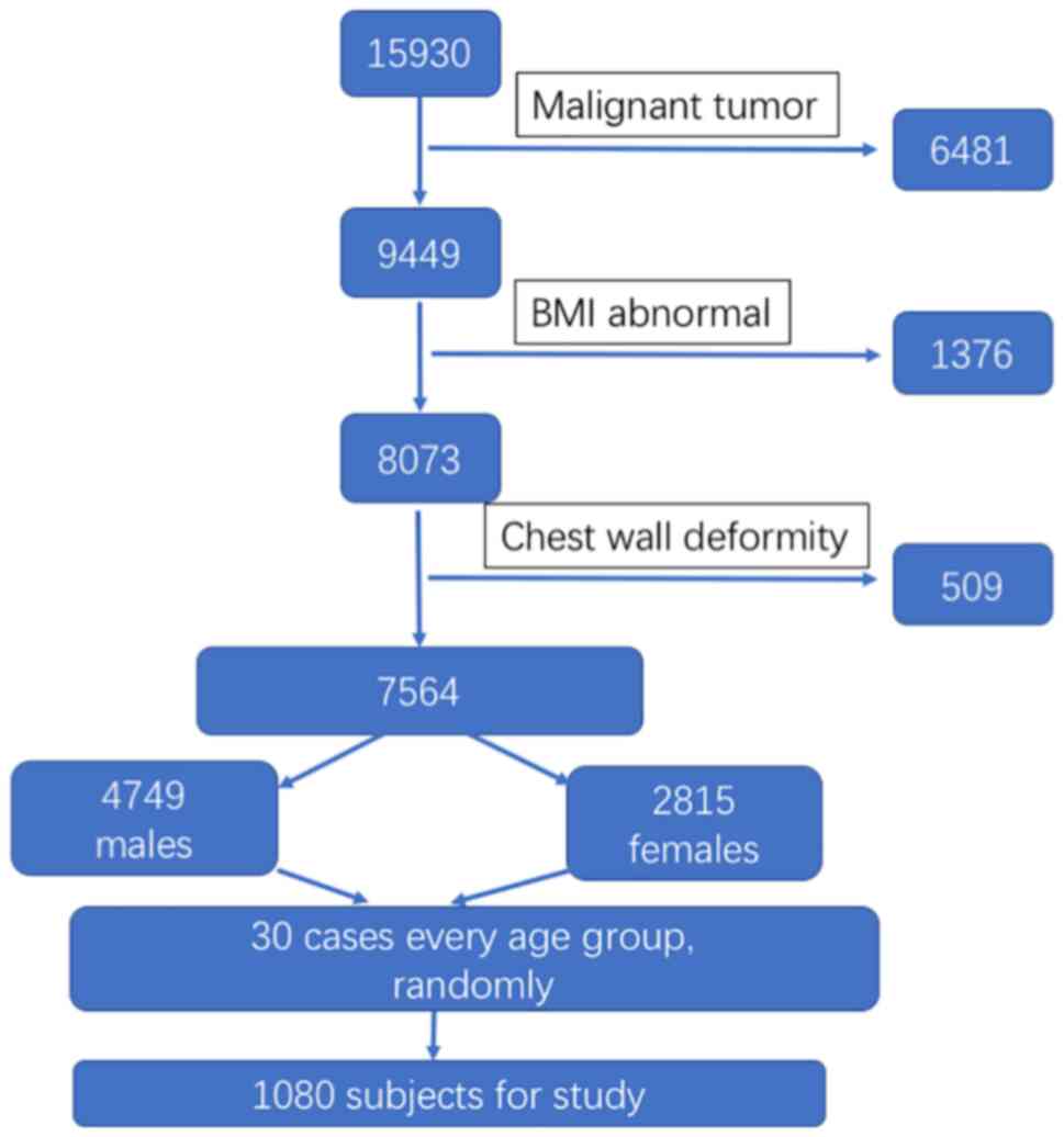
Inclusion criteria were as follows: i) 0-18 years of
age; ii) Undergone chest CT scan for suspicious pulmonary
infection, foreign body aspiration and trauma iii) The body weight
limited to ±20% of the standard reference; iv) Han nationality from
central China, including Hubei, Hunan, Henan, Anhui and Jiangxi
provinces.
Exclusion criteria were as follows: i) Those
patients with lung consolidation, pleural effusion, congenital or
acquired structural anomalies or congenital metabolic diseases; ii)
Patients who had had a history of thoracotomy or sternotomy; and
iii) Patients for whom the images obtained contained artifacts.
The entire cohort contained a total of 1,080 cases,
with 30 male and 30 female subjects in each age group (Fig. 1).
Multidetector-row CT (MDCT)
protocol
Non-contrast chest CT scans were obtained with the
patients in the supine position, and the scans were performed at
the end of an inspiration according to the pediatric protocol. CT
examinations were carried out following the MDCT protocol with
either a Discovery 750 HD (GE Medical Systems, LLC) or a SOMATOM
Definition AS (Siemens AG) CT system. Images were reconstructed
into lung and soft tissue mediastinal windows with a slice
thickness of 1.3 or 1.5 mm, and an interval of 1.3 mm or 1.5 mm,
respectively. All CT data was transferred to the workstations and
imaging analyses were performed on a picture archiving and
communication system workstation (Carestream Health, Inc.). Both
coronal and sagittal multiplanar reconstructions of CT images were
prepared.
Assessment of the images
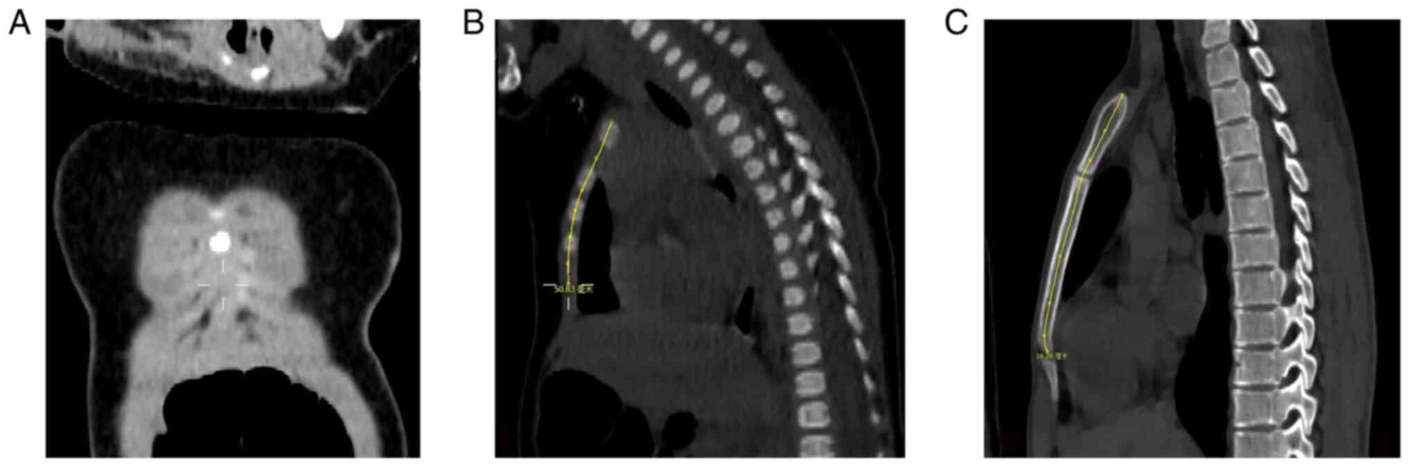
The length of the sternum before the ossification
centers completely merged into a single ossification center was
defined as the linear distance from the center of the suprasternal
notch or incisura jugularis (jugular notch) to the junction of the
6th/7th costal cartilage in the mid-sagittal plane (Fig. 2A and B), whereas the length of the sternum
after the ossification centers completely merged into a single
ossification center was defined as the linear distance from the
center of the suprasternal notch or incisura jugularis (jugular
notch) to the mesosterno-exiphoidal junction (Fig. 2C).
All the measurements were recorded by two different
radiologists (PL with >10 years of experience, and CC with >5
years of experience), and the mean values were used for subsequent
analysis.
Statistical analysis
Data were analyzed using SPSS version 20.0
statistical software (IBM Corp.). Sex-wise comparisons of the
length of the sternum were performed using the Mann-Whitney test.
Age or region group comparisons of the length of the sternum were
assessed using ANOVA, followed by Tukey's post hoc test. A further
regression analysis was performed, and the most suited regression
model was constructed. P<0.05 was considered to indicate a
statistically significant difference for all the statistical
data.
Results
A total of 1,080 patients were enrolled in the
present study. There were 540 females and 540 males who were
younger than 18 years old and without congenital or acquired
structural anomalies or congenital metabolic diseases, with 60 case
subjects in each age group.
Subjects in the present study were from central
China, including Hubei (n=761), Hunan (n=47), Henan (n=143), Anhui
(n=55) and Jiangxi (n=74) provinces. A general comparison was
conducted between subjects of Henan Province and Hubei Province
with a relatively larger number of subjects. No significant
differences were identified in the length of the sternum between
youngsters from the two regions (P=0.53).
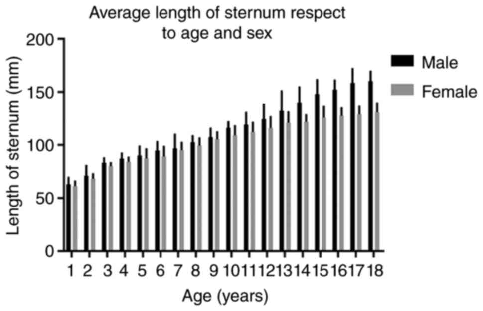
No significant differences were identified in the
length of the sternum in patients <14 years of age comparing
between the sexes. However, for the patients who were older than
14, the mean length of the sternum in males was longer compared
with that of females (Fig. 3 and
Table I).
 | Table IAverage length of sternum in respect
to age and sex. |
Table I
Average length of sternum in respect
to age and sex.
| | Mann-Whitney
Test |
|---|
| Age, years | N | Male Average length
of sternum (95% CI) (mm) | n | Female Average length
of sternum (95% CI) (mm) | Z-value | P-value |
|---|
| 1 | 30 | 62.96 (58.62,
67.54) | 30 | 61.05 (57.95,
64.50) | -1.134 | 0.257 |
| 2 | 30 | 71.10 (64.50,
77.77) | 30 | 68.32 (65.16,
71.40) | -1.134 | 0.257 |
| 3 | 30 | 83.30 (80.02,
86.38) | 30 | 80.03 (77.85,
82.21) | -1.814 | 0.070 |
| 4 | 30 | 87.26 (83.79,
90.73) | 30 | 84.50 (81.63,
87.36) | -1.436 | 0.151 |
| 5 | 30 | 90.51 (83.86,
96.15) | 30 | 87.38 (77.14,
89.63) | -1.361 | 0.174 |
| 6 | 30 | 94.79 (89.04,
100.55) | 30 | 89.28 (82.89,
95.67) | -1.285 | 0.199 |
| 7 | 30 | 96.84 (87.62,
106.07) | 30 | 95.25 (90.35,
100.16) | -0.378 | 0.705 |
| 8 | 30 | 102.51 (98.37,
106.65) | 30 | 99.63 (94.95,
104.31) | -1.097 | 0.273 |
| 9 | 30 | 107.44 (101.77,
113.12) | 30 | 105.40 (100.80,
110.01) | -0.605 | 0.545 |
| 10 | 30 | 116.01 (112.03,
120.00) | 30 | 108.85 (102.49,
115.22) | -1.890 | 0.059 |
| 11 | 30 | 119.26 (111.39,
127.13) | 30 | 112.19 (106.00,
118.38) | -1.436 | 0.151 |
| 12 | 30 | 124.46 (114.54,
134.37) | 30 | 115.98 (108.66,
123.29) | -1.512 | 0.131 |
| 13 | 30 | 132.32 (119.16,
145.48) | 30 | 120.93 (113.90,
127.97) | -1.209 | 0.226 |
| 14 | 30 | 140.10 (129.88,
150.33) | 30 | 121.79 (117.33,
126.26) | -2.873 | 0.004 |
| 15 | 30 | 148.11 (138.76,
157.47) | 30 | 125.79 (118.50,
133.08) | -2.987 | 0.003 |
| 16 | 30 | 152.31 (146.19,
158.43) | 30 | 127.47 (122.33,
132.61) | -3.780 | <0.001 |
| 17 | 30 | 158.52 (149.08,
167.95) | 30 | 128.91 (123.72,
134.10) | -3.780 | <0.001 |
| 18 | 30 | 160.31 (153.92,
166.69) | 30 | 130.84 (124.86,
136.82) | -3.704 | <0.001 |
| Total | 54 | 113.64 | 54 | 103.35 | -2.813 | 0.005 |
| | 0 | (109.20,
118.31) | 0 | (100.01,
106.59) | | |
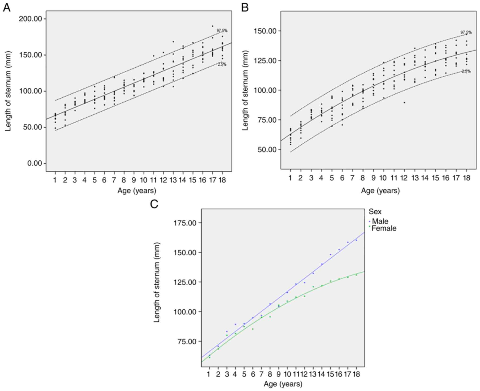
As demonstrated in Table II, sternal length associated
differently comparing between the sexes. In order to find the
association between age and the length of sternum for each sex,
different regression analysis was performed using linear,
logarithmic, inverse, quadratic and other regression models.
According to the value of R2, the most suitable
regression pattern was a linear model in males, and a quadratic
model in females (specifically, the regression equation for males
was y=5.616x+60.408; P<0.001; R2=0.889, whereas that
for females was y=-0.134x2+6.543x+56.805; P<0.001;
R2=0.890) (Fig. 4).
| Table IIFitness test in different regression
models for each sex. |
Table II
Fitness test in different regression
models for each sex.
| | Male | Female |
|---|
| | Model analysis | Parameter
analysis | Model analysis | Parameter
analysis |
|---|
| | R2 | P-value | B | P-value | R2 | P-value | B | P-value |
|---|
| Linear | 0.889 | <0.001 | 5.616 | <0.001 | 0.870 | <0.001 | 4.003 | <0.001 |
| Logarithmic | 0.773 | <0.001 | 34.867 | <0.001 | 0.847 | <0.001 | 26.300 | <0.001 |
| Inverse | 0.476 | <0.001 | -94.707 | <0.001 | 0.579 | <0.001 | -75.279 | <0.001 |
| Quadratic | 0.890 | <0.001 | 0.038 | 0.240 | 0.890 | <0.001 | -0.134 | <0.001 |
| Cubic | 0.891 | <0.001 | 0.005 | 0.481 | 0.890 | <0.001 | 0.0004 | 0.939 |
| Compound | 0.886 | <0.001 | 1.053 | <0.001 | 0.846 | <0.001 | 1.042 | <0.001 |
| Power | 0.850 | <0.001 | 0.335 | <0.001 | 0.889 | <0.001 | 0.281 | <0.001 |
| S | 0.589 | <0.001 | -0.964 | <0.001 | 0.667 | <0.001 | -0.841 | <0.001 |
| Growth | 0.886 | <0.001 | 0.051 | <0.001 | 0.846 | <0.001 | 0.041 | <0.001 |
| Exponential | 0.886 | <0.001 | 0.051 | <0.001 | 0.846 | <0.001 | 0.041 | <0.001 |
| Logistic | 0.886 | <0.001 | 0.950 | <0.001 | 0.846 | <0.001 | 0.960 | <0.001 |
Subsequently, to obtain an intuitive comparison of
the lengths of the sternum comparing between the two sexes, scatter
diagrams were drawn with age as the x-axis and the average length
of sternum as the y-axis, first using the most-fit formula for the
sexes (Fig. 4A and B), and then combining these curves
together into one chart (Fig.
4C).
Discussion
The sternum is a flat bone that extends vertically
through, and provides anterior support to, the thoracic cage.
Sternal development starts from the prenatal period and continues
throughout puberty (3,5). As the first study, to the best of our
knowledge, to have studied the sternum length in a normal Han
Chinese pediatric population, it was demonstrated that the length
of the sternum can be precisely estimated by age using our formulae
for the different sexes. Building up our knowledge on the sternum
is crucial for a proper understanding of the spectrum of the
deformities of the chest wall, giving rise to appropriate planning
for thoracic surgery and preventing misdiagnosis (3). However, given that the most recent
data available was published in the 1980s and the 1990s (12-14),
the reference value of sternum length in the normal Chinese
population may have changed over the course of the last 40
years.
Age estimation using the clavicular bone or teeth is
a well-standardized procedure in children and adolescents (15), although using the sternum may be a
possible alternative under certain conditions. According to the
present study, the length of the sternum is associated with age
under normal physiological conditions, but with different
regression patterns for the two different sexes. Boys eventually
develop a longer sternum, but before they reach 14 years of age,
boys have almost the same sternum length as girls. This finding may
be associated with the different growth curves of boys and
girls.
In the present study, the reference length of the
sternum with 95% confidence intervals has also been established.
Since numerous types of sternal fuse variations cause confusion for
the care provider in terms of estimating the age or in other
clinical practices, the length of the sternum may provide a
promising alternative for future applications.
Even though several studies have been performed that
were concerned with investigating the sternum length, the majority
of these were for forensic purposes in adults (16-18),
and our understanding of how the sternum develops throughout
childhood remains poor. A search of the literature in Web of
Science using as key words ‘length of sternum’ (or ‘sternum
length’) and ‘pediatric population’ (or
‘child’/ʻchildren’/ʻadolescent’) revealed a few studies that have
been published on sternum length in a normal pediatric population.
Canavese et al (19)
acquired the length of the sternum in 622 healthy individuals (406
girls and 216 boys) aged 6-18 years old in France using the optical
ORTEN system and it was revealed that the sternal length was
11.23±1.34 cm in girls and 10.61±1.15 cm in boys at the age of 5
years on average, attaining a mean value of 18.85±1.51 cm in girls
and 19.22±1.75 cm in boys at skeletal maturity. Veldre et al
(20) performed an anthropometric
cross-sectional study of 374 healthy schoolgirls aged 12-15 years
old from secondary schools of Tartu (Estonia), where the authors
systematized the data in each age group into 5 categories
(categories I-V) according to the level of correspondence between
body height and weight, and it was identified that the sternum
length did not differ significantly comparing two category
groupings (I-III and IV-V) in all age groups. Sandoz et al
(21) measured the volume of the
sternum in 48 children aged from 4 months to 15 years (22 girls and
26 boys) in the Necker Hospital (Paris, France), and their study
suggested that the volumes were significantly different between all
age groups, and the volume of the 15-year-old sternum was ~10 times
the volume at birth (21). The
research cohort of the present study was extracted from the
database of Union Hospital (Hubei, China), and the findings
suggested that the sternal length was 87.38 (95% CI: 77.14; 89.63)
mm in girls and 90.51 (95% CI: 83.86; 96.15) mm in boys at the age
of 5 years on average, attaining a mean value of 130.84 (95% CI:
124.86; 136.82) mm in girls and 160.31 (95% CI: 153.92; 166.69) mm
in boys at the age of 18 years. Compared with the results of
Canavese et al (19), the
subjects in the present study may have had shorter sternal length.
Therefore, whether or not the formulae can be applied for other
races requires further investigation.
In the present study, any associations between the
length of the sternum and body weight were not investigated, since
the case subjects were subjected to chest CT scans due to
suspicious pulmonary infection, foreign body aspiration or trauma,
all factors which may have influenced the body weight. The
objective of the present study was to investigate the ranges of
sternum length in a normal Chinese pediatric population of
different ages, and subjects were excluded if their body weight was
noted to have exceeded ±20% of the standard reference, as reported
by Zhang et al (22) and
Fang et al (23), to
minimize the potential influence of malnutrition and obesity.
Several previous studies have investigated the
correlation of the sternum length with body height (4,16,18,24-26).
The majority of these research groups, with the exception of the
studies by Menezes et al (24) and Yonguc et al (26), reported a poor correlation between
sternal measurements and stature. Furthermore, Chandrakanth et
al (27), having investigated
the length of the sternum and the stature of the non-fused or
partly fused sterna rather than the completely fused sternum, also
found that there was no significant correlation between these
parameters. Whether or not the length of the sternum is associated
with the body height remains controversial. Therefore, in the
present study, the weight of the subjects was strictly limited to
within the ±20% of the standard reference, and the association
between the stature and the sternal length was not studied.
There were a number of limitations associated with
the present study. First, it was not designed in a randomized and
perspective manner. There were only 30 cases in each subgroup,
which was a relatively small number of case subjects to include per
subgroup. Second, the number of each age group was not enough for
effective analysis on the region difference. Not having a fully
developed sternum in the younger age subgroups may have influenced
the results of length measurement. Moreover, people with different
genetic backgrounds and environmental factors may have different
growth curves, and therefore the sternum length could vary
according to nationality, or region.
Furthermore, it is important to realize that all
techniques used for age estimation can only provide a biological
age. Even age-matched individuals may show very different signs of
the sternal features due to extrinsic factors, including the health
status, nutrition and physical exercise, as these may have an
impact on physical development.
In conclusion, the present study comprised a
preliminary investigation to establish the normal ranges of the
length of the sternum, which has been shown to be important in
terms of estimating the age for a pediatric population. A more
complete understanding of the implications of these findings and
the practical importance of these results should emerge in the
future, although large-scale studies are required to confirm our
results.
Acknowledgements
The authors would like to thank Dr KKY Wong from
Division of Pediatric Surgery, Queen Mary Hospital, The University
of Hong Kong for language editing for this manuscript.
Funding
Funding: The present study was supported by the Public Welfare
Research, and special funds were received from National Health and
Family Planning of China (grant no. 201402007) and the Fundamental
Research Funds for the Central Universities (HUST; grant no.
2015LC023).
Availability of data and materials
The datasets used and/or analyzed during the current
study are available from the corresponding author on reasonable
request.
Authors' contributions
STT, XL, SL and PL conceptualized and designed the
study. STT and XL provided administrative support. SL, PL, YL, CC
and DY provided study materials or patients. SL, PL, YL, CC and DY
acquired data. YL and DY analyzed and interpreted the data. SL, PL
and YL confirmed the authenticity of all the raw data. All authors
prepared, edited and reviewed the manuscript. All authors read and
approved the final manuscript.
Ethics approval and consent to
participate
The present study was approved (approval no.
UHCT-IEC-SOP-016-03-01) by the Ethics Committee of Union Hospital,
Tongji Medical College, Huazhong University of Science and
Technology (Wuhan, China) and the procedures followed were in
accordance with the Helsinki Declaration of 1964, as revised in
2000.
Patient consent for publication
Not applicable.
Competing interests
The authors declare that they have no competing
interests.
References
|
1
|
Obermeyer RJ and Goretsky MJ: Chest wall
deformities in pediatric surgery. Surg Clin North Am. 92:669–684,
ix. 2012.PubMed/NCBI View Article : Google Scholar
|
|
2
|
Gumeler E, Akpinar E and Ariyurek OM: MDCT
evaluation of sternal development. Surg Radiol Anat. 41:281–286.
2019.PubMed/NCBI View Article : Google Scholar
|
|
3
|
Bayaroğulları H, Yengil E, Davran R,
Ağlagül E, Karazincir S and Balcı A: Evaluation of the postnatal
development of the sternum and sternal variations using
multidetector CT. Diagn Interv Radiol. 20:82–89. 2014.PubMed/NCBI View Article : Google Scholar
|
|
4
|
Tumram NK, Parchake SB, Bardale RV and
Dixit PG: Estimation of height from the length of the sternum in an
adult Indian population. Med Sci Law. 56:46–52. 2016.PubMed/NCBI View Article : Google Scholar
|
|
5
|
Oner Z, Turan MK, Oner S, Secgin Y and
Sahin B: Sex estimation using sternum part lenghts by means of
artificial neural networks. Forensic Sci Int. 301:6–11.
2019.PubMed/NCBI View Article : Google Scholar
|
|
6
|
Oldrini G, Harter V, Witte Y, Martrille L
and Blum A: Age estimation in living adults using 3D volume
rendered CT images of the sternal plastron and lower chest. J
Forensic Sci. 61:127–133. 2016.PubMed/NCBI View Article : Google Scholar
|
|
7
|
Fotiadis E, Grigoriadou A, Kapetanos G,
Kenanidis E, Pigadas A, Akritopoulos P and Samoladas E: The role of
sternum in the etiopathogenesis of Scheuermann disease of the
thoracic spine. Spine (Phila Pa 1976). 33:E21–E24. 2008.PubMed/NCBI View Article : Google Scholar
|
|
8
|
Madjarov JM, Katz MG, Kane PN, Madzharov S
and Robicsek F: Early surgical reconstruction of sternum with
longitudinal rigid polymer plating after acute chest trauma. Ann
Thorac Cardiovasc Surg. 24:324–327. 2018.PubMed/NCBI View Article : Google Scholar
|
|
9
|
Valente T, Bocchini G, Rossi G, Sica G,
Davison H and Scaglione M: MDCT prior to median re-sternotomy in
cardiovascular surgery: Our experiences, infrequent findings and
the crucial role of radiological report. Br J Radiol.
92(20170980)2019.PubMed/NCBI View Article : Google Scholar
|
|
10
|
Eisinger RS, Harris T, Rajderkar DA and
Islam S: Against the overgrowth hypothesis: Shorter costal
cartilage lengths in pectus excavatum. J Surg Res. 235:93–97.
2019.PubMed/NCBI View Article : Google Scholar
|
|
11
|
Park CH, Kim TH, Haam SJ and Lee S: Rib
overgrowth may be a contributing factor for pectus excavatum:
Evaluation of prepubertal patients younger than 10years old. J
Pediatr Surg. 50:1945–1948. 2015.PubMed/NCBI View Article : Google Scholar
|
|
12
|
Bahar N and Gan ZM: The relationship
between the sternal length and the stature in Han youngsters in
Xinjiang. Acta Academiae Medicinae, Xinjiang. 21:182–185. 1998.(In
Chinese).
|
|
13
|
Guang HZ, Gan ZM, Yang GZ, et al: The
relationship between the sternal length and the stature in young
people of Uygur nationality in Xinjiang. Acta Academiae Medicinae
Xinjiang. 12:143–148. 1989.(In Chinese).
|
|
14
|
Zheng JZ, Yang YT, Dang RL, et al: The
Relationship between the stature and the sternal length in Han
young students of Han nationality in Xian. Acta Anthropologica
Sinica. 4:268–274. 1985.(In Chinese).
|
|
15
|
Bassed RB: Advances in forensic age
estimation. Forensic Sci Med Pathol. 8:194–196. 2012.PubMed/NCBI View Article : Google Scholar
|
|
16
|
Marinho L, Almeida D, Santos A and Cardoso
HF: Is the length of the sternum reliable for estimating adult
stature? A pilot study using fresh sterna and a test of two methods
using dry sterna. Forensic Sci Int. 220:292.e1–4. 2012.PubMed/NCBI View Article : Google Scholar
|
|
17
|
Saraf A, Kanchan T, Krishan K, Ateriya N
and Setia P: Estimation of stature from sternum-Exploring the
quadratic models. J Forensic Leg Med. 58:9–13. 2018.PubMed/NCBI View Article : Google Scholar
|
|
18
|
Menezes RG, Nagesh KR, Monteiro FN, Kumar
GP, Kanchan T, Uysal S, Rao PP, Rastogi P, Lobo SW and Kalthur SG:
Estimation of stature from the length of the sternum in South
Indian females. J Forensic Leg Med. 18:242–245. 2011.PubMed/NCBI View Article : Google Scholar
|
|
19
|
Canavese F, Dimeglio A, Bonnel F, Corradin
M, Pereira B, Marcoul A and Charles YP: Thoracic cage volume and
dimension assessment by optoelectronic molding in normal children
and adolescents during growth. Surg Radiol Anat. 41:287–296.
2019.PubMed/NCBI View Article : Google Scholar
|
|
20
|
Veldre G, Stamm R and Koskel S: A
possibility of systematisation of anthropometric data of girls aged
12-15. Anthropol Anz. 60:369–382. 2002.PubMed/NCBI
|
|
21
|
Sandoz B, Badina A, Laporte S, Lambot K,
Mitton D and Skalli W: Quantitative geometric analysis of rib,
costal cartilage and sternum from childhood to teenagehood. Med
Biol Eng Comput. 51:971–979. 2013.PubMed/NCBI View Article : Google Scholar
|
|
22
|
Zhang YQ, Li H, Wu HH and Zong XN: Secular
trends in weight, height and weight for height among children under
7 years in nine cities of China, 1975-2015: Results from five
repeated cross-sectional surveys. BMJ Open.
9(e029201)2019.PubMed/NCBI View Article : Google Scholar
|
|
23
|
Fang H, Zhao L, Guo Q, et al: Trends of
height, weight and BMI in Chinese children and adolescents aged
6~17. Food and Nutrition in China. 27:16–20. 2021.(In Chinese).
|
|
24
|
Menezes RG, Kanchan T, Kumar GP, Rao PP,
Lobo SW, Uysal S, Krishan K, Kalthur SG, Nagesh KR and Shettigar S:
Stature estimation from the length of the sternum in South Indian
males: A preliminary study. J Forensic Leg Med. 16:441–443.
2009.PubMed/NCBI View Article : Google Scholar
|
|
25
|
Singh J, Pathak RK and Chavali KH:
Skeletal height estimation from regression analysis of sternal
lengths in a Northwest Indian population of Chandigarh region: A
postmortem study. Forensic Sci Int. 206:211.e1–8. 2011.PubMed/NCBI View Article : Google Scholar
|
|
26
|
Yonguc GN, Kurtulus A, Bayazit O, Adiguzel
E, Unal I, Demir S and Acar K: Estimation of stature and sex from
sternal lengths: An autopsy study. Anat Sci Int. 90:89–96.
2015.PubMed/NCBI View Article : Google Scholar
|
|
27
|
Chandrakanth HV, Kanchan T and Krishan K:
Effect of fusion status of sternum in stature estimation-A study
from South India. J Forensic Leg Med. 36:90–95. 2015.PubMed/NCBI View Article : Google Scholar
|