|
1
|
Kannus P, Parkkari J, Sievänen H, Heinonen
A, Vuori I and Järvinen M: Epidemiology of hip fractures. Bone. 18
(1 Suppl):S57–S63. 1996.PubMed/NCBI View Article : Google Scholar
|
|
2
|
Hayes WC, Myers ER, Robinovitch SN, Van
Den Kroonenberg A, Courtney AC and McMahon TA: Etiology and
prevention of age-related hip fractures. Bone. 18 (1
Suppl):77S–86S. 1996.PubMed/NCBI View Article : Google Scholar
|
|
3
|
Holt G, Smith R, Duncan K, Finlayson DF
and Gregori A: Early mortality after surgical fixation of hip
fractures in the elderly: An analysis of data from the scottish hip
fracture audit. J Bone Joint Surg Br. 90:1357–1363. 2008.PubMed/NCBI View Article : Google Scholar
|
|
4
|
Bartoníček J and Rammelt S: The history of
internal fixation of proximal femur fractures Ernst Pohl-the genius
behind. Int Orthop. 38:2421–2426. 2014.PubMed/NCBI View Article : Google Scholar
|
|
5
|
Usami T, Takada N, Nishida K, Sakai H,
Iwata H, Sekiya I, Ueki Y, Murakami H and Kuroyanagi G: Banding
with lesser trochanter fragment using nonabsorbable tape in
trochanteric femoral fractures. SICOT J. 7(33)2021.PubMed/NCBI View Article : Google Scholar
|
|
6
|
Socci AR, Casemyr NE, Leslie MP and
Baumgaertner MR: Implant options for the treatment of
intertrochanteric fractures of the hip: Rationale, evidence, and
recommendations. Bone Joint J. 99-B:128–133. 2017.PubMed/NCBI View Article : Google Scholar
|
|
7
|
Yu X, Wang H, Duan X, Liu M and Xiang Z:
Intramedullary versus extramedullary internal fixation for unstable
intertrochanteric fracture, a meta-analysis. Acta Orthop Traumatol
Turc. 52:299–307. 2018.PubMed/NCBI View Article : Google Scholar
|
|
8
|
Barrios C, Broström LA, Stark A and
Walheim G: Healing complications after internal fixation of
trochanteric hip fractures: The prognostic value of osteoporosis. J
Orthop Trauma. 7:438–442. 1993.PubMed/NCBI View Article : Google Scholar
|
|
9
|
Cheng CL, Chow SP, Pun WK and Leong JC:
Long-term results and complications of cement augmentation in the
treatment of unstable trochanteric fractures. Injury. 20:134–138.
1989.PubMed/NCBI View Article : Google Scholar
|
|
10
|
Kammerlander C, Gebhard F, Meier C, Lenich
A, Linhart W, Clasbrummel B, Neubauer-Gartzke T, Garcia-Alonso M,
Pavelka T and Blauth M: Standardised cement augmentation of the
PFNA using a perforated blade: A new technique and preliminary
clinical results. A prospective multicentre trial. Injury.
42:1484–1490. 2011.PubMed/NCBI View Article : Google Scholar
|
|
11
|
Fensky F, Nüchtern JV, Kolb JP, Huber S,
Rupprecht M, Jauch SY, Sellenschloh K, Püschel K, Morlock MM,
Rueger JM and Lehmann W: Cement augmentation of the proximal
femoral nail antirotation for the treatment of osteoporotic
pertrochanteric fractures-a biomechanical cadaver study. Injury.
44:802–807. 2013.PubMed/NCBI View Article : Google Scholar
|
|
12
|
Erhart S, Schmoelz W, Blauth M and Lenich
A: Biomechanical effect of bone cement augmentation on rotational
stability and pull-out strength of the proximal femur nail
antirotation™. Injury. 42:1322–1327. 2011.PubMed/NCBI View Article : Google Scholar
|
|
13
|
Lu JX, Huang ZW, Tropiano P, Clouet
D'Orval B, Remusat M, Dejou J, Proust JP and Poitout D: Human
biological reactions at the interface between bone tissue and
polymethylmethacrylate cement. J Mater Sci Mater Med. 13:803–809.
2002.PubMed/NCBI View Article : Google Scholar
|
|
14
|
Stańczyk M and van Rietbergen B: Thermal
analysis of bone cement polymerisation at the cement-bone
interface. J Biomech. 37:1803–1810. 2004.PubMed/NCBI View Article : Google Scholar
|
|
15
|
Boner V, Kuhn P, Mendel T and Gisep A:
Temperature evaluation during PMMA screw augmentation in
osteoporotic bone-an in vitro study about the risk of thermal
necrosis in human femoral heads. J Biomed Mater Res B Appl
Biomater. 90:842–848. 2009.PubMed/NCBI View Article : Google Scholar
|
|
16
|
Fliri L, Lenz M, Boger A and Windolf M: Ex
vivo evaluation of the polymerization temperatures during cement
augmentation of proximal femoral nail antirotation blades. J Trauma
Acute Care Surg. 72:1098–1101. 2012.PubMed/NCBI View Article : Google Scholar
|
|
17
|
Shin SJ and Lee JH and Lee JH: Influence
of hydroxyapatite stick on pedicle screw fixation in degenerative
lumbar spine: Biomechanical and radiologic study. Clin Spine Surg.
30:E819–E826. 2017.PubMed/NCBI View Article : Google Scholar
|
|
18
|
Hofmann A, Gorbulev S, Guehring T, Schulz
AP, Schupfner R, Raschke M, Huber-Wagner S and Rommens PM: CERTiFy
Study Group. Autologous iliac bone graft compared with biphasic
hydroxyapatite and calcium sulfate cement for the treatment of bone
defects in tibial plateau fract: A prospective, randomized,
open-label, multicenter study. J Bone Joint Surg Am. 102:179–193.
2020.PubMed/NCBI View Article : Google Scholar
|
|
19
|
Tami AE, Leitner MM, Baucke MG, Mueller
TL, van Lenthe GH, Müller R and Ito K: Hydroxyapatite particles
maintain peri-implant bone mantle during osseointegration in
osteoporotic bone. Bone. 45:1117–1124. 2009.PubMed/NCBI View Article : Google Scholar
|
|
20
|
Ohe M, Moridaira H, Inami S, Takeuchi D,
Nohara Y and Taneichi H: Pedicle screws with a thin hydroxyapatite
coating for improving fixation at the bone-implant interface in the
osteoporotic spine: Experimental study in a porcine model. J
Neurosurg Spine. 28:679–687. 2018.PubMed/NCBI View Article : Google Scholar
|
|
21
|
Reynolds KJ, Cleek TM, Mohtar AA and Hearn
TC: Predicting cancellous bone failure during screw insertion. J
Biomech. 46:1207–1210. 2013.PubMed/NCBI View Article : Google Scholar
|
|
22
|
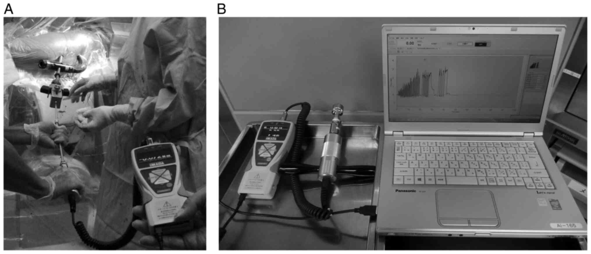
Suhm N, Hengg C, Schwyn R, Windolf M,
Quarz V and Hänni M: Mechanical torque measurement predicts load to
implant cut-out: A biomechanical study investigating DHS anchorage
in femoral heads. Arch Orthop Trauma Surg. 127:469–474.
2007.PubMed/NCBI View Article : Google Scholar
|
|
23
|
Ab-Lazid R, Perilli E, Ryan MK, Costi JJ
and Reynolds KJ: Does cancellous screw insertion torque depend on
bone mineral density and/or microarchitecture? J Biomech.
47:347–353. 2014.PubMed/NCBI View Article : Google Scholar
|
|
24
|
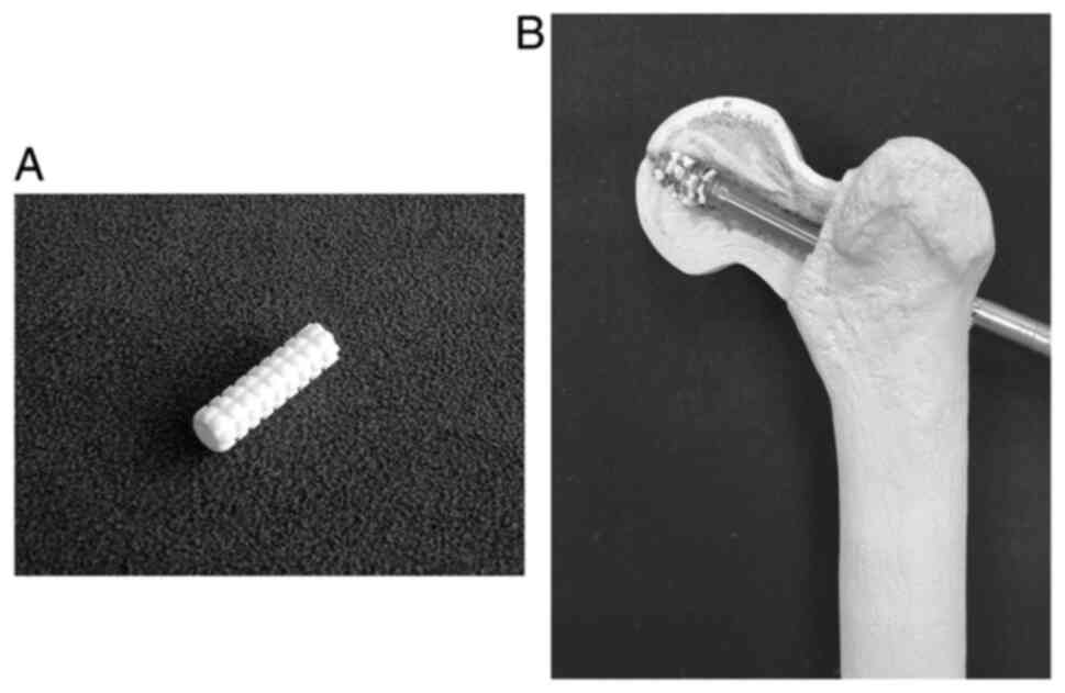
Iwata H, Takada N, Kuroyanagi G, Ikuta K,
Usami T, Sekiya I and Murakami H: Effect of hydroxyapatite tubes on
the lag screw intraoperative insertion torque for the treatment of
intertrochanteric femoral fractures. Injury. 52:3377–3381.
2021.PubMed/NCBI View Article : Google Scholar
|
|
25
|
Baumgaertner MR, Curtin SL, Lindskog DM
and Keggi JM: The value of the tip-apex distance in predicting
failure of fixation of peritrochanteric fractures of the hip. J
Bone Joint Surg Am. 77:1058–1064. 1995.PubMed/NCBI View Article : Google Scholar
|
|
26
|
Kane P, Vopat B, Heard W, Thakur N, Paller
D, Koruprolu S and Born C: Is tip apex distance as important as we
think? A biomechanical study examining optimal lag screw placement.
Clin Orthop Relat Res. 472:2492–2498. 2014.PubMed/NCBI View Article : Google Scholar
|
|
27
|
Pintus E, Sorbolini S, Albera A, Gaspa G,
Dimauro C, Steri R, Marras G and Macciotta NP: Use of locally
weighted scatterplot smoothing (LOWESS) regression to study
selection signatures in piedmontese and Italian brown cattle
breeds. Anim Genet. 45:1–11. 2014.PubMed/NCBI View Article : Google Scholar
|
|
28
|
Tamaddon M, Chen SM, Vanaclocha L, Hart A,
El-Husseiny M, Henckel J and Liu C: Decrease in local volumetric
bone mineral density in osteoarthritic joints is associated with
the increase in cartilage damage: A peripheral quantitative CT
study. Fron Mater. 4(37)2017.
|
|
29
|
Hasegawa K, Yamamura S and Dohmae Y:
Enhancing screw stability in osteosynthesis with hydroxyapatite
granules. Arch Orthop Trauma Surg. 117:175–176. 1998.PubMed/NCBI View Article : Google Scholar
|
|
30
|
Yamada M, Ueno T, Tsukimura N, Ikeda T,
Nakagawa K, Hori N, Suzuki T and Ogawa T: Bone integration
capability of nanopolymorphic crystalline hydroxyapatite coated on
titanium implants. Int J Nanomedicine. 7:859–873. 2012.PubMed/NCBI View Article : Google Scholar
|