Introduction
Colorectal cancer (CC) is the third most common
malignant tumor globally and accounts for ~9.7% of all malignant
tumors, with higher incidences in Europe, Australia and North
America (1). Surgical resection
combined with chemotherapy or radiotherapy remains the primary
treatment for CC (2). However, due
to the genetic differences between individuals, drug resistance
remains an issue (3). For example,
resistance to doxorubicin can occur due to decreased drug uptake or
increased drug efflux through drug transporters present on the cell
membrane. Moreover, cancer cells can develop mechanisms to detoxify
and eliminate doxorubicin from the cellular environment. Previous
studies have demonstrated that tumorigenesis is caused by gene
mutations in cells Such as TP53, which results in an unrestricted
cellular proliferation and resistance to apoptosis (4-6).
Autophagy is an apoptosis-like biological phenomenon. In the
initial stage of cancer, autophagy promotes the survival of normal
cells and inhibits carcinogenesis by removing damaged organelles
and DNA. In the advanced stage of cancer, autophagy provides
sufficient nutrients for proliferation and metabolism of tumor
cells and induces the survival of cancer cells, promote the
metastasis of cancer cells to distant locations and increase their
drug resistance (7). In addition,
autophagy is a research target for cancer therapy by affecting
cancer cells, stromal cells, and immune cells in the complex cancer
microenvironment (8).
Autophagy, also known as type II programmed cell
death, is a highly-conserved process of cellular destruction that
transfers intracellular substances (including proteins, lipids and
organelles) to lysosomes for degradation. The degraded
intracellular materials are then released from the lysosomes and
recycled in the cytosol (9).
Autophagy is crucial in various types of cancer, including breast,
lung, brain and CC (10). The
effect of autophagy on cancer development may depend on the cancer
type and the stage of cancer progression. In early stage cancer,
autophagy is often thought to have a tumor suppressor effect.
However, at a later stage, when the tumor microenvironment becomes
more hostile, autophagy can turn to promote tumor growth and
progression. The biological function of autophagy in cancer is
complex and is likely dependent on type of tumor, stage and genetic
context (11). The function of
autophagy changes at different stages of cancer. In its early
stages, autophagy may help suppress tumor growth by removing
damaged cell components and inhibiting genomic instability.
However, in later stages, autophagy can promote the survival of
cancer cells under stressful conditions, such as nutrient
deprivation or lack of oxygen, allowing them to adapt and grow. The
process of autophagy can be both tumor-promoting and
tumor-suppressing. Autophagy can suppress pathological processes,
such as tumor metastasis, by acting as an intracellular quality
control system. By removing damaged organelles, protecting against
oxidative stress, inhibiting inflammation, and regulating cell
death, autophagy acts as a defense mechanism that prevents or
limits tumor metastasis. By contrast, autophagy may help tumors
better adapt to adverse environments (12). For example, tumor cells can
activate autophagy as a protective mechanism against various
anti-cancer treatments, including chemotherapy and radiation
therapy. Autophagy enables tumor cells to survive and recover from
the induced stress, thereby promoting resistance to therapy.
However, tumors can outgrow their blood supply, leading to areas of
low oxygen levels (hypoxia). Hypoxia-induced autophagy enables
tumor cells to adapt to this stressful condition by promoting cell
survival and supporting angiogenesis, the formation of new blood
vessels.
Autophagy is a complex metabolic process, which is
regulated by autophagy-specific genes. Cleavage of light chain 3
(LC3)-I to LC3-II marks the beginning of autophagy (13). Subsequently, LC3-II binds to p62
(an adaptor protein) and promotes the autophagic degradation of
ubiquitinated protein aggregates (14). LC3 and p62 are biomarkers that are
commonly used for monitoring the levels of autophagy (15). LC3 is an indispensable component of
autophagosomes (16). It contains
the LC3A/B/C gene variants, with LC3B being most closely associated
with autophagy (17). Previously,
LC3 has been reported to serve a role in number of malignancies
including the brain, colorectal and melanoma (8,18,19).
However, previous studies have demonstrated opposing effects of the
LC3 expression level on the overall survival (OS) of patients with
different types of cancer (20,21).
A previous study demonstrated a positive association between LC3
and hepatocellular carcinoma (HCC), and reported that LC3 was
closely associated to the onset and progression of HCC (22). By contrast, another study
demonstrated that LC3 is a protective factor in patients with CC
(23). However, the relationship
between the LC3 expression level and the clinicopathological traits
of CC has not been reported. Therefore, the present meta-analysis
investigated the relationship between the LC3 expression level and
CC, and evaluated the prognostic effect of the LC3 expression level
on CC.
Materials and methods
Literature examination strategy
PubMed, Cochrane Library, Excerpta Medica Database,
China National Knowledge Infrastructure and Wanfang Data were used
to examine the literature. The cut-off date for publication
selection was February 2022. ‘LC3’ or ‘microtubule-associated
protein 1 light chain 3’ and ‘colorectal neoplasm’ or ‘colorectal
tumor’ or ‘colorectal cancer’ or ‘colorectal carcinoma’ were used
as search terms.
Selection criteria
Inclusion criteria included: i) Cohort or
case-control design; ii) patients were diagnosed according to
pathological criteria; iii) literature provided sufficient
clinicopathological and survival information to estimate the
association between LC3 and CC; and iv) the full text report was
issued in English or Chinese. Exclusion criteria included: i)
Animal or cell experiments, case reports, reviews, letters,
conference summaries or articles without full text; and ii)
republished articles with analogous datasets or subjects.
Data extraction
The data extracted from the articles that were
included in the present meta-analysis included the following
information: Author, year of publication, country, sample size,
patient characteristics (sex, age, tumor size, lymph node
metastasis, histological grade and TNM stage), detection method and
hazard ratio (HR) with 95% confidence interval (CI) for OS.
Quality evaluation
The Newcastle-Ottawa scale (NOS) was applied to
evaluate the quality of the articles (24). A score of ≥6 denotes a high-quality
study (Table I), scores of the 10
studies included in the present meta-analysis were all ≥6.
 | Table INewcastle-Ottawa scale for quality
assessment. |
Table I
Newcastle-Ottawa scale for quality
assessment.
| | Selection | Comparability | Outcome | |
|---|
| First author,
year | Exposed cohort | Non-exposed
cohort | Ascertainment of
exposure | Outcome of
interest | Appropriate control
used | Assessment of
outcome | Follow-up long
enough | Adequacy of
follow-up | Total score | (Refs.) |
|---|
| Park et al,
2013 | 1 | 1 | 1 | 1 | 1 | 1 | 0 | 0 | 6 | (25) |
| Choi et al,
2014 | 1 | 1 | 1 | 1 | 1 | 1 | 1 | 0 | 7 | (26) |
| Shim et al,
2016 | 1 | 1 | 1 | 1 | 1 | 1 | 1 | 1 | 8 | (27) |
| Schmitz et
al, 2016 | 1 | 1 | 1 | 1 | 2 | 1 | 1 | 0 | 8 | (28) |
| Wu et al,
2015 | 1 | 1 | 1 | 1 | 2 | 1 | 1 | 1 | 9 | (8) |
| Zhao et al,
2017 | 1 | 1 | 1 | 1 | 1 | 1 | 1 | 0 | 7 | (29) |
| Guo et al,
2019 | 1 | 1 | 1 | 1 | 1 | 1 | 1 | 1 | 8 | (30) |
| Wang et al,
2021 | 1 | 1 | 1 | 1 | 2 | 1 | 1 | 0 | 8 | (2) |
| Sui and Feng,
2012 | 1 | 1 | 1 | 1 | 2 | 1 | 0 | 0 | 7 | (31) |
| Li, 2018 | 1 | 1 | 1 | 1 | 2 | 1 | 0 | 0 | 7 | (32) |
Statistical analysis
Data were assessed using RevMan (version
5.4;Cochrane) and STATA (version 14.0; StataCorp LP). The HR and
95% CIs were extracted for survival analysis. Odds ratio (OR) and
95% CIs were used as outcome indices for dichotomous variables.
Mean differences and 95% Cis were used as outcome indicators for
continuous numerical variables. Since all studies included in the
present meta-analysis are from completely different groups, the
random-effect model was used to analyze data. Cochrane Q test is
used to assess whether the observed differences between study
results are merely due to chance. A significant P-value (<0.05)
indicates heterogeneity. In the event of significant heterogeneity,
a subgroup analysis was conducted to investigate the source.
Publication bias was evaluated using the Begg's test and funnel
plots. P<0.05 was considered to indicate a statistically
significant difference.
Results
Characteristics of included
studies
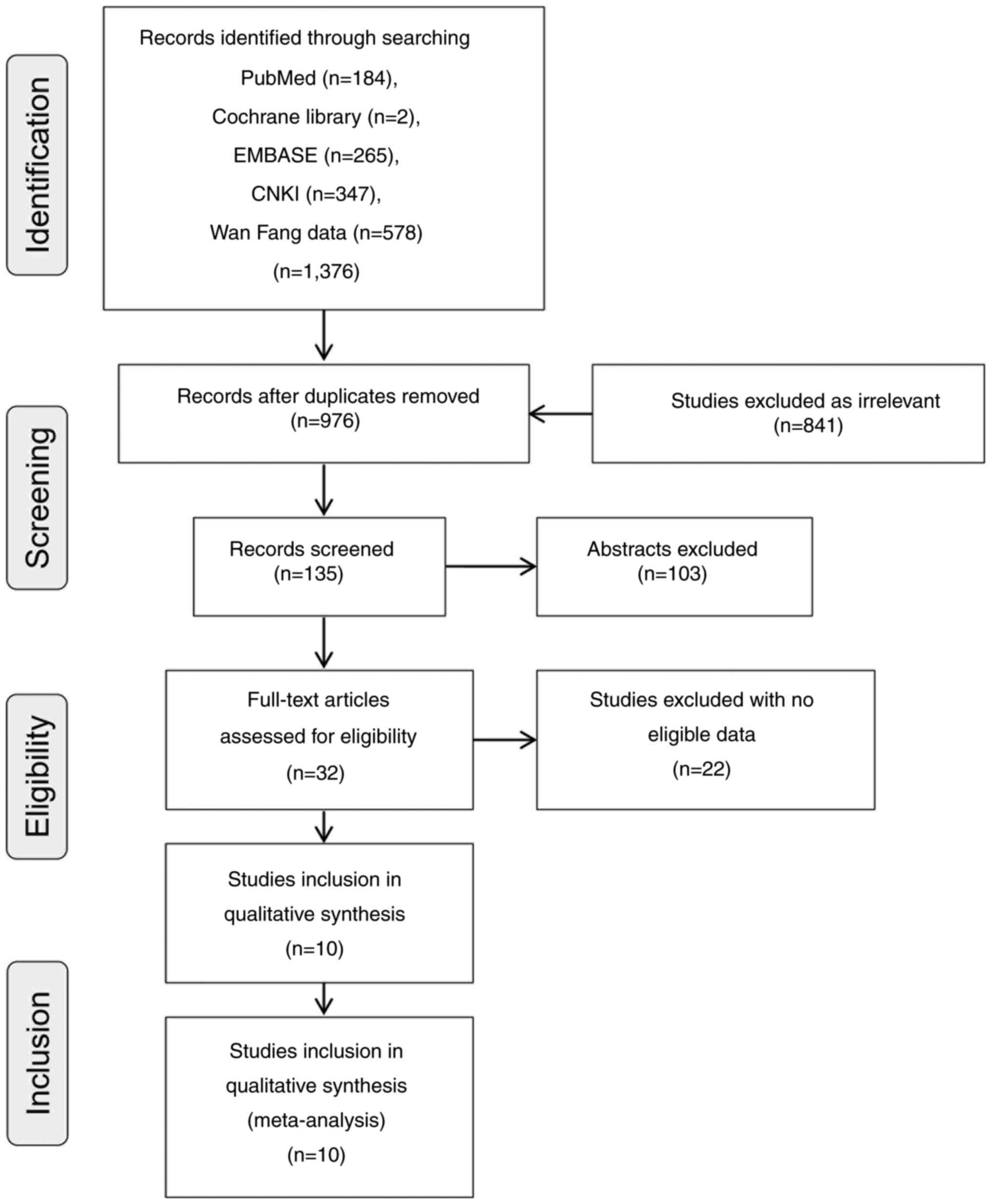
Through the literature search, a total of 1,376
relevant studies were obtained. After excluding repeated
literature, 976 studies remained. After eligibility evaluation, 10
studies were considered to meet the inclusion criteria of the
present meta-analysis (Fig. 1).
The characteristics of the included studies are presented in
Table I. The 10 included studies
were published between 2012 and 2021, of which eight were written
in English (2,8,25-30)
and 2 in Chinese (31,32). A total of 1,689 patients with CC
were included in the present meta-analysis. In all studies,
immunohistochemical staining was used to study expression of LC3.
NOS was used to assess the quality of the included studies, and all
10 studies were of high quality (Table II).
| Table IICharacteristics of the included
studies. |
Table II
Characteristics of the included
studies.
| | Sex | Age | Tumor size | Lymph node
metastasis | Histological
grade | TNM stage | |
|---|
| First author,
year | Country | Sample size | Male (+/-) | Female (+/-) | Older (+/-) | Middle aged
(+/-) | >5 cm (+/-) | ≤5 cm (+/-) | Negative (+/-) | Positive (+/-) | Poorly
differentiated (+/-) | Well and moderately
differentiated (+/-) | I and II (+/-) | III and IV
(+/-) | Method | OS data
provided | (Refs.) |
|---|
| Park et al,
2013 | USA | 178 | 99 | 79 | NA | NA | NA | NA | NA | NA | 59 | 119 | NA | NA | IHC | Yes | (25) |
| Choi et al,
2014 | South Korea | 263 | 141 | 122 | 122 | 141 | NA | NA | 99 (68/24) | 164 (118/39) | 41 (26/14) | 222 (160/49) | 105 (74/26) | 158 (112/37) | IHC | Yes | (26) |
| Shim et al,
2016 | South Korea | 101 | 69 | 32 | NA | NA | NA | NA | NA | NA | 7 (2/5) | 94 (42/49) | 52 | 49 | IHC | Yes | (27) |
| Schmitz et
al, 2016 | Germany | 127 | 66 | 61 | NA | NA | NA | NA | 56 (14/42) | 63 (20/43) | 26 (13/13) | 97 (22/75) | 15 (3/12) | 106 (32/74) | IHC | No | (28) |
| Wu et al,
2015 | China | 242 | 127 (113/14) | 115 (98/17) | 139 (120/19) | 103 (91/12) | 134 (120/14) | 108 (91/17) | 133 (107/26) | 109 (104/5) | 48 (43/5) | 194 (168/26) | 204 (178/26) | 38 (33/5) | IHC | Yes | (8) |
| Zhao et al,
2017 | China | 526 | 261 (105/156) | 265 (101/164) | 269 (106 163) | 257 (100/157) | NA | NA | NA | NA | 269 (55/214) | 257 (151/106) | 248 (125/123) | 278 (81/197) | IHC | No | (29) |
| Guo et al,
2019 | China | 68 | 44 (25/12) | 24 (15/4) | 13 (6/5) | 55 (34/11) | NA | NA | NA | NA | 18 (10/5) | 44 (30/8) | NA | NA | IHC | Yes | (30) |
| Wang et al,
2021 | China | 200 | 110 (97/13) | 90 (76/14) | 141 (121/20) | 59 (52/7) | NA | NA | 116 (98/18) | 84 (75/9) | 79 (69/10) | 121 (104/17) | 116 (98/18) | 84 (75/9) | IHC | No | (2) |
| Sui and Feng,
2012 | China | 115 | 62 (50/12) | 53 (45/8) | 75 (60/15) | 40 (35/5) | 101 (81/20) | 14 (14/0) | 73 (57/16) | 42 (38/4) | 5 (5/0) | 110 (90/20) | NA | NA | IHC | No | (31) |
| Li, 2018 | China | 69 | 42 (30/12) | 27 (16/11) | 51 (35/16) | 18 (11/7) | 32 (19/13) | 37 (27/10) | 49 (28/21) | 20 (18/2) | NA | NA | 46 (27/19) | 23 (19/4) | IHC | No | (32) |
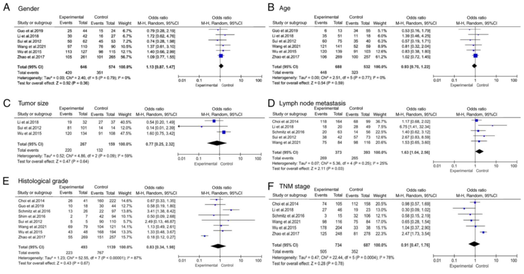
Meta-analysis of clinicopathological
features
The association between LC3 and various
clinicopathological traits of CC was evaluated. It was revealed
that LC3 expression was associated to histological grade [OR=0.82,
95% CI (0.43, 1.95), P<0.001] and TNM stage [OR=0.91, 95% CI
(0.47, 1.77), P<0.001]. However, no association was observed
between LC3 expression and sex [OR=1.14, 95% CI (0.90, 1.51);
P=0.678], age [OR=0.89, 95% CI (0.67, 1.20), P=0.663], tumor size
[OR=0.78, 95% CI (0.30, 2.34), P=0.090], or lymph node metastasis
[OR=2.05, 95% CI (1.19, 3.60), 0.250] (Table III).
| Table IIIMicrotubule associated protein 1
light chain 3 clinicopathological descriptions in patients with
colorectal cancer. |
Table III
Microtubule associated protein 1
light chain 3 clinicopathological descriptions in patients with
colorectal cancer.
| | Heterogeneity | |
|---|
| Clinicopathological
characteristic | Number of
studies | Number of
patients | Pooled OR(95%
CI) |
I2(%) | P-value | Model used |
|---|
| Sex | 6 | 1,208 | 1.14 (0.90,
1.51) | 0.0 | 0.678 | Random |
| Age | 6 | 1,208 | 0.89 (0.67,
1.20) | 0.0 | 0.663 | Random |
| Tumor size | 3 | 426 | 0.78 (0.30,
2.34) | 59.0 | 0.090 | Random |
| Lymph node
metastasis | 6 | 994 | 2.05 (1.19,
3.60) | 25.0 | 0.250 | Random |
| Histological
grade | 8 | 1,606 | 0.82 (0.43,
1.95) | 87.0 | <0.001 | Random |
| TNM stage | 6 | 1,407 | 0.91 (0.47,
1.77) | 78.0 | <0.001 | Random |
Subgroup evaluation
There was heterogeneity in four of the six features
studied, including tumor size (I2=59%, P=0.09), lymph
node metastasis (I2=25%, P=0.25), histological grade
(I2=87%, P<0.001) and TNM stage (I2=78%,
P<0.001; Table III). To
investigate the prospective sources of heterogeneity, a subgroup
analysis was performed. Because only three studies included tumor
size, there was insufficient information for further subgroup
analysis. Therefore, according to the sample size and NOS score,
only the subgroup analysis of lymph node metastasis, histological
grade and TNM stage was performed (Table IV).
| Table IVLymph node metastasis, histological
grading and TNM staging subgroup analyses. |
Table IV
Lymph node metastasis, histological
grading and TNM staging subgroup analyses.
| A, Lymph node
metastasis |
|---|
| | Heterogeneity | |
|---|
| Subgroup | Number of
studies | Number of
patients | Pooled OR (95%
CI) | I2
(%) | P-value | Model used |
|---|
| Sample, n | | | | | | |
|
>200 | 2 | 491 | 1.63 (1.04,
2.56) | 85.6 | 0.008 | Random |
|
≤200 | 4 | 503 | 2.04 (1.33,
3.26) | 19.2 | 0.287 | Random |
| NOS score | | | | | | |
|
>7 | 3 | 561 | 2.11 (0.99,
4.52) | 55.4 | 0.106 | Random |
|
≤7 | 3 | 433 | 2.22 (0.78,
6.30) | 65.2 | 0.057 | Random |
| B, Histological
grade |
| | Heterogeneity | |
| Subgroup | Number of
studies | Number of
patients | Pooled OR (95%
CI) | I2
(%) | P-value | Model used |
| Sample, n | | | | | | |
|
>200 | 3 | 1,017 | 0.48
(0.15,1.54) | 88.7 | <0.001 | Random |
|
≤200 | 5 | 589 | 1.37 (0.85,
2.41) | 48.6 | 0.100 | Random |
| NOS score | | | | | | |
|
>7 | 5 | 716 | 1.30 (0.84,
2.09) | 47.5 | 0.107 | Random |
|
≤7 | 3 | 890 | 0.39 (0.13,
1.20) | 80.0 | 0.007 | Random |
| C, TNM stage |
| | Heterogeneity | |
| Subgroup | Number of
studies | Number of
patients | Pooled OR (95%
CI) | I2
(%) | P-value | Model used |
| Sample, n | | | | | | |
|
>200 | 3 | 1,017 | 1.43 (0.68,
2.99) | 77.5 | 0.012 | Random |
|
≤200 | 3 | 390 | 0.91 (0.47,
1.76) | 0.0 | 0.583 | Random |
| NOS score | | | | | | |
|
>7 | 3 | 563 | 0.75 (0.42,
1.34) | 0.0 | 0.732 | Random |
|
≤7 | 3 | 844 | 1.02 (0.36,
2.85) | 87.4 | <0.001 | Random |
Upon classifying the data based on sample size,
there was an association between LC3 expression and lymph node
metastasis [OR=1.63, 95% CI (1.04-2.56)] as well as TNM stage
[OR=0.91, 95% CI (0.47-1.76)] in the subgroup with a small sample
size (n≤200), while no significant association was found in the
larger sample size subgroup (n>200). Heterogeneity was revealed
in the n>200 subgroups for lymph node metastasis, histological
grade and TNM stage, with I2 values of 85.6, 88.7, and
77.5%, respectively.
Based on the NOS score, heterogeneity was revealed
in both subgroups of lymph node metastasis (NOS>7,
I2=55.4%; NOS≤7, I2=65.2%), the low NOS score
subgroup of histological grade (NOS≤7, I2=80.0%) and the
low NOS score subgroup of TNM stage (NOS≤7, I2=87.4%).
No heterogeneity was revealed in the high NOS score subgroups of
histological grade (NOS>7, I2=47.5%) and TNM stage
(NOS>7, I2=0.0%).
Association between LC3 expression and
OS in CC
A total of 852 patients from five articles were
included to estimate OS value of LC3 expression in CC. No
significant heterogeneity (I2=45% and P=0.12) was
revealed (Fig. 2). The results
revealed that the overexpression of LC3 was a favorable factor for
OS in patients with CC [HR=0.56, 95% CI (0.39, 0.79)].
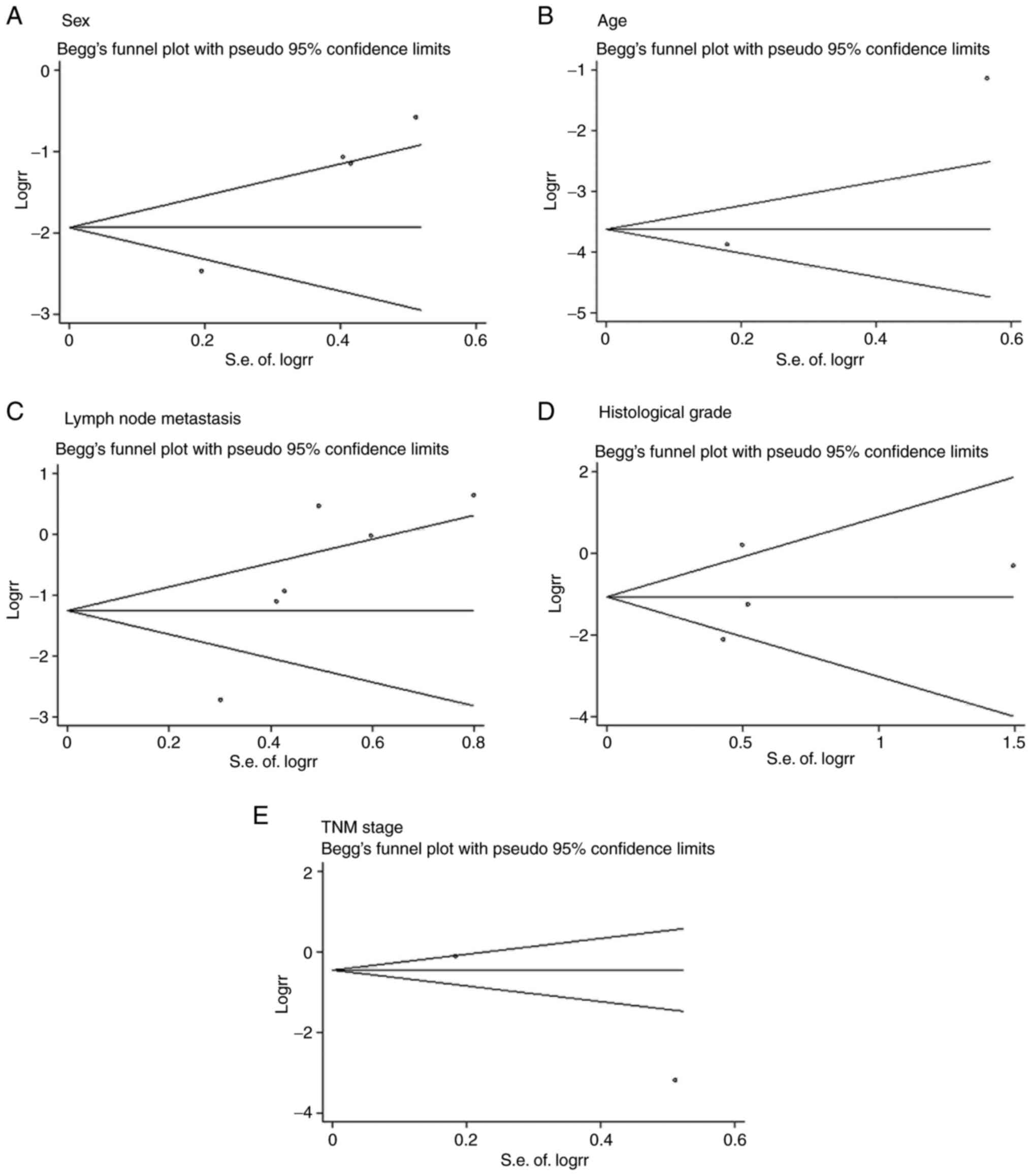
Publication bias
Begg's test was used to assess the potential
publication bias. No publication bias was revealed in sex
(P=0.308), age (P=1.000), lymph node metastasis (P=0.060),
histological grade (P=0.734) or TNM stage (P=1.000; Fig. 3). In addition, heterogeneity of
lymph node metastasis reduced from I2=53.8% to
I2=31.5% after the study by Wu et al (8) was removed.
Discussion
CC remains a medical, social and economic burden in
developed countries. Global burden of colorectal cancer in 2020 and
2040: incidence and mortality estimates from GLOBOCAN shows: CC
accounting for ~8% of cancer-associated mortalities worldwide
(33,34). More than 1.9 million new cases of
colorectal cancer and 930,000 deaths are estimated in 2020. CC is a
complex malignant tumor involving a variety of cellular signaling
pathways, including autophagy cascades. Beclin 1 and LC3 are the
most widely studied autophagy-associated proteins in CC (35,36).
A previous study revealed that LC3-I and LC3-II were scarce in
normal tissues but were strongly positive in ~70% of
adenocarcinomas and metastatic tumors (37). Consistent with the results of a
previous study (26), the present
study revealed that the expression of autophagy-associated proteins
maybe a novel prognostic indicator for patients with CC.
The present meta-analysis evaluated the association
between the LC3 expression level and the clinicopathological traits
and OS in patients with CC. In total, 10 studies with 1,689
patients were included. The findings of the present study indicated
that LC3 overexpression was positively associated to lymph node
metastasis in CC. Similar to the results of Li et al
(23), the present study also
suggested that LC3 was a protective marker for patients with CC. In
total, nine studies were included by Li et al (23), and only six of the studies were
associated with LC3 expression. Due to the lack of investigation on
the clinicopathological characteristics associated to the
expression of LC3 in patients with CC, the study by Li et al
(23) was limited, as the main
assessment was the association between LC3 and OS. Therefore, the
present meta-analysis investigated the relationship between the
expression of LC3 and the diagnosis of CC more comprehensively
based on the clinicopathological features of patients with CC (such
as tumor size, lymph node metastasis, histological grade and TNM
stage).
Furthermore, autophagy may also promote tumor growth
under stress conditions, such as hypoxia and starvation (38). Autophagy may have opposing roles in
different types of cancer. Previous studies have demonstrated that
LC3 expression is associated with developing HCCs (22,39).
In the present study, it was revealed that LC3 may increase the OS
of patients with CC.
The present study has reached seemingly
contradictory conclusions. It was demonstrated that overexpression
of LC3 was associated with lymph node metastasis, which is usually
regarded as an unfavorable factor (40). However, in the present study, the
high expression levels of LC3 were associated to a favorable OS
outcome. Thus, further studies on the role and mechanism of LC3 in
CC prognosis need to be conducted. Additionally, the present
meta-analysis has several limitations. Firstly, the number of
articles and patients included in the present study is small, and
further research is required in future. Secondly, the majority of
the included studies were conducted in China, which may lead to a
potential heterogeneity. Finally, The number of patients included
in the studies might have been relatively small, limiting the
statistical power and generalizability of the findings, and lymph
node metastasis was the only feature associated with LC3 expression
in CC.
In summary, the present analysis revealed that LC3
expression was only associated with lymph node metastasis in CC. At
the same time, LC3 expression seemed to be a protective indicator
for patients with CC. These seemingly contradictory findings need
to be verified using a larger sample size with the inclusion of
additional high-quality studies.
Acknowledgements
Not applicable.
Funding
Funding: No funding was received.
Availability of data and materials
The datasets used and/or analyzed during the current
study are available from the corresponding author on reasonable
request.
Authors' contributions
NS participated in methodology design,
investigation, data curation and writing the original draft. FH and
YS contributed to experiment design and writing the original draft.
JW contributed to the study design and wrote and edited the
manuscript. XC performed the literature review and prepared the
figures. LW participated in study conception, supervision, review
and editing of the manuscript. NS and LW confirm the authenticity
of all the raw data. All authors have read and approved the final
manuscript.
Ethics approval and consent to
participate
Not applicable.
Patient consent for publication
Not applicable.
Competing interests
The authors declare that they have no competing
interests.
References
|
1
|
Torre LA, Bray F, Siegel RL, Ferlay J,
Lortet-Tieulent J and Jemal A: Global cancer statistics, 2012. CA
Cancer J Clin. 65:87–108. 2015.PubMed/NCBI View Article : Google Scholar
|
|
2
|
Wang Y, Zhao Z, Zhuang J, Wu X, Wang Z,
Zhang B, Gao G, Zhang Y, Guo C and Xia Q: Prognostic value of
autophagy, microsatellite instability, and KRAS mutations in
colorectal cancer. J Cancer. 12:3515–3528. 2021.PubMed/NCBI View Article : Google Scholar
|
|
3
|
Hammond WA, Swaika A and Mody K:
Pharmacologic resistance in colorectal cancer: A review. Ther Adv
Med Oncol. 8:57–84. 2016.PubMed/NCBI View Article : Google Scholar
|
|
4
|
Bignell GR, Greenman CD, Davies H, Butler
AP, Edkins S, Andrews JM, Buck G, Chen L, Beare D, Latimer C, et
al: Signatures of mutation and selection in the cancer genome.
Nature. 463:893–898. 2010.PubMed/NCBI View Article : Google Scholar
|
|
5
|
Du L, Kim JJ, Shen J, Chen B and Dai N:
KRAS and TP53 mutations in inflammatory bowel disease-associated
colorectal cancer: A meta-analysis. Oncotarget. 8:22175–22186.
2017.PubMed/NCBI View Article : Google Scholar
|
|
6
|
Inoue A, Robinson FS, Minelli R, Tomihara
H, Rizi BS, Rose JL, Kodama T, Srinivasan S, Harris AL, Zuniga AM,
et al: Sequential administration of XPO1 and ATR inhibitors
enhances therapeutic response in TP53-mutated colorectal cancer.
Gastroenterology. 161:196–210. 2021.PubMed/NCBI View Article : Google Scholar
|
|
7
|
Koustas E, Sarantis P, Theoharis S, Saetta
AA, Chatziandreou I, Kyriakopoulou G, Giannopoulou I, Michelli M,
Schizas D, Papavassiliou AG and Karamouzis MV: Autophagy-related
proteins as a prognostic factor of patients with colorectal cancer.
Am J Clin Oncol. 42:767–776. 2019.PubMed/NCBI View Article : Google Scholar
|
|
8
|
Wu S, Sun C, Tian D, Li Y, Gao X, He S and
Li T: Expression and clinical significances of Beclin1, LC3 and
mTOR in colorectal cancer. Int J Clin Exp Pathol. 8:3882–3891.
2015.PubMed/NCBI
|
|
9
|
Levine B and Kroemer G: Autophagy in the
pathogenesis of disease. Cell. 132:27–42. 2008.PubMed/NCBI View Article : Google Scholar
|
|
10
|
Rabinowitz JD and White E: Autophagy and
metabolism. Science. 330:1344–1348. 2010.PubMed/NCBI View Article : Google Scholar
|
|
11
|
Kimmelman AC: The dynamic nature of
autophagy in cancer. Genes Dev. 25:1999–2010. 2011.PubMed/NCBI View Article : Google Scholar
|
|
12
|
Hanahan D and Weinberg RA: Hallmarks of
cancer: The next generation. Cell. 144:646–674. 2011.PubMed/NCBI View Article : Google Scholar
|
|
13
|
Kabeya Y, Mizushima N, Ueno T, Yamamoto A,
Kirisako T, Noda T, Kominami E, Ohsumi Y and Yoshimori T: LC3, a
mammalian homologue of yeast Apg8p, is localized in autophagosome
membranes after processing. EMBO J. 19:5720–5728. 2000.PubMed/NCBI View Article : Google Scholar
|
|
14
|
Pankiv S, Clausen TH, Lamark T, Brech A,
Bruun JA, Outzen H, Øvervatn A, Bjørkøy G and Johansen T:
p62/SQSTM1 binds directly to Atg8/LC3 to facilitate degradation of
ubiquitinated protein aggregates by autophagy. J Biol Chem.
282:24131–24145. 2007.PubMed/NCBI View Article : Google Scholar
|
|
15
|
Klionsky DJ, Abdalla F, Abeliovich H,
Abraham RT, Acevedo-Arozena A, Adeli K, Agholme L, Agnello M,
Agostinis P, Aguirre-Ghiso JA, et al: Guidelines for the use and
interpretation of assays for monitoring autophagy. Autophagy.
8:445–544. 2012.PubMed/NCBI View Article : Google Scholar
|
|
16
|
Maruyama Y, Sou YS, Kageyama S, Takahashi
T, Ueno T, Tanaka K, Komatsu M and Ichimura Y: LC3B is
indispensable for selective autophagy of p62 but not basal
autophagy. Biochem Biophys Res Commun. 446:309–315. 2014.PubMed/NCBI View Article : Google Scholar
|
|
17
|
Wu DH, Jia CC, Chen J, Lin ZX, Ruan DY, Li
X, Lin Q, Min-Dong Ma XK, Wan XB, et al: Autophagic LC3B
overexpression correlates with malignant progression and predicts a
poor prognosis in hepatocellular carcinoma. Tumour Biol.
35:12225–12233. 2014.PubMed/NCBI View Article : Google Scholar
|
|
18
|
Ghavami S, Shojaei S, Yeganeh B, Ande SR,
Jangamreddy JR, Mehrpour M, Christoffersson J, Chaabane W, Moghadam
AR, Kashani HH, et al: Autophagy and apoptosis dysfunction in
neurodegenerative disorders. Prog Neurobiol. 112:24–49.
2014.PubMed/NCBI View Article : Google Scholar
|
|
19
|
Segala G, David M, de Medina P, Poirot MC,
Serhan N, Vergez F, Mougel A, Saland E, Carayon K, Leignadier J, et
al: Dendrogenin A drives LXR to trigger lethal autophagy in
cancers. Nat Commun. 8(1903)2017.PubMed/NCBI View Article : Google Scholar
|
|
20
|
Cj P, Hv E, Vijayakurup V, R Menon G, Nair
S and Gopala S: High LC3/Beclin expression correlates with poor
survival in glioma: A definitive role for autophagy as evidenced by
in vitro autophagic flux. Pathol Oncol Res. 25:137–148.
2019.PubMed/NCBI View Article : Google Scholar
|
|
21
|
Zhu W, Pan X, Li F, Zhang Y and Lu X:
Expression of Beclin 1 and LC3 in FIGO stage I-II cervical squamous
cell carcinoma and relationship to survival. Tumour Biol.
33:1653–1659. 2012.PubMed/NCBI View Article : Google Scholar
|
|
22
|
Meng YC, Lou XL, Yang LY, Li D and Hou YQ:
Role of the autophagy-related marker LC3 expression in
hepatocellular carcinoma: A meta-analysis. J Cancer Res Clin Oncol.
146:1103–1113. 2020.PubMed/NCBI View Article : Google Scholar
|
|
23
|
Li JX, Yan Q, Liu N, Zheng WJ, Hu M, Yu
ZM, Zhou YD, Wang XW, Liang FX and Chen R: The prognostic value of
autophagy-related markers bclin-1 and LC-3 in colorectal cancers: A
systematic review and meta-analysis. Evid Based Complement Alternat
Med. 2020(8475840)2020.PubMed/NCBI View Article : Google Scholar
|
|
24
|
Stang A: Critical evaluation of the
Newcastle-Ottawa scale for the assessment of the quality of
nonrandomized studies in meta-analyses. Eur J Epidemiol.
25:603–605. 2010.PubMed/NCBI View Article : Google Scholar
|
|
25
|
Park JM, Huang S, Wu TT, Foster NR and
Sinicrope FA: Prognostic impact of Beclin 1, p62/sequestosome 1 and
LC3 protein expression in colon carcinomas from patients receiving
5-fluorouracil as adjuvant chemotherapy. Cancer Biol Ther.
14:100–107. 2013.PubMed/NCBI View Article : Google Scholar
|
|
26
|
Choi JH, Cho YS, Ko YH, Hong SU, Park JH
and Lee MA: Absence of autophagy-related proteins expression is
associated with poor prognosis in patients with colorectal
adenocarcinoma. Gastroenterol Res Pract.
2014(179586)2014.PubMed/NCBI View Article : Google Scholar
|
|
27
|
Shim BY, Sun S, Won HS, Lee MA, Hong SU,
Jung JH, Cho HM and Ko YH: Role of autophagy-related protein
expression in patients with rectal cancer treated with neoadjuvant
chemoradiotherapy. BMC Cancer. 16(207)2016.PubMed/NCBI View Article : Google Scholar
|
|
28
|
Schmitz KJ, Ademi C, Bertram S, Schmid KW
and Baba HA: Prognostic relevance of autophagy-related markers LC3,
p62/sequestosome 1, Beclin-1 and ULK1 in colorectal cancer patients
with respect to KRAS mutational status. World J Surg Oncol.
14(189)2016.PubMed/NCBI View Article : Google Scholar
|
|
29
|
Zhao H, Yang M and Zhao B: Beclin 1 and
LC3 as predictive biomarkers for metastatic colorectal carcinoma.
Oncotarget. 8:59058–59067. 2017.PubMed/NCBI View Article : Google Scholar
|
|
30
|
Guo GF, Wang YX, Zhang YJ, Chen XX, Lu JB,
Wang HH, Jiang C, Qiu HQ and Xia LP: Predictive and prognostic
implications of 4E-BP1, Beclin-1, and LC3 for cetuximab treatment
combined with chemotherapy in advanced colorectal cancer with
wild-type KRAS: Analysis from real-world data. World J
Gastroenterol. 25:1840–1853. 2019.PubMed/NCBI View Article : Google Scholar
|
|
31
|
Sui YQ and Feng YZ: Expression and
significance of autophagy related genes LC3 and Beclin-1 and
apoptosis related genes p53 and Bcl-2 in colorectal cancer. J Clin
Exp pathol. 28:282–286. 2012.
|
|
32
|
Li SM: Expression and significance of
Tricellulin, LC3 and Beclin1 in colorectal cancer. Shandong Med.
58:1–5. 2018.
|
|
33
|
Morgan E, Arnold M, Gini A, Lorenzoni V,
Cabasag CJ, Laversanne M, Vignat J, Ferlay J, Murphy N and Bray F:
Global burden of colorectal cancer in 2020 and 2040: Incidence and
mortality estimates from GLOBOCAN. Gut. 72:338–344. 2023.PubMed/NCBI View Article : Google Scholar
|
|
34
|
Brenner H, Kloor M and Pox CP: Colorectal
cancer. Lancet. 383:1490–1502. 2014.PubMed/NCBI View Article : Google Scholar
|
|
35
|
Chen Z, Li Y, Zhang C, Yi H, Wu C, Wang J,
Liu Y, Tan J and Wen J: Downregulation of Beclin 1 and impairment
of autophagy in a small population of colorectal cancer. Dig Dis
Sci. 58:2887–2894. 2013.PubMed/NCBI View Article : Google Scholar
|
|
36
|
Giatromanolaki A, Koukourakis MI, Harris
AL, Polychronidis A, Gatter KC and Sivridis E: Prognostic relevance
of light chain 3 (LC3A) autophagy patterns in colorectal
adenocarcinomas. J Clin Pathol. 63:867–872. 2010.PubMed/NCBI View Article : Google Scholar
|
|
37
|
Sato K, Tsuchihara K, Fujii S, Sugiyama M,
Goya T, Atomi Y, Ueno T, Ochiai A and Esumi H: Autophagy is
activated in colorectal cancer cells and contributes to the
tolerance to nutrient deprivation. Cancer Res. 67:9677–9684.
2007.PubMed/NCBI View Article : Google Scholar
|
|
38
|
Nagelkerke A, Bussink J, Geurts-Moespot A,
Sweep FC and Span PN: Therapeutic targeting of autophagy in cancer.
Part II: Pharmacological modulation of treatment-induced autophagy.
Semin Cancer Biol. 31:99–105. 2015.PubMed/NCBI View Article : Google Scholar
|
|
39
|
Degenhardt K, Mathew R, Beaudoin B, Bray
K, Anderson D, Chen G, Mukherjee C, Shi Y, Gélinas C, Fan Y, et al:
Autophagy promotes tumor cell survival and restricts necrosis,
inflammation, and tumorigenesis. Cancer Cell. 10:51–64.
2006.PubMed/NCBI View Article : Google Scholar
|
|
40
|
Kim JC, Lee KH, Yu CS, Kim HC, Kim JR,
Chang HM, Kim JH, Kim JS and Kim TW: The clinicopathological
significance of inferior mesenteric lymph node metastasis in
colorectal cancer. Eur J Surg Oncol. 30:271–279. 2004.PubMed/NCBI View Article : Google Scholar
|