Introduction
Vascular cognitive impairment (VCI) is a symptom of
cognitive impairment caused by a variety of cerebrovascular
diseases (such as cerebral infarction, cerebral hemorrhage) or
vascular risk factors (such as age, heredity, hypertension,
hyperlipidemia, diabetes, smoking, alcoholism, obesity and lack of
exercise) (1). It can occur alone
or is often in conjunction with neurodegenerative diseases, such as
Alzheimer's disease (2). Its
etiology is complex and diverse and includes brain
neurodegenerative diseases, cerebral ischemic and hemorrhagic
lesions (3,4), mainly manifested as memory decline,
thinking and language disorders and negative emotions (encompassing
anxiety, irritability, apathy and depressive mood) (5). The pathogenesis of VCI is ladder-like
and may eventually develop into dementia without timely
intervention and which, once formed, will be difficult to cure
(6,7). The incidence of VCI is increasing
year by year (8,9), resulting in a heavy burden on the
family and society, which has attracted increasing attention.
Ginkgo biloba extract 50 (GBE50) is an
extract of Ginkgo biloba leaf that is homologous to both
food and medicine, in which the mass fraction of ginkgolic acid is
<5 mg/kg, that of flavonol glycosides is >24%, that of
terpene lactones is >10% and that of flavonoids is >44%
(10,11). The Ginkgo biloba preparation
has been proved to have a positive effect on relieving ischemic
stroke (12) and improves VCI, but
its mechanism of action is not completely clear yet. Compared with
the traditional Ginkgo biloba leaf extract (13), GBE50 has more effective
ingredients, rapider effect, fewer potential allergenic components
and higher safety (14). The GBE50
dropping pill (GBE50DP) has the therapeutic effect of activating
blood circulation, removing blood stasis and clearing collaterals
(15). In the present study, the
intervention experiment of GBE50DP on VCI rats was conducted to
further understand the efficacy and mechanism of GBE50DP in
improving VCI.
Materials and methods
Reagents
GBE50DP was provided by Zhejiang Shangyao Jiuxu
Pharmaceutical Co., Ltd. Donepezil hydrochloride tablets were
provided by Chongqing Zein Biotechnology Co., Ltd. Pentobarbital
sodium was purchased from Merck KGaA. Superoxide dismutase (SOD),
malondialdehyde (MDA), glutathione peroxidase (GSH-Px), catalase
(CAT), IL-1β, IL-6 and TNF-α kits were provided by Jiangsu Jingmei
Biotechnology Co., Ltd. Terminal-deoxynucleotidyl transferase
mediated nick end labeling (TUNEL) kit was provided by Roche
Diagnostics GmbH. Rabbit two-step immunohistochemical kit (PV-6001)
and 3,3'-diaminobenzidine (DAB) were provided by Beijing Zhongshan
Jinqiao Biotechnology Co., Ltd. Caspase-3, Bcl-2, Bax, cytochrome
c (Cyto-C), PARP-1, β-actin monoclonal antibody, HRP-labeled
anti-rat and HRP-labeled anti-rabbit secondary antibody were
provided by Beijing Bioss Biotechnology Co., Ltd. Fas monoclonal
antibody was provided by Thermo Fisher Scientific, Inc. PVDF
membrane was acquired from Beijing Biotopped Technology Co., Ltd.
Enhanced chemiluminescence (ECL) reagent was purchased from Dalian
Meilun Biotechnology Co., Ltd. Tissue lysate and SDS-PAGE kits were
provided by Beyotime Biotechnology. A Bradford protein
quantification kit was provided by Beijing Lanjieke Technology Co.,
Ltd. MS-grade formic acid and acetonitrile were acquired from
Thermo Fisher Scientific, Inc. All other chemical reagents used
were of analytical grade.
Devices
Open field box (100x100x40 cm), Morris water maze
(diameter 150 cm, height 50 cm) and eight-arm maze (radial arm
29x6x15 cm) were provided by Anhui Zhenghua Biologic Apparatus
Facilities Co., Ltd. Centrifuges (5810R and 5424R) were acquired
from Eppendorf SE, a microplate reader (RT-6000) from Rayto Life
and Analytical Sciences Co., Ltd., a homogenizer (FA25) from Fluko
Shanghai Equipment Co., Ltd., an embedding machine (BM-VII) from
Xiaogan Hongye Medical Instrument Co., Ltd., a microtome (CM3050s)
from Leica Microsystems GmbH, an optical microscope (BX-53) from
Olympus Corporation, a fluorescence microscope (DM4000B) from Leica
Microsystems GmbH and a gel imaging system (C300) from Azure
Biosystems.
VCI rat model establishment and drug
administration
A total of 120 male Wistar rats (weighing 300-340 g;
9-11 weeks) were supplied by the Changchun Yisi Experimental Animal
Technology Co., Ltd. [license no., SCXK(Ji)-2023-0002], housed in a
temperature- and humidity-controlled environment (25˚C and 60%) and
given free access to water and a standard diet.
After 1 week of adaptation, the rats were randomly
divided into the control, model, positive (donepezil 0.45 mg/kg)
and GBE50DP low-, medium- and high-dose groups (50, 100 and 200
mg/kg, respectively), with 20 rats in each group. Anesthesia was
administered by intraperitoneal injection of 40 mg/kg sodium
pentobarbital. An incision was then made at the midline position in
the neck, and the bilateral common carotid arteries were isolated
and exposed. The control group was exposed without ligation, while
the other groups were permanently ligated (16,17)
and sutured. The drugs were prepared into suspension with 0.5%
sodium carboxymethyl cellulose aqueous solution for intragastric
administration once a day for 8 weeks, while the control and model
group were given the same volume of pure water.
Social behavior test
The experiment was carried out in a dark and quiet
environment within the open field box. The rats were placed
individually in the box for 5 min twice a day for 2 days, for the
adaptive training. At 1 h before the experiment, the tested rat was
isolated in a single cage to stimulate its social behavior. The
test was performed for 5 min, and the number and duration of
contacts were recorded.
Morris water maze test (18)
The inner wall of the Morris water maze was made
black, and four quadrants were distinguished by different markers.
A platform was placed in the center of one of the quadrants, 2 cm
below the water level, and the water temperature was controlled at
23.0±2.0˚C. Adaptive training: 1 Day before the experiment, the
rats were adapted to the environment in a non-platform pool and
swam freely for 1.5 min. Positioning navigation experiment: The
rats facing the pool wall were randomly placed in one of the three
quadrants without the platform. The time frame from their entering
the water to boarding the platform (the escape latency) was
recorded. The time frame was recorded as 1.5 min if the platform
was not found within 1.5 min, and the rats were then guided to the
platform and stayed for 10 sec so that they could perform spatial
learning and memory according to the markers. The experiments were
performed twice a day, in intervals of 4 h, for 5 days total. The
6th day was set as the test period. Space exploration experiment:
The platform was removed on the 6th day, and the residence time of
the rats in the target quadrant and the number of times they
crossed the original platform location within 1.5 min were
recorded.
Eight-arm maze test
The experiment was carried out in a quiet and
odorless environment. There was a removable door at the entrance of
each arm, and the arms 1, 2, 6 and 7 were set as the food arms.
There were 4 adaption tests before the test, 10 min for each per
day. On the first day, the bait was placed in the central area; on
the second day, it was placed in the central area and the entrance
of each arm; on the third day, it was placed on the top of each
arm; from the first day to the third day, three rats were placed in
the maze at the same time for exploration; on the fourth day, the
bait was placed only at the top of the food arm, was placed in the
maze individually for exploration. The rats were then trained for 5
days and fasted for 24 h before the training, with no restriction
on drinking water. During the training, only bait was placed in the
food arms. After the rats were placed in the central area for 15
sec, the doors of each arm were opened, and the foraging of all
arms was free for 10 min or completed in advance. Restricted feed
was given after each adaptation and training. The times that the
rats repeatedly entered the food arm that had been visited (the
working memory errors) and the times that the rats entered the
food-free arm (the reference memory errors) were recorded.
Sample collection and processing
After the rats were anesthetized with 3% sodium
pentobarbital (40 mg/kg, i.p.), they were fixed in the supine
position, and the chest cavity was quickly opened to expose the
heart. The perfusion needle was inserted into the aorta through the
apex of the left ventricle, and the right atrial appendage was cut
open simultaneously. First, 4˚C pre-cooled normal saline (~150 ml)
was rapidly perfused until the effluent was clear and the liver
turned white; then, 4˚C pre-cooled 4% paraformaldehyde (~200 ml)
was perfused for fixation. When the muscles of the rat's limbs and
tail became stiff, the rats were immediately decapitated to obtain
the brain. The hippocampal tissues were quickly separated on ice
and placed in 4% paraformaldehyde for fixation (the volume ratio of
fixative solution to tissue was >10:1), fixed at 4˚C for >24
h for subsequent experiments.
After the rats were anesthetized with 3% sodium
pentobarbital (40 mg/kg, i.p.), ~1.0 ml of blood was collected from
the orbital venous plexus and placed in a heparin sodium
anticoagulant tube. After confirming that the rats were in a deep
state of anesthesia (with disappearance of the toe clonus reflex),
the rats were immediately decapitated to obtain the brain, which
was quickly separated into hippocampal tissue on ice, placed in a
pre-frozen storage tube, rapidly frozen in liquid nitrogen, and
then transferred to -80˚C for storage. The blood samples were
centrifuged at 13,523 x g for 10 min at 4˚C to separate the
supernatant, which was obtained as the drug-containing plasma. The
plasma was frozen at -80˚C for storage. The animal experiment in
this study was approved by the Harbin University of Commerce Ethics
Committees (approval no. HSDYXY-2023035).
ELISA
The white transparent ‘herringbone’ hippocampus was
collected from the rat brain and quickly transferred to the ice.
The tissue was homogenized with pre-cooled PBS at a low temperature
until it was completely broken. Following centrifugation at 4˚C,
1,157 x g for 20 min, the supernatant was collected. The contents
of SOD (cat. no. JM-01793R1), MDA (cat. no. JM-10323R1), GSH-Px
(cat. no. JM-02173R1), CAT (cat. no. JM-10334R1), IL-1β (cat. no.
JM-01454R1), IL-6 (cat. no. JM-01597R1) and TNF-α (cat. no.
JM-01587R1) were detected by ELISA kits (Jiangsu Jingmei
Biotechnology Co., Ltd.) using the microplate reader.
Histopathological observation.
Hematoxylin and eosin (H&E) assay
The tissues were fixed using 4% paraformaldehyde
(4˚C for >24 h), dehydrated through a graded ethanol series (60%
for 4 h, 80% for 4 h, 90% for 4 h, 95% for 4 h twice and 100% for 2
h twice), cleared in xylene (5 min twice), embedded in paraffin
(soft wax at 52-54˚C for 30 min, twice; hard wax at 54-56˚C for 30
min, twice), sectioned at a thickness of 4.5 µm, dewaxed by xylene
and stained by H&E (19),
dehydrated by gradient ethanol and sealed. The damage of nerve
tissue in the CA1 region of the hippocampus was observed by optical
microscopy (BX-53; Olympus Corporation). For each sample, three
random fields of view were captured from this region.
TUNEL assay
Following the TUNEL kit instructions (20), the slices were dewaxed with xylene
and rehydrated through a graded ethanol series. The working
solution of protease K was added and incubated at 37˚C for 20 min.
The slices were rinsed with PBS, TUNEL reaction solution was added
and it was incubated in the dark for 60 min at 37˚C. The sections
were rinsed with PBS and sealed with an anti-fluorescence quencher.
Apoptosis was observed and calculated using a fluorescence
microscope (DM4000B; Leica Microsystems GmbH). The apoptosis rate
was calculated as: (number of TUNEL-positive cells/total number of
cells) x100.
Immunohistochemical method (21)
The tissues were fixed with 4% paraformaldehyde,
dehydrated, embedded in paraffin and then sectioned (4-µm
thickness), repaired with antigen (0.01 M citrate buffer, pH 6.0,
boiling water bath for 15 min). Endogenous peroxidase was
eliminated with 3% H2O2 (reagent 1 of PV-6001
kit; Zhongshan Jinqiao Biotechnology) at room temperature for 10
min, followed by three washes with PBS (3 min each). Sections were
incubated with the primary antibody (Caspase-3, bsm-61071R,
dilution 1:200; Bcl-2, bs-0032R, dilution 1:200; Bax, bsm-52316R,
dilution 1:200; Cyto-C, bsm-52050R, dilution 1:200; PARP-1,
bsm-52408R, dilution 1:200; β-actin, bsm-63325R, dilution 1:1,000;
Bioss Biotechnology; Fas, MA5-35308, dilution 1:200; Thermo Fisher
Scientific, Inc.) overnight at 4˚C, and then incubated with the
secondary antibody (HRP-conjugated goat anti-rabbit IgG; reagent 2
of PV-6001 kit) at 37˚C for 20 min. The slices were stained with
DAB (Zhongshan Jinqiao Biotechnology) for 5 min at room
temperature, counterstained with hematoxylin (room temperature, 20
sec), differentiated with hydrochloric acid alcohol, dehydrated
with gradient ethanol, returned to blue with tap water and sealed
with neutral gum. Images were captured using an optical microscopy
(BX-53; Olympus Corporation). The visual field was randomly
selected, and the average optical density in each visual field was
measured using ImageJ software (version 1.53a; National Institutes
of Health).
Western blotting (22)
Hippocampal tissue was collected, cut and ground in
liquid nitrogen. Lysis solution (RIPA lysis buffer; Beyotime
Biotechnology) was added to the tissue overnight at 4˚C. Following
centrifugation at 4˚C, 13,523 x g for 10 min, the supernatant was
collected and stored at -80˚C. The protein content was determined
and quantified (Bradford protein quantification kit, Lanjieke
Technology). The sample was mixed with the loading buffer and
denatured at 95˚C for 10 min (30 µg/lane). Gel electrophoresis was
performed (SDS-PAGE kit; Beyotime Biotechnology) at a voltage of 50
V. The protein bands were transferred to the PVDF membrane, sealed
with 5% skimmed milk powder for 1 h (room temperature), then
incubated with a primary antibody (Caspase-3, bsm-61071R, dilution
1:1,000; Bcl-2, bs-0032R, dilution 1:1,000; Bax, bsm-52316R,
dilution 1:1,000; Cyto-C, bsm-33193M, dilution 1:2,000; PARP-1,
bsm-52408R, dilution 1:500; β-actin, bsm-63325R, dilution 1:50,000;
Bioss Biotechnology; Fas, MA5-35308, dilution 1:1,000; Thermo
Fisher Scientific, Inc.) at 4˚C overnight, and incubated with
HRP-labeled secondary antibody (HRP-conjugated goat anti-rat,
bs-0293G-HRP, dilution 1:5,000; HRP-conjugated goat anti-rabbit,
bs-0295G-HRP, dilution 1:5,000; Bioss Biotechnology) at 37˚C for 1
h. The membranes were exposed with ECL (ECL reagent; Meilun
Biotechnology) and then images captured and analyzed using a gel
imaging system (C300; Azure Biosystems).
Liquid chromatography-tandem mass
spectrometry (LC-MS/MS) assay
After 8 weeks of intragastric administration of
GBE50DP, the orbital blood was collected, and the samples were
processed. A total of 200 µl plasma was taken, and 400 µl
pre-cooled acetonitrile was added, vortexed and mixed for 5 min,
centrifuged at 9,391 x g for 10 min at 4˚C, and the supernatant was
filtered using a 0.45 µm microporous membrane for injection
analysis. The chemical components of GBE50DP were analyzed
respectively in positive and negative ion modes by LC-MS/MS
technology, and the chemical components of blank plasma and
drug-containing plasma were compared.
The high-performance liquid chromatography (HPLC)
analysis was conducted on an e2695 system (Waters Corporation) with
an HSS T3 column (2.1x100 mm, 1.7 µm, Waters Corporation). The
mobile phase consisted of 0.1% formic acid-water and 0.1% formic
acid-acetonitrile with a flow rate of 0.3 ml/min according to the
gradient elution program: 5-20% B (0-3 min), 20-100% B (3-15 min).
The temperature of the column was 30˚C. The injection volume was
set at 5 µl.
Mass spectrometry (MS) detection was performed on a
Triple TOF 5600 system (Shanghai AB SCIEX Analytical Instrument
Trading Co.) equipped with an electrospray ionization. MS data
acquisition was achieved by the positive and negative ion modes,
and the ion source voltages were 5.5 and -4.5 kV, respectively. The
ion source temperature was 550˚C. The declustering potential was
40/-40 V, and the collision energy to 20/-20 V. Nitrogen was used
as an atomizer and auxiliary gas. The sprayer gas (Gas 1) and
heater gas (Gas 2) were both 55 psi, and the curtain gas was 35
psi. Information-dependent acquisition set eight peaks with
response values exceeding 100 cps for secondary mass spectrometry
scanning. Time-of-flight MS and tandem MS (MS/MS) were scanned
within the mass range of m/z 80-1,600 Da.
Statistical analysis
SPSS 22.0 statistical software (IBM Corp.) was used
for statistical analysis. Data are expressed as the mean ± standard
deviation. One-way analysis of variance followed by Tukey's post
hoc test was used for difference analysis between groups if the
data conformed to normal distribution. The rank sum test was used
for difference analysis between groups if the data did not conform
to normal distribution. P<0.05 was considered to indicate
statistical significance. Graphs were prepared using GraphPad Prism
6.0 software (Dotmatics).
Results
Results of social behavior test
As shown in Table
I, the number of contacts in the model group rats was (8±8)
times and the contact duration was 67.21±45.61 sec, both
significantly lower than those in the control group (16±4 times,
151.57±30.59 sec; P<0.001). Compared with the model group, the
number of contacts (12±6) times and contact duration (111.61±48.75)
sec in the GBE50DP high-dose group rats were significantly
increased (P<0.05).
 | Table IEffects of GBE50DP on the number and
duration of contacts (n=12). |
Table I
Effects of GBE50DP on the number and
duration of contacts (n=12).
| Group | Number of
contacts | Duration of
contacts (sec) |
|---|
| Control | 16±4 | 151.57±30.59 |
| Model | 8±8a |
67.21±45.61a |
| Donepezil | 13±6b |
130.42±49.31b |
| GBE50DP-L | 12±2c |
106.50±45.34c |
| GBE50DP-M | 12±5c | 96.26±32.84 |
| GBE50DP-H | 12±6c |
111.61±48.75c |
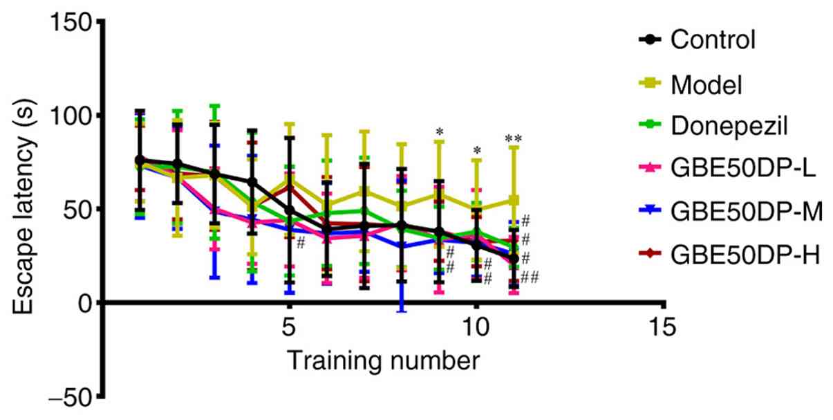
Results of Morris water maze test
The results of the positioning navigation experiment
are shown in Fig. 1. Compared with
the control group, the escape latency of rats in the model group
was significantly prolonged at the 9, 10 and 11th trials (P<0.05
or P<0.01). Compared with the model group, the escape latency of
rats in the donepezil group was significantly shortened at the 11th
trial (P<0.05), that of rats in the GBE50DP-L group was
significantly shortened at the 9 and 11th trials (P<0.05 or
P<0.01), that of rats in the GBE50DP-M group was significantly
shortened at the 5, 9, 10 and 11th trials (P<0.05), and that of
rats in the GBE50DP-H group was significantly shortened at the 10
and 11th trials (P<0.05).
The results of space exploration experiments are
shown in Table II. Compared with
the control group, the number of platform crossings of rats in the
model group was significantly decreased (P<0.05). Compared with
the model group, the number of platform crossings in the positive,
GBE50DP-M and GBE50DP-H groups was significantly increased
(P<0.05 or P<0.01).
| Table IIEffect of GBE50DP on the number of
crossing platforms (n=12). |
Table II
Effect of GBE50DP on the number of
crossing platforms (n=12).
| Group | Number of platforms
crossings |
|---|
| Control | 2.6±1.6 |
| Model |
1.4±1.0a |
| Donepezil |
2.8±1.3b |
| GBE50DP-L | 2.4±1.4 |
| GBE50DP-M |
2.6±1.2b |
| GBE50DP-H |
2.9±1.3c |
Results of eight-arm maze test
As shown in Table
III, compared with the control group, the average number of
working memory errors and reference memory errors of rats in the
model group was significantly increased (P<0.05), and the
foraging completion time was significantly prolonged (P<0.01).
Compared with the model group, the mean number of working memory
errors of rats in the positive, GBE50DP-L and -M groups was
significantly decreased (P<0.05), that of reference memory
errors of rats in the positive, GBE50DP-M and -H groups was
significantly decreased (P<0.05), and the foraging completion
time of the positive, GBE50DP-L and -H groups was significantly
decreased (P<0.05 or P<0.01).
| Table IIIEffect of GBE50DP on the indexes of
the eight-arm maze (n=12). |
Table III
Effect of GBE50DP on the indexes of
the eight-arm maze (n=12).
| Group | Number of working
memory errors | Number of reference
memory errors | Foraging completion
time (min) |
|---|
| Control | 1.9±0.8 | 2.5±1.3 | 5.04±2.28 |
| Model |
3.4±1.1a |
4.8±1.2a |
7.24±0.61b |
| Donepezil |
1.8±1.0c |
2.8±1.0c |
4.83±0.80c |
| GBE50DP-L |
2.0±0.8c | 3.6±1.5 |
5.27±1.59c |
| GBE50DP-M |
2.0±0.7c |
3.0±0.7c | 5.64±0.58 |
| GBE50DP-H | 2.3±1.0 |
2.8±0.5c |
4.64±1.22d |
Results of oxidative stress and
inflammatory marker detection in the rat hippocampus. Content of
oxidative stress indicators
As shown in Fig.
2A, compared with the control group, the SOD content in the
hippocampal tissue of the model group rats (30.31±3.67 ng/l) was
significantly decreased (P<0.01), the GSH-Px content (37.51±6.70
nmol/l) was significantly decreased (P<0.01), the CAT content
(22.59±1.72 ng/l) was significantly decreased (P<0.001), while
the MDA content (5.39±0.67 nmol/l) was significantly increased
(P<0.05). Compared with the model group, the SOD content
(40.32±7.88 ng/l) in the GBE50DP high-dose group was significantly
increased (P<0.01), the GSH-Px content (42.62±4.77 nmol/l) was
significantly increased (P<0.05), the CAT content (24.33±1.96
ng/l) was significantly increased (P<0.05), while the MDA
content (4.60±0.57 nmol/l) was significantly decreased
(P<0.05).
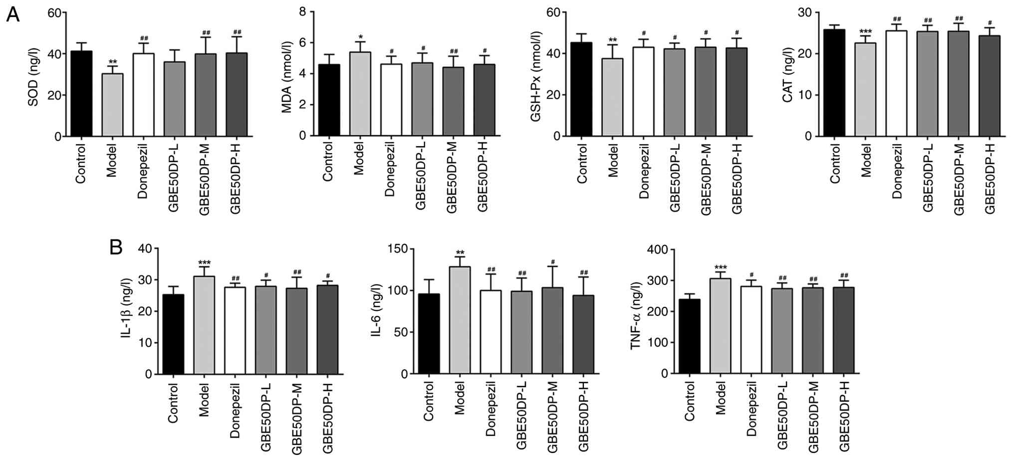 | Figure 2Effects of GBE50DP on oxidative
stress and inflammation in the hippocampus. (A) Effect of GBE50DP
on the contents of SOD, MDA, GSH Px and CAT in the hippocampus
(n=8); (B) Effect of GBE50DP on the contents of IL-1β, IL-6 and
TNF-α in the hippocampus (n=8). *P<0.05,
**P<0.01 and ***P<0.001 vs. Control
group; #P<0.05 and ##P<0.01 vs. Model
group. GBE50DP, Ginkgo biloba extract 50 dropping pill; SOD,
superoxide dismutase; MDA, malondialdehyde; GSH Px, glutathione
peroxidase; CAT, catalase; -L, low dose group; -M, medium dose
group; -H, high dose group. |
Content of inflammatory
indicators
As shown in Fig.
2B, compared with the control group, the IL-1β content
(31.08±3.03 ng/l) in the hippocampal tissue of the model group rats
was significantly increased (P<0.001), the IL-6 content
(128.50±12.07 ng/l) was significantly increased (P<0.01), and
the TNF-α content (306.16±21.47 ng/l) was significantly increased
(P<0.001). Compared with the model group, the IL-1β content
(28.21±1.37 ng/l) in the GBE50DP high-dose group was significantly
decreased (P<0.05), the IL-6 content (94.03±22.27 ng/l) was
significantly decreased (P<0.01) and the TNF-α content
(277.42±23.28 ng/l) was significantly decreased (P<0.01).
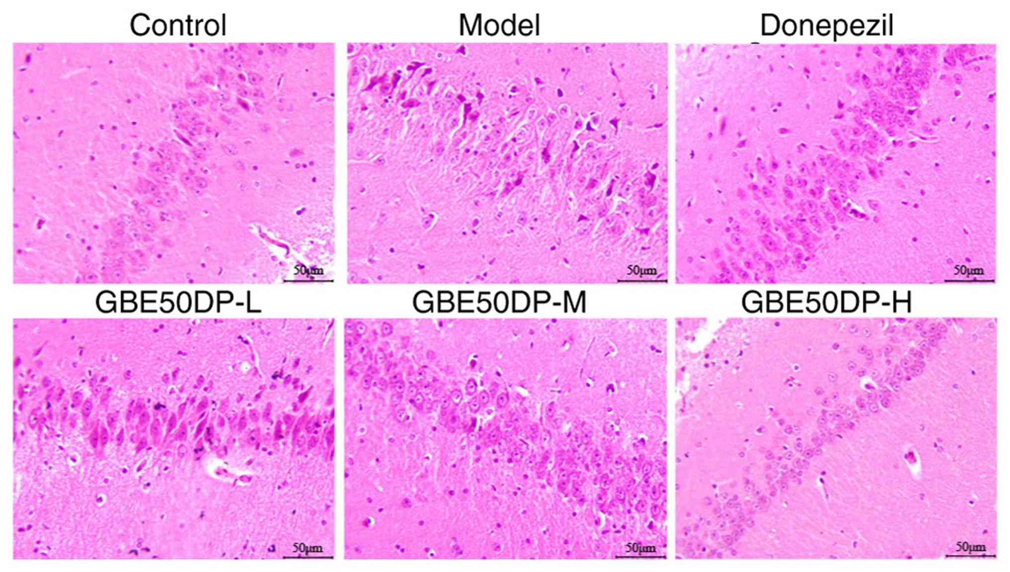
Morphological changes of neurons in
the CA1 region of the rat hippocampus. Results of H&E
assay
As shown in Fig. 3,
in the control group, the nerve cells were arranged neatly, the
cytoplasm and nucleus were stained clearly, and no inflammatory
cell infiltration, capillary dilation or congestion was observed.
Compared with the control group, the structure of the hippocampal
CA1 region of rats in the model group was loose, the arrangement of
nerve cells was irregular and the morphology was incomplete, the
cytoplasm was deeply stained, the nuclear condensation, apoptotic
bodies and glial cells were improved increased, the inflammatory
cells were scattered, and the capillaries were dilated. Compared
with the model group, the aforementioned pathological changes in
each administration group were improved to various degrees, showing
a tighter structure, a decrease in the number of apoptotic bodies
and glial cells, and an improvement in telangiectasia. Among them,
the improvement in the GBE50DP-H group was the most obvious,
followed by the positive group.
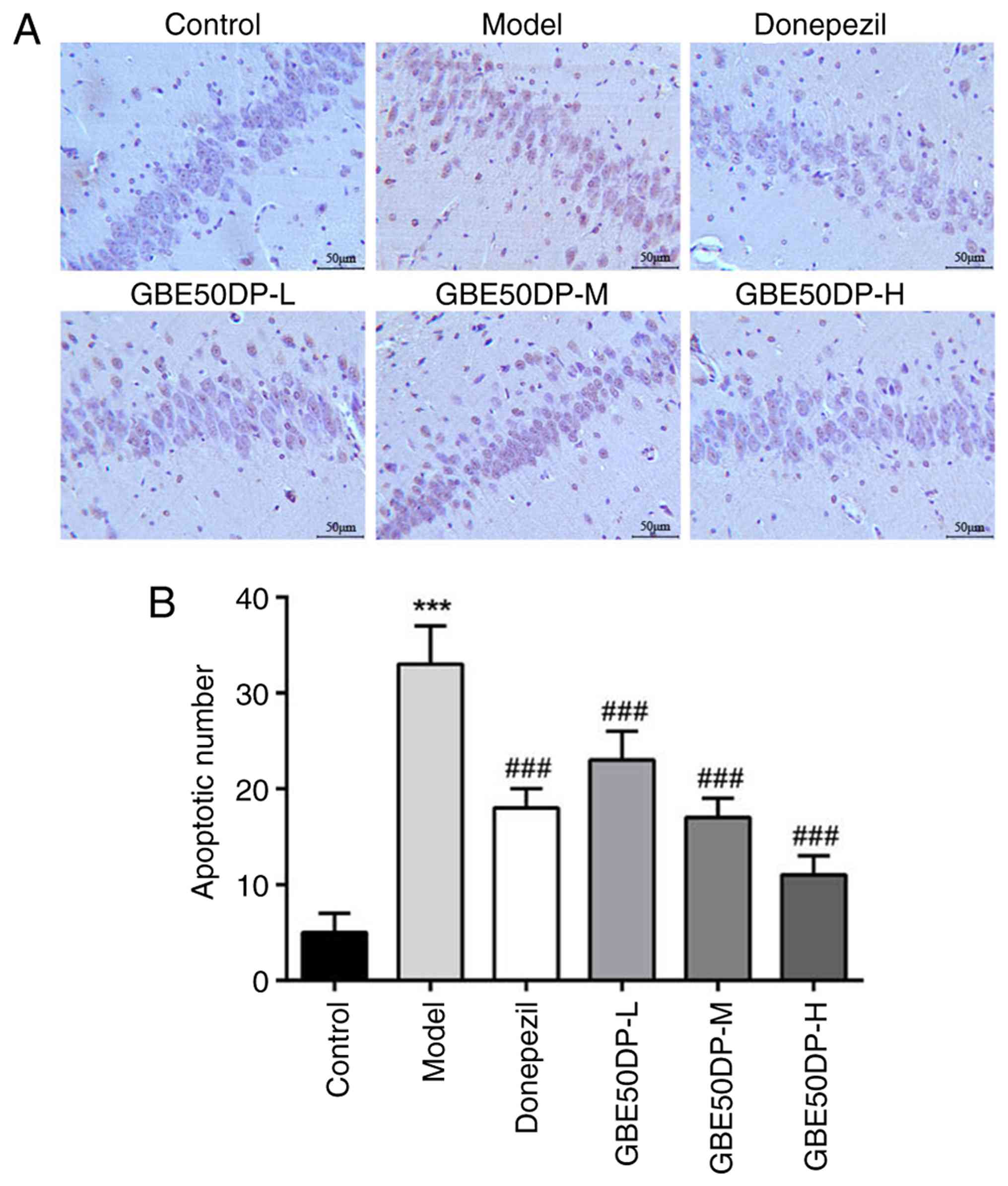
Results of the TUNEL assay
As shown in Fig. 4,
the number of apoptotic neurons in the CA1 region of the
hippocampus in the control group rats was 5±2, while in the model
group it was 33±4. Compared with the control group, the number of
apoptotic neurons in the model group was significantly increased
(P<0.001). Compared with the model group, the number of
apoptotic neurons in all treatment groups was significantly
decreased (P<0.001), with the GBE50DP high-dose group showing
the most pronounced change, having 11±2 apoptotic neurons.
Results of the immunohistochemical
assay
As shown in Fig. 5,
compared with the control group, the expression of Bcl-2 in the
hippocampal CA1 region of rats in the model group was significantly
decreased (P<0.001), while that of Cyto-C, Bax, Caspase-3, Fas
and PARP-1 was significantly increased (P<0.001). Compared with
the model group, the expression of Bcl-2 in the hippocampal CA1
region of rats in each administration group was significantly
increased (P<0.001), while that of Cyto-C, Bax, Caspase-3, Fas
and PARP-1 was significantly decreased (P<0.001), particularly
in the GBE50DP-H group.
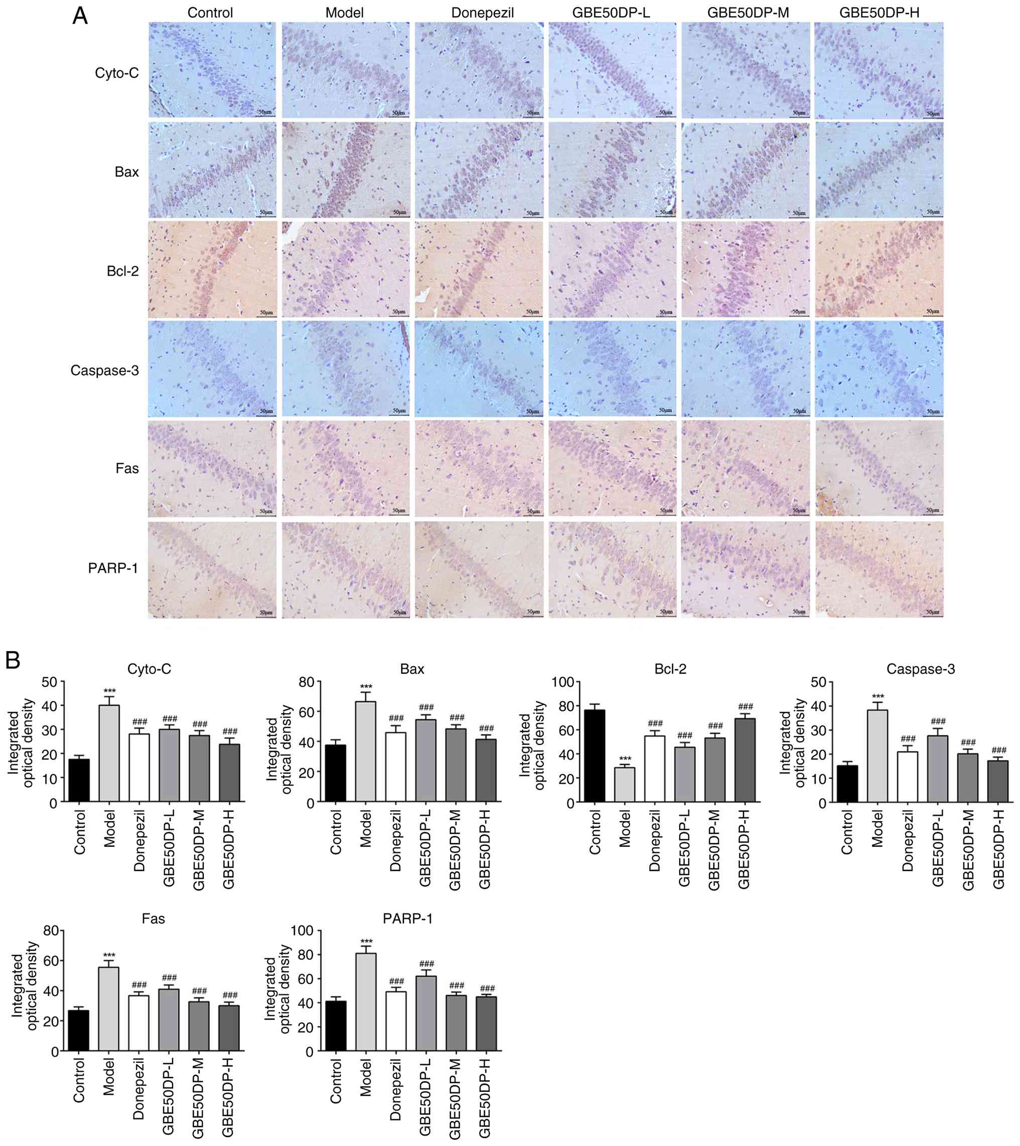 | Figure 5Effect of GBE50DP on the expression
of apoptosis-related proteins in the hippocampal CA1 region. (A)
Expression of Cyto-C, Bax, Bcl-2, Caspase-3, Fas and PARP-1 in the
hippocampal CA1 region (scale bar, 50, µm). (B) Integral optical
densities of Cyto-C, Bax, Bcl-2, Caspase-3, Fas, and PARP-1 in the
hippocampal CA1 region (n=3). ***P<0.001 vs. Control
group; ###P<0.001 vs. Model group. Cyto-C, cytochrome
c; PARP-1 Poly (ADP-ribose) polymerase 1; GBE50DP, Ginkgo
biloba extract 50 dropping pill; -L, low dose group; -M, medium
dose group; -H, high dose group. |
Results of western blot analysis
As shown in Fig. 6,
compared with the control group, the expression of Bcl-2 in the
hippocampal CA1 region of rats in the model group was significantly
decreased (P<0.05), while the expression of Cyto-C, Bax,
Caspase-3, Fas and PARP-1 were significantly increased (P<0.05
or P<0.01 or P<0.001). Compared with the model group, the
expression of Bcl-2 in the hippocampal CA1 region of rats in the
positive group and the GBE50DP-H group significantly was increased
(P<0.05), while that of Cyto-C, Bax, Caspase-3, Fas and PARP-1
in the positive and GBE50DP-H groups was significantly decreased
(P<0.05 or P<0.01).
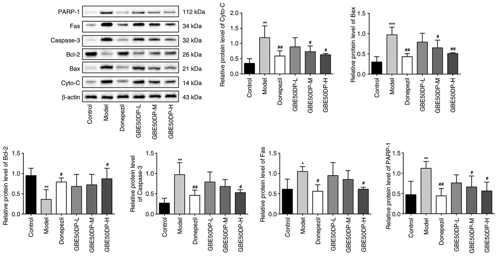 | Figure 6Relative expression levels of Cyto-C,
Bax, Bcl-2, Caspase-3, Fas and PARP-1 in the hippocampal CA1
region. Western blot bands and quantified bar charts of each
protein, n=3. *P<0.05, **P<0.01 and
***P<0.001 vs. Control group; #P<0.05
and ##P<0.01 vs. Model group. Cyto-C, cytochrome
c; PARP-1 Poly (ADP-ribose) polymerase 1; GBE50DP, Ginkgo
biloba extract 50 dropping pill; -L, low dose group; -M, medium
dose group; -H, high dose group. |
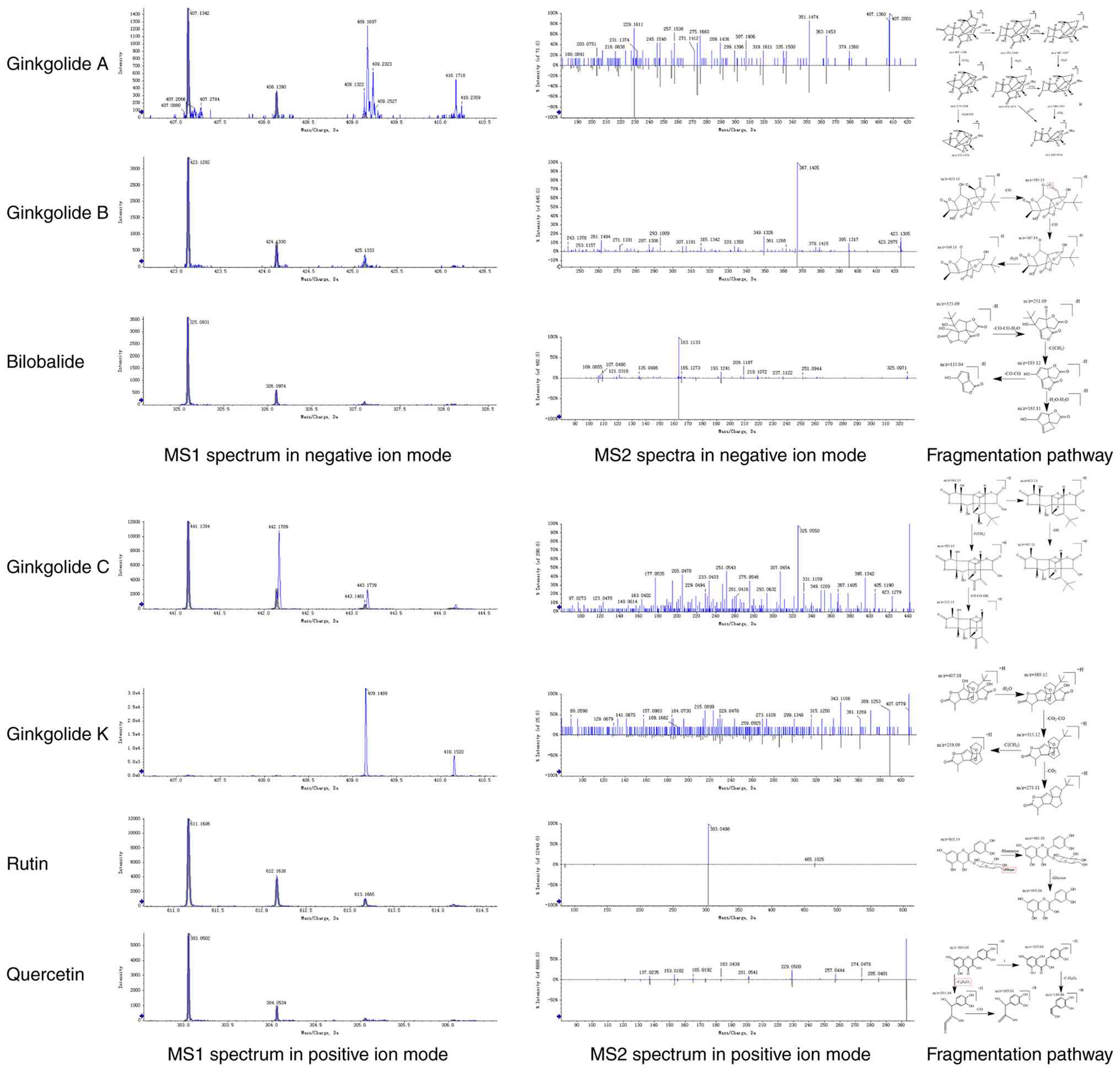
Analysis of components of
GBE50DP-containing plasma
The molecular formula of the compound in the
GBE50DP-containing plasma was speculated by ion peak and cleavage
law (see Fig. 7). The main
VCI-related compounds were speculated as the ginkgolide A, B, C, K,
bilobalide, rutin and quercetin.
Discussion
One of the clinical characteristics of VCI is social
disorder (23), which is
manifested by social withdrawal, decreased active communication,
weakened empathy and language communication disorders. The
experimental results of the present study showed that GBE50DP
(especially at high dose) can significantly shorten the escape
latency of VCI rats, increase the number of platform crossings, and
reduce errors in working memory and reference memory. This
suggested that it can effectively improve learning and memory
abilities, providing an experimental basis for clinical application
to improve the cognitive and social functions of VCI patients. In
addition, GBE50DP can significantly increase the activities of SOD
and GSH-Px in the hippocampus, reduce the levels of IL-1β and
TNF-α, increase the expression of Bcl-2 and decrease that of Bax
and Caspase-3. This indicated that it exerts anti-VCI effects
through multiple aspects, such as regulating oxidative stress,
neuroinflammation and apoptosis pathways, providing ideas for the
development of multi-target therapeutic drugs. At the same time,
GBE50DP can alleviate neuronal damage and apoptosis in the CA1
region of the hippocampus, suggesting that it may be applicable for
early intervention in VCI and delaying the progression of the
disease. In the present study, intragastric administration was
used, which was consistent with the clinical oral preparation
route, and the components of GBE50DP in the blood were clear, which
showed good potential for clinical transformation.
Ischemic brain injury is one of the strongest
stimuli that induces abnormal gene expression, causing
mitochondrial dysfunction and cellular apoptosis when the
accumulation of reactive oxygen species exceeds the cellular
clearance ability (24,25). The results of the in vivo
experiments demonstrated that, in the VCI model, the activity of
the crucial anti-oxidative enzymes (SOD, CAT and GSH-Px) in the
hippocampal CA1 region [the key structural region involved in
learning and memory (26)], was
inhibited due to the increase of oxidative stress caused by
vascular lesions, resulting in the increase of MDA [the production
of lipid peroxidation (27)] and
recruitment of the pro-inflammatory cytokines [IL-1β, IL-6 and
TNF-α (28)] through a synergistic
effect during ischemic injury (29). The expression changes of the
crucial nodes involved in the apoptotic signaling pathway exhibited
have proven those oxidative stress-induced apoptotic processes: The
Cyto-C released at the initial stage of apoptosis following the
vasculopathy in the VCI model, across the rupture of the
mitochondrial outer membrane induced by the increase of the
pro-apoptotic protein, Bax, and decrease of the anti-apoptotic
protein Bcl-2. Caspase-3 was then activated by the death receptor
Fas and cleaved multiple cell substrates, leading to apoptosis as
the key executor (30). The
excessive activation of PARP-1, as an DNA repair enzyme, depleted
cellular energy and promoted apoptosis (31,32).
The opposite tendency of all the aforementioned indicators
following the administration revealed that GBE50DP effectively
improved VCI symptoms by enhancing the activity of anti-oxidative
enzymes and inhibiting the apoptotic process, thereby protecting
cognitive function. This protective effect was reflected in the
pathological improvement, as demonstrated by morphological
observation using the staining methods.
Previous studies have shown that the Ginkgo
biloba extract can improve learning and memory abilities by
scavenging oxygen free radicals, enhancing antioxidant enzyme
activity and inhibiting neuroinflammation (33), and can safely and effectively
improve the mental state and daily living ability of VCI patients
(34). In the present study,
GBE50DP, as a high-purity Ginkgo biloba extract with >50%
total flavonoids and terpene lactones, has advantages in
neuroprotection (10). GBE50DP can
not only inhibit oxidative stress and inflammatory response, but
also regulate mitochondrial apoptotic pathways, such as
downregulate pro-apoptotic proteins Cyto-C and Bax and upregulate
anti-apoptotic protein Bcl-2, thereby reducing hippocampal neuron
damage. The high-dose group of GBE50DP was superior to the positive
drug donepezil in the number of crossing the platform, the time
required to complete foraging and the protection of neurons, which
may be associated with the following factors: GBE50DP has a higher
concentration of active ingredients and improved bioavailability;
the lipid-soluble component ginkgolide B can easily pass through
the blood-brain barrier and has a faster onset, and as a natural
platelet activating factor (PAF) antagonist, it can improve
cerebral microcirculation by inhibiting PAF activity (35); the antioxidant effect of rutin and
quercetin form a synergistic effect with the anti-inflammatory
effect of ginkgolides, and further regulate the mitochondrial
apoptosis pathway. The aforementioned results indicated that
GBE50DP has the advantage of improving VCI, suggesting that GBE50DP
is expected to be developed as a new preparation for clinical
treatment of VCI to expand its indications.
Studies have found that ginkgolide (including
ginkgolide A, B, C and K, etc.) and bilobalide belong to the
terpene lactones, which can regulate the expression of Bcl-2/Bax to
inhibit neuronal apoptosis (36)
and play a neuroprotective role in ischemic brain injury (37), thereby improving cognitive ability.
Among them, ginkgolide B is the strongest platelet-activating
factor antagonist found so far that has a significant
antithrombotic effect; ginkgolide A, K and bilobalide can scavenge
free radicals (38) and resist
cytotoxicity by anti-lipid peroxidation (39); ginkgolide C and bilobalide can
inhibit inflammatory responses (40,41).
Rutin and quercetin belong to the flavonoid family, which has
antioxidant effects such as scavenging free radicals, enhancing SOD
activity in brain tissue and reducing MDA content and can improve
mitochondrial function, as well as learning and memory ability
(42,43). Furthermore, the molecular targets
of the effective components would be clarified through molecular
interaction and molecular docking. Combined with pharmacokinetic
studies, the metabolic parameters of each component in vivo
and the interactions between components can then be analyzed to
clarify their efficacy, thereby verifying the pharmacodynamic
material basis and mechanism of GBE50DP.
In the present study, GBE50DP increased the
activities of SOD and GSH-Px and decreased the levels of IL-1β and
TNF-α, while the aforementioned inflammatory factors were often
highly expressed in the cerebrospinal fluid of clinical VCI
patients, suggesting that GBE50DP may improve cognitive function
through antioxidant and anti-inflammatory dual activities, which
provides an experimental basis for its application. It has been
reported that GBE50 can exert anti-inflammatory effects by
inhibiting the NF-κB signaling pathway (44) and can increase the activities of
SOD and GSH-Px (10). Nuclear
factor erythroid 2-related factor 2 (Nrf2)/heme oxygenase-1 (HO-1)
is a key upstream pathway for regulating these antioxidant enzymes.
The results of the present study were highly consistent with the
aforementioned mechanism. In addition, PARP-1 can regulate NF-κB
activity and promote the expression of inflammatory factors
(45). After GBE50DP downregulates
PARP-1, the inflammatory factors also decrease. Based on this
hypothesis, the protective effect of GBE50DP in VCI may not depend
on a single pathway, but may be achieved by activating the
Nrf2/HO-1 signaling pathway to combat oxidative stress, inhibiting
the NF-κB signaling pathway to alleviate neuroinflammation and
synergistically regulating the apoptotic pathway. Subsequent
studies can further verify the mechanism of GBE50DP by constructing
a cell injury model induced by H2O2 or
lipopolysaccharides (LPS), combining PARP-1 gene knockout animals
or specific inhibitors, and using a composite etiology VCI animal
model to provide a more substantial theoretical basis for its
clinical transformation.
In conclusion, long-term cerebral hypoperfusion
caused by blocking the cervical artery of rats can lead to
pathological changes such as hypoxia, neuronal damage, inflammatory
response and apoptosis in brain tissue, in turn leading to the
symptoms of cognitive impairment. GBE50DP can improve the social
behavior and ability of learning and memory, decrease oxidative
stress, inhibit inflammatory response, improve the pathological
morphology of the hippocampus, protect neurons and protect nerve
cells from apoptosis of VCI rats induced by cerebral hypoperfusion,
thereby enhancing the self-protection ability of the brain and
promoting the improvement of cognitive function. The effective
substances of GBE50DP in improving VCI has been preliminarily
determined. Those results provided an experimental basis for the
further understanding of the neuroprotective effects of GBE50DP and
helped reveal the potential of GBE50DP in the treatment of
cognitive impairment.
Acknowledgements
Not applicable.
Funding
Funding: The present study was supported by the Technical
Service Project of Zhejiang Shangyao Jiuxu Pharmaceutical Co., Ltd.
(grant no. HT20220012).
Availability of data and materials
The data generated in the present study may be
requested from the corresponding author. The data that support the
findings of this study have been deposited into CNSA with accession
number CNP0009414 (https://doi.org/10.26036/CNP0009414).
Authors' contributions
FLW performed the research and wrote the manuscript.
WLL and HL designed the study. FLW, WLL and HL analyzed the data
and revised the manuscript. FLW and HL confirm the authenticity of
all the raw data. All authors read and approved the final
manuscript.
Ethics approval and consent to
participate
The animal experiment in this study was approved by
the Harbin University of Commerce Ethics Committees (approval no.
HSDYXY-2023035).
Patient consent for publication
Not applicable.
Competing interests
Zhejiang Shangyao Jiuxu Pharmaceutical Co., Ltd.,
provided the GBE50DP free of charge, and the author HL is
affiliated to this company. The authors declare that they have no
other competing interests.
References
|
1
|
Zanon Zotin MC, Sveikata L, Viswanathan A
and Yilmaz P: Cerebral small vessel disease and vascular cognitive
impairment: From diagnosis to management. Curr Opin Neurol.
34:246–257. 2021.PubMed/NCBI View Article : Google Scholar
|
|
2
|
Cheng YW, Chiu MJ, Chen YF, Cheng TW, Lai
YM and Chen TF: The contribution of vascular risk factors in
neurodegenerative disorders: From mild cognitive impairment to
Alzheimer's disease. Alzheimers Res Ther. 12(91)2020.PubMed/NCBI View Article : Google Scholar
|
|
3
|
Markus HS and Joutel A: The pathogenesis
of cerebral small vessel diseases and vascular cognitive
impairment. Physiol Rev. 105:1075–1171. 2025.PubMed/NCBI View Article : Google Scholar
|
|
4
|
Rajeev V, Chai YL, Poh L, Selvaraji S,
Fann DY, Jo DG, De Silva TM, Drummond GR, Sobey CG, Arumugam TV, et
al: Chronic cerebral hypoperfusion: A critical feature in
unravelling the etiology of vascular cognitive impairment. Acta
Neuropathol Commun. 11(93)2023.PubMed/NCBI View Article : Google Scholar
|
|
5
|
Jiménez-Ruiz A, Aguilar-Fuentes V,
Becerra-Aguiar NN, Roque-Sanchez I and Ruiz-Sandoval JL: Vascular
cognitive impairment and dementia: A narrative review. Dement
Neuropsychol. 18(e20230116)2024.PubMed/NCBI View Article : Google Scholar
|
|
6
|
Rundek T, Tolea M, Ariko T, Fagerli EA and
Cemargo CJ: Vascular cognitive impairment (VCI). Neurotherapeutics.
19:68–88. 2022.PubMed/NCBI View Article : Google Scholar
|
|
7
|
Chang Wong E and Chang Chui H: Vascular
cognitive impairment and dementia. Continuum (Minneap Minn).
28:750–780. 2022.PubMed/NCBI View Article : Google Scholar
|
|
8
|
Pérez Palmer N, Trejo Ortega B and Joshi
P: Cognitive impairment in older adults: Epidemiology, diagnosis,
and treatment. Psychiatr Clin North Am. 45:639–661. 2022.PubMed/NCBI View Article : Google Scholar
|
|
9
|
Kalaria RN, Akinyemi RO, Paddick SM and
Ihara M: Current perspectives on prevention of vascular cognitive
impairment and promotion of vascular brain health. Expert Rev
Neurother. 24:25–44. 2024.PubMed/NCBI View Article : Google Scholar
|
|
10
|
Xia CY, Zhou MM, Dong XW, Zhao Y, Jiang
MF, Zhu GQ and Zhang ZX: Ginkgo biloba extract inhibits
hippocampal neuronal injury caused by mitochondrial oxidative
stress in a rat model of Alzheimer's disease. PLoS One.
19(e0307735)2024.PubMed/NCBI View Article : Google Scholar
|
|
11
|
Tang Z, Jiang DZ, Zhao XX and Zhang YW:
Comparison of different specifications of Ginkgo biloba
extract and discussion on specification improvement. Drug Stand
China. 21:21–25. 2020.
|
|
12
|
Liu J, Zhou J, Li WW, Chen YP and Wang J:
Effects of different doses of Ginkgo biloba extract on
cognitive function and neurotransmitter level in rats with vascular
cognitive impairment. Chin J Difficult Complicat Cases. 20:182–186.
2021.
|
|
13
|
Liu LM, Wang YT, Zhang JC and Wang SF:
Advances in the chemical constituents and chemical analysis of
Ginkgo biloba leaf, extract, and phytopharmaceuticals. J
Pharm Biomed Anal. 193(113704)2021.PubMed/NCBI View Article : Google Scholar
|
|
14
|
Li XY and Liu AJ: Chemical constitution
and pharmacological mechanism of Ginkgo biloba extract 50 in
the treatment of vascular related diseases. Chin Pharm J.
59:1074–1081. 2024.
|
|
15
|
Qian YY, Zhu GQ, Wang WJ and Gao Q:
Research progress on the clinical and pharmacological effects of
Ginkgo biloba extract 50 and its preparations. Chin Tradit
Patent Med. 43:998–1003. 2021.
|
|
16
|
Bhat JA and Kumar M: Neuroprotective
effects of theobromine in permanent bilateral common carotid artery
occlusion rat model of cerebral hypoperfusion. Metab Brain Dis.
37:1787–1801. 2022.PubMed/NCBI View Article : Google Scholar
|
|
17
|
Wu S, Huang R, Zhang R, Xiao C, Wang L,
Luo M, Song N, Zhang J, Yang F, Liu X and Yang W: Gastrodin and
gastrodigenin improve energy metabolism disorders and mitochondrial
dysfunction to antagonize vascular dementia. Molecules.
28(2598)2023.PubMed/NCBI View Article : Google Scholar
|
|
18
|
Shrief AI, Elshenawy DS, Elsukary AE,
Elekhtiar SA and Yahia OA: Behavioral and histological study on the
neuroprotective effect of thymoquinone on the cerebellum in
AlCl3-induced neurotoxicity in rats through modulation of oxidative
stress, apoptosis, and autophagy. J Mol Histol.
56(81)2025.PubMed/NCBI View Article : Google Scholar
|
|
19
|
Schneider MA, Heeb L, Beffinger MM,
Pantelyushin S, Linecker M, Roth L, Lehmann K, Ungethüm U, Kobold
S, Graf R, et al: Attenuation of peripheral serotonin inhibits
tumor growth and enhances immune checkpoint blockade therapy in
murine tumor models. Sci Transl Med. 13(eabc8188)2021.PubMed/NCBI View Article : Google Scholar
|
|
20
|
Yavari N, Nadia Sharifi Z, Rekabdar Y and
Movassaghi S: Protective effect of curcumin on CA1 region of
hippocampus in rat model of ischemia/reperfusion injury. Galen Med
J. 11(e1062)2022.PubMed/NCBI View Article : Google Scholar
|
|
21
|
Yu L, Jin W, Deng D, Wang Y, Chen Q, Zhang
Y, Wan H, Chen Y, Chen Y, He Y and Zhang L: Investigation of
anti-apoptotic effects and mechanisms of Astragaloside IV in a rat
model of cerebral ischemia-reperfusion injury. CNS Neurosci Ther.
31(e70209)2025.PubMed/NCBI View Article : Google Scholar
|
|
22
|
Fan LL, Fang H, Zheng JY, Qiu YH, Wu GL,
Cai YF, Chen YB and Zhang SJ: Taohong Siwu decoction alleviates
cognitive impairment by suppressing endoplasmic reticulum stress
and apoptosis signaling pathway in vascular dementia rats. J
Ethnopharmacol. 333(118407)2024.PubMed/NCBI View Article : Google Scholar
|
|
23
|
Khan MB, Hoda MN, Vaibhav K, Giri S, Wang
P, Waller JL, Ergul A, Dhandapani KM, Fagan SC and Hess DC: Remote
ischemic postconditioning: Harnessing endogenous protection in a
murine model of vascular cognitive impairment. Transl Stroke Res.
6:69–77. 2015.PubMed/NCBI View Article : Google Scholar
|
|
24
|
Li H, Liu Y, Lin LT, Wang XR, Du SQ, Yan
CQ, He T, Yang JW and Liu CZ: Acupuncture reversed hippocampal
mitochondrial dysfunction in vascular dementia rats. Neurochem Int.
92:35–42. 2016.PubMed/NCBI View Article : Google Scholar
|
|
25
|
Ogunro OB, Karigidi ME, Gyebi GA,
Turkistani A and Almehmadi AH: Tangeretin offers neuroprotection
against colchicine-induced memory impairment in Wistar rats by
modulating the antioxidant milieu, inflammatory mediators and
oxidative stress in the brain tissue. BMC Complement Med Ther.
25(40)2025.PubMed/NCBI View Article : Google Scholar
|
|
26
|
Rajabian A, Kioumarsi Darbandi Z, Aliyari
M, Saberi R, Amirahmadi S, Amini H, Salmani H, Youseflee P and
Hosseini M: Pioglitazone improves learning and memory in a rat
model of cholinergic dysfunction induced by scopolamine, the roles
of oxidative stress and neuroinflammation. Naunyn Schmiedebergs
Arch Pharmacol. 398:10221–10237. 2025.PubMed/NCBI View Article : Google Scholar
|
|
27
|
Tian Z, Ji X and Liu J: Neuroinflammation
in vascular cognitive impairment and dementia: Current evidence,
advances, and prospects. Int J Mol Sci. 23(6224)2022.PubMed/NCBI View Article : Google Scholar
|
|
28
|
Yu JY, Fang P, Wang C, Wang XX, Li K, Gong
Q, Luo BY and Wang XD: Dorsal CA1 interneurons contribute to acute
stress-induced spatial memory deficits. Neuropharmacology.
135:474–486. 2018.PubMed/NCBI View Article : Google Scholar
|
|
29
|
Li Y, Ma Y, Dang QY, Fan XR, Han CT, Xu SZ
and Li PY: Assessment of mitochondrial dysfunction and implications
in cardiovascular disorders. Life Sci. 306(120834)2022.PubMed/NCBI View Article : Google Scholar
|
|
30
|
Guo J, Yang N, Zhang J, Huang Y, Xiang Q,
Wen J, Chen Y, Hu T, Liu Q and Rao C: Neurotoxicity study of ethyl
acetate extract of Zanthoxylum armatum DC. on SH-SY5Y based on ROS
mediated mitochondrial apoptosis pathway. J Ethnopharmacol.
319(117321)2024.PubMed/NCBI View Article : Google Scholar
|
|
31
|
Wang S, Yang Y, Lin J, Zhang W, Yang C,
Zhang R, Zhou C, Zhang L, Wang X, Liu J, et al: Astragalin actives
autophagy and inhibits apoptosis of astrocytes in AD mice via
down-regulating Fas/Fasl-VDAC1 pathway. Free Radic Biol Med.
232:72–85. 2025.PubMed/NCBI View Article : Google Scholar
|
|
32
|
Jiao Y and Li G: PARP inhibitor PJ34
ameliorates cognitive impairments induced by transient cerebral
ischemia/reperfusion through its anti-inflammatory effects in a rat
model. Neurosci Lett. 764(136202)2021.PubMed/NCBI View Article : Google Scholar
|
|
33
|
DeFeudis FV and Drieu K: Ginkgo
biloba extract (EGb761) and CNS functions: Basic studies and
clinical applications. Curr Drug Targets. 1:25–58. 2000.PubMed/NCBI View Article : Google Scholar
|
|
34
|
Zhan M, Sun L, Liu J, Zeng Z, Shen W, Li
H, Wang Y, Han F, Shi J, Zeng X, et al: EGb in the treatment for
patients with VCI: A systematic review and meta-analysis. Oxid Med
Cell Longev. 2021(8787684)2021.PubMed/NCBI View Article : Google Scholar
|
|
35
|
Zhu Q and Liu D: Clinical efficacy and
mechanism of Ginkgo biloba extract in the treatment of
elderly ischemic cerebrovascular disease. Pak J Pharm Sci.
37:705–713. 2024.PubMed/NCBI
|
|
36
|
Hua J, Yin N, Yang B, Zhang J, Ding J, Fan
Y and Hu G: Ginkgolide B and bilobalide ameliorate neural cell
apoptosis in α-synuclein aggregates. Biomed Pharmacother.
96:792–797. 2017.PubMed/NCBI View Article : Google Scholar
|
|
37
|
Feng Z, Sun Q, Chen W, Bai Y, Hu D and Xie
X: The neuroprotective mechanisms of ginkgolides and bilobalide in
cerebral ischemic injury: A literature review. Mol Med.
25(57)2019.PubMed/NCBI View Article : Google Scholar
|
|
38
|
Ma S, Yin H, Chen L, Liu H, Zhao M and
Zhang X: Neuroprotective effect of ginkgolide K against acute
ischemic stroke on middle cerebral ischemia occlusion in rats. J
Nat Med. 66:25–31. 2012.PubMed/NCBI View Article : Google Scholar
|
|
39
|
Zhou LJ, Song W, Zhu XZ, Chen ZL, Yin ML
and Cheng XF: Protective effects of bilobalide on amyloid
beta-peptide 25-35-induced PC12 cell cytotoxicity. Acta Pharmacol
Sin. 21:75–79. 2000.PubMed/NCBI
|
|
40
|
Li B, Zhang B, Li Z, Li S, Li J, Wang A,
Hou J, Xu J and Zhang R: Ginkgolide C attenuates cerebral
ischemia/reperfusion-induced inflammatory impairments by
suppressing CD40/NF-κB pathway. J Ethnopharmacol.
312(116537)2023.PubMed/NCBI View Article : Google Scholar
|
|
41
|
Zhou JM, Gu SS, Wang HM, Zhou J, Wang ZZ
and Xiao W: Ginkgolides and bilobalide protect BV2 microglia cells
against OGD/reoxygenation injury by inhibiting TLR2/4 signaling
pathways. Cell Stress Chaperones. 21:1037–1053. 2016.PubMed/NCBI View Article : Google Scholar
|
|
42
|
Mao YJ, Feng YL, Wang MJ, Lyu ZY and Zhai
GY: Research progress on rutin derivatives. Zhongguo Zhong Yao Za
Zhi. 46:4654–4665. 2021.PubMed/NCBI View Article : Google Scholar : (In Chinese).
|
|
43
|
Park DJ, Shah FA and Koh PO: Quercetin
attenuates neuronal cells damage in a middle cerebral artery
occlusion animal model. J Vet Med Sci. 80:676–683. 2018.PubMed/NCBI View Article : Google Scholar
|
|
44
|
He GY, Yuan CG, Hao L, Xu Y and Zhang ZX:
GBE50 attenuates inflammatory response by inhibiting the p38 MAPK
and NF-κB pathways in LPS-stimulated microglial cells. Evid Based
Complement Alternat Med. 2014(368598)2014.PubMed/NCBI View Article : Google Scholar
|
|
45
|
Skaper SD: Poly(ADP-ribosyl)ation enzyme-1
as a target for neuroprotection in acute central nervous system
injury. Curr Drug Targets CNS Neurol Disord. 2:279–291.
2003.PubMed/NCBI View Article : Google Scholar
|