Introduction
Compared with hemodialysis (HD), peritoneal dialysis
(PD) provides multiple benefits, including greater treatment
flexibility, fewer dietary limitations and improved maintenance of
residual kidney function (1).
Globally, PD accounts for 10-11% of all dialysis modalities, with
marked regional variation and its utilization has increased
steadily over the past decade (2).
This increase has been largely attributable to health policy
measures encouraging home-based dialysis, supported by
international clinical guidelines such as those issued by Kidney
Disease: Improving Global Outcomes (3) which recognize PD as a key component
of patient-centered and sustainable management of end-stage kidney
disease. Consistent with these recommendations, national and
regional registry data have demonstrated a continued rise in both
incident and prevalent PD populations, highlighting the growing
role of PD in contemporary renal care (4).
Despite these developments, PD-associated
peritonitis (PDAP) remains a major complication of PD and a leading
cause of technique failure. Although advances in catheter
technology, connection devices, patient training and antimicrobial
management have contributed to a reduction in peritonitis
incidence, PDAP continues to be a predominant cause of
hospitalization, catheter removal and transfer to HD (5). Importantly, treatment failure not
only adversely affects individual patient outcomes but also
compromises long-term PD technique survival and the sustainability
of PD programs (6).
Current management strategies for PDAP primarily
rely on standardized empiric and pathogen-directed antibiotic
regimens (7). While effective for
many patients, these approaches do not fully account for the marked
heterogeneity in treatment response observed in routine clinical
practice. Existing guidelines, including the International Society
for Peritoneal Dialysis peritonitis recommendations (2016 and 2022
updates) (7,8), primarily focus on diagnosis and
antimicrobial management, but provide limited guidance on validated
indicators for predicting treatment failure, and no standardized
risk prediction tools have been established (5,9,10),
and clinicians often lack objective parameters to support timely
escalation or individualized management decisions. As the global PD
population continues to expand, there is an increasing clinical
need to more precisely identify determinants associated with
therapeutic failure in PDAP. Therefore, the present study aimed to
identify factors associated with unsuccessful treatment outcomes in
patients with PDAP, with the goal of enabling earlier intervention
and improving clinical prognosis.
Materials and methods
Study population
Patients diagnosed with PDAP and treated at the
Peritoneal Dialysis Center within the Department of Nephrology and
Rheumatology at Yongchuan Hospital (Chongqing, China) between May
2016 and December 2024 were enrolled in the present study. The
inclusion criteria were as follows: Patients aged ≥18 years who
were undergoing PD and had a confirmed diagnosis of PDAP. The
exclusion criteria comprised the following: i) Presence of
malignant tumors, hematologic malignancies, liver cirrhosis or
autoimmune diseases; ii) concurrent treatment with PD and HD or a
history of kidney transplantation; iii) history of acute or chronic
infection within the preceding 3 months; iv) occurrence of
thromboembolic events such as cerebral infarction, myocardial
infarction, pulmonary embolism or deep vein thrombosis within the
past 3 months; v) use of corticosteroids, immunosuppressive agents,
anticoagulants or antiplatelet drugs; and vi) incomplete clinical
data.
In the present study, PDAP episodes were considered
the unit of analysis. All patients were followed until
discontinuation of PD due to transfer to HD, kidney transplantation
or mortality, or until December 31, 2024, whichever occurred
first.
Data collection
Clinical data, including age and sex distribution,
baseline characteristics, PDAP episode information and treatment
outcomes were retrospectively extracted from the electronic medical
records of all eligible patients. A total of 72 patients who
experienced 103 PDAP episodes were included in the present study.
At the episode level, the mean age was 51.0 years, with a median of
52 years (range, 20-79 years; interquartile range, 44.5-56.0
years). Among the 103 PDAP episodes, 59 episodes (57.3%) occurred
in male patients and 44 episodes (42.7%) occurred in female
patients. The collected information included the following: i)
General information: Patient age, sex, primary renal disease,
comorbidities (hypertension and diabetes), dialysis vintage,
precipitating factors for PDAP and whether the episode represented
the first occurrence of peritonitis; ii) laboratory parameters:
White blood cell count, neutrophil count, lymphocyte count,
monocyte count, platelet count, hemoglobin, procalcitonin, serum
albumin, serum creatinine, blood urea nitrogen, serum phosphorus,
potassium, calcium, D-dimer, activated partial thromboplastin time,
total cholesterol, triglycerides, low-density lipoprotein and
microbiological culture results; and iii) calculated indices:
Systemic inflammatory response index (SIRI) calculated using the
following formula: SIRI=(neutrophil count x monocyte
count)/lymphocyte count.
Diagnosis and clinical outcomes of
PDAP
PDAP diagnosis was made according to the 2022
International Society for Peritoneal Dialysis guidelines for the
prevention and management of PDAP (7). PDAP was diagnosed when at least two
of the following three diagnostic criteria were met: i) Clinical
manifestations consistent with peritonitis, including abdominal
pain and/or cloudy dialysis effluent; ii) dialysate leukocyte count
>100/µl (or >0.1x10⁹/l), with a dwell time of ≥2 h
and polymorphonuclear leukocytes accounting for >50% of the
total leukocyte count; and iii) positive dialysate culture for
pathogenic microorganisms. PDAP treatment failure was defined as
the need for catheter removal or mortality within 30 days following
the onset of peritonitis (11).
Statistical analysis
Statistical analyses were performed using R software
(version 4.1.3; Posit Software, PBC). The normality of continuous
variables was assessed using the Shapiro-Wilk test, supplemented by
visual inspection of histograms and Q-Q plots. Normally distributed
continuous variables are presented as the mean ± SD and compared
using independent-sample t-tests, whereas non-normally distributed
variables are presented as medians (IQR) and were analyzed using
the Mann-Whitney U test. Categorical variables are reported as
counts and percentages. All categorical variables were compared
using Fisher's exact test with Monte Carlo simulation (10,000
replicates), which provides accurate P-value estimation regardless
of the expected counts in contingency table cells. Variable
selection was conducted using Least Absolute Shrinkage and
Selection Operator (LASSO) regression with the λ.1se criterion and
variables with non-zero coefficients were entered into a
generalized estimating equation (GEE) logistic regression model
with patient identity as the clustering variable to account for
repeated peritonitis episodes within the same patient. Receiver
operating characteristic (ROC) curve analysis was used to evaluate
predictive performance. A two-sided P<0.05 was considered to
indicate a statistically significant difference.
In the present study, the unit of analysis was the
peritonitis episode rather than the individual patient. As a number
of patients experienced >1 episode of peritonitis during the
present study period, GEE logistic regression with patient identity
as the clustering variable was used to account for intra-patient
association arising from repeated episodes. GEE logistic regression
analysis was performed to identify factors associated with
treatment failure. The present study primarily aimed to explore
episode-level risk factors.
Results
Baseline characteristics
Overall, 72 patients undergoing PD experienced a
total of 103 episodes of PDAP. Among these, 71 episodes (68.93%)
were classified as treatment successes and 32 (31.07%) as treatment
failures. The treatment success group showed a significantly higher
proportion of first-episode peritonitis compared with the treatment
failure group (P<0.05). By contrast, no significant differences
were observed between the groups with respect to sex, age, dialysis
duration, underlying renal disease, comorbid conditions or
precipitating causes of peritonitis (P>0.05). Detailed results
are presented in Table I.
 | Table IBaseline characteristics of 103
PD-associated peritonitis episodes. |
Table I
Baseline characteristics of 103
PD-associated peritonitis episodes.
| Variable | Treatment success
(n=71) | Treatment failure
(n=32) | P-value |
|---|
| Age, years
(IQR) | 52
(43.50-55.00) | 53
(46.50-63.00) | 0.255 |
| Men, n (%) | 40 (56.34) | 19 (59.38) | 0.773 |
| Primary kidney
disease, n (%) | | | 0.180 |
|
Hypertensive
nephropathy | 2 (2.82) | 2 (6.25) | |
|
Chronic
glomerulonephritis | 40 (56.34) | 11 (34.38) | |
|
Diabetic
nephropathy | 5 (7.04) | 4 (12.50) | |
|
Other | 24 (33.80) | 15 (46.88) | |
| Hypertension, n
(%) | 69 (97.18) | 29 (90.63) | 0.348 |
| Diabetes mellitus,
n (%) | 9 (12.68) | 8 (25.00) | 0.119 |
| First-onset
peritonitis, n (%) | 48 (67.61) | 12 (37.50) | 0.004 |
| Predisposing
factors, n (%) | | | 0.812 |
|
Diarrhea | 13 (18.31) | 7 (21.88) | |
|
Improper
exchange procedure | 4 (5.63) | 2 (6.25) | |
|
Constipation | 2 (2.82) | 2 (6.25) | |
|
Other | 52 (73.24) | 21 (65.63) | |
| PD duration, n
(%) | | | 0.648 |
|
<1
years | 23 (32.39) | 8 (25.00) | |
|
1-2
years | 17 (23.94) | 7 (21.88) | |
|
2-3
years | 9 (12.68) | 2 (6.25) | |
|
3-4
years | 12 (16.90) | 10 (31.25) | |
|
4-5
years | 5 (7.04) | 3 (9.38) | |
|
5-6
years | 5 (7.04) | 2 (6.25) | |
Laboratory characteristics
Significant differences (P<0.05) between the two
groups were observed for hemoglobin, serum albumin, serum
phosphorus, monocyte count, lymphocyte count, platelet count,
D-dimer level, SIRI and microbiological culture results. Detailed
data are presented in Table
II.
| Table IILaboratory characteristics of
peritoneal dialysis-associated peritonitis episodes in the
treatment success and failure groups. |
Table II
Laboratory characteristics of
peritoneal dialysis-associated peritonitis episodes in the
treatment success and failure groups.
| Variable | Treatment success
(n=71) | Treatment failure
(n=32) | P-value |
|---|
| Hemoglobin,
g/l | 106.73±22.87 | 92.94±16.74 | 0.003 |
| Serum albumin,
g/l | 32.10±5.22 | 28.06±6.85 | 0.001 |
| Serum phosphorus,
mmol/l | 1.27±0.41 | 1.51±0.57 | 0.037 |
| Serum calcium,
mmol/l | 2.18±0.18 | 2.12±0.33 | 0.315 |
| Total cholesterol,
mmol/l | 4.36±0.82 | 4.44±1.20 | 0.696 |
| White blood cell,
109/l | 8.20
(6.15-10.40) | 10.95
(6.57-14.30) | 0.119 |
| Neutrophil,
109/l | 6.44
(4.89-9.09) | 9.65
(5.09-12.69) | 0.105 |
| Monocyte,
109/l | 0.38
(0.28-0.52) | 0.50
(0.30-0.86) | 0.040 |
| Lymphocyte,
109/l | 0.74
(0.54-0.97) | 0.49
(0.39-0.80) | 0.005 |
| Platelet,
109/l | 194.00
(162.00-236.50) | 245.50
(182.25-300.25) | 0.006 |
| Serum creatinine,
µmmol/l | 842.00
(654.50-1027.00) | 899.50
(668.75-1304.50) | 0.312 |
| Serum urea,
mmol/l | 18.40
(14.24-22.17) | 16.90
(14.01-26.36) | 0.814 |
| Serum potassium,
mmol/l | 3.80
(3.35-4.10) | 3.65
(3.25-4.73) | 0.808 |
| D-dimer, µg/ml | 0.70
(0.47-1.15) | 1.58
(0.84-2.25) | 0.000 |
| APTT, sec | 26.20
(24.50-28.80) | 27.50
(25.67-30.92) | 0.119 |
| Triglycerides,
mmol/l | 1.19
(0.84-1.82) | 1.33
(0.97-1.58) | 0.477 |
| LDL, mmol/l | 2.15
(1.89-2.71) | 2.17
(1.90-2.84) | 0.845 |
| Procalcitonin,
ng/l | 1.20
(0.55-7.83) | 3.05
(0.54-12.26) | 0.285 |
| SIRI,
109/l | 3.21
(1.59-5.64) | 9.90
(2.56-17.67) | 0.005 |
| Infection type, n
(%) | | | 0.033 |
|
Gram-positive
strain | 31 (43.66) | 11 (34.38) | |
|
Gram-negative
strain | 11 (15.49) | 9 (28.13) | |
|
Culture-negative
peritonitis outcome | 21 (29.58) | 8 (25.00) | |
|
Polymicrobial
peritonitis | 8 (11.27) | 1 (3.13) | |
|
Fungi
peritonitis | 0 (0.00) | 3 (9.38) | |
Influencing factors of PDAP treatment
failure
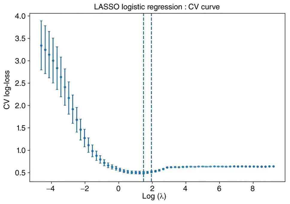
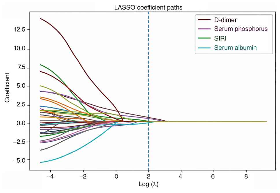
To identify candidate risk factors for treatment
failure, LASSO logistic regression with 10-fold cross-validation
was performed. Using the λ.1se rule, the optimal penalization
parameter was selected at λ.1se=7.038 (λ.min=4.406). At λ.1se, four
predictors retained non-zero coefficients and were therefore
selected for subsequent modeling: Serum albumin, serum phosphorus,
D-dimer and SIRI (Figs. 1 and
2).
Variables identified by LASSO regression, including
serum albumin, serum phosphorus, D-dimer and SIRI, were
subsequently incorporated into a GEE logistic regression model to
determine independent predictors of PDAP treatment failure. After
adjustment, higher serum phosphorus [odds ratio (OR)=3.16; 95% CI:
1.14-8.76; P=0.027], higher D-dimer (OR=3.57; 95% CI: 1.83-6.95;
P<0.001) and higher SIRI (OR=1.10; 95% CI: 1.05-1.15;
P<0.001) were independently associated with an increased risk of
treatment failure (Table
III).
| Table IIIGeneralized estimating equation
logistic regression analysis for predicting treatment failure in
peritoneal dialysis-associated peritonitis episodes. |
Table III
Generalized estimating equation
logistic regression analysis for predicting treatment failure in
peritoneal dialysis-associated peritonitis episodes.
| Variable | Odds ratio | 95% CI | P-value |
|---|
| Serum
phosphorus | 3.16 | 1.14-8.76 | 0.0266 |
| D-dimer | 3.57 | 1.83-6.95 | 0.0002 |
| SIRI | 1.10 | 1.05-1.15 | 0.0001 |
| Serum albumin | 0.91 | 0.82-1.01 | 0.0638 |
Predictive factors for treatment
failure in patients with PDAP
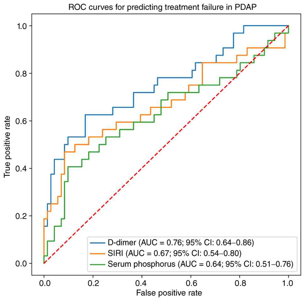
ROC curve analysis showed that the D-dimer level
achieved an area under the curve (AUC) value of 0.76 (95% CI:
0.64-0.86) for predicting treatment failure in PDAP. At the optimal
cutoff value of 1.39, the sensitivity was 62.5% and the specificity
was 83.1%, indicating an improved predictive performance compared
with SIRI (AUC=0.67; 95% CI: 0.55-0.80) and serum phosphorus
(AUC=0.64; 95% CI: 0.51-0.76). The corresponding ROC curves are
shown in Fig. 3.
Discussion
Elevated D-dimer, SIRI and serum phosphorus levels
were demonstrated to be independent predictors of treatment failure
in PDAP, with D-dimer level showing the strongest predictive value
in the present study.
Peritonitis is one of the most severe complications
of PD. Despite extensive investigation, to the best of our
knowledge, no universally accepted predictor of treatment failure
in PDAP has been established. A previous investigation indicated
that numerous parameters, including dialysis vintage, serum albumin
level, dialysate white blood cell count on day 5 and the causative
microorganism, may hold prognostic value for PDAP outcomes
(11). However, additional studies
have shown that these factors do not consistently predict treatment
outcomes (5,12). Previously, a number of hematologic
inflammation-based markers, including the neutrophil-to-lymphocyte
ratio (13), systemic
immune-inflammation index (14)
and SIRI (15) have been explored
as potential prognostic indicators of infectious and inflammatory
diseases.
D-dimer level is a biomarker reflecting a
hypercoagulable state and secondary fibrinolysis (16). Numerous studies have indicated that
coagulation parameters are associated with inflammatory responses
and may therefore serve as valuable markers of disease severity and
prognosis in infectious conditions (17-20).
In severe infections, endothelial damage triggers the exposure of
tissue factors and activates the extrinsic coagulation cascade,
triggering secondary fibrinolysis and generation of D-dimer as a
degradation product (21).
Concurrently, endothelial anticoagulant activity decreases, while
platelet activation and aggregation increase, ultimately leading to
microthrombus formation (22).
Theoretically, microvascular thrombosis may impair peritoneal
tissue perfusion and disturb microcirculatory homeostasis by
reducing capillary blood flow, inducing local hypoxia, promoting
endothelial dysfunction and triggering inflammatory responses
(23,24). Thereby limiting solute transfer and
reducing the delivery of antimicrobial agents and immune effector
cells to the infection site. Impaired clearance of metabolic
byproducts may further aggravate local tissue damage and microbial
growth, mechanisms similar to those previously described in
microcirculatory dysfunction during intra-abdominal
infection-associated sepsis (25).
In the present study, PDAP episodes with treatment failure
exhibited significantly higher D-dimer levels compared with those
with treatment success. GEE logistic regression analysis
demonstrated that D-dimer independently predicted treatment failure
in PDAP episodes. ROC curve analysis yielded an AUC of 0.76, with
an optimal cutoff value of 1.39, corresponding to 62.5% sensitivity
and 83.1% specificity, underscoring its potential clinical value
for the early identification of patients at an increased risk of
treatment failure.
SIRI is an integrated biomarker derived from
peripheral neutrophil, monocyte and lymphocyte counts (26). It reflects the imbalance between
pro-inflammatory mediators (neutrophils and monocytes) and immune
regulatory elements (lymphocytes) (27). During severe inflammatory
responses, neutrophil and monocyte counts typically increase,
whereas lymphocyte levels decrease, resulting in an overall
increase in SIRI values (28).
Such elevation reflects intensified systemic inflammation, a higher
infectious burden and impaired host immune defense (29). Consequently, a high SIRI may
indicate a pathophysiological state associated with greater disease
severity and a higher probability of therapeutic failure. Previous
evidence has demonstrated that SIRI is associated with all-cause
mortality among patients receiving PD (30). In addition, increased SIRI has been
identified as an independent predictor of both all-cause mortality
and cardiovascular mortality in patients with chronic kidney
disease (31). In the present
study, SIRI remained an independent determinant of treatment
failure in PDAP. Specifically, each one-unit increment in SIRI
corresponded to an ~10% higher risk of treatment failure. ROC
analysis indicated that SIRI exhibited a moderate discriminatory
performance in predicting treatment outcomes (AUC=0.67). Therefore,
SIRI may be more appropriately incorporated as part of a composite
risk stratification model, rather than used as a solitary
prognostic marker.
A previous study demonstrated that elevated serum
phosphorus concentrations are independently associated with an
increased risk of all-cause mortality among patients receiving PD
(32). However, limited evidence
exists regarding the association between serum phosphorus
concentrations and PDAP. In the present study, GEE logistic
regression analysis indicated that elevated serum phosphorus was
associated with PDAP treatment failure (OR=3.16; 95% CI: 1.14-8.76;
P=0.027). Fibroblast growth factor-23 (FGF-23), a hormone primarily
secreted by osteocytes that regulates phosphate and vitamin D
metabolism (33-35),
has been shown to increase in response to hyperphosphatemia and act
to reduce serum phosphorus levels (36). However, FGF-23 may also impair
immune function. Specifically, it can inhibit the chemotaxis,
phagocytosis and bactericidal activity of neutrophils (37) and macrophages (38) which serve key roles in the early
immune response to peritoneal infections. Consequently, impairment
of these immune functions may hinder effective pathogen elimination
and reduce the effectiveness of infection control. In addition,
FGF-23 promotes the production of pro-inflammatory cytokines,
including IL-6, TNF-α and C-reactive protein (39), potentially exacerbating
inflammatory responses and further impairing infection control by
promoting immune cell dysfunction and suppressing vitamin
D-mediated antimicrobial pathways (39). Persistent hyperphosphatemia has
also been recognized as an important contributor to
microinflammation in patients with chronic kidney disease (40). It can stimulate the production of
pro-inflammatory cytokines, such as IL6, IL-1β and TNF-α in
vascular smooth muscle cells, endothelial cells and various other
cell types (39,41). Notably, the relatively wide 95% CI
observed for serum phosphorus suggests limited precision in the
effect estimate, likely associated with the small sample size and
limited number of treatment-failure events. Variability in
phosphorus levels and residual confounding inherent to the
retrospective design may also have contributed to this finding.
Therefore, this result should be interpreted cautiously and
validated in future larger multicenter studies.
Furthermore, numerous studies have reported that
hypoalbuminemia is associated with adverse outcomes in patients
undergoing PD, including an increased incidence of peritonitis
(42), a higher risk of technique
failure (17) and reduced
long-term survival (43). In the
present study, serum albumin demonstrated a borderline inverse
association with treatment failure (OR=0.91; P=0.064). Despite this
finding having not reached statistical significance in the GEE
logistic regression model, the observed trend suggests that serum
albumin may represent an important clinical factor. Possible
explanations for the present result include serum albumin levels
being influenced by both nutritional and inflammatory conditions
and indirectly affecting infection outcomes by modulating immune
responses through mechanisms including maintenance of oncotic
pressure, antioxidant activity, binding and transport of
inflammatory mediators, and support of immune cell function
(44,45). When stronger inflammatory markers
such as D-dimer and SIRI were included in the GEE regression
analysis, the independent predictive value of albumin may have been
partially attenuated, thereby diminishing its statistical
significance. Nevertheless, the consistent protective trend
observed in the present study suggests that nutritional status may
remain an important contributor to treatment outcomes in PDAP.
The present study exhibits a number of limitations.
First, it was a single-center retrospective study with a relatively
limited sample size, including 103 PDAP episodes from 72 patients.
The limited sample size may have reduced statistical power of the
analysis and contributed to the relatively wide CIs observed for
certain variables, such as serum phosphorus. Second, as a number of
patients experienced multiple episodes of peritonitis during the
present study period, the analysis was performed at the episode
level. Although this approach allowed the evaluation of
episode-specific risk factors, repeated episodes occurring within
the same patient may have introduced intra-patient association. In
the present study, GEE logistic regression was applied to account
for within-patient association. Despite this, residual association
among recurrent episodes may still have existed, and future studies
with larger sample sizes should aim to further explore more
advanced statistical approaches, such as mixed-effects models, to
better account for associations among recurrent episodes. Third,
although the number of variables included in the multivariable
model was limited and selected using LASSO regression, the
relatively small number of treatment failure events (n=32) may
still raise concerns regarding model stability. Although only four
predictors were included in the final model, the event-per-variable
(EPV) ratio was 8, slightly below the commonly recommended
threshold of ≥10 EPV for logistic regression models (46), thus the findings should be
interpreted cautiously and validated in larger studies.
In summary, the present retrospective study examined
elevated D-dimer, SIRI and serum phosphorus levels as independent
predictors of treatment failure in PDAP, with D-dimer demonstrating
the strongest predictive performance. These findings suggest that
readily available laboratory indicators reflecting coagulation and
systemic inflammation may provide useful information for the early
identification of PDAP episodes at increased risk of treatment
failure. Further large-scale multicenter prospective studies are
warranted to validate these findings and to develop integrated
predictive models incorporating inflammatory, coagulation, and
microbiological indicators to improve risk stratification and
clinical management of PDAP.
Acknowledgements
Not applicable.
Funding
Funding: The present study was supported by the Natural Science
Foundation of Chongqing, China (grant no.
CSTB2023NSCQ-MSX1103).
Availability of data and materials
The data generated in the present study may be
requested from the corresponding author.
Authors' contributions
YJ and YL conceived and designed the present study.
YJ, XL and TP collected, analyzed and interpreted the data. YJ
wrote the manuscript and YL provided critical revision. YJ and YL
confirm the authenticity of all the raw data. All authors read and
approved the final version of the manuscript.
Ethics approval and consent to
participate
The present study was conducted in accordance with
the Declaration of Helsinki and was approved by the Ethics
Committee of Yongchuan Hospital affiliated to Chongqing Medical
University (Chongqing, China; approval no. 2024EC0066).
Patient consent for publication
Not applicable.
Competing interests
The authors declare that they have no competing
interests.
References
|
1
|
Andreoli MCC and Totoli C: Peritoneal
dialysis. Rev Assoc Med Bras (1992). 66 (Suppl 1):S37–S44.
2020.PubMed/NCBI View Article : Google Scholar
|
|
2
|
Thongprayoon C, Wathanavasin W,
Suppadungsuk S, Davis PW, Miao J, Mao MA, Craici IM, Qureshi F and
Cheungpasitporn W: Assessing global and regional public interest in
home dialysis modalities from 2004 to 2024. Front Nephrol.
4(1489180)2024.PubMed/NCBI View Article : Google Scholar
|
|
3
|
Perl J, Brown EA, Chan CT, Couchoud C,
Davies SJ, Kazancioğlu R, Klarenbach S, Liew A, Weiner DE, Cheung
M, et al: Home dialysis: Conclusions from a Kidney Disease:
Improving Global Outcomes (KDIGO) Controversies Conference. Kidney
Int. 103:842–858. 2023.PubMed/NCBI View Article : Google Scholar
|
|
4
|
Cho Y, Cullis B, Ethier I, Htay H, Jha V,
Arruebo S, Caskey FJ, Damster S, Donner JA, Levin A, et al: Global
structures, practices, and tools for provision of chronic
peritoneal dialysis. Nephrol Dial Transplant. 39 (Suppl
1):ii18–ii25. 2024.PubMed/NCBI View Article : Google Scholar
|
|
5
|
Liu X, Qin A, Zhou H, He X, Cader S, Wang
S, Tang Y and Qin W: Novel predictors and risk score of treatment
failure in peritoneal Dialysis-related peritonitis. Front Med
(Lausanne). 8(639744)2021.PubMed/NCBI View Article : Google Scholar
|
|
6
|
Perl J, Davies SJ, Lambie M, Pisoni RL,
McCullough K, Johnson DW, Sloand JA, Prichard S, Kawanishi H,
Tentori F and Robinson BM: The peritoneal dialysis outcomes and
practice patterns study (PDOPPS): Unifying efforts to inform
practice and improve global outcomes in peritoneal dialysis. Perit
Dial Int. 36:297–307. 2016.PubMed/NCBI View Article : Google Scholar
|
|
7
|
Li PK, Chow KM, Cho Y, Fan S, Figueiredo
AE, Harris T, Kanjanabuch T, Kim YL, Madero M, Malyszko J, et al:
ISPD peritonitis guideline recommendations: 2022 update on
prevention and treatment. Perit Dial Int. 42:110–153.
2022.PubMed/NCBI View Article : Google Scholar
|
|
8
|
Li PKT, Szeto CC, Piraino B, de Arteaga J,
Fan S, Figueiredo AE, Fish DN, Goffin E, Kim YL, Salzer W, et al:
ISPD peritonitis recommendations: 2016 update on prevention and
treatment. Perit Dial Int. 36:481–508. 2016.PubMed/NCBI View Article : Google Scholar
|
|
9
|
Mao Y, Xiao D, Deng S and Xue S:
Development of a clinical risk score system for peritoneal
dialysis-associated peritonitis treatment failure. BMC Nephrol.
24(229)2023.PubMed/NCBI View Article : Google Scholar
|
|
10
|
Li J, Liu Y, Lu Y, Fu C, Ye Z and Zhang Z:
Dynamic changes in serum albumin and ferritin indicate higher risk
of early-onset peritonitis in peritoneal dialysis patients. Ther
Apher Dial: Jan 2, 2025 (Epub ahead of print). doi:
10.1111/1744-9987.14244.
|
|
11
|
Meng L, Zhu X, Yang L, Li X, Cheng S, Guo
S, Zhuang X, Zou H and Cui W: Development and validation of a
prediction model for treatment failure in peritoneal
dialysis-associated peritonitis patients: A multicenter study. Nan
Fang Yi Ke Da Xue Xue Bao. 42:546–553. 2022.PubMed/NCBI View Article : Google Scholar : (In Chinese).
|
|
12
|
Htay H, Cho Y, Pascoe EM, Darssan D,
Nadeau-Fredette AC, Hawley C, Clayton PA, Borlace M, Badve SV, Sud
K, et al: Center effects and peritoneal dialysis peritonitis
outcomes: Analysis of a national registry. Am J Kidney Dis.
71:814–821. 2018.PubMed/NCBI View Article : Google Scholar
|
|
13
|
Su N, Zheng Y, Zhang X, Tang X, Tang LW,
Wang Q, Chen X, Wang X, Wen Y, Feng X, et al:
Platelet-to-lymphocyte ratio and the first occurrence of
peritonitis in peritoneal dialysis patients. BMC Nephrol.
23(415)2022.PubMed/NCBI View Article : Google Scholar
|
|
14
|
Li G, Yu J, Jiang S, Wu K, Xu Y, Lu X,
Wang Y, Lin J, Yang X, Li Z, et al: Systemic immune-inflammation
index was significantly associated with all-cause and
cardiovascular-specific mortalities in patients receiving
peritoneal dialysis. J Inflamm Res. 16:3871–3878. 2023.PubMed/NCBI View Article : Google Scholar
|
|
15
|
Yang Y, Xu Y, Liu S, Lu P, Zhou H and Yang
M: The systemic inflammation indexes predict all-cause mortality in
peritoneal dialysis patients. Renal failure.
45(2160348)2023.PubMed/NCBI View Article : Google Scholar
|
|
16
|
Tripodi A and Di Micco P: How we manage a
high D-dimer. Haematologica. 109:933–943. 2024.PubMed/NCBI View Article : Google Scholar
|
|
17
|
Cerda-Mancillas MC, Santiago-Germán D,
Andrade-Bravo B, Pedraza-Olivares F, Valenzo-Hernández F,
Leaños-Miranda A and Isordia-Salas I: D-dimer as a biomarker of
severity and adverse outcomes in patients with community acquired
pneumonia. Arch Med Res. 51:429–435. 2020.PubMed/NCBI View Article : Google Scholar
|
|
18
|
Schupp T, Weidner K, Rusnak J, Jawhar S,
Forner J, Dulatahu F, Brück LM, Hoffmann U, Kittel M, Bertsch T, et
al: D-Dimer levels and the disseminated intravascular coagulation
score to predict severity and outcomes in sepsis or septic shock.
Clin Lab: May 1, 2023 (Epub ahead of print). doi:
10.7754/Clin.Lab.2022.221015.
|
|
19
|
Dumache R, Muresan CO, Laitin SMD,
Ivanovic N, Chisalita A, Herlo A, Marinescu A, Lazureanu EV and Cut
TG: COVID-19 organ injury pathology and D-Dimer expression
patterns: A retrospective analysis. Diagnostics (Basel).
15(1860)2025.PubMed/NCBI View Article : Google Scholar
|
|
20
|
Balaceanu LA and Dina I: D-dimers in
advanced liver cirrhosis: Useful biomarker or not? Am J Med Sci.
368:415–423. 2024.PubMed/NCBI View Article : Google Scholar
|
|
21
|
Fiusa MM, Carvalho-Filho MA,
Annichino-Bizzacchi JM and De Paula EV: Causes and consequences of
coagulation activation in sepsis: An evolutionary medicine
perspective. BMC Medicine. 13(105)2015.PubMed/NCBI View Article : Google Scholar
|
|
22
|
Doganyigit Z, Eroglu E and Akyuz E:
Inflammatory mediators of cytokines and chemokines in sepsis: From
bench to bedside. Hum Exp Toxicol.
41(9603271221078871)2022.PubMed/NCBI View Article : Google Scholar
|
|
23
|
Levi M and van der Poll T: Inflammation
and coagulation. Crit Care Med. 38 (Suppl 2):S26–S34.
2010.PubMed/NCBI View Article : Google Scholar
|
|
24
|
De Backer D, Orbegozo Cortés D, Donadello
K and Vincent JL: Pathophysiology of microcirculatory dysfunction
and the pathogenesis of septic shock. Virulence. 5:73–79.
2014.PubMed/NCBI View Article : Google Scholar
|
|
25
|
Iba T, Helms J and Levy JH: Sepsis-induced
coagulopathy (SIC) in the management of sepsis. Ann Intensive Care.
14(148)2024.PubMed/NCBI View Article : Google Scholar
|
|
26
|
Qi Q, Zhuang L, Shen Y, Geng Y, Yu S, Chen
H, Liu L, Meng Z, Wang P and Chen Z: A novel systemic inflammation
response index (SIRI) for predicting the survival of patients with
pancreatic cancer after chemotherapy. Cancer. 122:2158–2167.
2016.PubMed/NCBI View Article : Google Scholar
|
|
27
|
Wei Y, Wang T, Li G, Feng J, Deng L, Xu H,
Yin L, Ma J, Chen D and Chen J: Investigation of systemic
immune-inflammation index, neutrophil/high-density lipoprotein
ratio, lymphocyte/high-density lipoprotein ratio, and
monocyte/high-density lipoprotein ratio as indicators of
inflammation in patients with schizophrenia and bipolar disorder.
Front Psychiatry. 13(941728)2022.PubMed/NCBI View Article : Google Scholar
|
|
28
|
Agnello L, Giglio RV, Bivona G, Scazzone
C, Gambino CM, Iacona A, Ciaccio AM, Lo Sasso B and Ciaccio M: The
value of a complete blood count (CBC) for sepsis diagnosis and
prognosis. Diagnostics (Basel). 11(1881)2021.PubMed/NCBI View Article : Google Scholar
|
|
29
|
Wang Z, Zhang W, Chen L, Lu X and Tu Y:
Lymphopenia in sepsis: A narrative review. Crit Care.
28(315)2024.PubMed/NCBI View Article : Google Scholar
|
|
30
|
Li J, Li Y, Zou Y, Chen Y, He L, Wang Y,
Zhou J, Xiao F, Niu H and Lu L: Use of the systemic inflammation
response index (SIRI) as a novel prognostic marker for patients on
peritoneal dialysis. Ren Fail. 44:1227–1235. 2024.PubMed/NCBI View Article : Google Scholar
|
|
31
|
Gu L, Xia Z, Qing B, Wang W, Chen H, Wang
J, Chen Y, Gai Z, Hu R and Yuan Y: Systemic inflammatory response
index (SIRI) is associated with all-cause mortality and
cardiovascular mortality in population with chronic kidney disease:
Evidence from NHANES (2001-2018). Front Immunol.
15(1338025)2024.PubMed/NCBI View Article : Google Scholar
|
|
32
|
Lopes MB, Karaboyas A, Zhao J, Johnson DW,
Kanjanabuch T, Wilkie M, Nitta K, Kawanishi H, Perl J and Pisoni
RL: PDOPPS Steering Committee. Association of single and serial
measures of serum phosphorus with adverse outcomes in patients on
peritoneal dialysis: Results from the international PDOPPS. Nephrol
Dial Transplant. 38:193–202. 2023.PubMed/NCBI View Article : Google Scholar
|
|
33
|
Liu CT, Lin YC, Lin YC, Kao CC, Chen HH,
Hsu CC and Wu MS: Roles of serum calcium, phosphorus, PTH and ALP
on mortality in peritoneal dialysis patients: A nationwide,
population-based longitudinal study. Sci Rep. 7(33)2017.PubMed/NCBI View Article : Google Scholar
|
|
34
|
Huo Z, Liu D, Ye P, Zhang Y, Cao L, Gong
N, Dou X, Ren C, Zhu Q, Li D, et al: Longer serum phosphorus time
in range is associated with lower mortality risk among peritoneal
dialysis patients: A multicenter cohort study. BMC Nephrol.
25(117)2024.PubMed/NCBI View Article : Google Scholar
|
|
35
|
White KE and Ix JH: Regulation of FGF23
production and phosphate metabolism. Nat Rev Nephrol. 19:185–193.
2023.PubMed/NCBI View Article : Google Scholar
|
|
36
|
Fitzpatrick EA, Han X, Xiao Z and Quarles
LD: Role of fibroblast growth Factor-23 in innate immune responses.
Front Endocrinol (Lausanne). 9(320)2018.PubMed/NCBI View Article : Google Scholar
|
|
37
|
Rossaint J, Oehmichen J, Van Aken H,
Reuter S, Pavenstädt HJ, Meersch M, Unruh M and Zarbock A: FGF23
signaling impairs neutrophil recruitment and host defense during
CKD. J Clin Invest. 126:962–974. 2016.PubMed/NCBI View
Article : Google Scholar
|
|
38
|
Han X, Li L, Yang J, King G, Xiao Z and
Quarles LD: Counter-regulatory paracrine actions of FGF-23 and
1,25(OH)2 D in macrophages. FEBS Lett. 590:53–67. 2016.PubMed/NCBI View Article : Google Scholar
|
|
39
|
Raju S and Saxena R: Hyperphosphatemia in
kidney failure: Pathophysiology, challenges, and critical role of
phosphorus management. Nutrients. 17(1587)2025.PubMed/NCBI View Article : Google Scholar
|
|
40
|
Yamada S, Tokumoto M, Tatsumoto N,
Taniguchi M, Noguchi H, Nakano T, Masutani K, Ooboshi H, Tsuruya K
and Kitazono T: Phosphate overload directly induces systemic
inflammation and malnutrition as well as vascular calcification in
uremia. Am J Physiol Renal Physiol. 306:F1418–F1428.
2014.PubMed/NCBI View Article : Google Scholar
|
|
41
|
Voelkl J, Lang F, Eckardt KU, Amann K,
Kuro OM, Pasch A, Pieske B and Alesutan I: Signaling pathways
involved in vascular smooth muscle cell calcification during
hyperphosphatemia. Cell Mol Life Sci. 76:2077–2091. 2019.PubMed/NCBI View Article : Google Scholar
|
|
42
|
Zha D, Yang X and Xi H: Association of
hypoalbuminemia with the risk of peritoneal dialysis-associated
peritonitis in peritoneal dialysis patients: A meta-analysis. Blood
Purif: Feb 10, 2025 (Epub ahead of print). doi:
10.1159/000543693.
|
|
43
|
Zhang L, Cao T, Li Z, Wen Q, Lin J, Zhang
X, Guo Q, Yang X, Yu X and Mao H: Clinical outcomes of peritoneal
dialysis patients transferred from hemodialysis: A matched
case-control study. Perit Dial Int. 33:259–266. 2013.PubMed/NCBI View Article : Google Scholar
|
|
44
|
Arques S: Human serum albumin in
cardiovascular diseases. Eur J Intern Med. 52:8–12. 2018.PubMed/NCBI View Article : Google Scholar
|
|
45
|
Soeters PB, Wolfe RR and Shenkin A:
Hypoalbuminemia: Pathogenesis and clinical significance. JPEN J
Parenter Enteral Nutr. 43:181–193. 2019.PubMed/NCBI View Article : Google Scholar
|
|
46
|
van Smeden M, Moons KGM, de Groot JAH,
Collins GS, Altman DG, Eijkemans MJ and Reitsma JB: Sample size for
binary logistic prediction models: Beyond events per variable
criteria. Stat Methods Med Res. 28:2455–2474. 2018.PubMed/NCBI View Article : Google Scholar
|