Introduction
Non-Hodgkin lymphoma (NHL) is a heterogeneous
malignancy of the lymphatic system, and dozens of subtypes have
been identified to date (1). With
the aging of the global population, the incidence burden of NHL is
expected to increase substantially, with the number of newly
diagnosed cases projected to rise from ~545,000 cases in 2020 to
778,000 cases by 2040(2). Among
patients with NHL, 30-40% are of the most prevalent subtype,
diffuse large B-cell lymphoma (DLBCL) (3). The incidence of DLBCL varies
geographically and increases substantially with age, ranging from
~2.3 to 13.8 cases per 100,000 person-years (4). Elderly individuals, particularly
those aged >60 years, are more frequently affected (5). Gene damage, epigenetic instability
and long-term immunological activation all contribute to its
etiology (6). Together, these
elements alter interactions within the tumor microenvironment,
B-cell differentiation and survival signal transduction. A notable
portion of patients continue to develop primary resistance or
relapse despite marked advancements in molecular profiling
(7). Rituximab with
cyclophosphamide, doxorubicin, vinblastine and prednisone is the
standard first-line treatment (8).
For high-risk disease subgroups, such as patients with aggressive
biological characteristics or a poor response to
immunochemotherapy, the results are still disappointing (9).
A notable factor influencing stress adaptation in
cancer is autophagy, a conserved lysosome-dependent catabolic
process (10). Autophagy promotes
metabolic flexibility, limits oxidative stress and modifies
immune-related signals by removing damaged organelles and protein
aggregates (11). Impaired
autophagy has been associated with worse outcomes following
immunochemotherapy in DLBCL. According to Xu et al (12) the paired box 5-driven long
non-coding RNA (lncRNA) ARRDC1-AS1 enhances autophagy by regulating
autophagy-related gene 5 (ATG5) through the sequestration of
microRNA-2355-5p, thereby promoting the development of DLBCL.
Furthermore, research suggests that the combination of rituximab
and chidamide might reduce treatment resistance by increasing the
expression of autophagy-associated proteins (13). By specifically regulating the
stability of carcinogenic signaling proteins, autophagy can also be
involved in the biological processes of DLBCL. In the MCD subtype
of DLBCL, characterized by MYD88L265P and CD79B
mutations, TANK-binding kinase 1 (TBK1)-dependent selective
autophagy can target and degrade the ubiquitinated
MYD88L265P protein, and bruton tyrosine kinase (BTK)
inhibitors appear to enhance this autophagic clearance, potentially
leading to therapeutic benefits (14). All of these outcomes point to
autophagy as a therapeutically useful and physiologically marked
process in DLBCL.
A fundamental member of the ribosomal P protein
family, ribosomal protein lateral stalk subunit P0 (RPLP0),
is necessary for ribosome stability and translational elongation
(15). There is growing evidence
that numerous cancer types, such as lung and prostate cancer,
exhibit abnormal expression of RPLP0 (16,17).
The clinical prognosis and tumor progression are intimately linked
to this imbalance. Reactive oxygen species (ROS) accumulate as a
result of inhibition of RPLP family proteins, which initiates
autophagy, reduces proliferation and induces G2 phase
arrest, according to findings (18). These findings suggest that
RPLP0-associated translational processes regulate autophagy
and redox homeostasis in cancer cells. The PI3K/AKT/mTOR signaling
cascade is a key oncogenic hub that integrates cellular stress,
growth factor signals and nutrient availability (19). While PI3K/AKT/mTOR activation
typically inhibits autophagic activity, pathway inhibition might
increase autophagic flux and encourage cell death. The data
presented suggest that RPLP0 may influence autophagy in
DLBCL via the PI3K/AKT/mTOR pathway, with implications for
biomarker development and patient stratification.
DLBCL may develop due to oxidative stress and
altered mitochondrial activity, according to prior research
(20,21). The autophagy system helps maintain
cellular homeostasis under stressful conditions. Moreover, the
AKT/mTOR signaling pathway is known to regulate cell survival and
autophagy in response to environmental cues. The aim of the present
study was to clarify the role of RPLP0 in regulating these
cellular processes in DLBCL. Based on the aforementioned evidence,
we hypothesized that RPLP0 promotes DLBCL cell survival by
maintaining redox homeostasis and activating the AKT/mTOR pathway,
thereby suppressing stress-induced autophagy. Investigating these
pathways will shed light on the molecular causes of DLBCL and may
reveal potential therapeutic targets.
Materials and methods
Data selection and differential
expression analysis
The Gene Expression Omnibus Database (https://www.ncbi.nlm.nih.gov/gds/) and The Cancer
Genome Atlas (TCGA) database (https://www.cancer.gov/ccg/research/genome-sequencing/tcga)
provided the gene expression data for this present investigation.
TCGA-DLBC dataset was selected, which includes 48 DLBCL tumor
samples from TCGA and 929 normal control samples from the
Genotype-Tissue Expression database (https://gtexportal.org/), and the GSE12453 dataset,
containing 11 tumor samples and 25 normal samples (22). Differential expression analysis of
genes in the two datasets was performed using the R package ‘Limma’
(version 3.54.2; Bioconductor; T Core Team) (23). Genes were screened based on fold
change (FC), with FC>2 considered as upregulated differentially
expressed genes (DEGs) and FC<0.5 considered as downregulated
DEGs, with a significance criterion of P<0.05. Intersection
analysis was then performed on the upregulated and downregulated
DEGs to obtain key intersection genes utilizing Bioinformatics
& Evolutionary Genomics (https://bioinformatics.psb.ugent.be/webtools/Venn/).
Protein-protein interaction (PPI)
network and selection of hub genes
To assess the potential functional relevance of key
intersecting genes, the authors analyzed the STRING database
(https://string-db.org/) to construct a PPI
network. The top 15 genes were further analyzed for their
interaction networks using the Module Clustering Coefficient (MCC),
Edge Percolated Component (EPC) and Maximum Neighborhood Component
(MNC) algorithms in the CytoHubba plugin of Cytoscape software
(version 3.10.1; Cytoscape Consortium). Subsequently, the top 15
genes from these three algorithms were subjected to intersection
analysis to obtain candidate genes. Then, gene expression analysis
of these candidate genes was performed on TCGA-DLBC and GSE12453
datasets using the Sanger website (http://vip.sangerbox.com/home.html). By analyzing gene
expression levels in the normal and tumor groups, the present study
identified the pivotal genes.
Cell culture
Human normal lymphoblastic GM12878 and DLBCL cell
lines (OCI-LY7 and U2932) were acquired from Changsha Abiwei
Biotechnology Co., Ltd. and the DLBCL cell line OCI-LY8 was
purchased from Shanghai Enzyme-linked Biotechnology Co., Ltd.
OCI-LY8 cells were cultured in DMEM medium (Gibco; Thermo Fisher
Scientific, Inc.). GM12878, OCI-LY7 and U2932 cells were cultured
under the same conditions in RPMI 1640 medium (Gibco; Thermo Fisher
Scientific, Inc.). In all media, 10% fetal bovine serum (Gibco;
Thermo Fisher Scientific, Inc.) and 1% penicillin-streptomycin
(Gibco; Thermo Fisher Scientific, Inc.) were included. The cells
were cultivated in an incubator with 5% CO2 at 37˚C for
24 h before subsequent experiments.
Cell transfection and treatment
Lipofectamine 2000 (Invitrogen; Thermo Fisher
Scientific, Inc.) was used for cell transfection in accordance with
the manufacturer's recommendations. For RPLP0 knockdown
studies, cells were transfected with three RPLP0-specific
small interfering RNAs (siRNAs): si-RPLP0-1 (sense,
5'-GCUAAGGUUGAAGCCAAGGAA-3'; and antisense,
5'-UUCCUUGGCUUCAACCUUAGC-3'), si-RPLP0-2 (sense,
5'-UGAUCAAGACUGGAGACAAAG-3' and antisense,
5'-CUUUGUCUCCAGUCUUGAUC-3') and si-RPLP0-3 (sense,
5'-GCUGCUGAACAUGCUCAACAU-3' and antisense,
5'-AUGUUGAGCAUGUUCAGCCAGC-3') or a non-targeting control siRNA
(si-NC; sense, 5'-UUCUCCGAACGUGUCACGUTT-3' and antisense,
5'-ACGUGACACGUUCGGAGAATT-3') at a final concentration of 50 nM. For
overexpression studies, cells were transfected with the
corresponding empty vector or a pcDNA3.1(+)-RPLP0 expression
plasmid generated using the pcDNA3.1(+) backbone (Invitrogen;
Thermo Fisher Scientific, Inc.) with 2 µg plasmid DNA used per well
in a 6-well plate. The plasmid construct was verified by DNA
sequencing. For subsequent investigations, cells were harvested 48
h after transfection.
DLBCL cells require special treatment for autophagy
studies. 3-Methyladenine (3-MA; Sigma-Aldrich; Merck KGaA) is an
autophagy inhibitor that acts in the early stages of the autophagy
pathway. To block autophagy, DLBCL cells were treated with 10 mM
3-MA for 48 h. Rapamycin (RAPA) is a typical mTOR inhibitor. DLBCL
cells were treated with 25 nM RAPA (Sigma-Aldrich; Merck KGaA) for
24 h for further analysis. A traditional ROS scavenger,
N-acetyl-L-cysteine (NAC; Sigma-Aldrich; Merck KGaA), lowers
intracellular ROS levels by directly interacting with ROS through
redox processes. To regulate ROS levels, DLBCL cells were treated
with 5 mM NAC for 2 h before subsequent assays.
Reverse transcription-quantitative PCR
(RT-qPCR)
Total RNA was extracted from OCI-LY7 and OCI-LY8
cells in the indicated treatment groups using TRIzol®
reagent (Takara Bio, Inc.). Important procedures, including qPCR
amplification, cDNA synthesis and RNA quality assessment, were
carried out in accordance with the techniques outlined in earlier
research (24). Specifically, cDNA
was synthesized using the PrimeScript RT reagent Kit with gDNA
Eraser (Takara Bio, Inc.). The reaction procedure included the
removal of genomic DNA at 42˚C for 2 min, followed by reverse
transcription at 37˚C for 15 min, and enzyme inactivation at 85˚C
for 5 sec. qPCR amplification was performed using TB Green Premix
Ex Taq II (Takara Bio, Inc.). The thermocycling conditions were as
follows: Initial denaturation at 95˚C for 30 sec, followed by 40
cycles of denaturation at 95˚C for 5 sec and annealing/extension at
60˚C for 30 sec. Primer sequences are as follows: RPLP0:
Forward: 5'-CGTCCTCGTGGAAGTGACAT-3', reverse:
5'-CTTGGAGCCCACATTGTCTG-3', GAPDH: forward:
5'-CATGTTGCAACCGGGAAGGA-3', reverse: 5'-ATCACCCGGAGGAGAAATCG-3'.
mRNA expression was normalized using the endogenous reference
GAPDH. The 2-ΔΔCq method
was employed for relative quantification (25).
Western blotting (WB)
RIPA lysis buffer (Beyotime Biotechnology)
containing phosphatase and protease inhibitors was used to extract
total protein from cells. Following determination of protein
content using a BCA Protein Assay Kit (Beyotime Biotechnology), in
accordance with procedures described in previous studies, 30 µg of
protein per lane was separated using 8-15% SDS-PAGE gels according
to the molecular weights of the target proteins and transferred
onto polyvinylidene fluoride membranes. The membranes were blocked
with 5% bovine serum albumin for 1 h at room temperature in
Tris-buffered saline containing 0.1% Tween-20 (TBST) (26). The membranes were incubated
overnight with the primary antibody at 4˚C. The following are the
primary antibodies utilized in this investigation: RPLP0 (cat. no.
ab192866; 1:1,000), thioredoxin (TXN; cat. no. ab133524; 1:10,000),
heat shock protein family A member 1A (HSPA1A; cat. no. ab5439;
1:1,000), heat shock protein family B member 1 (HSPB1; cat. no.
ab2790; 1:1,000), microtubule-associated protein 1 light chain 3
alpha (MAP1LC3A; cat. no. ab52628; 1:50,000), ATG5 (cat. no.
ab108327; 1:1,000), cleaved caspase-3 (cat. no. ab32351; 1:2,000),
caspase-3 (cat. no. ab184787; 1:2,000), phosphorylated (p)-AKT1
(cat. no. 66444-1-Ig; 1:2,000), AKT1 (cat. no. 10176-2-AP;
1:2,000), p-mTOR (cat. no. ab109268; 1:1,000), mTOR (cat. no.
ab109268; 1:1,000) and GAPDH (cat. no. ab181602; 1:10,000). Among
them, p-AKT1 and AKT1 antibody were derived from Wuhan Sanying
Biotechnology. By contrast, the other primary antibodies were
derived from Abcam. Following three washes with TBST containing
0.1% Tween-20, the membrane was incubated for 1 h at room
temperature with goat anti-rabbit (cat. no. ab6721; 1:2,000; Abcam)
and anti-mouse secondary antibodies (cat. no. ab6789; 1:2,000;
Abcam) conjugated to horseradish peroxidase. Immobilon Western
Chemiluminescent HRP Substrate (Merck KGaA) was used for
chemiluminescent detection. Image Lab software (version 6.1;
Bio-Rad Laboratories, Inc.). was installed to measure band
intensity.
Colony formation assay
OCI-LY7 and OCI-LY8 cells were transfected with
si-NC or si-RPLP0-1. Following transfection, cells in the
logarithmic growth phase were collected, resuspended in complete
culture medium, and seeded into 6-well plates at a density of 1x10³
cells per well. The cells were cultured at 37˚C in a humidified
atmosphere containing 5% CO2 for 14 days, with the
culture medium replaced every 3 days. After incubation, 4%
paraformaldehyde was added to fix the cells for 15 min. After
fixation, colonies were stained with nitroblue tetrazolium chloride
for 30 min at room temperature. A cell cluster containing at least
50 cells was defined as a colony. Images were acquired using a
chemiluminescence imaging system (Bio-Rad Laboratories, Inc.), and
the colonies were counted using ImageJ software (version 1.53;
National Institutes of Health).
Cell Counting Kit-8 (CCK-8) assay
96-well plates were seeded with DLBCL cells at a
density of 1,000 cells per well. After cell collection, 10 µl CCK-8
reagent (Dojindo Laboratories, Inc.) was added to 100 µl culture
medium. After 2 h incubation at 37˚C, an iMark™
Microplate Absorbance Reader (Bio-Rad Laboratories, Inc.) was
employed to quantify the optical density (OD) at 450 nm to identify
cell growth.
Flow cytometry
As directed by the manufacturer, apoptosis was
measured with an Annexin V-FITC/PI apoptosis detection kit (cat.
no. ab14085; Abcam). Briefly, OCI-LY7 and OCI-LY8 cells were
collected, cleaned in cold PBS and then reconstituted in 500 µl of
1X binding buffer. Annexin V-FITC (5 µl) was used to stain the
cells at room temperature for 10 min in the dark, followed by PI (5
µl) for 5 min. Samples were analyzed on a FACSCalibur flow
cytometer (BD Biosciences). Flow cytometric data were analyzed
using FlowJo software (version 10.8.1; BD Biosciences).
For cell cycle analysis, cells were collected and
fixed in pre-chilled 70% ethanol at -20˚C overnight. Following
fixation, cells were washed with PBS, treated with RNase A (100
µg/ml), and stained with PI (50 µg/ml) for 30 min in the dark. A
FACSCalibur flow cytometer was used to quantify DNA content, and
the proportions of cells in different cell cycle phases were
analyzed using FlowJo software (version 10.8.1; BD
Biosciences).
Mitochondrial membrane potential (MMP)
assay
A JC-1 staining kit (cat. no. ab113850; Abcam) was
used to assess MMP in OCI-LY7 and OCI-LY8 cells according to the
manufacturer's instructions. Cells were cultured with the JC-1
working solution for 20 min at 37˚C in the dark after the specified
treatments. Following incubation, the cells were immediately
examined under a fluorescence microscope (Nikon Corporation) after
being cleaned with PBS. Red JC-1 aggregates indicate intact ΔΨm,
whereas increased green JC-1 monomers reflect mitochondrial
depolarization.
ROS level assay
2',7'-dichlorodihydrofluorescein diacetate (DCFH-DA;
Beyotime Biotechnology) was used to measure intracellular ROS
levels in accordance with the manufacturer's directions. OCI-LY7
and OCI-LY8 cells were transfected with si-RPLP0-1 or si-NC,
seeded on coverslips in 6-well plates, and incubated under standard
culture conditions. Following the specified incubation period,
cells were treated with DCFH-DA solution (10 µM) for 20 min in the
dark at 37˚C after being rinsed with serum-free media. Excess probe
was removed by washing three times with serum-free medium. DAPI (1
µg/ml) for 5 min was used to counterstain the nuclei, and a
fluorescence microscope (Nikon Corporation) was used to capture
fluorescence images rapidly.
Transmission electron microscope
(TEM)
Cells with si-NC or si-RPLP0-1 were collected
and fixed in 2.5% glutaraldehyde overnight at 4˚C. Following a
phosphate buffer wash, cells were dehydrated by a graded ethanol
series, post-fixed for 1 h with 1% osmium tetroxide, and then
embedded in epoxy resin. The resin blocks were polymerized at 60˚C
for 48 h before ultrathin sectioning. After cutting and mounting
ultrathin slices (70 nm thick) on copper grids, they were
sequentially stained with 2% uranyl acetate for 15 min and lead
citrate for 10 min at room temperature. Mitochondrial morphology
was observed with an HT7700 TEM (Hitachi Ltd.).
Determination of NAD+/NADH
ratio
Intracellular NAD+/NADH ratios were
measured using a commercial NAD+/NADH Assay Kit (cat.
no. S0175, Beyotime Biotechnology) as directed by the manufacturer.
Briefly, cells were harvested and lysed on ice in the
NAD+/NADH extraction buffer provided in the kit. The
supernatants were collected after centrifuging the lysates at
12,000 x g for 10 min at 4˚C. For total NAD (NAD+ +
NADH) measurement, an aliquot of each extract was directly
subjected to the enzymatic cycling reaction. To assess NADH, a
parallel aliquot was chilled on ice after a 30 min incubation at
60°C to break down NAD+. Reaction mixtures
were prepared according to the kit protocol, and absorbance at 450
nm was measured using an iMark microplate reader (Bio-Rad
Laboratories, Inc.). NAD+ levels were assessed as total
NAD minus NADH, and the NAD+/NADH ratio was computed as
(total NAD-NADH)/NADH. Values were normalized to equal cell numbers
(or total protein) across samples.
Statistical analysis
The R program (version 4.1.0; Posit Software, PBC)
was used for the statistical analysis. Statistical significance was
defined as P<0.05. Differences between groups were assessed
using an unpaired Student's t-test (two groups), and one-way ANOVA
with Tukey's post hoc test were employed to compare multiple
groups. Each experiment was conducted at least three times, and the
mean ± SD was used to describe the results.
Results
Identification and characterization of
hub overlapping DEGs in DLBCL
In the present study, 6,338 upregulated and 1,115
downregulated DEGs were first identified from TCGA-DLBCL dataset
(Fig. 1A). Additionally, from the
GSE12453 dataset, 604 upregulated and 18 downregulated DEGs were
detected (Fig. 1B). Venn diagram
analysis was performed to identify overlapping DEGs between the two
datasets, revealing 396 upregulated DEGs and 0 downregulated DEGs
(Fig. 1C and D). PPI network analyses of these
overlapping upregulated DEGs were conducted using the MCC, MNC and
EPC algorithms. This analysis identified the top 15 most highly
associated genes for each algorithm (Fig. 1E-G). Among these, six candidate
genes (RPS11, RPL27, RPS18, RPS5,
RPLP0 and RPS3) were selected as potential biomarkers
(Fig. 1H). Expression analysis of
these candidate genes in DLBCL-related datasets revealed that,
compared with normal controls, all six genes exhibited notably
higher expression in the tumor group (Fig. 1I and J). The present investigation examined
RPLP0 because it is an essential component of the large
ribosomal subunit and vital for protein synthesis.
 | Figure 1Overlapping DEGs in DLBCL found in
the GSE12453 and TCGA-DLBCL datasets. (A) A volcano plot shows the
upregulated (orange) and downregulated (green) DEGs from TCGA-DLBCL
dataset. (B) The volcano plot shows the upregulated (orange) and
downregulated (green) DEGs found in the GSE12453 dataset. (C) Venn
diagram showing the overlap of upregulated DEGs between TCGA-DLBCL
and GSE12453 datasets. (D) Venn diagram illustrating the overlap of
downregulated DEGs between the two datasets. PPI network analysis
of the overlapping upregulated DEGs. The top 15 genes were selected
using the (E) MCC, (F) MNC and (G) EPC algorithms. (H) The six
candidate genes were identified from the MCC, MNC and EPC
algorithms. (I and J) Expression of the six candidate genes
(RPS11, RPL27, RPS18, RPS5,
RPLP0 and RPS3) in DLBCL and normal groups in the (I)
TCGA-DLBCL dataset and (J) GSE12453 datasets.
****P<0.0001. DLBCL, diffuse large B-cell lymphoma;
DEGs, differentially expressed genes; PPI, protein-protein
interaction; MCC, maximal clique centrality; MNC, maximum
neighborhood component; EPC, edge percolated component; TCGA, The
Cancer Genome Atlas. |
Expression and knockdown efficiency of
RPLP0 in DLBCL cell lines
The GM12878 cell line and DLBCL cell lines (OCI-LY7,
OCI-LY8 and U2932) were subjected to RT-qPCR and WB analyses to
assess RPLP0 levels in DLBCL. The results showed that RPLP0
expression was elevated in DLBCL cell lines, with OCI-LY7 and
OCI-LY8 cells exhibiting the highest levels (Fig. 2A-C). Three distinct siRNA
constructs targeting RPLP0 were transfected into OCI-LY7 and
OCI-LY8 cells to investigate the function of RPLP0.
RPLP0 knockdown markedly reduced RPLP0 mRNA and protein
levels by RT-qPCR and WB analyses, with si-RPLP0-1 showing
the most effective knockdown (Fig.
2D-H).
 | Figure 2Expression and knockdown efficiency
of RPLP0 in DLBCL cell lines. (A) RT-qPCR analysis of
RPLP0 expression in the GM12878 (normal B cell), OCI-LY7,
OCI-LY8 and U2932 cell lines. (B and C) WB analysis of RPLP0
expression in the GM12878 (normal B cell), OCI-LY7, OCI-LY8 and
U2932 cell lines. Knockdown of RPLP0 using three different
siRNA constructs (si-RPLP0-1, si-RPLP0-2 and
si-RPLP0-3) in (D) OCI-LY7 and (E) OCI-LY8 cells. The
efficiency of RPLP0 knockdown was assessed by RT-qPCR. The
x-axis represents the siRNA constructs used (si-RPLP0-1,
si-RPLP0-2 and si-RPLP0-3), and the y-axis shows
relative mRNA levels. (F) The efficiency of RPLP0 knockdown
was assessed by WB. The y-axis shows relative protein levels in (G)
OCI-LY7 and (H) OCI-LY8. *P<0.05,
**P<0.01 and ***P<0.001. DLBCL, diffuse
large B-cell lymphoma; RT-qPCR, reverse transcription-quantitative
PCR; WB, western blot; si, small interfering RNA; NC, negative
control. |
RPLP0 knockdown disrupts
proliferation, cell cycle and apoptosis in DLBCL cell lines
Colony formation assays assessed the effects of
RPLP0 knockdown on cell proliferation. The findings showed
that, compared with the control group, silencing RPLP0
reduced the ability of OCI-LY7 and OCI-LY8 cells to proliferate
(Fig. 3A and B). The effects of RPLP0 knockdown
on the cell cycle distribution of OCI-LY7 and OCI-LY8 cells were
next evaluated by flow cytometry. The outcomes revealed a
concurrent build-up of cells in the G2 phase and a
notable decline in the proportion of cells in the S phase (Fig. 3C and D). Additionally, apoptosis experiments
were conducted to determine if RPLP0 knockdown affected cell
death. However, there were no appreciable variations in the rates
of apoptosis between the si-NC control group and the RPLP0
knockdown group in DLBCL cell lines (Fig. 3E and F). These outcomes imply that RPLP0
mainly controls DLBCL cell proliferation through cell cycle
modulation.
RPLP0 knockdown disrupts redox
homeostasis and induces mitochondrial dysfunction in DLBCL
cells
The impact of RPLP0 knockdown on oxidative
damage repair markers in DLBCL cell lines was further examined.
According to WB analysis, si-RPLP0-1 markedly decreased TXN,
HSPA1A and HSPB1 protein expression compared with the control group
(Fig. 4A-C). This is supported by
the notable increase in the NAD+/NADH ratio following
RPLP0 knockdown (Fig. 4D).
Next, the authors utilized JC-1 staining to measure MMPs. Cells
with silenced RPLP0 showed decreased JC-1 aggregates and
increased JC-1 monomers, indicating mitochondrial depolarization.
Quantitative analysis of the red/green fluorescence ratio further
confirmed a marked reduction in membrane potential compared with
the si-NC group (Fig. 4E-F).
Additionally, the si-RPLP0-1 group exhibited greater green
fluorescence intensity in DCFH-DA staining, and quantitative
analysis of ROS fluorescence intensity revealed a marked increase,
suggesting enhanced intracellular ROS accumulation (Fig. 4G-H). These outcomes imply that
RPLP0 deficiency worsens mitochondrial function and
oxidative damage in DLBCL cells.
 | Figure 4Effects of RPLP0 knockdown on
oxidative stress and mitochondrial function in OCI-LY7 and OCI-LY8
cell lines. (A) Representative western blotting images showing the
expression of TXN, HSPA1A and HSPB1 in OCI-LY7 and OCI-LY8 cells
following RPLP0 knockdown. GAPDH was used as the loading
control. (B) Quantitative analysis of TXN, HSPA1A and HSPB1 protein
expression in OCI-LY7 cells. (C) Quantitative analysis of TXN,
HSPA1A and HSPB1 protein expression in OCI-LY8 cells. (D)
Measurement of the NAD+/NADH ratio in OCI-LY7 and
OCI-LY8 cells following si-RPLP0-1 transfection. The x-axis
represents the treatment groups (si-NC vs. si-RPLP0-1) and
the y-axis shows the NAD+/NADH ratio. (E) Mitochondrial
membrane potential analysis by JC-1 staining in
RPLP0-knockdown OCI-LY7 and OCI-LY8 cell. Scale bar: 50 µm.
(F) Quantitative analysis of the JC-1 red/green fluorescence ratio.
(G) ROS levels in OCI-LY7 and OCI-LY8 cells following
si-RPLP0-1 transfection, measured by
7'-dichlorodihydrofluorescein diacetate fluorescence. Scale bar, 50
µm. (H) Quantitative analysis of relative reactive oxygen species
fluorescence intensity. *P<0.05,
**P<0.01 and ***P<0.001. ROS, reactive
oxygen species; si, small interfering RNA; NC, negative control;
RPLP0, ribosomal protein lateral stalk subunit P0; TXN,
thioredoxin; HSPA1A, heat shock protein family A member 1A; HSPB1,
heat shock protein family B member 1. |
RPLP0 deletion alters autophagy
regulation and mitochondrial morphology in DLBCL cells
The transformation of LC3-I into LC3-II is a crucial
stage in the creation of autophagosomes. In OCI-LY7 and OCI-LY8
cells transfected with si-RPLP0-1, the protein levels of LC3
and ATG5 were measured using WB to investigate the effect of
RPLP0 knockdown on autophagy. The present findings
demonstrated that RPLP0 knockdown increased the LC3-II/I
ratio and upregulated ATG5 protein expression in both cell lines
(Fig. 5A-C). TEM was subsequently
used to examine the mitochondrial morphology of
RPLP0-silenced cells. The findings revealed notable signs of
autophagy, including an increase in the number of autophagic
vacuoles in the field of view and quantification confirmed a
notable increase in the number of autolysosomes per field (Fig. 5D and E). Next, the autophagy inhibitor 3-MA (5
mM) was added to RPLP0-silenced OCI-LY7 and OCI-LY8 cells.
According to WB analysis, RPLP0 knockdown increased the
LC3-II/I ratio but had no discernible effect on caspase-3
expression. Nevertheless, 3-MA treatment reversed the effects of
RPLP0 knockdown, obviously reducing the LC3-II/I ratio and
upregulating cleaved caspase-3 expression (Fig. 5F-H). The present findings suggest
that RPLP0 knockdown increases autophagy in DLBCL cells by
modulating ATG5 and LC3-II expression.
 | Figure 5Impact of RPLP0 knockdown on
autophagy markers, mitochondrial morphology and the effects of 3-MA
treatment in OCI-LY7 and OCI-LY8 cells. (A) WB analysis of
autophagy-related proteins LC3 and ATG5 in OCI-LY7 and OCI-LY8
cells transfected with si-RPLP0-1. (B) Quantitative analysis
of the LC3-II/LC3-I ratio in OCI-LY7 and OCI-LY8 cells. (C)
Quantitative analysis of ATG5 protein expression in OCI-LY7 and
OCI-LY8 cells. (D) Transmission electron microscopy images showing
increased autophagic vacuoles in RPLP0-silenced cells
compared with controls, with (E) quantitative analysis of the
number of autolysosomes per field. Scale bar, 2 µm. (F) WB analysis
of LC3-I, LC3-II, cleaved Caspase-3 and Caspase-3 expression in
OCI-LY7 and OCI-LY8 cells treated with 3-MA (5 mM) following
si-RPLP0-1 transfection. (G) Quantitative analysis of the
LC3-II/LC3-I ratio in the indicated treatment groups. (H)
Quantitative analysis of Caspase-3 protein expression in the
indicated treatment groups. *P<0.05,
**P<0.01, ***P<0.001,
****P<0.0001 and ns, not significant. WB, western
blotting; 3-MA, 3-methyladenine; RPLP0, ribosomal protein
lateral stalk subunit P0; si, small interfering RNA; NC, negative
control; ATG5, autophagy-related gene 5. |
Overexpression of RPLP0 activates
AKT/mTOR signaling and inhibits autophagy in DLBCL cells
The transfection efficiency of the RPLP0
overexpression plasmid was evaluated via RT-qPCR and WB in
subsequent experiments. Investigations demonstrated that levels of
RPLP0 mRNA and protein expression of both cell lines were
markedly increased after RPLP0 overexpression (Fig. 6A-C). The effects of RPLP0
overexpression on total and phosphorylated AKT and mTOR were
examined by WB analysis. The present findings indicated that
although there were no appreciable changes in the overall
expression levels of AKT and mTOR, RPLP0 overexpression
increased the levels of p-AKT and p-mTOR (Fig. 6D-F). According to these data,
RPLP0 may trigger the AKT/mTOR pathway in DLBCL cells. The
impact of RPLP0 overexpression and the mTOR inhibitor RAPA
(25 nM) on LC3 and ATG5 expression in OCI-LY7 and OCI-LY8 cells was
further investigated. The present results demonstrated that
RPLP0 overexpression reduced the LC3-II/I ratio and
decreased ATG5 levels. However, co-treatment with RAPA partially
reversed these effects, markedly restoring the LC3-II/I ratio and
ATG5 protein levels (Fig. 6G-I).
These results indicate that RPLP0 overexpression activates
the AKT/mTOR pathway, thereby inhibiting stress-induced autophagy
in DLBCL cells.
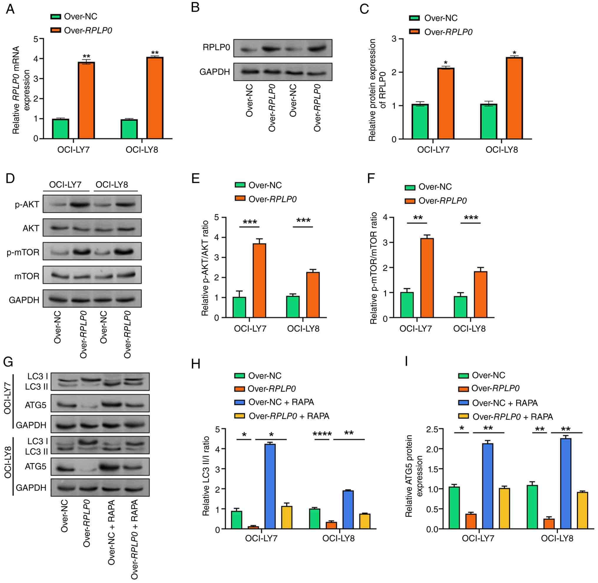 | Figure 6Effects of RPLP0
overexpression on the AKT/mTOR pathway and autophagy in OCI-LY7 and
OCI-LY8 cell lines. (A) RT-qPCR analysis of RPLP0 expression
in OCI-LY7 and OCI-LY8 cells transfected with the RPLP0
overexpression plasmid. The x-axis represents the treatment groups,
and the y-axis shows relative mRNA expression levels. (B) WB
analysis of RPLP0 expression in OCI-LY7 and OCI-LY8 cells
transfected with the RPLP0 overexpression plasmid. (C)
Quantitative analysis of RPLP0 protein expression in OCI-LY7 and
OCI-LY8 cells. (D) Representative WB images showing the expression
of phosphorylated AKT, total AKT, phosphorylated mTOR and total
mTOR in OCI-LY7 and OCI-LY8 cells following RPLP0
overexpression. GAPDH was used as the loading control. (E)
Quantitative analysis of the phosphorylated AKT/total AKT ratio.
(F) Quantitative analysis of the phosphorylated mTOR/total mTOR
ratio. (G) Representative WB images showing LC3-I, LC3-II and ATG5
expression in OCI-LY7 and OCI-LY8 cells following RPLP0
overexpression, with or without RAPA treatment (25 nM). GAPDH was
used as the loading control. (H) Quantitative analysis of the
LC3-II/LC3-I ratio in the indicated treatment groups. (I)
Quantitative analysis of ATG5 protein expression in the indicated
treatment groups. *P<0.05, **P<0.01,
***P<0.001 and ****P<0.0001. RT-qPCR,
reverse transcription-quantitative PCR; RAPA; rapamycin, WB,
western blotting; ATG5, autophagy-related gene 5. |
RPLP0 modulates ROS-induced AKT/mTOR
signaling and autophagy in DLBCL cells
NAC is a well-known ROS scavenger that specifically
reduces intracellular ROS levels by directly reacting with ROS in
redox reactions. To explore the role of RPLP0 in ROS
regulation and its impact on downstream signaling, the authors
investigated the effects of RPLP0 knockdown and NAC
treatment (5 mM) on the AKT/mTOR signaling pathway in OCI-LY7 and
OCI-LY8 cell lines. RPLP0 knockdown notably decreased p-AKT
and p-mTOR, according to WB analysis. However, upon NAC treatment,
the levels of p-AKT and p-mTOR were restored to control levels,
while the total protein expression of AKT and mTOR remained
unaffected (Fig. 7A-C). The WB
findings then demonstrated that RPLP0 knockdown increased
the LC3-II/I ratio and ATG5 expression. Following NAC therapy, ATG5
expression returned to baseline, and the LC3-II/I ratio normalized
(Fig. 7D-F). Additionally, the
CCK-8 assay was employed to measure cell viability. The present
results showed that NAC therapy could restore the considerable
decline in cell viability caused by RPLP0 knockdown
(Fig. 7G and H). According to these findings,
RPLP0 controls the buildup of ROS in DLBCL, thereby
activating the AKT/mTOR signaling pathway and preventing oxidative
stress-induced autophagy.
 | Figure 7Effects of RPLP0 knockdown and
NAC treatment on AKT/mTOR signaling, autophagy, and cell viability
in DLBCL cell lines. (A) Representative WB images and quantitative
analysis of AKT/mTOR pathway-related proteins in OCI-LY7 and
OCI-LY8 cells after RPLP0 knockdown, with or without NAC
treatment (5 mM). (B) Quantitative analysis of the phosphorylated
AKT/total AKT ratio in the indicated treatment groups. (C)
Quantitative analysis of the phosphorylated mTOR/total mTOR ratio
in the indicated treatment groups. (D) Representative WB images and
quantitative analysis of LC3 and autophagy-related gene 5
expression in OCI-LY7 and OCI-LY8 cells after RPLP0
knockdown, with or without NAC treatment. (E) Quantitative analysis
of the LC3-II/LC3-I ratio in the indicated treatment groups. (F)
Quantitative analysis of autophagy-related protein 5 expression in
the indicated treatment groups. (G and H) Cell viability analysis
using CCK-8 assays in (G) OCI-LY7 and (H) OCI-LY8 cells following
RPLP0 knockdown and NAC treatment. The x-axis represents the
treatment groups, and the y-axis shows relative cell viability.
*P<0.05, **P<0.01,
***P<0.001 and ****P<0.0001. DLBCL,
diffuse large B-cell lymphoma; WB, western blot; NAC,
N-acetyl-L-cysteine; CCK-8, Cell Counting Kit-8; RPLP0, ribosomal
protein lateral stalk subunit P0; si, small interfering RNA; NC,
negative control; p-, phosphorylated. |
Discussion
By combining TCGA-DLBCL and GSE12453 datasets, 396
overlapping upregulated genes were found. Six ribosome-related
candidate genes (RPS11, RPL27, RPS18,
RPS5, RPLP0 and RPS3) were found to be
markedly differentially expressed in DLBCL based on PPI network
analysis. Experiments on cellular function revealed that
RPLP0 knockdown reduced cell survival and colony-forming
capacity. Concurrently, RPLP0 knockdown altered the cell
cycle distribution, decreasing S phase and increasing G2
phase accumulation, whereas apoptosis levels remained constant.
Mechanistically, RPLP0 knockdown caused mitochondrial
depolarization, elevated intracellular ROS levels and disrupted
redox equilibrium. RPLP0 knockdown caused autophagy (LC3-II
accumulation and ATG5 overexpression) and decreased the levels of
oxidative stress-related proteins (TXN, HSPA1A and HSPB1).
Conversely, RPLP0 overexpression reduced autophagy markers
and increased AKT/mTOR phosphorylation, which RAPA partially
reversed. Importantly, scavenging ROS with NAC restored AKT/mTOR
activity, normalized autophagy-related proteins and rescued the
viability of RPLP0-silenced cells. These results indicate
that RPLP0 promotes DLBCL cell survival by coordinating the
ROS-dependent AKT/mTOR signaling pathway to limit stress-induced
autophagy.
DLBCL is a highly aggressive malignant tumor
characterized by rapid proliferation and immense biosynthetic
demands; therefore, ribosome biogenesis and protein translation are
important for maintaining malignant growth (27). Consistent with this, ribosomal gene
signatures have shown prognostic value in DLBCL, providing a basis
for risk stratification and potential therapeutic targets. A
previous study supports the functional relevance of ribosomal
proteins in the biology of DLBCL. RPS6 is upregulated in
DLBCL, and its knockdown suppresses proliferation (28). Low-dose RAPA modulates 5'TOP
mRNA translation, a process regulated by RPS6, linking
RPS6 to aberrant protein synthesis in DLBCL. In the present
study, the authors integrated DLBCL-related datasets and identified
six consistently upregulated ribosome-related candidate genes
(RPS11, RPL27, RPS18, RPS5,
RPLP0 and RPS3). Although these genes have not yet
been fully studied in DLBCL, cumulative evidence from other
malignancies highlights their pro-tumorigenic potential. For
example, aberrant upregulation of RPS3 has been linked to
cancer progression and treatment resistance through the NF-κB
pathway, while RPL27 has been reported to promote colorectal
cancer progression; its depletion impairs cell cycle progression
and cell proliferation and attenuates the malignant phenotype
(29,30). Notably, RPLP0 is also
associated with different cancers, including cervical cancer,
breast cancer, lung adenocarcinoma and gastric cancer, with high
expression correlating with tumorigenesis and poor prognosis
(31-33).
A mechanistic study suggests that the upstream tumor suppressor
phospholipase A and acyltransferase 4 can regulate RPLP0 to
inhibit proliferation and survival, and may regulate key cell cycle
checkpoints through CDK2 and p21-related pathways (34). Consistent with these observations,
the present data show that RPLP0 is notably upregulated in
DLBCL cells, and its knockdown inhibits proliferation and induces
G2 phase arrest, supporting RPLP0 as a
potentially clinically relevant functional hub gene in DLBCL.
ROS serve a regulatory role in cellular activity,
but excessive levels can lead to oxidative stress, which damages
cellular structure and function (35). Modulating ROS may be a potential
treatment approach for DLBCL, as the loss of mitochondrial membrane
potential during apoptosis is frequently accompanied by elevated
ROS levels (36). To preserve
intracellular redox homeostasis, TXN is a crucial redox-regulating
protein (37). Classic indicators
of heat shock and protein toxicity stress, HSPA1A and HSPB1, can
prevent irreversible protein aggregation under stressful conditions
(38,39). According to Feng et al
(40) DCZ0358 suppresses cell
growth and induces G0/G1 cell cycle arrest in
DLBCL by impairing mitochondrial function and increasing ROS
levels. The JNK signaling pathway is then triggered, encouraging
apoptosis. Additionally, prior research has demonstrated that
downregulation of ribosomal P complex proteins (such as RPLP0,
RPLP1 or RPLP2) results in the buildup of ROS, which in turn causes
endoplasmic reticulum stress and the unfolded protein response,
ultimately triggering autophagy (41). According to research by Meng et
al (42) RPLP0 is
overexpressed in hepatocellular carcinoma (HCC) and is associated
with a bad prognosis. RPLP0 regulates the
ROS-JAK2/STAT3-c-Myc pathway, forming a positive feedback loop with
c-Myc to promote HCC progression. Consistent with these
observations, the present data demonstrate that RPLP0
knockdown in DLBCL downregulates TXN, HSPA1A and HSPB1, thereby
altering the cellular redox state and elevating the
NAD+/NADH ratio. Additionally, RPLP0 knockdown
causes mitochondrial depolarization, which eventually results in a
large build-up of ROS. Notably, despite these pro-apoptotic
signals, apoptosis remained unchanged, which the authors attribute
to concurrent activation of protective autophagy, which buffers
against cell death, as supported by 3-MA and NAC experiments. In
conclusion, the present results imply that RPLP0 serves an
essential role in maintaining redox homeostasis and mitochondrial
integrity in DLBCL cells and may be a potential therapeutic
target.
A number of studies have shown that autophagy
reduces oxidative stress by eliminating damaged mitochondria,
thereby averting oxidative stress-induced cellular damage (43,44).
According to Zhang et al (45) patients with DLBCL had considerably
higher levels of the autophagy marker p62 than those with reactive
lymphoid hyperplasia, and higher p62 expression was associated with
a shorter 2-year progression-free survival. Chen et al
(46) demonstrated that artesunate
(ART) induces autophagy, ferroptosis, apoptosis and cell cycle
arrest in DLBCL cells by blocking the STAT3 signaling pathway.
Furthermore, ART-induced apoptosis was mitigated by suppression of
autophagy and ferroptosis, underscoring the complex interactions
between these cellular mechanisms. To engage in autophagy, LC3, a
crucial protein on the autophagosome membrane, changes from LC3-I
to LC3-II (47). ATG5 promotes the
development and growth of the autophagosomal membrane by
interacting with ATG12 and ATG16L1(48). In the present investigation,
RPLP0 knockdown in DLBCL cells increased ATG5 expression and
elevated the LC3-II/I ratio. TEM revealed an increased number of
autophagosomes, suggesting activation of autophagy. Furthermore,
treatment with the autophagy inhibitor 3-MA markedly reduced the
LC3-II/I ratio. These outcomes suggest that RPLP0 deficiency
may control autophagy, which may help DLBCL cells manage oxidative
stress and mitochondrial malfunction.
The PI3K/AKT/mTOR signaling pathway is a well-known
regulatory system for autophagy that is key for several biological
processes (49). Studies have
shown that the loss of ribosomal proteins reduces mTOR
phosphorylation in cancer cells, thereby affecting autophagy and
the regulation of proliferation (50,51).
According to research by Wang et al (52) research, macrophage-capping protein
is upregulated in DLBCL and activates the PI3K/AKT signaling
pathway to promote cell invasion and proliferation. Similarly, Li
et al (53) noted that in
pancreatic ductal adenocarcinoma, ROS activate AMPK to trigger
autophagy while inhibiting mTORC1. The authors' research
demonstrates that tailored selective autophagy can reduce the risk
of adverse effects. Du et al (54) found that plumbagin (PL) increased
ROS generation and inhibited the growth of DLBCL cells.
Additionally, PL decreased the expression of p-Akt, p-PI3K, Bcl-2
and p-mTOR while increasing the expression of cleaved Caspase-3 and
Bax. These findings suggest that PL induces apoptosis in DLBCL
cells by triggering oxidative stress, which in turn inactivates the
PI3K/AKT/mTOR signal pathway. In the present study, RPLP0
overexpression was shown to activate the AKT/mTOR signaling
pathway, thereby suppressing autophagy and increasing cell
survival. Conversely, RPLP0 knockdown increases autophagy,
suppresses AKT/mTOR phosphorylation and causes ROS buildup in DLBCL
cells. It is noteworthy that NAC can reverse alterations in
autophagy and restore AKT/mTOR function. These findings emphasize
the therapeutic promise of RPLP0, revealing its critical
role in regulating cell survival in DLBCL by orchestrating the
autophagy process, thereby modulating the AKT/mTOR signaling
pathway.
Several limitations of the present study should be
acknowledged. First, all functional experiments were performed only
in DLBCL cell lines, without in vivo validation using animal
models. Second, the present findings are primarily derived from
cell-based assays and public database analyses and lack direct
validation with independent clinical DLBCL specimens. Third, the
present study did not investigate whether RPLP0 exerts
differential effects across other DLBCL subtypes, as only germinal
center B-cell-like-subtype cell lines were used. Future studies
will establish DLBCL xenograft mouse models, validate RPLP0
expression and its clinical relevance using larger patient cohorts,
and explore its subtype-specific roles using additional cell lines
and clinical samples from different molecular subtypes.
In conclusion, the present research examined the
vital role of RPLP0 in controlling autophagy, mitochondrial
activity and oxidative stress in DLBCL. Functional studies
demonstrated that RPLP0 knockdown led to G2 cell
cycle arrest and reduced cell growth. By controlling mitochondrial
function, RPLP0 knockdown also made oxidative stress worse
in DLBCL. Additionally, RPLP0 overexpression can stimulate
the AKT/mTOR signaling pathway and suppress autophagy in DLBCL
cells, whereas RPLP0 deficiency can enhance autophagy.
Notably, by triggering the ROS-induced AKT/mTOR signaling pathway,
RPLP0 markedly contributes to the survival of DLBCL cells
under oxidative stress. These discoveries may guide the development
of therapies targeting the RPLP0-related pathway and deepen
understanding of DLBCL.
Acknowledgements
Not applicable.
Funding
Funding: No funding was received.
Availability of data and materials
The data generated in the present study may be
requested from the corresponding author.
Authors' contributions
Conception and design of the research was performed
by SW, XY and JT. Acquisition of data was performed by SW and XY.
Analysis and interpretation of data was the responsibility of SW,
XY, RM, BH, DS and NM. RM, BH, SS and NM performed the statistical
analysis. SW and XY drafted the manuscript. JT revised the
manuscript for important intellectual content. All authors have
read and approved the final version of the manuscript. SW and XY
confirm the authenticity of all the raw data.
Ethics approval and consent to
participate
Not applicable.
Patient consent for publication
Not applicable.
Competing interests
The authors declare that they have no competing
interests.
Use of artificial intelligence tools
During the preparation of this work, artificial
intelligence tools were used to improve the readability and
language of the manuscript or to generate images, and subsequently,
the authors revised and edited the content produced by the
artificial intelligence tools as necessary, taking full
responsibility for the ultimate content of the present
manuscript.
References
|
1
|
Shi X, Zhuo H, Du Y, Nyhan K, Ioannidis J
and Wallach JD: Environmental risk factors for non-Hodgkin's
lymphoma: Umbrella review and comparison of meta-analyses of
summary and individual participant data. BMJ Med.
1(e000184)2022.PubMed/NCBI View Article : Google Scholar
|
|
2
|
Chu Y, Liu Y, Fang X, Jiang Y, Ding M, Ge
X, Yuan D, Lu K, Li P, Li Y, et al: The epidemiological patterns of
non-Hodgkin lymphoma: Global estimates of disease burden, risk
factors, and temporal trends. Front Oncol.
13(1059914)2023.PubMed/NCBI View Article : Google Scholar
|
|
3
|
Mafra A, Laversanne M, Gospodarowicz M,
Klinger P, De Paula Silva N, Piñeros M, Piñeros M,
Steliarova-Foucher E, Bray F and Znaor A: Global patterns of
non-Hodgkin lymphoma in 2020. Int J Cancer. 151:1474–1481.
2022.PubMed/NCBI View Article : Google Scholar
|
|
4
|
Garg M, Takyar J, Dhawan A, Saggu G,
Agrawal N, Hall A, Raut M and Ryland KE: Diffuse large B-cell
lymphoma (DLBCL): A structured literature review of the
epidemiology, treatment guidelines, and real-world treatment
patterns. Blood. 140 (Suppl 1):S12106–S12107. 2022.
|
|
5
|
Berhan A, Almaw A, Damtie S and Solomon Y:
Diffuse large B cell lymphoma (DLBCL): Epidemiology,
pathophysiology, risk stratification, advancement in diagnostic
approaches and prospects: Narrative review. Discov Oncol.
16(184)2025.PubMed/NCBI View Article : Google Scholar
|
|
6
|
He MY and Kridel R: Treatment resistance
in diffuse large B-cell lymphoma. Leukemia. 35:2151–2165.
2021.PubMed/NCBI View Article : Google Scholar
|
|
7
|
Walker JS, Wenzl K, Novak JP, Stokes ME,
Hopper MA, Dropik AR, Siminski MS, Bock AM, Sarangi V, Ortiz M, et
al: Integrated genomics with refined cell-of-origin subtyping
distinguishes subtype-specific mechanisms of treatment resistance
and relapse in diffuse large B-cell lymphoma. Blood Cancer J.
15(120)2025.PubMed/NCBI View Article : Google Scholar
|
|
8
|
Candelaria M and Dueñas-Gonzalez A:
Rituximab in combination with cyclophosphamide, doxorubicin,
vincristine, and prednisone (R-CHOP) in diffuse large B-cell
lymphoma. Ther Adv Hematol. 12(2040620721989579)2021.PubMed/NCBI View Article : Google Scholar
|
|
9
|
Susanibar-Adaniya S and Barta SK: 2021
Update on diffuse large B cell lymphoma: A review of current data
and potential applications on risk stratification and management.
Am J Hematol. 96:617–629. 2021.PubMed/NCBI View Article : Google Scholar
|
|
10
|
Vietri M, Miranda MR, Amodio G, Ciaglia T,
Bertamino A, Campiglia P, Remondelli P, Vestuto V and Moltedo O:
The link between endoplasmic reticulum stress and lysosomal
dysfunction under oxidative stress in cancer cells. Biomolecules.
15(930)2025.PubMed/NCBI View Article : Google Scholar
|
|
11
|
Zuo H, Chen C and Sa Y: Therapeutic
potential of autophagy in immunity and inflammation: Current and
future perspectives. Pharmacol Rep. 75:499–510. 2023.PubMed/NCBI View Article : Google Scholar
|
|
12
|
Xu H, Yu X, Yang Z, Song Q, Cheng S, He Z
and Dai L: PAX5-activated lncRNA ARRDC1-AS1 accelerates the
autophagy and progression of DLBCL through sponging miR-2355-5p to
regulate ATG5. Life Sci. 286(119932)2021.PubMed/NCBI View Article : Google Scholar
|
|
13
|
Kong YL, Pan BH, Liang JH, Zhu HY, Wang L,
Xia Y, Wu JZ, Fan L, Li JY and Xu W: Chidamide, a histone
deacetylase inhibitor, inhibits autophagy and exhibits therapeutic
implication in chronic lymphocytic leukemia. Aging (Albany NY).
12:16083–16098. 2020.PubMed/NCBI View Article : Google Scholar
|
|
14
|
Mouhssine S, Maher N, Matti BF, Alwan AF
and Gaidano G: Targeting BTK in B cell malignancies: From mode of
action to resistance mechanisms. Int J Mol Sci.
25(3234)2024.PubMed/NCBI View Article : Google Scholar
|
|
15
|
Yan B, Guo J, Lai W, Chen Y, Wang Z, Peng
Y and Chang S: The multifaceted roles of ribosomal P. complex in
cancer. World J Surg Oncol. 23(388)2025.PubMed/NCBI View Article : Google Scholar
|
|
16
|
Chang C and Xu M: miR-4731-5p enhances
apoptosis and alleviates epithelial-mesenchymal transition through
targeting RPLP0 in non-small-cell lung cancer. J Oncol.
2022(3793318)2022.PubMed/NCBI View Article : Google Scholar
|
|
17
|
Pérez-Gómez JM, Porcel-Pastrana F, De La
Luz-Borrero M, Montero-Hidalgo AJ, Gómez-Gómez E, Herrera-Martínez
AD, Guzmán-Ruiz R, Malagón MM, Gahete MD and Luque RM: LRP10, PGK1
and RPLP0: Best reference genes in periprostatic adipose tissue
under obesity and prostate cancer conditions. Int J Mol Sci.
24(15140)2023.PubMed/NCBI View Article : Google Scholar
|
|
18
|
Zhou J, Li XY, Liu YJ, Feng J, Wu Y, Shen
HM and Lu GD: Full-coverage regulations of autophagy by ROS: From
induction to maturation. Autophagy. 18:1240–1255. 2022.PubMed/NCBI View Article : Google Scholar
|
|
19
|
Omolekan TO, Chamcheu JC, Buerger C and
Huang S: PI3K/AKT/mTOR signaling network in human health and
diseases. Cells. 13(1500)2024.PubMed/NCBI View Article : Google Scholar
|
|
20
|
Gomez Solsona B, Horn H, Schmitt A, Xu W,
Bucher P, Heinrich A, Kalmbach S, Kreienkamp N, Franke M, Wimmers
F, et al: Inhibition of glutaminase-1 in DLBCL potentiates
venetoclax-induced antitumor activity by promoting oxidative
stress. Blood Adv. 7:7433–7444. 2023.PubMed/NCBI View Article : Google Scholar
|
|
21
|
Caserta S, Martino EA, Skafi M, Alvaro ME,
Bruzzese A, Amodio N, Lucia E, Olivito V, Labanca C, Mendicino F,
et al: Management of primary refractory diffuse large B-cell
lymphoma in patients unsuitable for CAR T-cell therapy. Eur J
Haematol. 116:771–784. 2026.PubMed/NCBI View Article : Google Scholar
|
|
22
|
Brune V, Tiacci E, Pfeil I, Döring C,
Eckerle S, Van Noesel CJ, Klapper W, Falini B, von Heydebreck A,
Metzler D, et al: Origin and pathogenesis of nodular
lymphocyte-predominant Hodgkin lymphoma as revealed by global gene
expression analysis. J Exp Med. 205:2251–2268. 2008.PubMed/NCBI View Article : Google Scholar
|
|
23
|
Ritchie ME, Phipson B, Wu D, Hu Y, Law CW,
Shi W and Smyth GK: limma powers differential expression analyses
for RNA-sequencing and microarray studies. Nucleic Acids Res.
43(e47)2015.PubMed/NCBI View Article : Google Scholar
|
|
24
|
Hata A and Hayama M: Selection of RT-PCR
assays and reliability of viral gene detection in water samples
containing RT-PCR inhibitors. Water Air Soil Pollut.
236(618)2025.
|
|
25
|
Livak KJ and Schmittgen TD: Analysis of
relative gene expression data using real-time quantitative PCR and
the 2(-Delta Delta C(T)) method. Methods. 25:402–408.
2001.PubMed/NCBI View Article : Google Scholar
|
|
26
|
Xiang Y, Zheng Y, Liu S, Liu G, Li Z and
Dong W: Comparison of the sensitivity of western blotting between
PVDF and NC membranes. Sci Rep. 11(12022)2021.PubMed/NCBI View Article : Google Scholar
|
|
27
|
Bram Ednersson S, Stenson M, Stern M,
Enblad G, Fagman H, Nilsson-Ehle H, Hasselblom S and Andersson PO:
Expression of ribosomal and actin network proteins and
immunochemotherapy resistance in diffuse large B cell lymphoma
patients. Br J Haematol. 181:770–781. 2018.PubMed/NCBI View Article : Google Scholar
|
|
28
|
Hagner PR, Mazan-Mamczarz K, Dai B, Balzer
EM, Corl S, Martin SS, Zhao XF and Gartenhaus RB: Ribosomal protein
S6 is highly expressed in non-Hodgkin lymphoma and associates with
mRNA containing a 5' terminal oligopyrimidine tract. Oncogene.
30:1531–1541. 2011.PubMed/NCBI View Article : Google Scholar
|
|
29
|
Liang ZX, Liu HS, Xiong L, Yang X, Wang
FW, Zeng ZW, He XW, Wu XR and Lan P: A novel NF-κB regulator
encoded by circPLCE1 inhibits colorectal carcinoma progression by
promoting RPS3 ubiquitin-dependent degradation. Mol Cancer.
20(103)2021.PubMed/NCBI View Article : Google Scholar
|
|
30
|
Park SY, Seo D, Jeon EH, Park JY, Jang BC,
Kim JI, Im SS, Lee JH, Kim S, Cho CH and Lee YH: RPL27 contributes
to colorectal cancer proliferation and stemness via PLK1 signaling.
Int J Oncol. 63(93)2023.PubMed/NCBI View Article : Google Scholar
|
|
31
|
Xu C, Lu Z, Hou G and Zhu M: Exploring the
function and prognostic value of RPLP0, RPLP1 and RPLP2 expression
in lung adenocarcinoma. J Mol Histol. 55:1079–11091.
2024.PubMed/NCBI View Article : Google Scholar
|
|
32
|
Teller A, Jechorek D, Hartig R, Adolf D,
Reißig K, Roessner A and Franke S: Dysregulation of apoptotic
signaling pathways by interaction of RPLP0 and cathepsin X/Z in
gastric cancer. Pathol Res Pract. 211:62–70. 2015.PubMed/NCBI View Article : Google Scholar
|
|
33
|
Lin D, Fu R, Xi E and Bai Y (eds):
Computational prediction of biological signatures for candidate
driver genes associated with ovarian cancer. 2021 IEEE
International Conference on Bioinformatics and Biomedicine (BIBM).
IEEE, 2021.
|
|
34
|
Wang CH, Wang LK, Wu CC, Chen ML, Lee MC,
Lin YY and Tsai FM: The ribosomal protein RPLP0 mediates
PLAAT4-induced cell cycle arrest and cell apoptosis. Cell Biochem
Biophys. 77:253–260. 2019.PubMed/NCBI View Article : Google Scholar
|
|
35
|
Murphy MP, Bayir H, Belousov V, Chang CJ,
Davies KJA, Davies MJ, Dick TP, Finkel T, Forman HJ,
Janssen-Heininger Y, et al: Guidelines for measuring reactive
oxygen species and oxidative damage in cells and in vivo. Nat
Metab. 4:651–662. 2022.PubMed/NCBI View Article : Google Scholar
|
|
36
|
Wu C, Chen S, Wu Z, Xue J, Zhang W, Wang
S, Zhao X and Wu S: Chidamide and orelabrutinib synergistically
induce cell cycle arrest and apoptosis in diffuse large B-cell
lymphoma by regulating the PI3K/AKT/mTOR pathway. J Cancer Res Clin
Oncol. 150(98)2024.PubMed/NCBI View Article : Google Scholar
|
|
37
|
Muri J and Kopf M: The thioredoxin system:
Balancing redox responses in immune cells and tumors. Eur J
Immunol. 53(e2249948)2023.PubMed/NCBI View Article : Google Scholar
|
|
38
|
Kumari B: Cellular stress responses and
associated diseases: A focus on heat shock proteins. Cell Biochem
Biophys. 83:2793–2804. 2025.PubMed/NCBI View Article : Google Scholar
|
|
39
|
Bonavita R, Vitale F, Verdicchio LV,
Williams SV, Caporaso MG, Fleming A and Renna M: Small HSPs at the
crossroad between protein aggregation, autophagy and unconventional
secretion: Clinical implications and potential therapeutic
opportunities in the context of neurodegenerative diseases. Front
Cell Dev Biol. 13(1538377)2025.PubMed/NCBI View Article : Google Scholar
|
|
40
|
Feng Q, Hu K, Hu H, Lu Y, Zhang H, Wang G,
Zhang Q, Xu Z, Gao X, Jia X, et al: Berberine derivative DCZ0358
induce oxidative damage by ROS-mediated JNK signaling in DLBCL
cells. Int Immunopharmacol. 125(111139)2023.PubMed/NCBI View Article : Google Scholar
|
|
41
|
Du X, Wei H, Zhang B, Pang LK, Zhao R,
Zhang XD and Yao W: Unveiling the prognostic implications of RPLP1
upregulation in osteosarcoma. Am J Cancer Res. 13:4822–4831.
2023.PubMed/NCBI
|
|
42
|
Meng Y, Yuan L, Meng G, Huang H, Huang X,
Xu X and Peng X: c-Myc-regulated RPLP0 via the ROS-mediated
JAK2/STAT3 positive feedback loop facilitates hepatocellular
carcinoma malignancy progression. Int J Oncol.
68(13)2026.PubMed/NCBI View Article : Google Scholar
|
|
43
|
Liu X, Hussain R, Mehmood K, Tang Z, Zhang
H and Li Y: Mitochondrial-endoplasmic reticulum
communication-mediated oxidative stress and autophagy. Biomed Res
Int. 2022(6459585)2022.PubMed/NCBI View Article : Google Scholar
|
|
44
|
Prakash R, Fauzia E, Siddiqui AJ, Yadav
SK, Kumari N, Shams MT, Naeem A, Praharaj PP, Khan MA, Bhutia SK,
et al: Oxidative stress-induced autophagy compromises stem cell
viability. Stem Cells. 40:468–478. 2022.PubMed/NCBI View Article : Google Scholar
|
|
45
|
Zhang YL, Ma MX, Xing LN, Zhang JN, Guo XN
and Qiao SK: Downregulation of autophagy is associated with poor
clinical outcome after immunochemotherapy in patients with diffuse
large B-cell lymphoma. Exp Hematol. 139(104638)2024.PubMed/NCBI View Article : Google Scholar
|
|
46
|
Chen Y, Wang F, Wu P, Gong S, Gao J, Tao
H, Shen Q, Wang S, Zhou Z and Jia Y: Artesunate induces apoptosis,
autophagy and ferroptosis in diffuse large B cell lymphoma cells by
impairing STAT3 signaling. Cell Signal. 88(110167)2021.PubMed/NCBI View Article : Google Scholar
|
|
47
|
Iriondo MN, Etxaniz A, Varela YR,
Ballesteros U, Hervás JH, Montes LR, Goñi FM and Alonso A: LC3
subfamily in cardiolipin-mediated mitophagy: A comparison of the
LC3A, LC3B and LC3C homologs. Autophagy. 18:2985–3003.
2022.PubMed/NCBI View Article : Google Scholar
|
|
48
|
Wei F, Qin N, Chen Y, Liu Z, Zhao X, Yu X,
Feng Z, Wang Y, Yang A and Cui H: ATG16L1 membrane recruitment in
autophagy. Crit Rev Biochem Mol Biol. 60:107–122. 2025.PubMed/NCBI View Article : Google Scholar
|
|
49
|
Huang J, Chen L, Wu J, Ai D, Zhang JQ,
Chen TG and Wang L: Targeting the PI3K/AKT/mTOR signaling pathway
in the treatment of human diseases: Current status, trends, and
solutions. J Med Chem. 65:16033–16061. 2022.PubMed/NCBI View Article : Google Scholar
|
|
50
|
Yi X, Zhang C, Liu B, Gao G, Tang Y, Lu Y,
Pan Z, Wang G and Feng W: Ribosomal protein L22-like1 promotes
prostate cancer progression by activating PI3K/Akt/mTOR signalling
pathway. J Cell Mol Med. 27:403–411. 2023.PubMed/NCBI View Article : Google Scholar
|
|
51
|
Liu L, Wu Y, Liu K, Zhu M, Guang S, Wang
F, Liu X, Yao X, He J and Fu C: The absence of the ribosomal
protein Rpl2702 elicits the MAPK-mTOR signaling to modulate
mitochondrial morphology and functions. Redox Biol.
73(103174)2024.PubMed/NCBI View Article : Google Scholar
|
|
52
|
Wang G, Liu H, An L, Hou S and Zhang Q:
CAPG facilitates diffuse large B-cell lymphoma cell progression
through PI3K/AKT signaling pathway. Hum Immunol. 83:832–842.
2022.PubMed/NCBI View Article : Google Scholar
|
|
53
|
Li J, Chen X, Kang R, Zeh H, Klionsky DJ
and Tang D: Regulation and function of autophagy in pancreatic
cancer. Autophagy. 17:3275–3296. 2021.PubMed/NCBI View Article : Google Scholar
|
|
54
|
Du J, Ye T, Li P, Yu Y, Fan F, Zhang R and
Shen N: Plumbagin induces apoptosis in diffuse large B-cell
lymphoma by modulating the ROS-PI3K-Akt-mTOR signaling pathway.
Crit Rev Eukaryot Gene Expr. 35:13–25. 2025.PubMed/NCBI View Article : Google Scholar
|

















