Introduction
Ovarian cancer (OC) is a lethal malignancy of the
female reproductive system, characterized by insidious onset,
pronounced aggressiveness and poor prognosis. It ranks among the
leading causes of cancer-related mortality in women worldwide.
Currently, the standard therapeutic regimens for OC include
surgery, platinum-based chemotherapy combined with paclitaxel
(PTX), radiotherapy, and targeted therapy (1). Although the initial response rate to
first-line treatment is relatively high, most patients with
advanced OC ultimately experience disease recurrence, metastasis
and acquired chemoresistance. Consequently, the 5-year survival
rate for patients with OC remains constrained at 30-40% (1). Therefore, there remains a need to
identify novel and effective therapeutic targets for OC and to
expedite the development of anti-OC drugs.
Traditional Chinese medicine has demonstrated
notable efficacy in alleviating clinical symptoms of OC, mitigating
the toxicity and side effects associated with radiotherapy and
chemotherapy and improving the quality of life of affected patients
(2-4).
Gramine, also known as 3-(N, N-dimethylaminomethyl) indole
(5), is primarily extracted from
the rhizomes of Arundo donax L., a perennial grass species
belonging to the genus Arundo within the family Poaceae.
Gramine features a distinctive eterocyclic skeleton and exhibits a
broad spectrum of biological activities, including antiviral,
antioxidant, antitumor and neuroprotective effects, thereby
indicating considerable promise in terms of druggability and
therapeutic potential (6). Several
structural analogs of gramine, including the marketed drugs
sumatriptan and rizatriptan, have been successfully developed for
clinical use. In the context of antitumor research, structural
optimization based on gramine scaffold has yielded derivatives with
notable activity against gastric cancer. Among these, the
derivative designated ’16 h’ has been shown not only to induce cell
apoptosis and arrest the cell cycle at the G2/M phase, but also to
suppress metastatic and proliferative capabilities of cancer cells
(7). In oral squamous cell
carcinoma, gramine was reported to repress angiogenesis and
provoked apoptosis through downregulation of TGF-β signaling
(8). In addition, gramine has been
shown to attenuate EGFR-mediated inflammation and cell
proliferation by regulating NF-κB and STAT3 signaling pathways
(9). These findings collectively
indicate that gramine possesses considerable antitumor potential.
However, the pharmacological effects and molecular mechanism of
gramine on OC have not yet been elucidated.
PDK1 is an upstream serine/threonine protein kinase
that regulates protein kinase B (PKB/C-AKT). PDK1 phosphorylates
the T-loop region of AGC family kinases, thereby regulating cell
proliferation, invasive metastasis and apoptosis (10). PDK1 drives OC malignancy through
multiple mechanisms, positioning it as a potential prognostic
marker and therapeutic target (11-13).
Additionally, PDK1 is associated with chemoresistance in OC cells
(13). These data indicate that
interfering with PDK1 signaling pathway can provide potential value
for the treatment of OC. The present study aimed to explore the
anti-tumor effects of gramine in OC cells and clarify the
underlying mechanism involving the PDK1 signaling pathway.
Materials and methods
Cell culture and drugs
The human OC cell lines OV-90 and SK-OV-3, and
normal ovarian epithelial IOSE80 cells were purchased form Zhong
Qiao Xin Zhou Biotechnology Co., Ltd. Cisplatin-resistant SK-OV-3
cells (SK-OV-3-R) were obtained from Zhejiang Meisen Cell
Technology Co Ltd. All cells were maintained in DMEM (Gibco; Thermo
Fisher Scientific, Inc.) supplemented with 10% fetal bovine serum
(FBS; Gibco; Thermo Fisher Scientific, Inc.) and 1%
penicillin/streptomycin (Beijing Solarbio Science & Technology
Co., Ltd.), and cultured at 37˚C in a humidified atmosphere
containing 5% CO2. Gramine (HY-N0166, purity: 99.80%),
Cisplatin (HY-17394, purity: 99.30%) and dimethyl sulfoxide (DMSO;
HY-Y0320C, purity: 99.98%) were purchased from MedChemExpress. DMSO
was used as the vehicle, and the final concentration of DMSO in all
cell culture systems was maintained <0.1% (v/v) to exclude
solvent cytotoxicity.
Establishment of PDK-stably
transfected cells
Recombinant lentiviruses encoding
3-phosphoinositide-dependent protein kinase-1 (PDK1) (backbone:
pCDH-CMV-MCS-EF1α-Puro) and empty vector control were purchased
from Shanghai GenePharma Co., Ltd. Lentiviral particles were
generated via the third-generation lentiviral packaging system in
293T cells (American Type Culture Collection). For transfection in
10-cm culture dishes, the mass ratio of transfer plasmid:packaging
plasmid (psPAX2):envelope plasmid (pMD2.G) was 4:3:1, with a total
plasmid amount of 8 µg per dish. Lentivirus-containing supernatant
was collected after transfection. Target cells were infected with
PDK1-overexpressing lentivirus (PDK1-OV) or empty vector lentivirus
(EV) at a multiplicity of infection of 10 and incubated at 37˚C for
48 h. After transduction, the cells were cultured for 8 days before
subsequent experimentation. Stable transfectants were then selected
and maintained using 2 µg/ml puromycin (P9620; MilliporeSigma).
Cell counting kit-8 (CCK-8)
Cell viability was evaluated using the CCK-8 kit
(Beyotime Biotechnology). Cells plated in 96-well plates were
treated with various concentrations of gramine (12.5, 25, 50, 100
or 200 µM) or/and cisplatin (5, 10, 20, 40 or 80 µM) at 37˚C for 24
h. Subsequently, the culture medium was substituted with 10% CCK-8
solution prepared in 10% FBS-supplemented medium, and the plates
were incubated at 37˚C for 3 h. The optical density was then
measured at 450 nm using a microplate reader (Thermo Fisher
Scientific, Inc.). Each group contained 6 replicate wells, and the
assay was repeated 3 times biologically.
Colony formation assay
Cells (5x10²) plated in 12-well plates
were allowed to grow for 5 days, followed by treatment with gramine
for another 3 days. Thereafter, the colonies were fixed and stained
with 0.1% crystal violet (Beijing Solarbio Science & Technology
Co., Ltd.) dissolved in methanol at room temperature for 30 min.
Images were captured under a light microscope. Colonies containing
>50 cells were quantified using ImageJ software version 1.8.0
(National Institutes of Health). A total of 3 biological replicates
were performed, and each group had 3 parallel plates.
5-ethynyl-2'-deoxyuridine (EdU) and
TdT-mediated dUTP nick-end labeling (TUNEL) staining assays
For the EdU assay, cells grown on 12-well glass
slides were treated with gramine at 37˚C for 24 h, followed by
incubation with 10 µM EdU at 37˚C for 3 h. Cells were then fixed
with 4% paraformaldehyde, permeabilized with 0.3% Triton X-100 at
room temperature, and stained using the Click Reaction Mixture
(Beyotime Biotechnology) according to the manufacturer's protocol.
For the TUNEL assay, cells were fixed and permeabilized as
aforementioned and subsequently incubated with the TUNEL reaction
mixture at 37˚C (Beyotime Biotechnology). Nuclei were dyed with
DAPI (Beijing Solarbio Science & Technology Co., Ltd.).
Fluorescent images were visualized using a fluorescence microscope
(Zeiss GmbH). The percentage of EdU-positive cells was quantified
using ImageJ software. For each sample, at least 3 randomly
selected microscopic fields were analyzed, with 3 biological
replicates for each group.
Wound healing assay
Confluent cell monolayers were scratched using a 200
µl pipette tip to create a uniform wound gap. Cells were then
cultured in serum-free medium and treated with gramine for 24 h.
Wound images were imaged at 0 and 24 h using an inverted microscope
(Nikon Corporation). The wound areas were measured using ImageJ
software, and the wound closure rate was calculated by the formula:
Wound closure rate (%)=(Area 0 h-Area 24
h)/Area 0 h x100. For each sample, at least 3
randomly selected microscopic fields were analyzed, with 3
biological replicates for each group.
Transwell invasion assay
Transwell inserts (8-µm pore size, Corning, Inc.)
were coated with 0.2% Matrigel (Corning, Inc.) and incubated at
37˚C for 2 h to allow gel solidification. Cells (5x10⁴)
suspended in 200 µl of serum-free medium were introduced to the
upper chamber, and 600 µl of complete medium containing 10% FBS was
placed in the lower chamber as a chemoattractant. After overnight
incubation to allow cell attachment, gramine was added to the upper
chamber and the cells were incubated at 37˚C for 24 h. Non-invading
cells on the upper surface of the membrane were carefully erased
using cotton swabs. Cells that had invaded through the membrane and
adhered to the lower surface were fixed with 4% paraformaldehyde at
room temperature for 30 min, followed by staining with 0.1% crystal
violet (Beyotime Biotechnology) at room temperature for 15 min.
Images were acquired using a light microscope (Zeiss GmbH), and the
numbers of invaded cells were quantified using ImageJ software. For
each sample, at least 3 randomly selected microscopic fields were
analyzed, with 3 biological replicates for each group.
Western blotting
Cells treated with gramine for 24 h were lysed in
RIPA lysis buffer (Beijing Solarbio Science & Technology Co.,
Ltd.) supplemented with PMSF (Beijing Solarbio Science &
Technology Co., Ltd.) and phosphatase inhibitors (Bimake). Protein
quantification was performed via the BCA assay, and identical
quantities of protein (20 µg) were loaded into each lane for
western blot analysis. Protein samples were then separated by 10%
SDS-PAGE and transferred onto PVDF membranes (MilliporeSigma).
After blocking with 5% non-fat milk for 1 h at room temperature,
the membranes were incubated with primary antibodies at 4˚C
overnight, followed by incubation with anti-rabbit (1:5,000;
SA00001-2; Proteintech Group, Inc.) or anti-mouse (1:5,000;
SA00001-1; Proteintech Group, Inc.) secondary antibodies for 1 h at
room temperature. The primary antibodies used were as follows: Ki67
(1:2,000; 28074-1-AP; Proteintech Group, Inc.), Bax (1:20,000;
50599-2-Ig; Proteintech Group, Inc.), Bcl-2 (1:5,000; 68103-1-Ig;
Proteintech Group, Inc.), PARP1 (1:1,000; 13371-1-AP; Proteintech
Group, Inc.), cleaved PAPR1 (1:5,000; 60555-1-Ig; Proteintech
Group, Inc.), N-cadherin (1:20,000; 22018-1-AP; Proteintech Group,
Inc.), E-cadherin (1:20,000; 20874-1-AP; Proteintech Group, Inc.),
vimentin (1:20,000; 10366-1-AP; Proteintech Group, Inc.), GAPDH
(1:50,000; 60004-1-Ig; Proteintech Group, Inc.), PDK1 (1:1,000;
A0834; Abclonal Biotech Co., Ltd.), AKT (1:1,000; 4691; Cell
Signaling Technology, Inc.) and p-AKT (Thr308) (1:1,000; 9275; Cell
Signaling Technology, Inc.). Protein bands were visualized using an
enhanced chemiluminescence detection system (Thermo Fisher
Scientific, Inc.) with Amersham Imager 680 (GE Healthcare) in
auto-exposure mode. Band densities were quantified with ImageJ:
p-AKT (Thr308) signals were normalized to total AKT, and all other
protein bands were normalized to GAPDH. A total of 3 independent
biological replicates were performed.
Statistical analysis
Data are presented as the mean ± standard deviation.
Statistical analyses were performed using GraphPad Prism software
version 8.3.1 (Dotmatics). Normality of the data were first
assessed using the Shapiro-Wilk test. Comparisons between two
groups were conducted using two-tailed Student's t-test, while
comparisons among multiple groups were tested by one-way analysis
of variance followed by Tukey's post hoc test. P<0.05 was
considered to indicate statistically significant difference.
Results
Gramine suppressed cell proliferation
and provoked apoptosis in OC cells
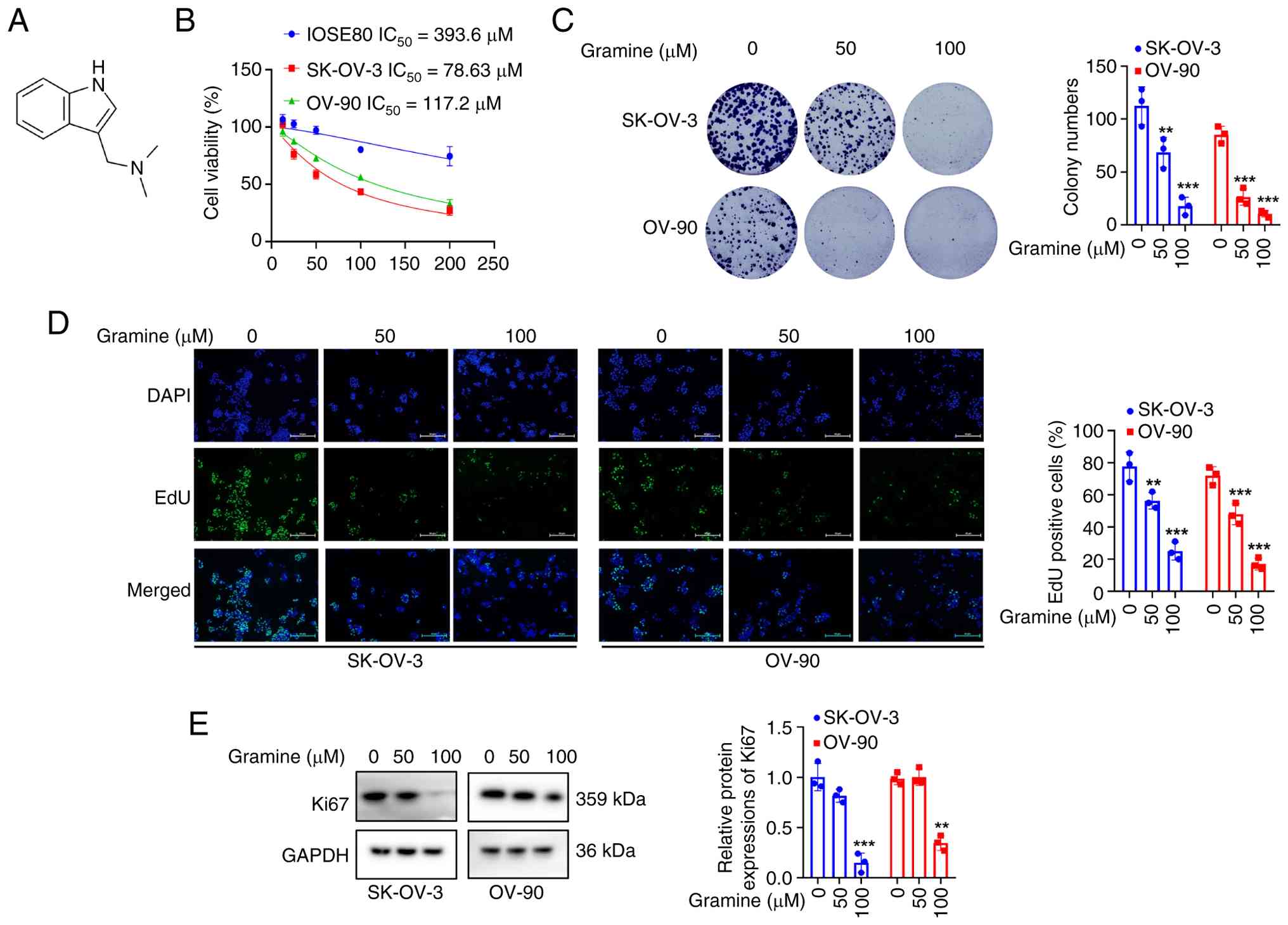
Fig. 1A depicts the
chemical structure of gramine. The biological effects of gramine
were evaluated in two OC cell lines (SK-OV-3 and OV-90) and normal
ovarian epithelial IOSE80 cells. As shown in Fig. 1B, gramine triggered a
dose-dependent suppression of cell viability across all three cell
lines, with half-maximal inhibitory concentration (IC50)
values of 78.63 µM (SK-OV-3), 117.2 µM (OV-90) and 393.6 µM
(IOSE80). The markedly lower IC50 of gramine in OC cells
compared with IOSE80 cells suggest a degree of selective
cytotoxicity against malignant ovarian cells. Consistent with the
viability data, gramine dose-dependently diminished the
colony-forming capacity in OC cells (Fig. 1C). Based on these results and its
minimal cytotoxicity toward normal ovarian epithelial cells,
concentrations of 50 and 100 µM gramine were selected for the
subsequent investigations. Additionally, the proportion of
EdU-positive cells (Fig. 1D) and
the expression level of Ki67 protein (Fig. 1E) were notably decreased following
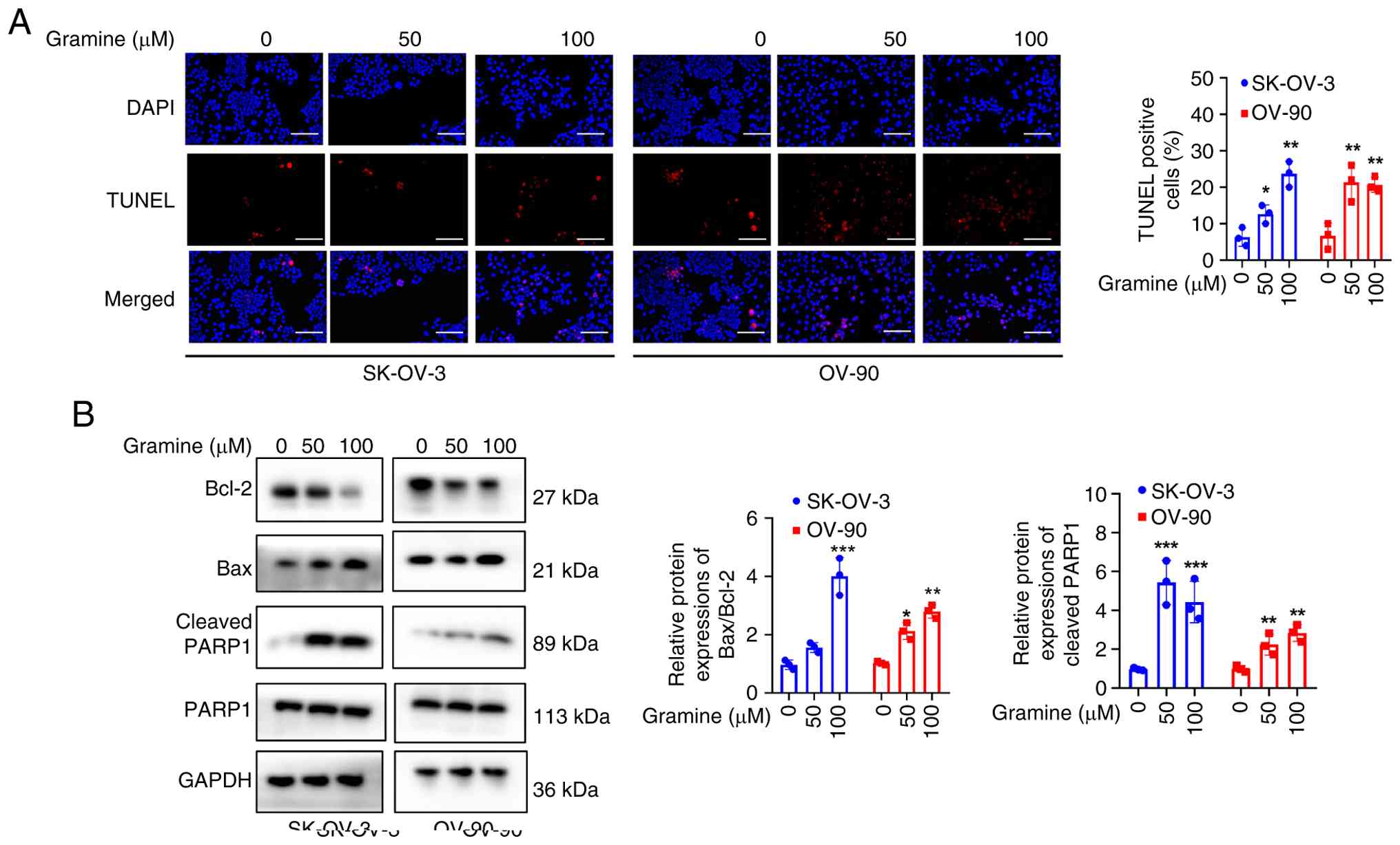
gramine administration. Next, it was explored whether gramine
induced apoptotic cell death in OC cells. As shown in Fig. 2A, an increased number of
TUNEL-positive cells was observed in gramine-treated cells.
Concurrently, elevated ratios of BAX/Bcl-2 and cleaved PARP1/PAPR1
were detected after gramine exposure (Fig. 2B). These data suggest that gramine
inhibits OC cell proliferation and promotes apoptosis.
Gramine inhibited cell migration and
invasion in OC cells
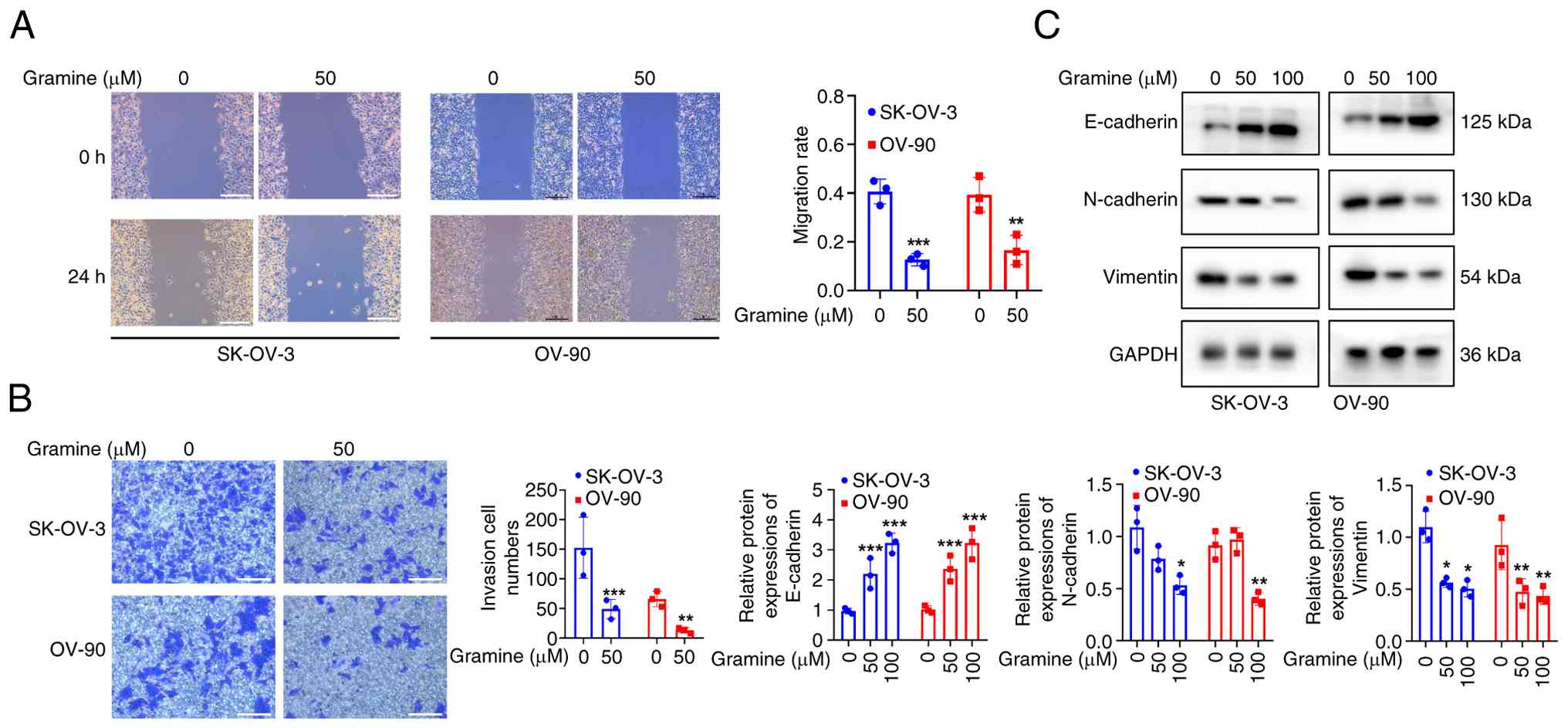
Then it was assessed whether gramine affects the
metastatic potential of OC cells. As shown in Fig. 3A and B, wound healing and Transwell invasion
assays revealed that gramine led to reduced migration and invasion
capacities, respectively. Western blot analysis further
demonstrated that gramine downregulated the expressions of
mesenchymal markers N-cadherin and vimentin, while concomitantly
upregulating the epithelial biomarker E-cadherin (Fig. 3C). These findings suggest that
gramine attenuates the invasive capacity of OC cells.
PDK1 overexpression impaired the
suppressive effects of gramine on OC cell growth
Given the vital role of PDK1 in the tumorigenesis of
OC, the present study sought to investigate whether gramine impedes
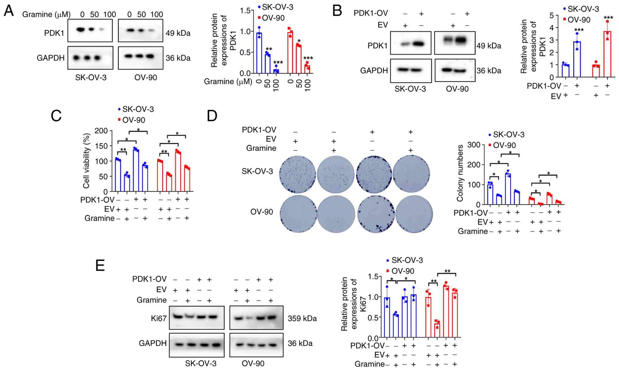
ovarian cancer cell growth by suppressing PDK1. Western blot
analysis revealed that gramine exposure for 24 h markedly reduced
PDK1 protein levels in OC cells in a concentration-dependent manner
(Fig. 4A). To further clarify the
functional role of PDK1 in gramine-mediated cellular responses,
lentiviral transfection was performed to generate stable
PDK1-overexpressing OC cell lines for subsequent rescue experiments
(Fig. 4B). Functional assays
validated that PDK1 overexpression effectively counteracted the
suppressive effects of gramine on OC cell proliferation.
Specifically, following gramine treatment, cells with increased
PDK1 expression exhibited higher cellular viability (Fig. 4C), enhanced colony-forming capacity
(Fig. 4D) and elevated Ki67 levels
(Fig. 4E) compared with control
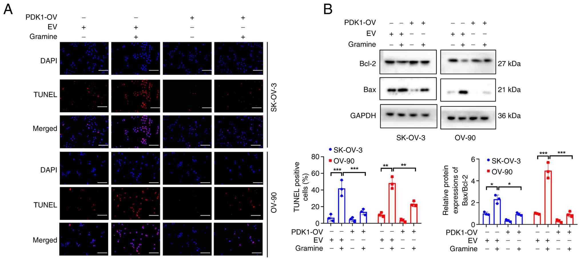
cells. Furthermore, PDK1 overexpression partially mitigated the
pro-apoptotic property of gramine. Although the proportion of
TUNEL-positive cells did not reach statistical significance,
ectopic PDK1 expression tended to abrogate gramine-induced
apoptosis (Fig. 5A). Consistently,
the increased Bax/Bcl-2 ratio induced by gramine was also rescued
upon PDK1 overexpression (Fig.
5B). These data collectively suggest that gramine-mediated
suppression of OC cell growth is associated with PDK1
downregulation.
PDK1/AKT signaling was involved in
anti-migration and invasion effects of gramine in OC cells
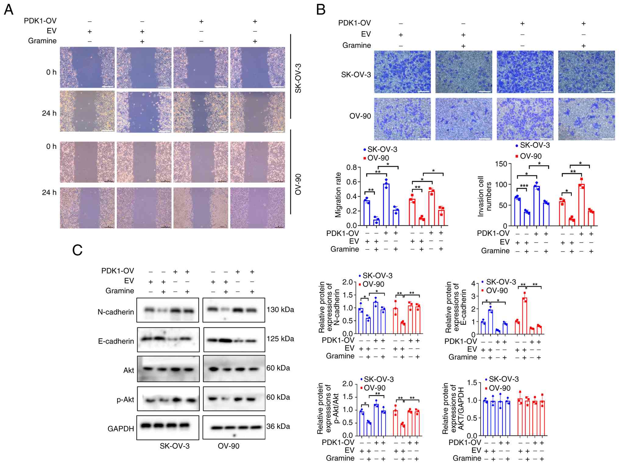
Ectopic overexpression of PDK1 potentiated the
migratory and invasive capacities of OC cells (Fig. 6A and B). Consistent with this phenotypic
change, PDK1 overexpression also upregulated N-cadherin while
downregulating E-cadherin protein levels (Fig. 6C). Notably, the inhibitory effects
of gramine on cell migration and invasion were substantially
abrogated following PDK1 overexpression (Fig. 6A and B). Similarly, PDK1 enrichment
counteracted gramine-mediated N-cadherin reduction and E-cadherin
elevation (Fig. 6C). In addition,
PDK1 overexpression also effectively restored the level of p-AKT
(Thr308), which was suppressed by gramine treatment (Fig. 6C). Together, these data suggest
that PDK1/AKT cascade contributes to the anti-tumor activity of
gramine in OC cells.
Gramine downregulates PDK1/AKT and is
associated with enhanced cisplatin sensitivity in OC cells
It was explored whether gramine could alleviate
cisplatin resistance in OC cells. As depicted in Fig. 7A, SK-OV-3-R cells exhibited
significant tolerance to cisplatin compared with parental SK-OV-3
cells, even at the maximum tested concentration of 80 µM,
confirming successful establishment of the cisplatin-resistant cell
model. Functional assays revealed that combined treatment with
gramine (50 µM) and cisplatin significantly enhanced the
sensitivity of SK-OV-3-R cells to cisplatin, as evidenced by
reduced cell viability and colony-forming potential (Fig. 7B and C). Moreover, the co-treatment group
presented an increased proportion of TUNEL-positive cells and
impaired migration and invasion capacities (Fig. 7D and E). Mechanistically, western blot results
demonstrated that combined application of gramine and cisplatin
induced a more pronounced reduction in PDK1 and p-AKT (Thr308)
proteins abundance compared with the cisplatin monotherapy
(Fig. 7F). These data suggest that
gramine may increase cisplatin responsiveness in SK-OV-3-R cells,
an effect associated with reduced PDK1/AKT signaling.
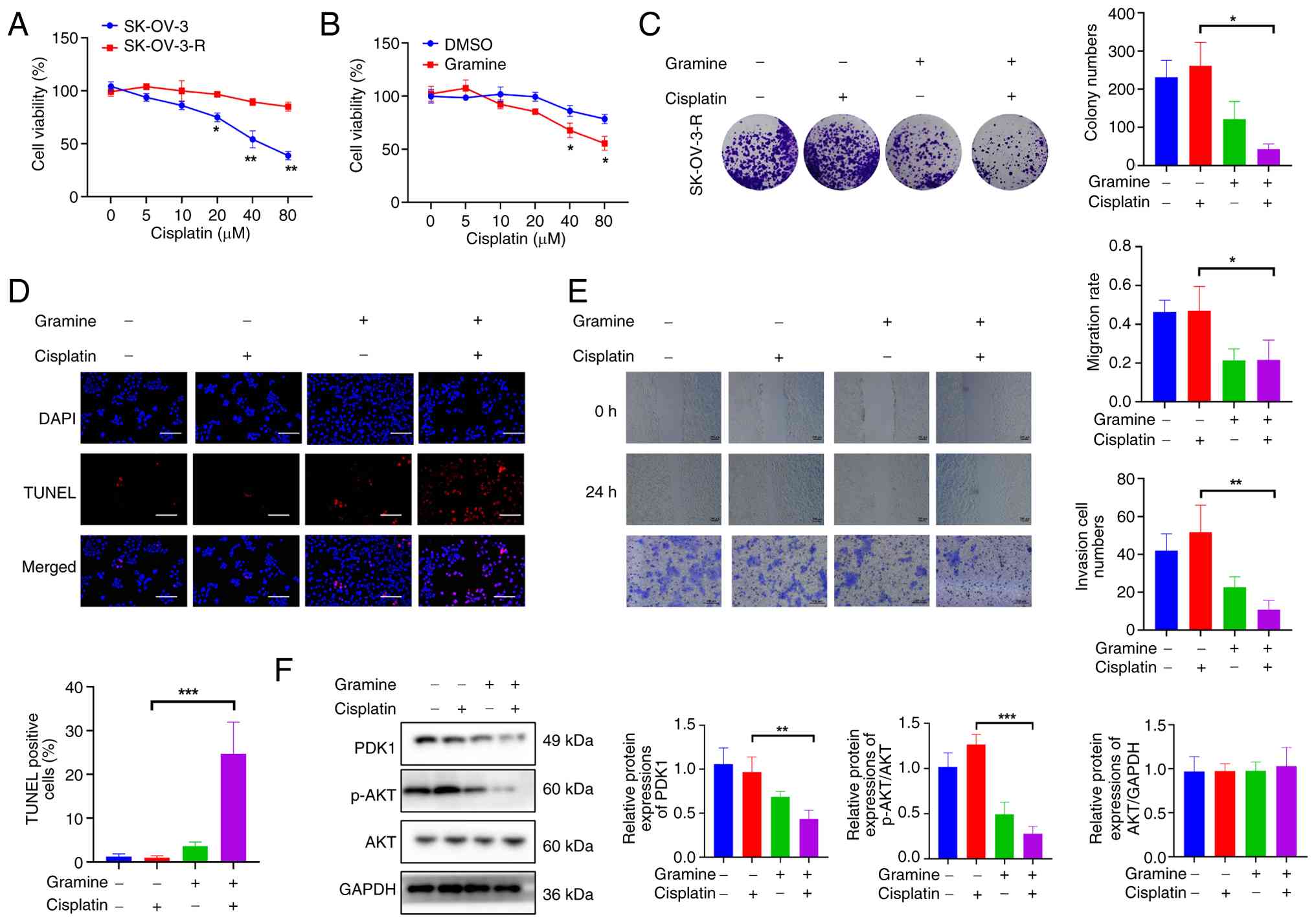 | Figure 7Gramine downregulates PDK1/AKT linked
to cisplatin sensitization of OC cells. (A) CCK-8 assay detecting
the effect of cisplatin on cell viability. SK-OV-3 and SK-OV-3-R
cells were incubated with cisplatin (5, 10, 20, 40, 80 µM) for 24
h. *P<0.05, **P<0.01 vs. the untreated
control. (B) CCK-8 assay detecting the effect of gramine and
cisplatin on cell viability. SK-OV-3-R cells were incubated with
gramine (50 µM) and cisplatin (5, 10, 20, 40, 80 µM) for 24 h.
*P<0.05 vs. the DMSO group at the identical cisplatin
concentration. (C) Clone formation assay assessing the effect of
gramine and cisplatin on colony formation ability. (D) TUNEL assay
evaluating the effect of gramine and cisplatin on cell apoptosis.
Scale bar, 200 µm. (E) Wound healing and transwell invasion assays
detecting the effect of gramine and cisplatin on the abilities of
cell migration and invasion respectively. Scale bar, 200 µm. (F)
Western blotting assay determining the effect of gramine and
cisplatin on the expressions of PDK1 and p-AKT (Thr308). SK-OV-3-R
cells were incubated with gramine (50 µM) and cisplatin (80 µM) for
24 h. *P<0.05, **P<0.01,
***P<0.001 vs. the cisplatin group (C-F). Data
represent the mean ± SD of three independent experiments. CCK-8,
cell counting kit-8. |
Discussion
In the present study, it was found that gramine
reduced PDK1 protein levels in a dose-dependent manner, however,
whether this reflects direct molecular interaction or an indirect
consequence remains unresolved. Based on current evidence, the
interpretation that PDK1 downregulation is more likely an indirect
result of cellular stress rather than direct binding and
inhibition. In the present study, biophysical binding assays were
not performed to confirm a physical interaction between gramine and
PDK1. In addition, the relatively high micromolar concentrations
required for observable effects are consistent with a pleiotropic
stress response (14).
Furthermore, the incomplete rescue by PDK1 overexpression implies
that gramine may perturb more upstream signaling nodes (such as
PI3K activity or PTEN function) (15,16)
or induce broader cellular stress (oxidative or endoplasmic
reticulum stress), which secondarily reduces PDK1 expression
(17). Definitive clarification
will require reverse transcription-quantitative PCR analysis of
PDK1 mRNA, assessment of ubiquitin-proteasome degradation
and direct binding studies.
Among the various cellular phenotypes elicited by
gramine, PDK1 overexpression exerted the most pronounced rescue
effect on cell proliferation and colony formation, where it
partially but consistently reversed gramine-induced growth
inhibition. By contrast, the suppression of cell migration and
invasion was weakly and incompletely restored by PDK1
overexpression. This disparity suggests that the anti-migratory and
anti-invasive activities of gramine are independent of the PDK1/AKT
axis, whereas its impact on proliferation is more tightly coupled
to this pathway. In addition, proliferation-blocking controls were
not included in the present study and time-matched viability tests
aligned with the wound-healing incubation period were not
performed. Therefore, anti-proliferative effects cannot be fully
separated from the specific migratory inhibition in the current
dataset. Notably, none of the phenotypes were fully reversed by
PDK1 overexpression, indicating that gramine does not act solely
through PDK1 inhibition and that additional targets or parallel
pathways are involved.
Full AKT activation requires both PDK1-mediated
phosphorylation at Thr308 and mTORC2-mediated phosphorylation at
Ser473 (18,19). The present data showed that gramine
markedly inhibits AKT phosphorylation, but it cannot yet be
concluded that this inhibition occurs exclusively through PDK1.
While PDK1 is the primary kinase for AKT-Thr308, changes in PI3K
activity, activation of AKT phosphatases (such as PP2A or PHLPP)
(20) or perturbation of mTORC2
function could equally affect AKT phosphorylation (21). Moreover, published evidence
suggests that gramine may influence multiple kinases or receptor
pathways (9,22). Therefore, the present conclusion
should be cautiously framed as ‘gramine suppresses the PDK1/AKT
signaling axis’, leaving the question of whether AKT inhibition is
strictly PDK1-dependent as an open issue. Future cross-validation
using selective PI3K inhibitors or AKT phosphatase modulators will
help delineate the specificity of this effect.
Cisplatin-based chemotherapy is the first-line
treatment for OC, while its resistance markedly limits the efficacy
of chemotherapy in OC (23). In
the present study, cisplatin-resistant OC cells were used to
preliminarily observe that gramine enhances cisplatin sensitivity,
associating this effect with PDK1 downregulation and AKT
inactivation. However, this conclusion remains preliminary and
insufficiently validated. The major limitations of the present
study include: i) The absence of quantitative synergy metrics, such
as combination index or isobologram analysis, preventing
determination of whether the effect is synergistic, additive or
merely additive; ii) a lack of PDK1 rescue experiments in resistant
cells, which are essential to confirm that sensitization is indeed
mediated by PDK1 inhibition; and iii) failure to compare the
magnitude of PDK1/AKT suppression between sensitive and resistant
cells, or to exclude alternative mechanisms such as modulation of
drug efflux pumps (such asABCB1) (24) or apoptosis regulators (such as p53)
(25). Therefore, the current data
only support that the combination of gramine and cisplatin warrants
further investigation, and fall far short of establishing a
definitive sensitization mechanism. Future work should
systematically address these gaps, including evaluation in animal
models and primary cells.
In addition, further limitations must be
acknowledged to contextualize the present in vitro findings.
First, all functional and mechanistic experiments were conducted
solely in OC cell lines, with no in vivo animal models or
clinical specimen validation to assess the activity of gramine
under physiological conditions. Second, there is a lack of
biophysical binding or target engagement experiments, so it the
direct molecular interaction between gramine and PDK1 cannot be
confirmed to distinguish direct inhibition from indirect
stress-triggered PDK1 downregulation. Third, partial rescue by PDK1
overexpression prevents full establishment of a causal
PDK1/AKT-dependent mechanism for the anti-tumor activity of
gramine, leaving PDK1-independent pathways uncharacterized. Fourth,
the present evaluation of cisplatin responsiveness lacks
quantitative synergy metrics, IC50 shift data and PDK1
rescue validation in resistant cells, which are necessary to
confirm gramine-mediated cisplatin sensitization. Lastly, the
relatively high micromolar concentrations of gramine used to
produce cellular phenotypes raise concerns about potential
non-specific off-target cytotoxicity. Future work will address all
these experimental gaps to further validate and expand our
preliminary observations.
In summary, gramine effectively inhibits OC cell
proliferation, migration and invasion in vitro, and shows
preliminary potential to sensitize cells to cisplatin, with these
effects being at least partially attributable to suppression of the
PDK1/AKT pathway. The present findings provide a valuable starting
point for deeper investigation but establishing gramine as a
credible PDK1-targeting candidate or a clinically applicable
sensitizer will require a substantially stronger body of in
vitro and in vivo evidence.
Acknowledgements
Not applicable.
Funding
Funding: The present study was supported by the Wenzhou
Municipal Science and Technology Bureau (grant no. Y2023903).
Availability of data and materials
Data generated in the present study may be requested
from the corresponding author.
Authors' contributions
HW, conceptualized the study, performed the
literature search and experiments, and wrote the original draft. RZ
and BW performed the analysis and interpretation of data, and
reviewed and edited the manuscript. SH and HC performed the
analysis and interpretation of data, and supervised the study. HW
and RZ confirm the authenticity of all the raw data. All authors
read and approved the final version of the manuscript.
Ethics approval and consent to
participate
Not applicable.
Patient consent for publication
Not applicable.
Competing interests
The authors declare no that they have no competing
interests.
References
|
1
|
Caruso G, Weroha SJ and Cliby W: Ovarian
cancer: A review. JAMA. 334:1278–1291. 2025.PubMed/NCBI View Article : Google Scholar
|
|
2
|
Nie DY, Zhang H, Wang H, Song W, Yuan D,
Zhu D, Mao G, Guo T and Lin M: Artesunate triggers ferroptosis in
ovarian cancer via GP130-mediated IL-6/STAT3/OTUB1/SLC711 axis
disruption. Phytomedicine. 148(157269)2025.PubMed/NCBI View Article : Google Scholar
|
|
3
|
Chen G, Yuan D, Li J, Mao G, Zhang Y, Guo
T and Lin M: Polyphyllin H inhibits malignant progression of
ovarian cancer in patient-derived xenograft mouse models by
regulating CGN/RhoA/Rock2 axis: An experimental research. Int J
Surg. 111:7842–7856. 2025.PubMed/NCBI View Article : Google Scholar
|
|
4
|
Wu F, Liu Y, Luo GQ, Huang DH, Liang WL,
Cao SX, Niu J, Kurihara H, Zhang XH, Li YF, et al: Chaihu Shugan
San formula alleviates psychological stress-induced ovarian cancer
susceptibility by inhibiting ubiquitin degradation of TLR2 in
macrophages. Phytomedicine. 145(156967)2025.PubMed/NCBI View Article : Google Scholar
|
|
5
|
Lajarín-Cuesta R, Arribas RL, Nanclares C,
García-Frutos EM, Gandía L and de Los Ríos C: Design and synthesis
of multipotent 3-aminomethylindoles and 7-azaindoles with enhanced
protein phosphatase 2A-activating profile and neuroprotection. Eur
J Med Chem. 157:294–309. 2018.PubMed/NCBI View Article : Google Scholar
|
|
6
|
Hong Y, Hu HY, Xie X, Sakoda A, Sagehashi
M and Li FM: Gramine-induced growth inhibition, oxidative damage
and antioxidant responses in freshwater cyanobacterium Microcystis
aeruginosa. Aquat Toxicol. 91:262–269. 2009.PubMed/NCBI View Article : Google Scholar
|
|
7
|
Zhang XH, Guo Q, Wang HY, Li YH, Khamis
MY, Ma LY, Wang B and Liu HM: Gramine-based structure optimization
to enhance anti-gastric cancer activity. Bioorg Chem.
107(104549)2021.PubMed/NCBI View Article : Google Scholar
|
|
8
|
Ramu A, Kathiresan S and Ali Ahmed B:
Gramine inhibits angiogenesis and induces apoptosis via modulation
of TGF-β signalling in 7,12 dimethylbenz[a]anthracene (DMBA)
induced hamster buccal pouch carcinoma. Phytomedicine. 33:69–76.
2017.PubMed/NCBI View Article : Google Scholar
|
|
9
|
Ramu A, Kathiresan S, Ramadoss H, Nallu A,
Kaliyan R and Azamuthu T: Gramine attenuates EGFR-mediated
inflammation and cell proliferation in oral carcinogenesis via
regulation of NF-κB and STAT3 signaling. Biomed Pharmacother.
98:523–530. 2017.PubMed/NCBI View Article : Google Scholar
|
|
10
|
Bayascas JR: PDK1: The major transducer of
PI 3-kinase actions. Curr Top Microbiol Immunol. 346:9–29.
2010.PubMed/NCBI View Article : Google Scholar
|
|
11
|
Zheng NN, Wei JQ, Wu DP, Xu Y and Guo JP:
Master kinase PDK1 in tumorigenesis. Biochim Biophys Acta Rev
Cancer. 1878(188971)2023.PubMed/NCBI View Article : Google Scholar
|
|
12
|
Wang JJ, Siu MK, Jiang YX, Leung TH, Chan
DW, Cheng RR, Cheung AN, Ngan HY and Chan KK: Aberrant upregulation
of PDK1 in ovarian cancer cells impairs CD8+ T cell
function and survival through elevation of PD-L1. Oncoimmunology.
8(e1659092)2019.PubMed/NCBI View Article : Google Scholar
|
|
13
|
Zhang M, Cong Q, Zhang XY, Zhang MX, Lu YY
and Xu CJ: Pyruvate dehydrogenase kinase 1 contributes to cisplatin
resistance of ovarian cancer through EGFR activation. J Cell
Physiol. 234:6361–6370. 2019.PubMed/NCBI View Article : Google Scholar
|
|
14
|
Altintas DM, Cerqua M, Comoglio PM and
Chaveroux C: The Janus framework of the integrated stress response:
From homeostasis to maladaptation. Life Sci Alliance.
9(e202503523)2025.PubMed/NCBI View Article : Google Scholar
|
|
15
|
Lac V, Verhoef L, Aguirre-Hernandez R,
Nazeran TM, Tessier-Cloutier B, Praetorius T, Orr NL, Noga H, Lum
A, Khattra J, et al: Iatrogenic endometriosis harbors somatic
cancer-driver mutations. Hum Reprod. 34:69–78. 2019.PubMed/NCBI View Article : Google Scholar
|
|
16
|
Cousin S, Grellety T, Toulmonde M,
Auzanneau C, Khalifa E, Laizet Y, Tran K, Le Moulec S, Floquet A,
Garbay D, et al: Clinical impact of extensive molecular profiling
in advanced cancer patients. J Hematol Oncol. 10(45)2017.PubMed/NCBI View Article : Google Scholar
|
|
17
|
Yan T, Ma X, Guo L and Lu R: Targeting
endoplasmic reticulum stress signaling in ovarian cancer therapy.
Cancer Biol Med. 20:748–764. 2023.PubMed/NCBI View Article : Google Scholar
|
|
18
|
Scheid MP, Marignani PA and Woodgett JR:
Multiple phosphoinositide 3-kinase-dependent steps in activation of
protein kinase B. Mol Cell Biol. 22:6247–6260. 2002.PubMed/NCBI View Article : Google Scholar
|
|
19
|
Sarbassov DD, Guertin DA, Ali SM and
Sabatini DM: Phosphorylation and regulation of Akt/PKB by the
rictor-mTOR complex. Science. 307:1098–1101. 2005.PubMed/NCBI View Article : Google Scholar
|
|
20
|
Liao Y and Hung MC: Physiological
regulation of Akt activity and stability. Am J Transl Res. 2:19–42.
2010.PubMed/NCBI
|
|
21
|
Duan J, Zhang Z, Du J, Zhang J, Li M and
Li C: Esomeprazole alleviates cisplatin resistance by inhibiting
the AKT/mTOR pathway in ovarian cancer cells. Onco Targets Ther.
16:425–440. 2023.PubMed/NCBI View Article : Google Scholar
|
|
22
|
Zhang J, Jia Q, Li N, Gu L, Dan W and Dai
J: Recent developments of gramine: Chemistry and biological
activity. Molecules. 28(5695)2023.PubMed/NCBI View Article : Google Scholar
|
|
23
|
Torri V, Harper PG, Colombo N, Sandercock
J and Parmar MK: Paclitaxel and cisplatin in ovarian cancer. J Clin
Oncol. 18:2349–2351. 2000.PubMed/NCBI
|
|
24
|
Skinner KT, Palkar AM and Hong AL:
Genetics of ABCB1 in cancer. Cancers (Basel).
15(4236)2023.PubMed/NCBI View Article : Google Scholar
|
|
25
|
Yan X, Fraser M, Qiu Q and Tsang BK:
Over-expression of PTEN sensitizes human ovarian cancer cells to
cisplatin-induced apoptosis in a p53-dependent manner. Gynecol
Oncol. 102:348–355. 2006.PubMed/NCBI View Article : Google Scholar
|