Introduction
Endothelial cells (ECs) derived from vascular
progenitor cells are responsible for angiogenesis and the events of
wound healing (1). They are
characterized by the expression of vascular endothelial-cadherin
(VE-cadherin, CDH5) (2–4), vascular endothelial growth factor
receptor 2 (VEGFR2, KDR) (5,6) and
CD31 (also called platelet endothelial cell adhesion molecule-1,
PECAM-1) (7). As blood supply is
crucial for wound healing and tissue regeneration, it is important
to determine how the progenitors of ECs differentiate into vascular
cells in order to establish a practical strategy for regenerative
therapy (1).
For regenerative therapy, biologically active
soluble factors such as cytokines and growth factors are being
evaluated for clinical use in the regeneration of periodontal
tissue damaged or lost as a result of periodontitis. Among these
factors, fibroblast growth factor (FGF)-2 is a multifunctional
growth factor that exerts a variety of effects, including the
induction of proliferation and differentiation in a wide range of
mesodermal and neuro-ectodermal cells. Moreover, FGF-2 is one of
the most potent angiogenesis inducers (8). Therefore, we investigated whether
FGF-2 induces EC-specific markers in cultured dental pulp (DP)
cells in vitro.
Dental pulp (DP) tissue is a non-hematopoietic
connective tissue that is almost completely surrounded by hard
tissue (9). After tooth
maturation, DP tissues act only in a reparative capacity in
response to general mechanical erosion or disruption and dentinal
degradation caused by bacteria. Recently, dental pulp
progenitor/stem cells (DPSCs) have been shown to be capable of
differentiating into osteoblasts, adipocytes and neural cells in
vivo (10). However, despite
extensive investigation of DPSCs, the characteristics and
properties of postnatal stem cells derived from DP cells from
deciduous teeth have not yet been sufficiently studied in terms of
the expression of the phenotype of endothelial cells and the
regulation of differentiation in DP cells.
In the present study, we demonstrated that
VE-cadherin, VEGFR2 and CD31 mRNA are expressed in cultured DP
cells upon treatment with FGF-2. We also demonstrated CD31 protein
expression in DP cell cultures using Western blot analysis. This is
the first report regarding the inducible expression of the
endothelial cell phenotype by DP cells derived from human deciduous
teeth.
Materials and methods
Reagents
FGF-2 was obtained from R&D Systems
(Minneapolis, MN, USA). Anti-CD31 monoclonal antibody for the
Western blot analysis was obtained from Cell Signaling Technology,
Inc. (Danvers, MA, USA).
Cell culture
The dental pulp (DP) tissues were obtained from the
crown and root of healthy human deciduous teeth from three donors,
aged 7–8 years. Informed consent was obtained from the donors’
parents prior to tooth extraction, which was carried out in our
hospital during the course of orthodontic treatment. The study
protocol was approved by the Ethics Committee of Iwate Medical
University, School of Dentistry (no. 01101).
DP tissues were cut into pieces using a surgical
blade and digested with collagenase (2 mg/ml) at 37°C for 30 min.
The tissues were then washed with Dulbecco’s phosphate-buffered
saline (PBS), placed on culture dishes and maintained in α-modified
minimum essential medium (α-MEM) (Gibco BRL, Gaithersburg, MD, USA)
supplemented with 10% fetal bovine serum (FBS) (Gibco BRL).
Fibroblastic cells that outgrew from the DP tissues were used as DP
cells. When the cells reached confluence, they were detached with
0.2% trypsin and 0.02% EDTA (4 Na) in PBS and subcultured at a 1:4
split ratio. Experiments were performed using 4th passage cells
cultured in α-MEM supplemented with 10% FBS in the absence or
presence of 10 ng/ml FGF-2 for 2 days. The cultures were maintained
at 37°C in a humidified atmosphere of 5% CO2 in air.
Isolation of total RNA
Total RNA was extracted from the cultured DP cells
using Isogen (Nippon Gene, Tokyo, Japan) as previously described
(11,12). The pellet of total RNA was washed
briefly with 75% ethanol, resuspended in 30 μl of
diethylpyrocarbonate (DEPC)-treated water, and stored at −80°C. The
concentration of total RNA was determined spectrophotometrically by
measuring the optical density at 260 nm.
Quantitative real-time reverse
transcription-polymerase chain reaction
Using a PrimeScript RT reagent kit (Takara Shuzo,
Kyoto, Japan), 1 μg of RNA sample was reverse-transcribed to
first-strand cDNA according to the manufacturer’s protocol. A
Thermal Cycler Dice Real-Time system (Takara Shuzo) was used for
the two-step reverse transcription-polymerase chain reaction. cDNA
was amplified with SYBR Premix ExTaq and specific oligonucleotide
primers for target sequences encoding parts of VE-cadherin, VEGFR2
and CD31. The primers (listed in Table
1) were designed based on the cDNA sequences of human mRNA for
VE-cadherin, VEGFR2, CD31 and glyceraldehyde 3-phosphate
dehydrogenase (GAPDH). Amplification conditions consisted of 10 sec
at 95°C followed by 40 cycles at 95°C for 5 sec and 60°C for 30
sec, with a final 15 sec at 95°C and 30 sec at 60°C in the Thermal
Cycler Dice Real-Time system.
 | Table I.Primers used in the quantitative
real-time reverse transcription-polymerase chain reaction. |
Table I.
Primers used in the quantitative
real-time reverse transcription-polymerase chain reaction.
| Gene name | Primer | Oligonucleotide
sequence (5′-3′) |
|---|
| VE-cadherin | Forward |
GAGACCTCATCAGCCTTGGGATAG |
| Reverse |
CTGGATTTGCCAGCATTTGAGA |
| VEGFR2 | Forward |
CCAGGCAACGTAAGTGTTCGAG |
| Reverse |
GGGACCCACGTCCTAAACAAAG |
| CD31 | Forward |
GACGTGCAGTACACGGAAGTTCA |
| Reverse |
GTGCATCTGGCCTTGCTGTC |
| GAPDH | Forward |
GCACCGTCAAGGCTGAGAAC |
| Reverse |
TGGTGAAGACGCCAGTGGA |
Western blot analysis of cell surface
CD31 expression in DP cells
After treatment with FGF-2 for 21 days, DP cells
were washed twice with PBS and then treated with lysis buffer [10
mM HEPES-KOH (pH 7.5), 100 mM KCL and 0.1% NP-40]. Protein
concentration in the cell lysate was measured using a BioRad
Protein Assay kit (BioRad, Hercules, CA). Each sample containing
equal amounts of protein was separated by 10% SDS-polyacrylamide
gel electrophoresis (SDS-PAGE) and transferred to a polyvinylidene
difluoride membrane (Millipore, Bedford, MA). After being blocked
with 5% skim milk in Tris-buffered saline containing 0.1% Tween-20
(TBST), the membrane was incubated with mouse anti-human CD31
antibodies and subsequently with anti-mouse secondary antibodies
(Zymed Laboratories Inc., San Francisco, CA). Specific protein
bands on the membrane were detected using an enhanced AP conjugate
substrate kit (BioRad Laboratories, CA) as previously described
(11,12).
Statistical analysis
Results are expressed as the means ± SEM.
Statistical significance was determined by one-way analysis of
variance and Bonferroni comparisons between pairs of groups. Data
with P-values <0.01 were considered statistically
significant.
Results
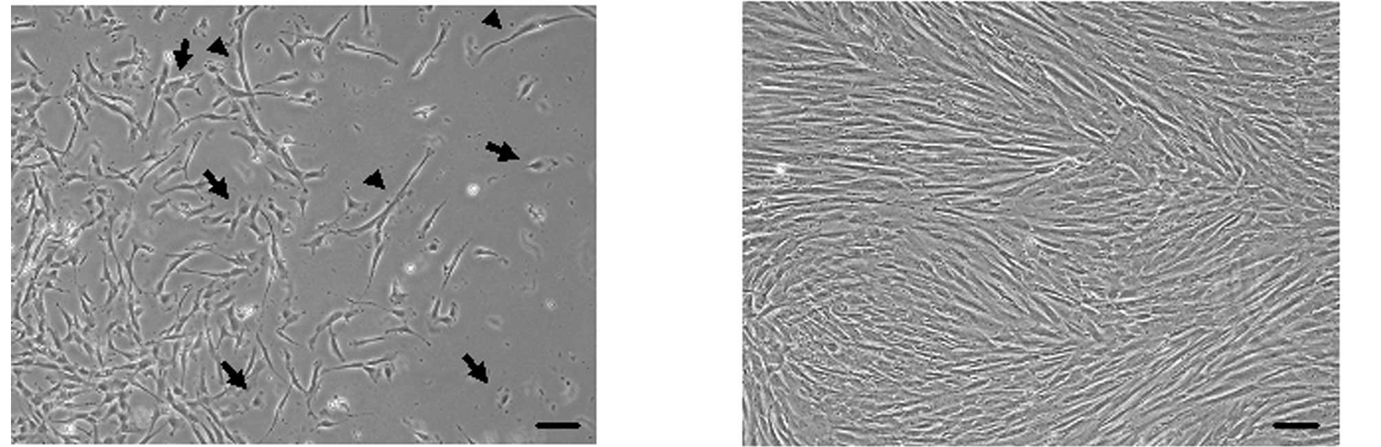
Varying cell morphology of DP cells
derived from deciduous teeth in primary culture
Using phase-contrast microscopy, DP cells were
observed to differentiate into various types of cells 10 days after
isolation from DP tissues (Fig.
1). Most of the DP cells derived from deciduous teeth exhibited
a fibroblastic cell morphology (Fig.
1A). Some of the cells showed polygonal-like epithelial cell
and mature osteoblast morphologies (Fig. 1A, arrow). A few cells showed a
senescent fibroblastic cell-like morphology (Fig. 1A, arrowhead). After reaching
confluence and undergoing subculture, it was no longer possible to
distinguish between these cell morphologies (Fig. 1B).
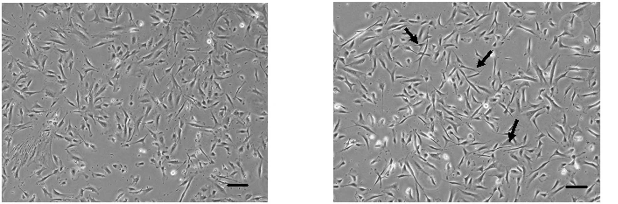
FGF-2-induced morphological changes in DP
cells after 2 days of treatment
DP cells were cultured in control media (Fig. 2A) or in the presence of FGF-2
(Fig. 2B), and reached
subconfluence after 2 days of culture. FGF-2-treated DP cells
reached confluence and exhibited an altered morphology of long and
thin spindle-shaped fibroblasts (Fig.
2B).
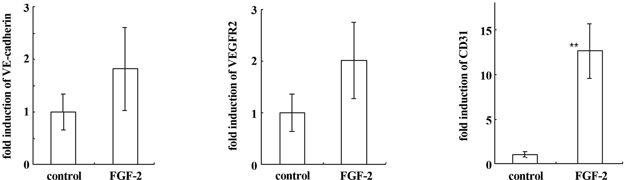
FGF-2-induced EC-specific markers in DP
cells after 2 days of treatment
In DP cells cultured in the presence of FGF-2,
VE-cadherin and VEGFR2 mRNA expression was increased, though not
significantly (Fig. 3A and B).
However, CD31 expression was significantly induced by treatment
with FGF-2 (Fig. 3C).
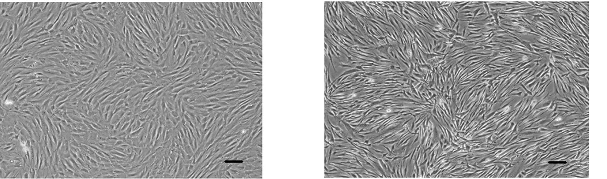
FGF-2-induced morphological changes in DP
cells after 3 weeks of treatment
After three weeks of culture with FGF-2, DP cells
reached confluence and exhibited a long and thin spindle-shaped
fibroblastic morphology (Fig. 4B)
compared to the cells cultured in control media (Fig. 4A).
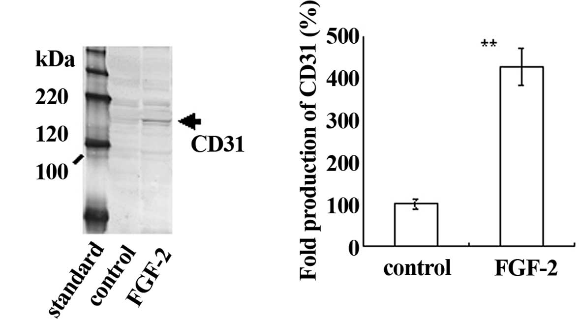
FGF-2-induced CD31 expression in DP
cells
To determine whether CD31 protein was induced in DP
cells cultured with FGF-2 (Fig.
5), Western blot analysis was conducted. CD31 expression was
not detected after 2 days of treatment with FGF-2 (data not shown),
nor after 2 weeks of culture. After treatment with FGF-2 for 3
weeks, CD31 expression was detected by Western blot analysis as
compared with the control (Fig.
5A). Therefore, the production of CD31 was induced in DP cells
by FGF-2 treatment (Fig. 5B).
Discussion
Tissue regeneration and homeostasis in response to
pathological and environmental changes such as dental injury (e.g.,
preparation of a deep cavity) are thought to depend in large part
upon angiogenesis in DP tissue. DP tissues exist in the pulp
chamber surrounding dentin, and thus are likely to play an key role
in protection against dental caries (9). DP cells were shown to have biological
characteristics in common with bone marrow mesenchymal cells,
suggesting that multipotent stem cells are present in DP tissue
(10). However, it remains unclear
whether DP cells derived from human deciduous teeth give rise to
the endothelial cell (EC) lineage in vitro. To investigate
DP tissue regeneration and homeostasis, it is crucial to determine
whether DP cells have the ability to differentiate into ECs.
In the present study, we used DP cells derived from
human deciduous teeth to investigate the effects of FGF-2 on the
expression of markers specific for mature ECs: VE-cadherin
(2–4), VEGFR2 (5,6) and
CD31 (7). Previous findings have
demonstrated that DP cells unstimulated by FGF-2 basally express EC
markers such as CD31, though very weakly (13,14).
In agreement with these findings, using real-time PCR we observed
that DP cells without FGF-2 treatment expressed VE-cadherin, VEGFR2
or CD31. Unexpectedly, the expression of VE-cadherin and VEGFR2 was
increased in DP cells cultured in the presence of FGF-2. The
expression of CD31 was also significantly increased in DP cells
cultured with FGF-2.
Compared with mRNA expression, CD31 protein
production showed relatively small changes in the DP cells.
However, treatment with FGF-2 was sufficient to induce CD31 protein
expression. As shown in Fig. 5, in
DP cells treated with FGF-2, CD31 production was increased by
approximately 4-fold compared with the control group. The amount of
protein, which is determined not only at the mRNA level but also by
multiple processes of protein synthesis and degradation, might be a
critical factor.
Here, we demonstrated for the first time that DP
cells derived from human deciduous teeth inducibly express the
EC-specific markers VE-cadherin, VEGFR2 and CD31 upon treatment
with FGF-2 in vitro. These findings may aid in understanding
the regeneration of DP tissue through the induction of
angiogenesis.
Acknowledgements
This work was supported in part by
Grants-in-Aid for Scientific Research (no. 18592026 to A.I., no.
19791370 to N.C. and no. 18592239 to T.H.) from the Ministry of
Education, Culture, Sports, Science and Technology of Japan, by the
Open Research Project and High-Tech Research Project from the
Ministry of Education, Culture, Sports, Science and Technology of
Japan, by the Akiyama Foundation (to T.H., 2005), and by a grant
from the Keiryokai Research Foundation (no. 100 to N.C., 2008; no.
106 to T.H., 2009).
References
|
1.
|
Bartold PM, Shi S and Gronthos S: Stem
cells and periodontal regeneration. Periodontol 2000. 40:164–172.
2006. View Article : Google Scholar : PubMed/NCBI
|
|
2.
|
Dejana E: Endothelial cell-cell junctions:
happy together. Nat Rev Mol Cell Biol. 5:261–270. 2004. View Article : Google Scholar : PubMed/NCBI
|
|
3.
|
Cavallaro U, Liebner S and Dejana E:
Endothelial cadherins and tumor angiogenesis. Exp Cell Res.
312:659–667. 2006. View Article : Google Scholar : PubMed/NCBI
|
|
4.
|
Carmeliet P, Lampugnani MG, Moons L,
Breviario F, Compernolle V, Bono F, Balconi G, Spagnuolo R,
Oosthuyse B, Dewerchin M, Zanetti A, Angellilo A, Mattot V, Nuyens
D, Lutgens E, Clotman F, de Ruiter MC, Gittenberger-de Groot A,
Poelman R, Lupu F, Herbert JM, Collen D and Dejana E: Targeted
deficiency or cytosolic truncation of VE-cadherin gene in mice
impairs VEGF-mediated endothelial survival and angiogenesis. Cell.
98:147–157. 1999. View Article : Google Scholar : PubMed/NCBI
|
|
5.
|
Lampugnani MG, Orsenigo F, Gagliani MC,
Tacchetti C and Dejana E: Vascular endothelial cadherin controls
VEGFR-2 internalization and signaling from intracellular
compartments. J Cell Biol. 174:593–604. 2006. View Article : Google Scholar : PubMed/NCBI
|
|
6.
|
Yamaguchi TP, Dumont DJ, Conlon RA,
Breitman ML and Rossant J: flk-1, an flt-related receptor tyrosine
kinase, is an early marker for endothelial cell precursors.
Development. 118:489–498. 1993.PubMed/NCBI
|
|
7.
|
Hristov M, Erl W and Weber PC: Endothelial
progenitor cells: mobilization, differentiation, and homing.
Arterioscler Thromb Vasc Biol. 23:1185–1189. 2003. View Article : Google Scholar : PubMed/NCBI
|
|
8.
|
Ferrara N and Davis-Smyth T: The biology
of vascular endothelial growth factor (Review). Endocr Rev.
18:4–25. 1997. View Article : Google Scholar
|
|
9.
|
Orchardson R and Cadden SW: An update on
the physiology of the dentine-pulp complex (Review). Dent Update.
28:208–209. 2001.PubMed/NCBI
|
|
10.
|
Miura M, Gronthos S, Zhao M, Lu B, Fisher
LW, Robey PG and Shi S: SHED: stem cells from human exfoliated
deciduous teeth. Proc Natl Acad Sci USA. 100:5807–5812. 2003.
View Article : Google Scholar : PubMed/NCBI
|
|
11.
|
Hasegawa T, Yoshimura Y, Kikuiri T, Yawaka
Y, Takeyama S, Matsumoto A, Oguchi H and Shirakawa T: Expression of
receptor activator of NF-kappa B ligand and osteoprotegerin in
culture of human periodontal ligament cells. J Periodont Res.
37:405–411. 2002. View Article : Google Scholar : PubMed/NCBI
|
|
12.
|
Hasegawa T, Kikuiri T, Takeyama S,
Yoshimura Y, Mitome M, Oguchi H and Shirakawa T: Human periodontal
ligament cells derived from deciduous teeth induce
osteoclastogenesis in vitro. Tissue Cell. 34:44–51. 2002.
View Article : Google Scholar : PubMed/NCBI
|
|
13.
|
Trubiani O, Isgro A, Zini N, Antonucci I,
Aiuti F, Di Primio R, Nanci A, Caputi S and Paganelli R: Functional
interleukin-7/interleukin-7Ralpha, and SDF-1alpha/CXCR4 are
expressed by human periodontal ligament derived mesenchymal stem
cells. J Cell Physiol. 214:706–713. 2008. View Article : Google Scholar : PubMed/NCBI
|
|
14.
|
Nagatomo K, Komaki M, Sekiya I, Sakaguchi
Y, Noguchi K, Oda S, Muneta T and Ishikawa I: Stem cell properties
of human periodontal ligament cells. J Periodontal Res. 41:303–310.
2006. View Article : Google Scholar : PubMed/NCBI
|