Introduction
Cancer is an increasing global concern, and breast
cancer remains the most frequently diagnosed cancer affecting
females, accounting for >20% of all cancer cases in women
worldwide (1). Breast cancer is
categorized based on cellular markers that reflect the available
targeted therapies. Triple-negative breast cancer (TNBC) is a
subtype of breast cancer characterized by the suppressed expression
levels of the estrogen receptor (ER), progesterone receptor (PR)
and human epidermal growth factor receptor 2 (HER2). TNBC accounts
for ~10-20% of breast cancer cases and is a highly aggressive
disease with frequent early relapses and a very poor overall
survival rate (2). Therefore, in
addition to its prevention, the development of novel treatment
options for this type of breast cancer is crucial due to the
limited number of available treatments.
Previous epidemiological studies have suggested that
diets rich in cruciferous vegetables, such as broccoli, cabbage and
kale reduce the risk of developing a number of common types of
cancer, including breast cancer (3,4).
Sulforaphane (SFN) is an isothiocyanate derivative generated by the
hydrolytic conversion of glucoraphanin, a sulfur-containing
compound found in cruciferous vegetables (5). Recently, SFN was shown to be
effective in preventing breast cancer at different stages of
carcinogenesis by increasing the levels of antioxidants and phase
II detoxifying enzymes via the activation of the nuclear factor
erythroid 2-related factor 2 (6,7). In
addition to its chemopreventive effects, SFN has been found to
exert anti-proliferative effects on various human breast cancer
cell lines that are representative of a wide range of breast cancer
phenotypes by inducing apoptosis, cell cycle arrest and exhibiting
anti-angiogenic capacity (6-8).
Therefore, SFN may have the potential to prevent and treat all
subtypes of breast cancer.
Molecular aberrations in the epidermal growth factor
receptor (EGFR)/phosphoinositide 3-kinase (PI3K)/protein kinase B
(Akt)/mechanistic target of rapamycin (mTOR) pathway are well-known
pathognomonic abnormalities in breast cancer across various
subtypes and are commonly observed in TNBC (9). A subset of TNBC (~18%) is known to
express EGFR and is associated with a poor prognosis (10). This signaling pathway is also
activated in TNBC cells by the stimulation with non-receptor
tyrosine kinases, such as the Src oncoprotein (11), which in turn triggers PI3K
activation, followed by the phosphorylation of Akt and mTOR.
Moreover, it has been demonstrated that the loss of phosphatase and
tensin homolog (PTEN), a tumor suppressor gene that inhibits
cell proliferation by inhibiting the PI3K signaling pathway, is a
frequent event that occurs in half of TNBC cases (12), and is associated with aggressive
behavior and a poor prognosis in patients with TNBC (13). Thus, it is conceivable that the
oncogenic activation of the PI3K/Akt/mTOR pathway may be induced in
TNBC cells, either by the overexpression/activation of various
upstream tyrosine kinases, activating mutations of the PI3K
catalytic subunit α, or the loss of function of PTEN. Currently,
clinical drugs targeting PI3K/Akt/mTOR signaling have not yet been
successfully developed. It has been shown that SFN inhibits the
Akt/mTOR pathway, resulting in the decreased survival of
phenotypically different breast cancer cells (14). Therefore, SFN may be potentially
useful in the treatment of patients with TNBC; however, its precise
inhibitory mechanisms remain poorly understood in human TNBC cells
presenting an overactivated signaling pathway downstream of
EGFR.
Thus, the present study investigated the cellular
and molecular mechanisms of the growth-inhibitory activity of SFN
against the MDA-MB-468 TNBC cell line exhibiting the activation of
the PI3K/Akt/mTOR signaling pathway due to high levels of EGFR
expression with the concomitant deletion of PTEN (15,16).
In addition, the in vivo activity of SFN was examined using
a mouse xenograft model in order to determine the potential
clinical application of SFN in the prevention and treatment of this
type of breast cancer. To the best of our knowledge, the present
study is the first to demonstrate the in vivo antitumor
activity of SFN against MDA-MB-468 TNBC cells overexpressing EGFR
with the co-deletion of PTEN.
Materials and methods
Cell culture and chemicals
The present study was performed using the MDA-MB-468
TNBC cells purchased from the American Type Culture Collection
(HTB-132) supplied by Summit Pharmaceuticals International Co. The
origins of the cell line and its hormone receptor and HER2 status
have been previously described (17). The MDA-MB-468 cells lack PTEN
repressors (16) and possess high
EGFR levels (15). This cell line
was cultured in RPMI-1640 medium (FUJIFILM Wako Pure Chemical
Corporation) supplemented with 10% fetal bovine serum (FBS; Cosmo
Bio Co., Ltd.), 100 IU/ml penicillin and 100 µg/ml streptomycin
(Thermo Fisher Scientific, Inc.) in a humidified atmosphere of 95%
air and 5% CO2 at 37˚C. SFN for use in the in
vitro experiments was purchased from Sigma-Aldrich; Merck KGaA
and stored at -20˚C. A 225 mM stock solution was prepared by
dissolving the original SFN with dimethyl sulfoxide (DMSO;
Sigma-Aldrich; Merck KGaA) and diluted with RPMI-1640 immediately
prior to experimental use. The final concentration of DMSO for all
experiments and treatments (including vehicle controls, where no
SFN was added) was maintained at ≤0.002%. These concentrations of
DMSO were confirmed to be non-cytotoxic for at least 72 h of
consecutive treatment (data not shown).
Determination of growth
inhibition
The anti-proliferative effects of SFN on the growth
of MDA-MB-468 cells were assessed using the Cell Counting Kit-8
(CCK-8; Dōjindo Laboratories, Inc.) according to the manufacturer's
protocol. Briefly, 2,000 cells/100 µl suspension were seeded into
each well of a 96-well plate (Corning Inc.). Following 24 h of
incubation, 100 µl SFN at various concentrations (0-4.5 µM) was
added and cells were further cultured for up to 72 h. The culture
medium was then replaced with CCK-8 solution, and incubated for 2 h
at 37˚C. The relative number of viable cells was determined by
comparing the absorbance (490 nm) of the treated cells with the
corresponding absorbance of vehicle-treated cells taken as 100%,
using Infinite 200 Pro, Tecan Trading AG. The IC50 value
was defined as the concentration at which cell viability was
inhibited by 50%.
Cell cycle analysis and measurement of
apoptosis
At different time points (0, 24, 48 h ) following
treatment with 2 µM SFN (an approximate IC50
concentration), floating and trypsinized adherent cells were
combined, fixed in 70% ethanol for 2 h at 4˚C and stored at 4˚C
prior to use in cell cycle analysis. Following the removal of
ethanol by centrifugation at 500 x g for 5 min in 4˚C, the cells
were washed with PBS and stained with a solution containing RNase A
(10 µ1/ml) and propidium iodide (PI; 50 µg/ml; Sigma-Aldrich; Merck
KGaA) for 30 min at room temperature. Cell cycle analyses were
performed on a Gallios flow cytometer with Kaluza ver. 1.2 software
(Beckman Coulter, Inc.). Each cell cycle phase was classified based
on each histogram, and the percentages were calculated. The extent
of apoptosis was determined by measuring the sub G0/G1 population
detected by flow cytometry in the same manner as described
above.
Western blot analysis of signaling
proteins involved in cell growth and apoptosis
Following treatment with 2 µM SFN, the cells were
washed with ice-cold PBS and scraped into 0.5 ml lysis buffer
(Bio-Rad Laboratories, Inc.). Then protein concentration was
determined using the Bradford method. Proteins (10 µg/lane) were
resolved by 4-15% sodium dodecyl sulfate-polyacrylamide gel
electrophoresis (SDS-PAGE; Bio-Rad Laboratories, Inc.) and
electrotransferred onto a polyvinylidene difluoride membrane (GE
Healthcare; Cytiva). Non-specific binding sites were blocked by
incubating the membranes in blocking buffer (Nacalai Tesque, Inc.)
at room temperature for 30 min. The membranes were then incubated
overnight at 4˚C with primary antibodies against Akt (cat. no.
9272; 1:200), phosphorylated (p-)Akt (Ser473; cat. no. 4058;
1:200), mTOR (cat. no. 2983; 1:2,000), p-mTOR (Ser2448; cat. no.
2971; 1:2,000), B-cell lymphoma (Bcl)-2 (cat. no. 2876; 1:1,000),
Bcl-2-associated X (Bax; cat. no. 2772; 1:1,000) or β-actin (cat.
no. 4967; 1:500) (all from Cell Signaling Technology, Inc.). The
membranes were hybridized with horseradish peroxidase-conjugated
secondary antibody (cat. no. 7074; 1:1,000, Cell Signaling
Technology, Inc.) for 1 h at room temperature. Immunoblots were
developed using enhanced chemiluminescence system (GE Healthcare;
Cytiva) and quantified using a Fusion Fx Imaging System (Vilber).
The density ratios are shown at the bottom of the bands (in graphs)
as a relative ratio vs. the untreated control.
Apoptosis was assessed by poly (ADP-ribose)
polymerase (PARP) cleavage detected using western blot analysis
with PARP antibody (cat. no. 9542; 1:1,000, Cell Signaling
Technology, Inc.) using the aforementioned conditions. PARP is a
substrate for certain caspases that are activated during the early
stages of apoptosis. These proteases cleave PARP to fragments of
~89 and 24 kDa. The detection of the 89 kDa PARP fragment with
anti-PARP serves as an early marker of apoptosis.
In vivo tumor xenograft model
All animal procedures were performed in accordance
with the protocols approved by the Institutional Animal Care and
Use Committee of the Nakamura Gakuen University (Approval no.
2018-1). Athymic nude mice (BALB/cAJcl-nu/nu) were obtained from
CLEA Japan, Inc. and housed at the Nakamura-Gakuen Animal Center
under the following conditions: A temperature of 24˚C, 40% humidity
and a 12-h-light/-dark cycle, with free access to food and
water.
A xenograft model of human TNBC was established by
the subcutaneous dorsal flank injections of MDA-MB-468 cells
(~2.5x10⁶) into 8 female nude mice (4 weeks of age). At
1 week prior to implantation, the 8 female nude mice (4 weeks of
age) were divided into two groups, each consisting of 4 mice. A 100
µl SFN (LKT Laboratories, Inc.) solution or PBS (vehicle control)
were orally administered daily using polyurethane tubes
(FCR&Bio Co., Ltd.) from 1 week prior to the inoculation of the
tumor cells to the end of the experiment. The vehicle control group
was treated with 100 µl PBS, and the SFN group with 100 µl of 1 mM
SFN solution prepared by dissolving SFN with PBS immediately prior
to administration. This oral concentration of SFN (1 mM) was
arbitrarily determined by preliminary experiments (data not shown).
Initially, 6 µM SFN were applied daily by referring to two previous
in vivo experiments that used 5.6-6.0 µM SFN from LKT
Laboratories, Inc. (18,19). The concentration of SFN
administered daily was gradually increased until effects on tumor
growth were observed, and finally found that 1 mM SFN was a
sufficient dose for attenuating tumor growth (17.7 µg
SFN/mouse/day). Food intake and body weight were monitored during
the experiment. Tumor size was measured every week using calipers,
and tumor volume was calculated using the following formula: Tumor
volume (mm3)=[length (mm)]x[width (mm)] 2x0.52. At the
end of the study period (7 weeks following tumor cell
implantation), the mice were anesthetized using an intraperitoneal
injection of pentobarbital (75 mg/kg) followed by euthanasia via
exsanguination, and the tumors were removed, weighed and processed
for pathological analysis.
Hematoxylin and eosin (H&E)
staining
The tumors were fixed in 10% buffered formalin
solution at least for 24 h at room temperature until paraffin
embedding. Subsequently, the paraffin-embedded tissue blocks were
cut into 3-µm-thick sections. The sections were then stained with
H&E (FUJI FILM Wako Pure Chemical Corporation) each for 10 min
at room temperature. Histopathological images were obtained using
an Olympus FSX100 all-in-one inverted microscope (Olympus
Corporation).
Statistical analyses
Statistical analyses were performed using
statistical software (IBM SPSS Statistics version 25). Data from at
least three independent experiments performed in triplicate are
presented as the mean ± standard deviation (SD). ANOVA was used to
compare changes over time in cytotoxicity experiments After having
tested for normality, ANOVA was used for parametric data, and the
Mann-Whitney U test for non-parametric data. Comparisons among
multiple groups were first performed using one-way ANOVA. If the
results revealed significant differences, comparisons were
performed using Dunnett's t-test. The Mann-Whitney U test was used
to compare two groups, the SF-treated group and the non-treated
group, in animal experiments. Statistical tests were two-tailed,
and a P-value <0.05 was considered to indicate a statistically
significant difference.
Results
Effects of SFN on cell proliferation
and survival
To determine the effects of SFN on cell growth and
survival, MDA-MB-468 cells were treated with various concentrations
of SFN for 72 h. As shown in Fig.
1, SFN exhibited concentration-dependent antitumor activity
against the MDA-MB-468 cells. The 50% inhibitory concentration
(IC50) was 1.8±0.4 µM following 72 h of exposure.
Time-course analysis of the effects of
SFN on cell cycle progression and apoptosis
To examine whether the inhibitory effects observed
in the cytotoxicity assays reflect the arrest or delay of cell
cycle progression or apoptotic cell death, the cells were treated
with 2 µM SFN, and cell cycle progression and apoptosis were
evaluated by fluorescence-activated cell sorting (FACS) analysis.
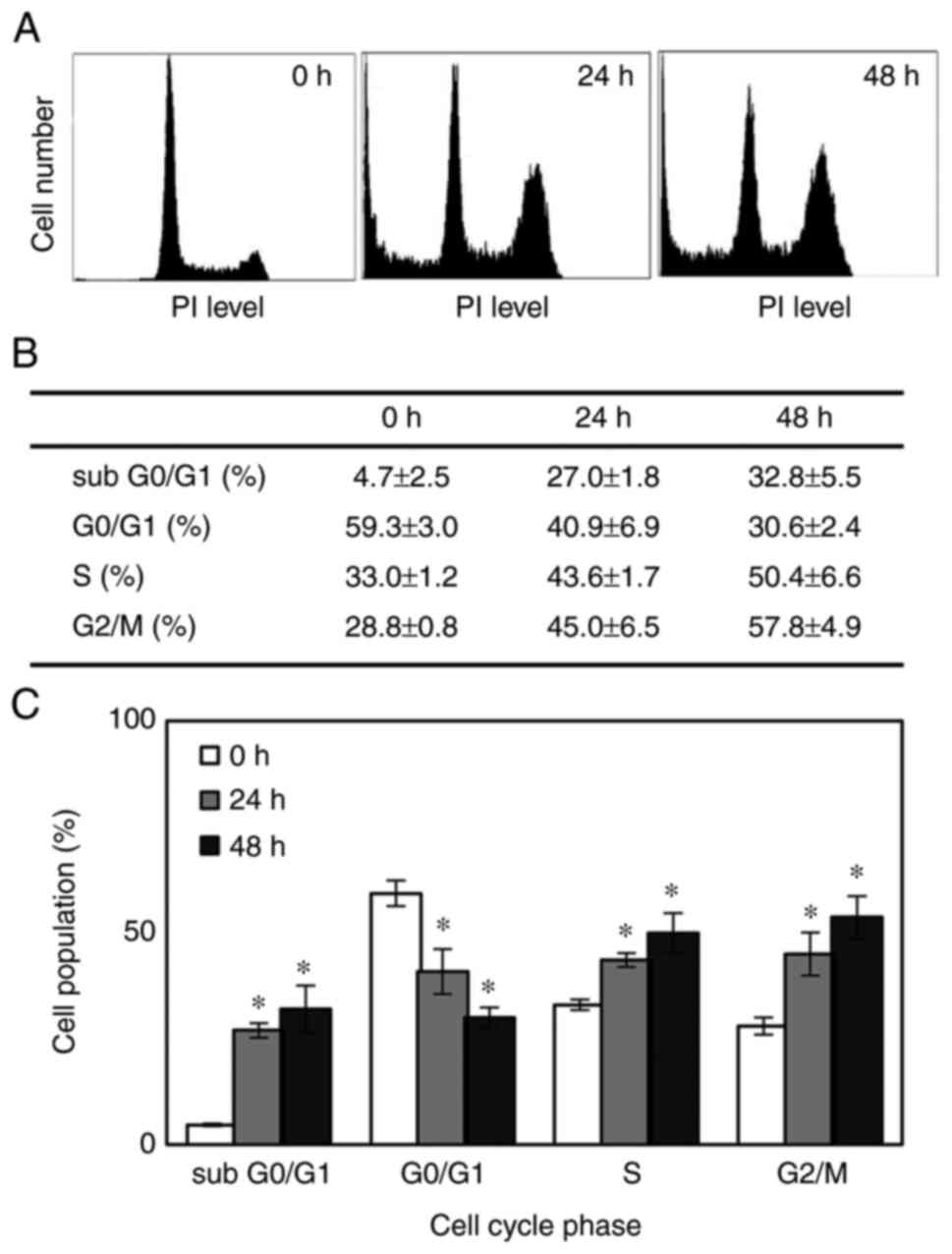
Representative cell cycle distributions following consecutive
treatment with SFN at the indicated time points are shown in
Fig. 2A. When the MDA-MB-468 cells
were treated with 2 µM SFN, the proportion of cells in the S and
G2/M phases significantly increased from 33 and 28.8% at the
beginning of the treatment to 43.6 and 45.0%, respectively, with a
corresponding decrease in the number of cells in the G0/G1 phase
following 24 h of exposure. The percentage of cells in each cell
cycle phase was not significantly altered after 48 h consecutive
exposure compared with the 24-h time point (Fig. 2B and C). Therefore, SFN arrested the cell cycle
at the S phase and more predominantly at the G2/M phase.
The sub G0/G1 cell population, which represents
apoptotic cells, abruptly increased from 4.7 to 27.0 and 32.8%
following 24 and 48 h of exposure, respectively (Fig. 2B and C), increasing by almost 7-fold following
48 h of exposure. Furthermore, the cleavage of PARP, which serves
as an early marker of apoptosis, was demonstrated at 48 h
post-treatment (Fig. 3A). The
dissociation between the appearance of the initiation of the
apoptotic cellular event (cell population at the sub G0/G1 phase)
and the cleavage of PARP may be explained by the difference in
their detection times. These data indicated that the observed
SFN-induced growth decline appeared to be due to the combined
effects of the progressive expansion of the apoptotic cell
population and the S/G2/M arrest of the cell cycle.
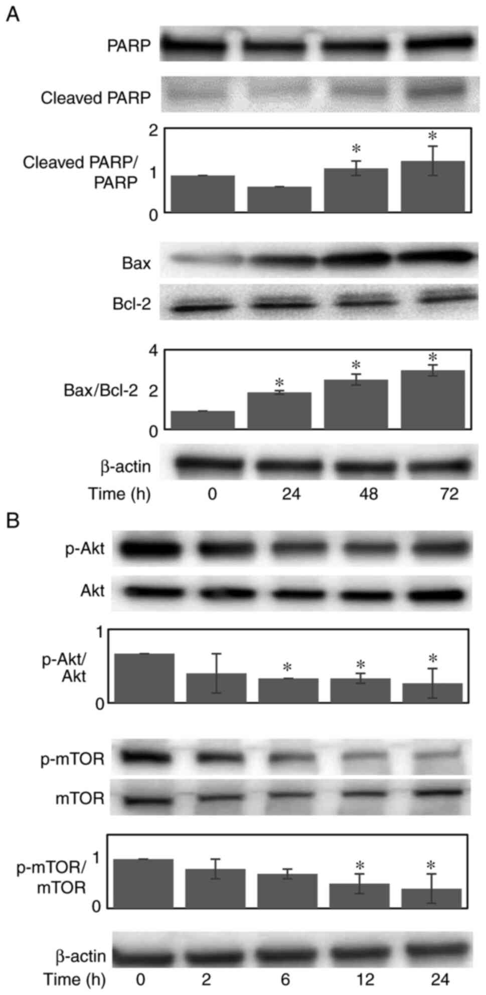 | Figure 3Effect of SFN on the activation of
signaling molecules for cell proliferation and apoptosis/survival.
Cells were treated with 2 µM SFN for the indicated periods of time
and harvested for western blot analyses. (A) Representative western
blots for the effects of SFN on apoptotic Bax, anti-apoptotic Bcl-2
and PARP. (B) Representative western blots are shown for total and
phosphorylated Akt, p-Akt (ser473), mTOR and p-mTOR (ser2448).
β-actin was used as the internal control. The column bars of
cleaved PARP/PARP, Bax/Bcl-2, p-Akt/Akt, and p-mTOR/mTOR ratios at
the indicated time points are also shown. The vertical bars
indicate the mean expression level ± SD of three independent
experiments. *P<0.05, significant difference vs. the
control (0 h). SFN, sulforaphane; Bax, B-cell lymphoma
(Bcl)-2-associated X; Bcl-2, B-cell lymphoma-2; Akt, protein kinase
B; mTOR, mechanistic target of rapamycin; PARP, poly(ADP-ribose)
polymerase; p-, phosphorylated. |
Effects of SFN on the expression of
pro- and anti-apoptotic proteins
To clarify the apoptotic mechanisms induced by SFN,
the protein expression levels of anti-apoptotic Bcl-2 and
pro-apoptotic Bax were examined (Fig.
3A). Upon treatment with 2 µM SFN, the expression of Bax was
increased in a time-dependent manner, whereas the protein
expression of Bcl-2 remained unaltered. Thus, the Bax/Bcl-2 ratio
increased up to 3.2-fold following 72 h of consecutive treatment.
These results suggest that the increased expression of Bax plays a
causative role in SFN-induced apoptosis.
Effects of SFN on the activation of
Akt/mTOR signaling molecules
Since the activation of PI3K/Akt/mTOR, a major
signaling pathway involved in cell proliferation and survival, is
considered to be activated in MDA-MB-468 TNBC cells due to their
biological features, including the overexpression of EGFR and the
co-deletion of PTEN (15,16), the present study examined the
effects of SFN on the expression and activation (phosphorylation)
of these proteins. Upon treatment with 2 µM SFN, the
phosphorylation of Akt and mTOR was substantially inhibited in a
time-dependent manner (Fig. 3B).
These data thus indicated that the SFN-induced reduction in cell
proliferation and survival appeared to be mediated by the
inactivation of Akt and mTOR.
Effects of SFN on MDA-MB-468 cells
xenotransplanted into nude mice
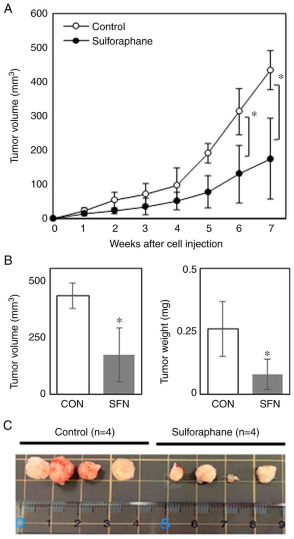
An in vivo experiment was conducted using
nude mice xenotransplanted with MDA-MB-468 cells to determine
whether the inhibitory effects of SFN on tumor development are
observed when administered orally to mice. Although the tumors grew
rapidly at ~5 weeks following cell inoculation in control mice, the
oral administration of SFN significantly suppressed tumor growth
from 5 weeks following inoculation (Fig. 4A), reducing the tumor size and
tumor weight by ~60 and 70%, respectively, compared with the
untreated control mice at 7 weeks following cell inoculation
(Fig. 4B). Images of the xenograft
tumors excised from four individual mice in each group at the end
of experiment are depicted in Fig.
4C. Although individual tumors varied in size, the tumor sizes
appeared to be evidently smaller in the SFN-treated group compared
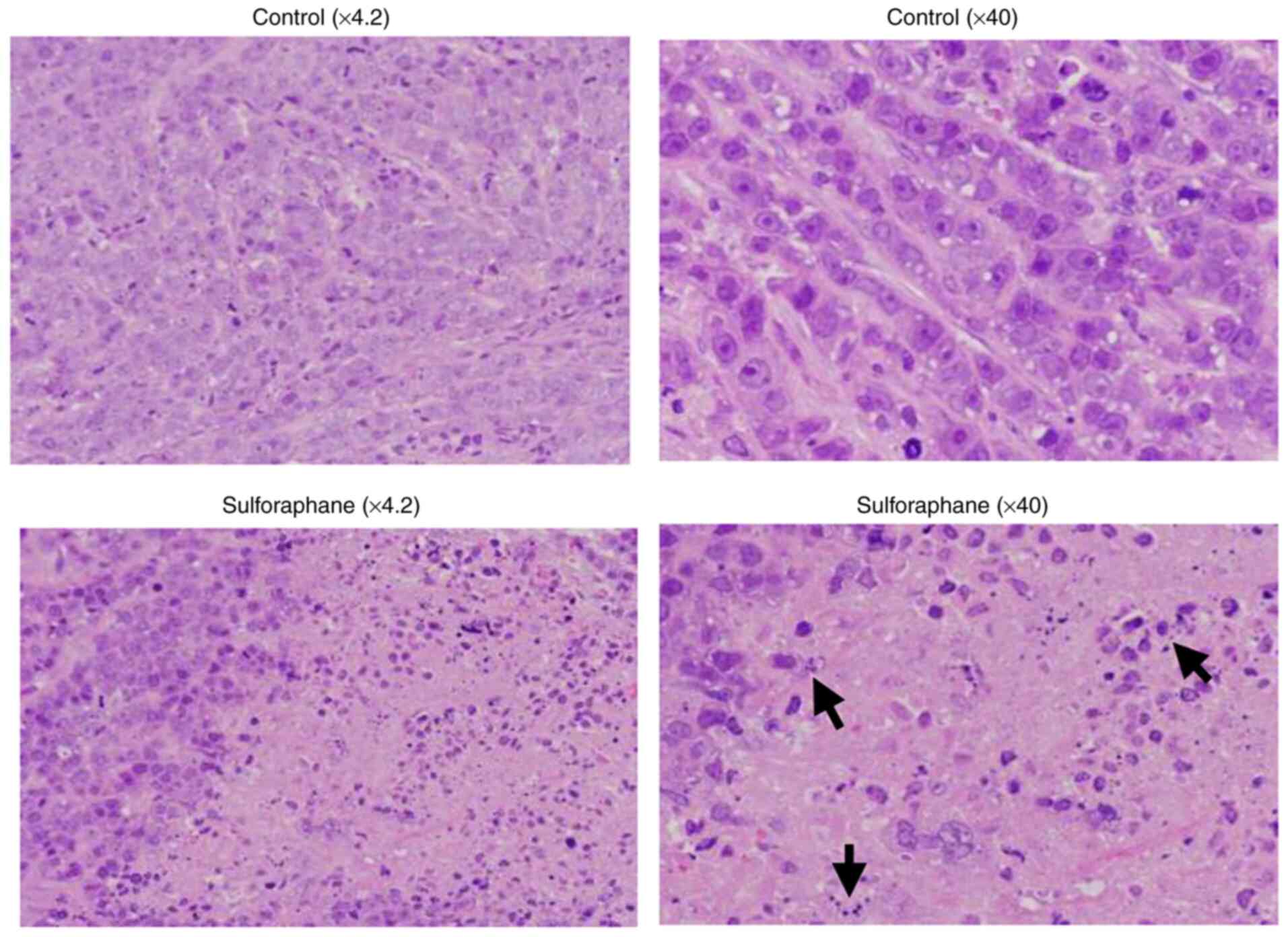
with the control group. Representative images of tumor tissue
sections stained with H&E are presented in Fig. 5. The tumor specimens from
SFN-treated mice exhibited a degenerated tumor cell appearance with
pyknotic nuclei, resembling apoptotic cells (Fig. 5). Furthermore, no significant
differences were observed in body weight and food consumption
between the two groups (Table I),
suggesting that SFN did not exert any or minimal adverse effects at
the oral concentration used in the present study. These data thus
indicated that SFN substantially inhibited TNBC tumor growth in
vivo without exerting any apparent toxic effects.
 | Table IBody weight and total food
consumption of mice xenotransplanted with MDA-MB-468 cells. |
Table I
Body weight and total food
consumption of mice xenotransplanted with MDA-MB-468 cells.
| Parameter | Control (n=4) | Sulforaphane
(n=4) |
P-valuea |
|---|
| Body
weightb (g) | 22.5±1.82 | 23.5±1.61 | 0.468 |
| Food
intakec (g) | 210.4±26.72 | 213.3±27.41 | 0.356 |
Discussion
SFN is widely recognized as a promising
chemopreventive agent with effects against numerous types of human
cancers through a variety of mechanisms (20). Moreover, SFN has been reported to
potentially prevent breast cancer development and recurrence
(7). In the present study, it was
found that SFN inhibited the proliferation of MDA-MB-468 TNBC cells
with an IC50 value of ~2 µM following 72 h of exposure.
This IC50 value appears to be quite low compared with
those of various phytochemicals tested in the authors' laboratory
for TNBC (21-24),
indicating that SFN is a promising candidate for preventing TNBC
among various phytochemicals.
Moreover, the present in vivo experiment
using nude mice xenotransplanted with MDA-MB-468 cells revealed
that the per os administration of SFN evidently attenuated
tumor growth over a period of 7 weeks, reducing the tumor size by
60% compared with the control. Adverse effects to major organs were
considered negligible as there were no significant differences in
body weight gain and the consumption of food between the two
groups, although this is indirect evidence of the adverse effects.
More precisely, the examinations of hematological and biochemical
toxicities are required in nude mice. A similar result has been
reported using MDA-MB-231 TNBC xenografts (25); however, to the best of our
knowledge, the present study is the first to demonstrate the in
vivo antitumor activity of SFN against MDA-MB-468 cells.
Moreover, SFN has been reported to synergistically enhance the
efficacy of several anticancer drugs, including cisplatin (26), doxorubicin (25,27)
and paclitaxel (28) in various
types of human cancer cells. Therefore, co-treatment with
chemotherapeutic agents and SFN may reduce the administered doses,
thereby alleviating the adverse effects of anticancer agents.
Furthermore, it has been reported that human subjects who ingested
100 g of broccoli daily as a soup exhibited a peak plasma
concentration of ~2-7 µM SFN metabolites, including free SFN
(29). Thus, it is conceivable
that consuming a diet rich in cruciferous vegetables, such as
broccoli sprouts, may reduce the risk and development of TNBC and
may potentially be useful for the treatment of TNBC.
Cell cycle checkpoints are crucial for controlling
the mechanisms that ensure the proper execution of cell cycle
events. SFN has been shown to modulate cell cycle progression in
several cellular models, such as prostate, colon, breast and
bladder cancers, arresting cells in the G1 (30,31)
or the G2/M phase (32-34),
depending on the cell type, the treatment concentration and the
duration of exposure (35). In the
present study, SFN inhibited the proliferation of MDA-MB-468 TNBC
cells by inducing S/G2/M cell cycle arrest, causing a blockade of
cell cycle entry into mitosis, as previously shown in different
TNBC cell lines, including MDA-MB-231(36). Furthermore, the sub G0/G1 cell
population, which represents apoptotic cells, increased, followed
by the cleavage of PARP, which serves as a marker of cells
undergoing apoptosis. Therefore, these data indicate that the
inhibitory effects of SFN observed in cytotoxicity assays reflect
the combination of SFN-induced S/G2/M cell cycle arrest and
apoptotic cell death.
Although SFN has been found to induce the apoptosis
of a variety of breast cancer cells, the mechanisms through which
SFN induces apoptosis varies between different cells. The present
study demonstrated that SFN upregulated the protein expression of
pro-apoptotic Bax, which has been shown to induce apoptosis by
promoting the release of cytochrome c, as a result of its
translocation from the cytosol to the mitochondria (37). SFN has also been reported to induce
the downregulation of anti-apoptotic Bcl-2 protein in several
breast cancer cell lines (36). A
previous study using MDA-MB-468 cells reported that Bcl-2 levels
decreased in a concentration-dependent manner from 5 µM (36). In the present study, however, Bcl-2
expression was not altered. This may be due to the concentration
that we used for the experiment in which the effect of SFN on
apoptosis signaling proteins was evaluated at a concentration of 2
µM. Nonetheless, the resultant increase in the Bax/Bcl-2 ratio may
play an important role in SFN-induced apoptosis in MDA-MB-468 cells
at such a low level of SFN.
Studies on the mechanisms underlying the anticancer
activities of SFN have indicated that its regulatory effects on the
tumor cell cycle, apoptosis and angiogenesis are mediated by the
modulation of the related signaling pathways (6-8).
MDA-MB-468 TNBC cells are devoid of PTEN, which antagonizes the
activity of PI3K, and exhibit high levels of EGFR (15,16),
which functions upstream of PI3K. Akt plays a critical role in
controlling survival by directly phosphorylating mTOR at
Ser2448(38), leading to an
increase in downstream molecules (39). Upon treatment of the MDA-MB-468
cells with SFN, Akt activity was inhibited with a simultaneous
decrease in mTOR activity, indicating that SFN substantially
inhibited the PI3K/Akt/mTOR pathway even in cells with the
overactivation of the downstream pathway caused by the
overexpression of EGFR and co-deletion of PTEN. Since EGFR
overexpression and loss of PTEN are frequently occurring events in
TNBC cases, and are associated with aggressive behavior and a poor
prognosis of patients with TNBC (10,12,13),
SFN appears to be a promising target drug acting against survival
signaling downstream of PI3K in these patients. Moreover, such
activities of SFN appear to be crucial as mitogenic and
anti-apoptotic potentials are driven by the activation of
intracellular signaling molecules upstream of PI3K in almost all
cancer cells.
Despite recent advances in breast cancer treatment,
breast cancer recurrence is a major problem and the principal cause
of breast cancer-related deaths. Emerging evidence suggests the
existence of cancer stem cells, a population of cells capable of
self-renewal and initiating tumor growth, which might be
responsible for breast cancer recurrence (40). Recently, SFN has gained immense
attention due to its wide safety profile and ability to target
heterogeneous populations of cancer cells, including cancer stem
cells. Accordingly, SFN has been shown to reduce the tumor volume
in TNBC stem-like cells (MDA-MB-231-Luc-D3H1 cell line)
administered daily by intraperitoneal injection (41). Moreover, Burnett et al
(42) reported that the
intraperitoneal injection of SFN enhanced the anticancer activity
of taxanes against TNBC by killing cancer stem cells. In the
present study, the oral administration of SFN inhibited the growth
of xenotransplanted MDA-MB-468 TNBC tumors consisting of a
population of in vivo selected and thus highly tumorigenic
cells resembling cancer stem cells. The major difference between
the two aforementioned previous studies and the present study is
the design of the administration route of SFN. The present
experimental design mimics the ordinary method of SFN uptake
included in dietary vegetables. Therefore, absorption routes via
either the intestine or peritoneal membrane may greatly affect the
pharmacokinetics of SFN administered via either route. To the best
of our knowledge, the present study is also the first to
demonstrate the in vivo antitumor activity of SFN against
the MDA-MB-468 TNBC cell line exhibiting the overactivation of the
signaling pathway downstream of EGFR due to EGFR overexpression and
the deletion of PTEN, as opposed to SUM149 cells, which possess
tumor suppressor BRCA1 mutation (43).
In conclusion, the data of the present study suggest
that SFN may prove to be potentially useful, not only for the
prevention and treatment, but also for the reduction of the
recurrence of TNBC.
Acknowledgements
The authors would like to thank Mrs. Takako Higuchi,
Graduate School of Health and Nutritional Sciences, Nakamura Gakuen
University for providing technical assistance.
Funding
Funding: The present study was supported by the Japan Society
for the Promotion of Science (JSPS) KAKENHI (Grants-in-Aid for
Scientific Research; grant nos. 15K00864 and 26750059). The present
study was also supported by the 2019 Cancer Research Fund of the
Fukuoka Foundation for Sound Health.
Availability of data and materials
The datasets used and/or analyzed during the current
study are available from the corresponding author on reasonable
request.
Authors' contributions
AY, MO and SN designed the study. AY and MO
conducted the research. AY, MO, and MT analyzed the data. MO and SN
wrote the manuscript. All authors had the primary responsibility
for the final content, and read and approved the final manuscript.
All authors confirmed the authenticity of the raw data.
Ethics approval and consent to
participate
All animal procedures were performed in accordance
with the protocols approved by the Institutional Animal Care and
Use Committee of the Nakamura Gakuen University (no. 2018-1).
Patient consent for publication
Not applicable.
Competing interests
The authors declare that they have no competing
interests.
References
|
1
|
Sung H, Ferlay J, Siegel RL, Laversanne M,
Soerjomataram I, Jemal A and Bray F: Global cancer statistics 2020:
GLOBOCAN estimates of incidence and mortality worldwide for 36
cancers in 185 countries. CA Cancer J Clin. 71:209–249.
2021.PubMed/NCBI View Article : Google Scholar
|
|
2
|
Kumar P and Aggarwal R: An overview of
triple-negative breast cancer. Arch Gynecol Obstet. 293:247–269.
2016.PubMed/NCBI View Article : Google Scholar
|
|
3
|
Liu X and Lv K: Cruciferous vegetables
intake is inversely associated with risk of breast cancer: A
meta-analysis. Breast. 22:309–313. 2013.PubMed/NCBI View Article : Google Scholar
|
|
4
|
Higdon JV, Delage B, Williams DE and
Dashwood RH: Cruciferous vegetables and human cancer risk:
Epidemiologic evidence and mechanistic basis. Pharmacol Res.
55:224–236. 2007.PubMed/NCBI View Article : Google Scholar
|
|
5
|
Fahey JW, Wehage SL, Holtzclaw WD, Kensler
TW, Egner PA, Shapiro TA and Talalay P: Protection of humans by
plant glucosinolates: Efficiency of conversion of glucosinolates to
isothiocyanates by the gastrointestinal microflora. Cancer Prev Res
(Phila). 5:603–611. 2012.PubMed/NCBI View Article : Google Scholar
|
|
6
|
Jabbarzadeh Kaboli P, Afzalipour
Khoshkbejari M, Mohammadi M, Abiri A, Mokhtarian R, Vazifemand R,
Amanollahi S, Yazdi Sani S, Li M, Zhao Y, et al: Targets and
mechanisms of sulforaphane derivatives obtained from cruciferous
plants with special focus on breast cancer-contradictory effects
and future perspectives. Biomed Pharmacother.
121(109635)2020.PubMed/NCBI View Article : Google Scholar
|
|
7
|
Kuran D, Pogorzelska A and Wiktorska K:
Breast cancer prevention-is there a future for sulforaphane and its
analogs? Nutrients. 12(1559)2020.PubMed/NCBI View Article : Google Scholar
|
|
8
|
Clarke JD, Dashwood RH and Ho E:
Multi-targeted prevention of cancer by sulforaphane. Cancer Lett.
269:291–304. 2008.PubMed/NCBI View Article : Google Scholar
|
|
9
|
Costa RLB, Han HS and Gradishar WJ:
Targeting the PI3K/AKT/mTOR pathway in triple-negative breast
cancer: A review. Breast Cancer Res Treat. 169:397–406.
2018.PubMed/NCBI View Article : Google Scholar
|
|
10
|
Rimawi MF, Shetty PB, Weiss HL, Schiff R,
Osborne CK, Chamness GC and Elledge RM: Epidermal growth factor
receptor expression in breast cancer association with biologic
phenotype and clinical outcomes. Cancer. 116:1234–1242.
2010.PubMed/NCBI View Article : Google Scholar
|
|
11
|
Lou L, Yu Z, Wang Y, Wang S and Zhao Y:
c-Src inhibitor selectively inhibits triple-negative breast cancer
overexpressed Vimentin in vitro and in vivo. Cancer Sci.
109:1648–1659. 2018.PubMed/NCBI View Article : Google Scholar
|
|
12
|
Dean SJ, Perks CM, Holly JM, Bhoo-Pathy N,
Looi LM, Mohammed NA, Mun KS, Teo SH, Koobotse MO, Yip CH and
Rhodes A: Loss of PTEN expression is associated with IGFBP2
expression, younger age, and late stage in triple-negative breast
cancer. Am J Clin Pathol. 141:323–333. 2014.PubMed/NCBI View Article : Google Scholar
|
|
13
|
Beg S, Siraj AK, Prabhakaran S, Jehan Z,
Ajarim D, Al-Dayel F, Tulbah A and Al-Kuraya KS: Loss of PTEN
expression is associated with aggressive behavior and poor
prognosis in Middle Eastern triple-negative breast cancer. Breast
Cancer Res Treat. 151:541–553. 2015.PubMed/NCBI View Article : Google Scholar
|
|
14
|
Pawlik A, Wiczk A, Kaczynska A,
Antosiewicz J and Herman-Antosiewicz A: Sulforaphane inhibits
growth of phenotypically different breast cancer cells. Eur J Nutr.
52:1949–1958. 2013.PubMed/NCBI View Article : Google Scholar
|
|
15
|
Liu T, Yacoub R, Taliaferro-Smith LD, Sun
SY, Graham TR, Dolan R, Lobo C, Tighiouart M, Yang L, Adams A and
O'Regan RM: Combinatorial effects of lapatinib and rapamycin in
triple-negative breast cancer cells. Mol Cancer Ther. 10:1460–1469.
2011.PubMed/NCBI View Article : Google Scholar
|
|
16
|
Lu Y, Lin YZ, LaPushin R, Cuevas B, Fang
X, Yu SX, Davies MA, Khan H, Furui T, Mao M, et al: The
PTEN/MMAC1/TEP tumor suppressor gene decreases cell growth and
induces apoptosis and anoikis in breast cancer cells. Oncogene.
18:7034–7045. 1999.PubMed/NCBI View Article : Google Scholar
|
|
17
|
Neve RM, Chin K, Fridlyand J, Yeh J,
Baehner FL, Fevr T, Clark L, Bayani N, Coppe JP, Tong F, et al: A
collection of breast cancer cell lines for the study of
functionally distinct cancer subtypes. Cancer Cell. 10:515–527.
2006.PubMed/NCBI View Article : Google Scholar
|
|
18
|
Singh SV, Warin R, Xiao D, Powolny AA,
Stan SD, Arlotti JA, Zeng Y, Hahm ER, Marynowski SW, Bommareddy A,
et al: Sulforaphane inhibits prostate carcinogenesis and pulmonary
metastasis in TRAMP mice in association with increased cytotoxicity
of natural killer cells. Cancer Res. 69:2117–2125. 2009.PubMed/NCBI View Article : Google Scholar
|
|
19
|
Singh AV, Xiao D, Lew KL, Dhir R and Singh
SV: Sulforaphane induces caspase-mediated apoptosis in cultured
PC-3 human prostate cancer cells and retards growth of PC-3
xenografts in vivo. Carcinogenesis. 25:83–90. 2004.PubMed/NCBI View Article : Google Scholar
|
|
20
|
Jiang X, Liu Y, Ma L, Ji R, Qu Y, Xin Y
and Lv G: Chemopreventive activity of sulforaphane. Drug Des Devel
Ther. 12:2905–2913. 2018.PubMed/NCBI View Article : Google Scholar
|
|
21
|
Takeshima M, Ono M, Higuchi T, Chen C,
Hara T and Nakano S: Anti-proliferative and apoptosis-inducing
activity of lycopene against three subtypes of human breast cancer
cell lines. Cancer Sci. 105:252–257. 2014.PubMed/NCBI View Article : Google Scholar
|
|
22
|
Wakimoto R, Ono M, Takeshima M, Higuchi T
and Nakano S: Differential anticancer activity of pterostilbene
against three subtypes of human breast cancer cells. Anticancer
Res. 37:6153–6159. 2017.PubMed/NCBI View Article : Google Scholar
|
|
23
|
Ono M, Takeshima M, Nishi A, Higuchi T and
Nakano S: Genistein suppresses v-Src-driven proliferative activity
by arresting the cell-cycle at G2/M through increasing p21 level in
Src-activated human gallbladder carcinoma cells. Nutr Cancer.
73:1471–1479. 2021.PubMed/NCBI View Article : Google Scholar
|
|
24
|
Chen C, Ono M, Takeshima M and Nakano S:
Antiproliferative and apoptosis-inducing activity of nobiletin
against three subtypes of human breast cancer cell lines.
Anticancer Res. 34:1785–1792. 2014.PubMed/NCBI
|
|
25
|
Yang F, Wang F, Liu Y, Wang S, Li X, Huang
Y, Xia Y and Cao C: Sulforaphane induces autophagy by inhibition of
HDAC6-mediated PTEN activation in triple negative breast cancer
cells. Life Sci. 213:149–157. 2018.PubMed/NCBI View Article : Google Scholar
|
|
26
|
Gong TT, Liu XD, Zhan ZP and Wu QJ:
Sulforaphane enhances the cisplatin sensitivity through regulating
DNA repair and accumulation of intracellular cisplatin in ovarian
cancer cells. Exp Cell Res. 393(112061)2020.PubMed/NCBI View Article : Google Scholar
|
|
27
|
Bose C, Awasthi S, Sharma R, Beneš H,
Hauer-Jensen M, Boerma M and Singh SP: Sulforaphane potentiates
anticancer effects of doxorubicin and attenuates its cardiotoxicity
in a breast cancer model. PLoS One. 13(e0193918)2018.PubMed/NCBI View Article : Google Scholar
|
|
28
|
Kim SH, Park HJ and Moon DO: Sulforaphane
sensitizes human breast cancer cells to paclitaxel-induced
apoptosis by downregulating the NF-κB signaling pathway. Oncol
Lett. 13:4427–4432. 2017.PubMed/NCBI View Article : Google Scholar
|
|
29
|
Gasper AV, Al-Janobi A, Smith JA, Bacon
JR, Fortun P, Atherton C, Taylor MA, Hawkey CJ, Barrett DA and
Mithen RF: Glutathione S-transferase M1 polymorphism and metabolism
of sulforaphane from standard and high-glucosinolate broccoli. Am J
Clin Nutr. 82:1283–1291. 2005.PubMed/NCBI View Article : Google Scholar
|
|
30
|
Chiao JW, Chung FL, Kancherla R, Ahmed T,
Mittelman A and Conaway CC: Sulforaphane and its metabolite mediate
growth arrest and apoptosis in human prostate cancer cells. Int J
Oncol. 20:631–636. 2002.PubMed/NCBI View Article : Google Scholar
|
|
31
|
Shan Y, Sun C, Zhao X, Wu K, Cassidy A and
Bao Y: Effect of sulforaphane on cell growth, G(0)/G(1) phase cell
progression and apoptosis in human bladder cancer T24 cells. Int J
Oncol. 29:883–888. 2006.PubMed/NCBI
|
|
32
|
Tang L and Zhang Y: Dietary
isothiocyanates inhibit the growth of human bladder carcinoma
cells. J Nutr. 134:2004–2010. 2004.PubMed/NCBI View Article : Google Scholar
|
|
33
|
Jackson SJ and Singletary KW: Sulforaphane
inhibits human MCF-7 mammary cancer cell mitotic progression and
tubulin polymerization. J Nutr. 134:2229–2236. 2004.PubMed/NCBI View Article : Google Scholar
|
|
34
|
Parnaud G, Li P, Cassar G, Rouimi P,
Tulliez J, Combaret L and Gamet-Payrastre L: Mechanism of
sulforaphane-induced cell cycle arrest and apoptosis in human colon
cancer cells. Nutr Cancer. 48:198–206. 2004.PubMed/NCBI View Article : Google Scholar
|
|
35
|
Lenzi M, Fimognari C and Hrelia P:
Sulforaphane as a promising molecule for fighting cancer. Cancer
Treat Res. 159:207–223. 2014.PubMed/NCBI View Article : Google Scholar
|
|
36
|
Pledgie-Tracy A, Sobolewski MD and
Davidson NE: Sulforaphane induces cell type-specific apoptosis in
human breast cancer cell lines. Mol Cancer Ther. 6:1013–1021.
2007.PubMed/NCBI View Article : Google Scholar
|
|
37
|
Dewson G and Kluck RM: Mechanisms by which
Bak and Bax permeabilise mitochondria during apoptosis. J Cell Sci.
122:2801–2808. 2009.PubMed/NCBI View Article : Google Scholar
|
|
38
|
Chiang GG and Abraham RT: Phosphorylation
of mammalian target of rapamycin (mTOR) at Ser-2448 is mediated by
p70S6 kinase. J Biol Chem. 280:25485–25490. 2005.PubMed/NCBI View Article : Google Scholar
|
|
39
|
Nitulescu GM, Van De Venter M, Nitulescu
G, Ungurianu A, Juzenas P, Peng Q, Olaru OT, Grădinaru D, Tsatsakis
A, Tsoukalas D, et al: The Akt pathway in oncology therapy and
beyond (review). Int J Oncol. 53:2319–2331. 2018.PubMed/NCBI View Article : Google Scholar
|
|
40
|
Butti R, Gunasekaran VP, Kumar TVS,
Banerjee P and Kundu GC: Breast cancer stem cells: Biology and
therapeutic implications. Int J Biochem Cell Biol. 107:38–52.
2019.PubMed/NCBI View Article : Google Scholar
|
|
41
|
Castro NP, Rangel MC, Merchant AS,
MacKinnon G, Cuttitta F, Salomon DS and Kim YS: Sulforaphane
suppresses the growth of triple-negative breast cancer stem-like
cells in vitro and in vivo. Cancer Prev Res (Phila). 12:147–158.
2019.PubMed/NCBI View Article : Google Scholar
|
|
42
|
Burnett JP, Lim G, Li Y, Shah RB, Lim R,
Paholak HJ, McDermott SP, Sun L, Tsume Y, Bai S, et al:
Sulforaphane enhances the anticancer activity of taxanes against
triple negative breast cancer by killing cancer stem cells. Cancer
Lett. 394:52–64. 2017.PubMed/NCBI View Article : Google Scholar
|
|
43
|
Elstrodt F, Hollestelle A, Nagel JH, Gorin
M, Wasielewski M, van den Ouweland A, Merajver SD, Ethier SP and
Schutte M: BRCA1 mutation analysis of 41 human breast cancer cell
lines reveals three new deleterious mutants. Cancer Res. 66:41–45.
2006.PubMed/NCBI View Article : Google Scholar
|