TGF-β1 and IGF-1 influence the re-differentiation capacity of human chondrocytes in 3D pellet cultures in relation to different oxygen concentrations

  • Authors:
    • Anika Jonitz
    • Katrin Lochner
    • Thomas Tischer
    • Doris Hansmann
    • Rainer Bader
  • View Affiliations

  • Published online on: June 25, 2012     https://doi.org/10.3892/ijmm.2012.1042
  • Pages: 666-672
Metrics: Total Views: 0 (Spandidos Publications: | PMC Statistics: )
Total PDF Downloads: 0 (Spandidos Publications: | PMC Statistics: )


Abstract

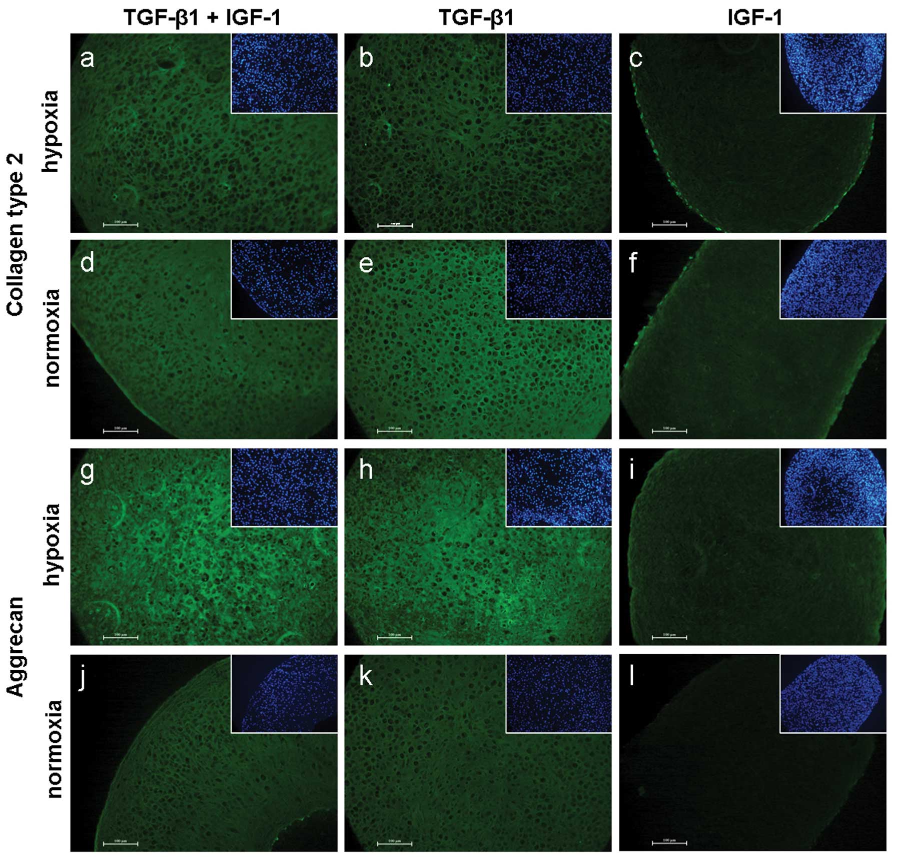
To prevent de-differentiation of chondrocytes in vitro, the 3D environment, growth factors and different oxygen concentrations were considered. In this in vitro study, we quantified the influence of insulin-like growth factor (IGF)-1 and/or transforming growth factor (TGF)-β1 under differing oxygen (5/21% O2) levels on the proliferation and synthesis rates of hyaline extracellular matrix (ECM) components in chondrogenic pellet cultures. Human chondrocytes isolated from articular cartilage were transferred into conical tubes to form pellets. Pellets were stimulated with TGF-β1 and/or IGF-1. After 2 and 5 weeks of cultivation the DNA concentration and expression of pro-collagen type 1, type 2 and aggrecan were analysed. Under hypoxia the DNA content remained stable. In contrast, under normoxia, cells showed an increase of DNA concentration after stimulation with TGF-β1/IGF-1 and TGF-β1. Nevertheless, DNA contents under normoxia did not reach the values of hypoxic-cultivated cells. Under both culture conditions a reduced synthesis of pro-collagen type 1 could be determined. Although the expression of pro-collagen type 2 was significantly higher under normoxia, a decrease in the case of TGF-β1/IGF-1- and IGF-1-stimulated cells was observed. Under hypoxia pro-collagen type 2 contents remained stable or increased for TGF-β1/IGF-1-stimulated cells. Furthermore, incubation with growth factors resulted in aggrecan accumulation under hypoxia, while a reduced expression under normoxia could be determined for TGF-β1/IGF-1- and IGF-1-stimulated cells. Our results demonstrate that the treatment with growth factors causes differences in the expression of ECM compounds within pellet cultures. While under normoxia TGF-β1 alone leads to a positive effect of the expression of hyaline cartilage-specific ECM components, an additive effect of both growth factors was only determined under hypoxia.
View Figures
View References

Related Articles

Journal Cover

September 2012
Volume 30 Issue 3

Print ISSN: 1107-3756
Online ISSN:1791-244X

Sign up for eToc alerts

Recommend to Library

Copy and paste a formatted citation
x
Spandidos Publications style
Jonitz A, Lochner K, Tischer T, Hansmann D and Bader R: TGF-β1 and IGF-1 influence the re-differentiation capacity of human chondrocytes in 3D pellet cultures in relation to different oxygen concentrations. Int J Mol Med 30: 666-672, 2012
APA
Jonitz, A., Lochner, K., Tischer, T., Hansmann, D., & Bader, R. (2012). TGF-β1 and IGF-1 influence the re-differentiation capacity of human chondrocytes in 3D pellet cultures in relation to different oxygen concentrations. International Journal of Molecular Medicine, 30, 666-672. https://doi.org/10.3892/ijmm.2012.1042
MLA
Jonitz, A., Lochner, K., Tischer, T., Hansmann, D., Bader, R."TGF-β1 and IGF-1 influence the re-differentiation capacity of human chondrocytes in 3D pellet cultures in relation to different oxygen concentrations". International Journal of Molecular Medicine 30.3 (2012): 666-672.
Chicago
Jonitz, A., Lochner, K., Tischer, T., Hansmann, D., Bader, R."TGF-β1 and IGF-1 influence the re-differentiation capacity of human chondrocytes in 3D pellet cultures in relation to different oxygen concentrations". International Journal of Molecular Medicine 30, no. 3 (2012): 666-672. https://doi.org/10.3892/ijmm.2012.1042