Introduction
Aberrant accumulation of protein aggregates is a key
characteristic of several age-related degenerative disorders
(1). During life, cells are
chronically exposed to oxidative stress resulting from
mitochondrial inefficiency or dysfunction, which can cause a
variety of oxidative modifications to proteins such as thiolation,
glycation, phosphorylation and deamidation (2). The oxidatively damaged proteins are
prone to form into tangled aggregates by non-enzymatic reaction
unless they are degraded by the ubiquitin-proteasome system in a
timely manner (3).
Transglutaminase 2 (TGase2) is a member of a family
of enzymes that post-translationally modify proteins by catalyzing
an acyl transfer reaction between the γ-carboxamide group of
protein glutamine residues and the ɛ-amino group of lysine residues
(protein cross-linking), or polyamines (protein polyamination)
(4). The transamidation activity
of TGase2 is calcium-dependent and produces irreversible protein
polymers that are resistant to proteolytic degradation (5). Thus, TGase2 plays a crucial role in
the formation of insoluble protein aggregates and has been
implicated in the pathogenesis of many diseases termed
‘conformational diseases’ (4).
For example, TGase2 may contribute to the formation of crystallin
polymers in age-related cataracts (6), the aggregation of huntingtin protein
with an expanded polyglutamine domain as occurs in Huntington’s
disease (7), and the accumulation
of insoluble neurofibrillary tangles and β-amyloid plaques in
Alzheimer’s disease (8,9). We recently demonstrated that
reactive oxygen species (ROS) activate intracellular TGase2 in
various cell types (10) and that
the transforming growth factor β (TGFβ) signaling pathway is
involved in TGase2 activation (11). However, the role of TGase2 in
cellular responses to other stressors remain to be elucidated.
The accumulation of misfolded proteins in the
endoplasmic reticulum (ER) can trigger a specific stress response
called the unfolded protein response (UPR) (12). Accordingly, several chemical
inhibitors of protein folding procedures such as tunicamycin (TM;
inhibitor of N-glycosylation), dithiothreitol (DTT) or
β-mercaptoethanol (β-ME; permeable reducing agents), and
thapsigargin (TG; inhibitor of the Ca2+ pump in ER)
induce the UPR response. The UPR pathway is important for
regulation of normal cellular homeostasis and may also play key
roles in the pathology of conformational diseases (13). Cells can employ different UPR
programs depending on the level of ER stress (14). In response to moderate stress,
cells reduce the cellular burden of improperly folded proteins by
attenuating de novo protein synthesis through
phosphorylation of the protein translation initiation factor 2
(eIF2α) and by inducing the expression of chaperone proteins,
including several glucose response proteins (GRPs). By contrast,
sustained and unresolved ER stress may trigger programmed cell
death through activation of activating transcription factor 4
(ATF4), ATF6, CCAAT/enhance-binding protein homologous protein
(CHOP) and caspases (12,13,15,16). Moreover, at the cellular level, ER
stress induces an increase in intracellular Ca+
concentration and ROS generation (14,17). Of note, these intracellular
conditions are known to activate in situ transamidation
activity of TGase2 (10,11,18,19), suggesting that the aggregate
formation of misfolded proteins is accelerated by TGase2-mediated
protein modifications. Thus, it is reasonable to investigate the
likely relationship between protein misfolding stress and TGase2
activity. In the present study, we found that ER stress induces
TGase2 activation in various cell types, including lens epithelial
cells, and that the activated enzyme plays a critical role in the
formation of protein aggregates.
Materials and methods
Cell culture
Human lens epithelial (HLE-B3), erythroleukemia
(K562), cervical carcinoma (HeLa), and neuroblastoma (SH-SY5Y) cell
lines were cultured as previously described (10). For UPR activators, cells were
treated with culture media containing β-ME (Sigma, St. Louis, MO,
USA; 7.5 mM), DTT (Sigma; 3 mM), TG (Sigma; 1 mM) or TM (Sigma; 5
μg/ml) for the indicated times and then maintained in culture until
analysis. KCC009, a specific chemical inhibitor of TGase2 (20), was added at a concentration of 125
μM to inhibit transamidation activity. To differentiate SH-SY5Y
cells, the cells were treated with 5 μM retinoic acid (RA) (Sigma)
for 1 day before induction of ER stress.
Measurement of intracellular calcium
Ca2+ levels were measured by fluorimetry
using the Fluo-4AM (Molecular Probes, Carlsbad, CA, USA).
Approximately 3×10⁴ cells were grown overnight in a
96-well microplate. After exposure to ER stress, the cells were
incubated with 100 μl of assay buffer (Hanks’ balanced salt
solution in 20 mM HEPES, pH 7.4) containing 5 μM Fluo-4AM at 37°C
for 30 min and for an additional 30 min at room temperature. The
cells were then washed with the assay buffer 4 times, and then the
intensity of fluorescence was measured using a fluorescence
microplate reader (Cary Eclipse; Varian, Palo Alto, CA, USA) with
excitation set at 488 nm and emission set at 516 nm. After reading,
the cells were stained with crystal violet (Sigma) to normalize the
fluorescence value. Intracellular Ca2+ levels were
expressed as the ratio of values in ER stress-exposed cells to that
of untreated cells. EGTA (1.5 mM) and BAPTA-AM (20 μM, Molecular
Probes) were used for calcium chelation.
In situ transamidation assays
In situ TGase2 activity was measured by
determining the biotinylated pentylamine (BP) incorporated into
cellular proteins. Cells were incubated with 1 mM BP (Pierce,
Rockford, IL, USA) for 1 h prior to harvesting, and then the cell
extracts were prepared by sonication in phosphate-buffered saline
(PBS) with a protease inhibitor cocktail, followed by
centrifugation (14,000 × g, 10 min at 4°C). For the solid-phase
microtiter plate assay, cell extracts (0.2 mg/ml, 50 μl/well) in
coating buffer (50 mM Tris-Cl, pH 7.5, 150 mM NaCl, 5 mM EGTA, 5 mM
EDTA) were added to each well of a 96-well microtiter plate. In
situ TGase2 activity was evaluated by determining the
incorporated BP using HRP-conjugated streptavidin (Pierce),
followed by reaction with O-phenylenediamine dihydrochloride
(Sigma). Assays were quantified by measuring the absorbance at 490
nm on a microplate spectrophotometer (Molecular Devices). In
situ TGase2 activities were normalized by subtracting values
representing endogenous biotin-conjugated proteins that were
obtained without the addition of biotinylated pentylamine. In
situ TGase2 activity was presented as folds of activation,
compared to non-treated experiments. Western blot analysis was
performed by subjecting the cell extracts (30 μg) to SDS-PAGE using
a 12% gel, and the proteins were then transferred to nitrocellulose
membranes. The proteins incorporated with BP were probed with
HRP-conjugated streptavidin, followed by chemiluminescence
detection (Pierce).
Western blotting
The cell extracts (30 μg) were separated on 12%
SDS-PAGE gels following preparation in RIPA lysis buffer (Santa
Cruz Biotechnology, Inc., Santa Cruz, CA, USA). Protein levels were
assessed by probing with monoclonal antibodies specific for TGase2
(21) and β-actin (Santa Cruz
Biotechnology, Inc.). To investigate the TGase2-catalyzed
crosslinking of lens proteins, whole cell extracts were prepared by
sonication in homogenate buffer (50 mM Tris-Cl, pH 6.8, 6 M urea,
2% SDS, 40 mM DTT and a protease inhibitor cocktail) and further
centrifuged at 12,000 × g for 10 min at 4°C. Proteins were
quantified using the BCA method (Pierce), resolved on 6–15%
SDS-PAGE gels, and subsequently analyzed using antibodies specific
for αB-crystallin (Stressgen) and vimentin (Santa Cruz
Biotechnology, Inc.). For the solubility experiments, cell extracts
were separated into the water-soluble and water-insoluble fractions
as previously described (11).
Cell viability assay
Cell viability after treatment with UPR stress was
determined by MTT assay (Sigma) according to the manufacturer’s
protocol. The reduction of MTT reagent was quantified after 4 h by
measuring the absorbance at 570 nm on a microplate
spectrophotometer (Molecular Devices).
Statistical analysis
All data on TGase2 activity and cell viability were
analyzed using one-way or two-way ANOVA with Bonferroni post-tests.
All analyses were performed using GraphPad Prism 5.0 statistical
software (GraphPad Software, La Jolla, CA, USA). Statistical
significance was defined as p<0.05 or p<0.01.
Results
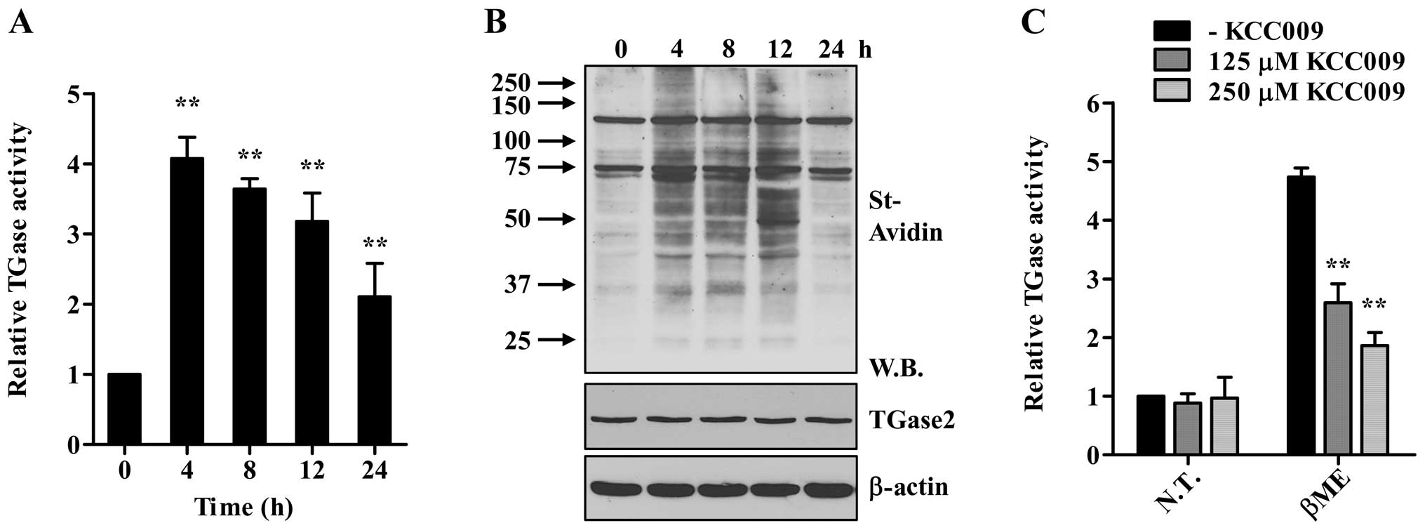
The aberrant activation of TGase2 accelerates the
pathological misfolding/aggregation of proteins (10,11). To test whether ER stress activates
TGase2, we measured the intracellular transamidation activity in
the HLE-B3 cells following treatment with β-ME. In situ
activity of TGase2 was monitored by incubating the cells with BP
and by measuring the BP-incorporated proteins in the cell extracts
using a well plate or SDS-PAGE assay. As shown in Fig. 1A and B, treatment with β-ME
significantly increased intracellular TGase2 activity, peaking at 4
h after treatment. The level of TGase2 protein was little affected
under this condition (Fig. 1B),
suggesting that latent TGase2 present under normal culture
conditions was activated by ER stress as previously observed in the
case of the oxidative stress (10,11). In addition, treatment with KCC009,
a chemical inhibitor for TGase2 abrogated the β-ME-induced increase
of TGase2 activity (Fig. 1C).
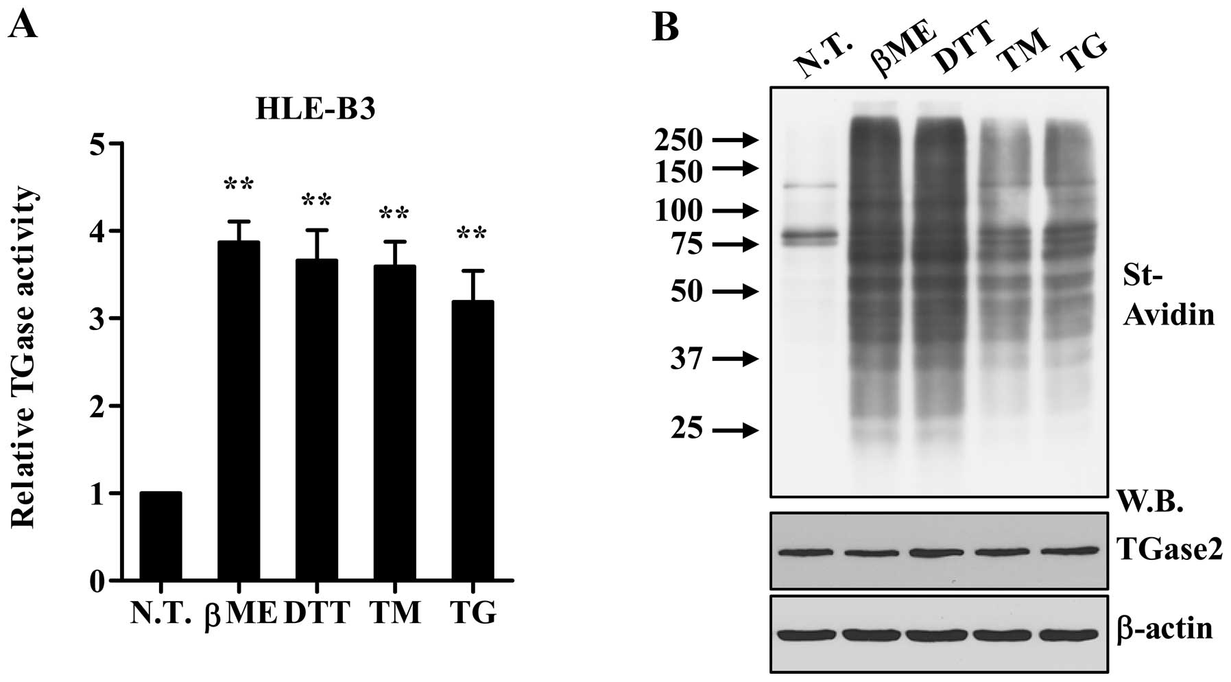
We next evaluated whether other ER stress-causing
agents affect the TGase2 activity in HLE-B3 cells. As shown in
Fig. 2, in situ TGase2
activity was similarly increased with little change in the protein
levels following treatment with most UPR activators, including DTT,
TG, and TM. Supra-physiological stressors, such as DTT and β-ME,
rapidly increased the TGase2 activity at ~4 h. By contrast,
treatment with TG or TM reached a peak of induced enzyme activity
by 24 h, indicating that each ER stress exhibited distinct TGase2
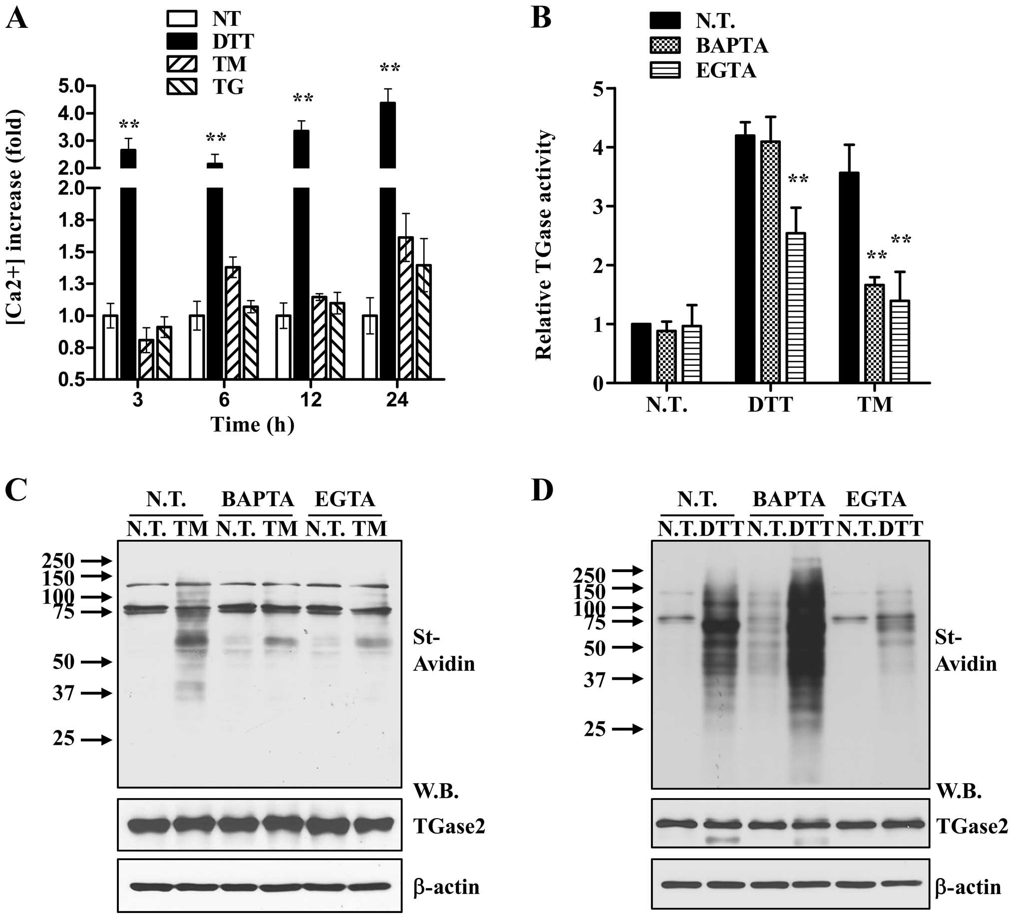
activation kinetics. Since the transamidation activity of TGase2 is
calcium-dependent (22),
intracellular calcium concentrations were measured following
treatment with different ER stress inducers. In correlation with
the observed time-dependent rise in TGase2 activity, Ca+
concentrations in HLE-B3 cells rapidly increased with DTT, but
increased more gradually over 24 h following TG or TM treatment
(Fig. 3A). In addition,
Ca2+ chelation by BAPTA-AM or EGTA abrogated the effect
of TM on TGase2 activity (Fig. 3B and
C), indicating that an elevated intracellular Ca2+
concentration is required for enzyme activation. The increased
TGase2 activity stimulated by DTT was partially attenuated by the
presence of EGTA; however it was little affected by treatment with
BAPTA-AM (Fig. 3B and D). The
TGase2 protein level was also similar in all of the conditions
tested. Therefore, these results demonstrate that UPR increases
intracellular calcium ion concentrations that activate TGase2.
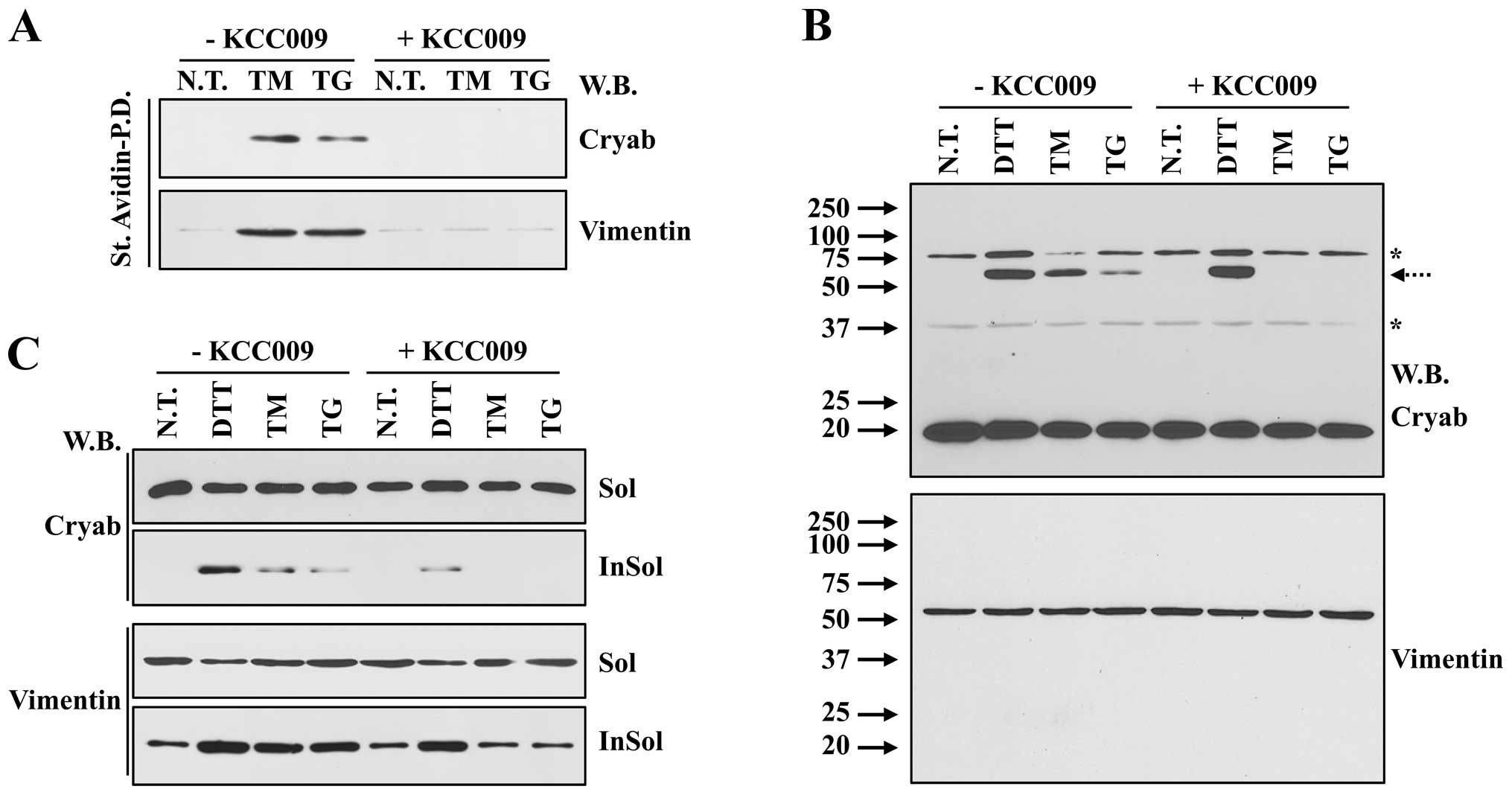
To gain insight into the potential
pathophysiological roles of TGase2, we evaluated whether TGase2
modifies individual lens proteins upon ER stress. To address this
issue, proteins cross-linked with BP by TGase2 were isolated using
streptavidin pull-down and subjected to western blot analysis. BP
modification of αB-crystallin and vimentin was observed
specifically in cells treated with TM or TG, but it was inhibited
by treatment with KCC009 (Fig.
4A). Previous studies report that the lens proteins modified by
TGase2 in response to oxidative stress or TGFβ stimulation were
dimerized and showed decreased water solubility (10,11). Similarly, treatment with each UPR
activator tested led to the formation of high-molecular-weight
protein of αB crystallin, but not vimentin (Fig. 4B). When the cell extracts were
separated into water soluble and insoluble fractions, both αB
crystallin and vimentin proteins in the water-insoluble fraction
increased in the cells exposed to ER stress (Fig. 4C). Moreover, KCC009 significantly
inhibited the protein cross-linking and water insolubility of lens
proteins stimulated by treatment with TG and TM, but not DTT
(Fig. 4B and C). These results
demonstrate that the activation of TGase2 under ER stress
conditions plays a pivotal role in the formation of water-insoluble
aggregates of lens proteins.
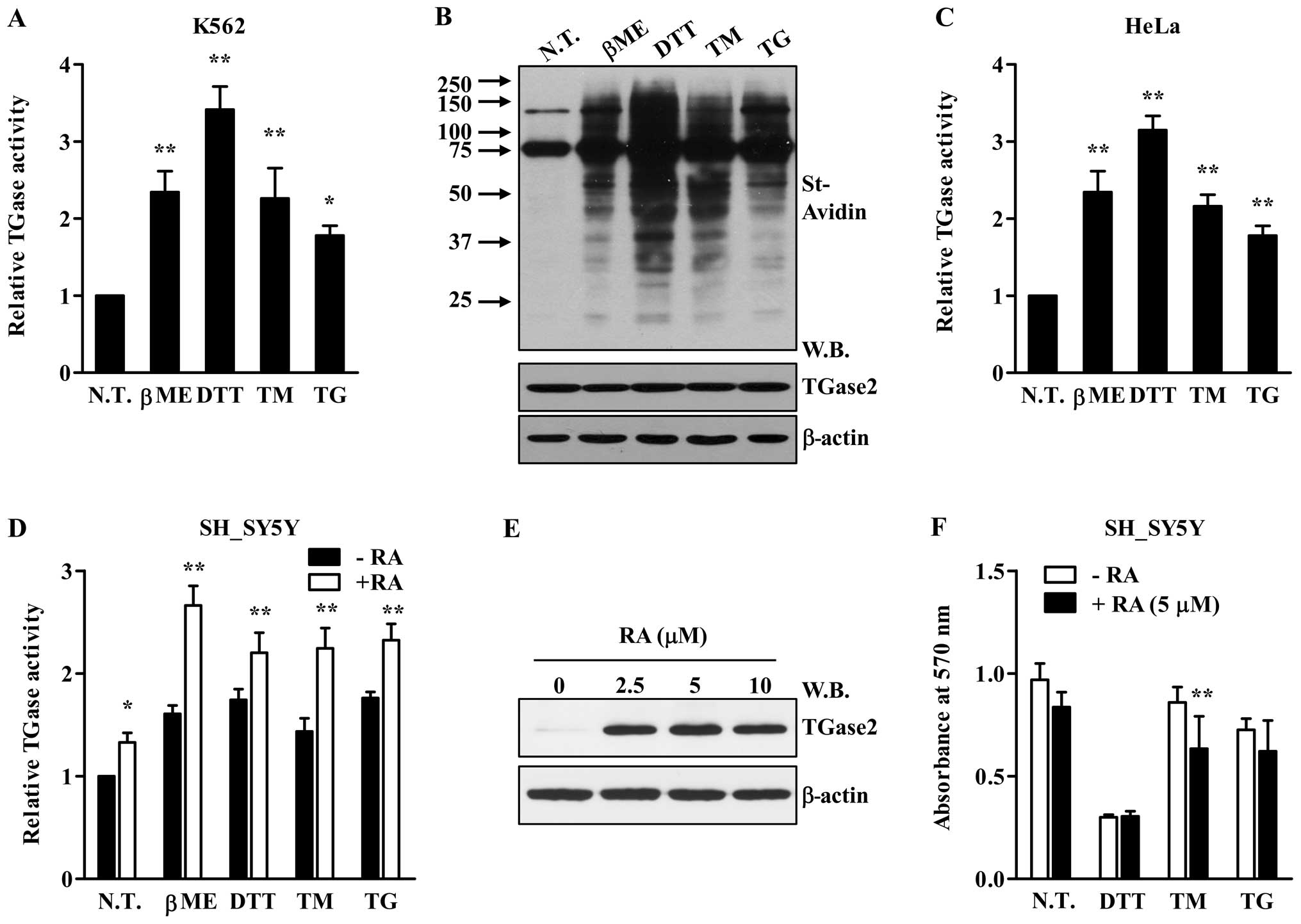
Next, we investigated whether ER stress activates
TGase2 in other types of cells. As shown in Fig. 5, ER stress inducers increased
TGase2 intracellular activity in most of the cell types evaluated,
including human erythrocarcinoma (K562; Fig. 5A and B), cervical cancer (HeLa;
Fig. 5C) and neuroblastoma
(SH-SY5Y; Fig. 5D) cell lines.
These results indicate that the activation of TGase2 is not
specific to lens epithelial cells. Moreover, treatment of SH-SY5Y
cells with retinoic acid (RA) induced the expression of TGase2, and
it may play an important role in cell survival (23–25). When SH-SY5Y cells were pre-treated
with RA, the protein level of TGase2 increased (Fig. 5E). Under this condition, ER stress
further increased in situ TGase activity in SH-SY5Y cells
(Fig. 5D). When cell viability
was evaluated following exposure to ER stress, little significant
difference in cell death was noted between non-treated and
RA-treated SH-SY5Y cells (Fig.
5F), suggesting that UPR-induced cell death was independent of
TGase2-mediated protein aggregation.
Discussion
The present study demonstrated that ER stress
activates transglutaminase 2 (TGase2) in several cell types which
subsequently plays a causal role in the formation and accumulation
of intracellular protein aggregates. The TGase enzyme family
consists of 8 enzymes, TGase1 to TGase7, and Factor XIIIa. Each
TGase isoenzyme shows a restricted pattern of tissue distribution
where it plays a specific function, such as the formation of the
barrier structure in skin (TGase1, TGase3 and TGase5), formation of
fibrin aggregates in blood clots (coagulation Factor XIIIa), and
generation of the post-coital plug of seminal fluid (TGase4)
(26–28). By contrast, TGase2 is unique among
the TGase family members in its ubiquitous tissue expression and
widespread subcellular localization (22). In the lens epithelium, TGase2 is
the major TGase isoform and was found to induce the formation of
lens protein aggregates in response to UV-irradiation and oxidative
stress in a lens organ culture model of cataract (11,29). In the present study, we showed
that several of the ER stress-causing agents tested increased the
in situ transamidation reaction in lens epithelial cells
(Figs. 1 and 2). Moreover, ER stress-induced protein
aggregation was reduced by treatment with a chemical inhibitor for
TGase2 (Fig. 1C). These results
demonstrate that unfolded or misfolded proteins produced by
oxidative stress activate TGase2 leading in the accumulation of
protein aggregates.
Previous studies have shown that intracellular
TGase2 activity does not correlate with its protein expression
level and that TGase2 activation is cell type-dependent (10). Under normal culture conditions,
intracellular TGase2 activity was not observed in lens epithelial
cells despite of the high TGase2 protein level (Figs. 1 and 2), indicating that control of this
enzyme is tightly regulated in the intracellular environment. The
transamidation reaction of TGase2 is dependent on the intracellular
calcium level (30). UPR
activators evaluated here increased the intracellular concentration
of Ca2+, and Ca2+ chelation prevented TGase2
activation by UPR (Fig. 3A and
B), indicating that the rise in intracellular Ca2+
could explain the observed increase in TGase2 activity despite of
little change in its protein level. However, DTT treatment
activated TGase2 even in the presence of BAPTA-AM (Fig. 3B and C), suggesting that other
cellular factor(s) may be involved in the regulation of TGase2
activity in response to DTT treatment.
In the present study, we employed a lens epithelial
cell line to investigate the role of TGase2 in the formation of
intracellular aggregates of misfolded proteins. For this purpose,
lens tissue may provide several advantages since lens epithelial
cells have a much simpler protein complexity and accumulate
oxidatively modified proteins without degradation (31). A single layer of lens epithelial
cells differentiates into fiber cells, which make up the lamellae
inside the lens without replacing the older fiber cells (31). The misfolding of lens proteins
such as crystallins and vimentin is easily monitored using
dimerization and water-solubility as assessed by western blotting
(Fig. 4). Moreover, lens protein
aggregation can be easily detected by the development of lens
opacity (11). Thus, ex
vivo lens organ culture and in vivo animal models may be
excellent systems for the development of pharmaceuticals that
inhibit the protein aggregation caused by a variety of forms of
cellular stress.
Our results showed that TGase2 was also activated by
ER stress in other cell types including neuroblastoma cells. Of
note, it is known that TGase2 post-translationally modifies several
aggregation-prone proteins such as amyloid β-peptide, tau,
α-synuclein and huntingtin (7–9,32).
In particular, treatment with cystamine, a TGase inhibitor
(33), or ablation of TGase2
(34) delays the onset of
neurological symptoms and improves the life expectancy of
Huntington’s disease model mice, suggesting that aberrant
activation of TGase2 may be involved in the formation of insoluble
aggregates in neurons through protein modification during ageing.
Several studies have indicated that the induction of TGase2 during
RA-mediated differentiation plays a protective role against
neuroblastoma cell death following exposure to excitotoxic or
inflammatory stress (23,24). However, another study reported
that TGase2 enhanced β-amyloid 1–42-induced apoptosis (25). In the present study, RA
pre-treatment had little effect on the viability of SH-SY5Y cells
exposed to ER stress (Fig. 5F).
Thus, further study on the unknown cellular function(s) of
TGase2-mediated protein aggregation under stress conditions is
required to precisely understand the pathophysiological role of
this enzyme.
Acknowledgements
This study was partly supported by a grant from the
Basic Science Research Program through the National Research
Foundation of Korea (NRF) funded by the Ministry of Education,
Science, and Technology (2012R1A1A1004438), and grants from the
Korean Health Technology R&D Project, Ministry of Health and
Welfare, Republic of Korea (A120301 to D.-M.S. and A100190 to
I.-G.K.).
References
|
1
|
Hartl FU, Bracher A and Hayer-Hartl M:
Molecular chaperones in protein folding and proteostasis. Nature.
475:324–332. 2011. View Article : Google Scholar : PubMed/NCBI
|
|
2
|
Lin MT and Beal MF: Mitochondrial
dysfunction and oxidative stress in neurodegenerative diseases.
Nature. 443:787–795. 2006. View Article : Google Scholar : PubMed/NCBI
|
|
3
|
Bence NF, Sampat RM and Kopito RR:
Impairment of the ubiquitin-proteasome system by protein
aggregation. Science. 292:1552–1555. 2001. View Article : Google Scholar : PubMed/NCBI
|
|
4
|
Lorand L and Graham RM: Transglutaminases:
crosslinking enzymes with pleiotropic functions. Nat Rev Mol Cell
Biol. 4:140–156. 2003. View
Article : Google Scholar : PubMed/NCBI
|
|
5
|
Klöck C and Khosla C: Regulation of the
activities of the mammalian transglutaminase family of enzymes.
Protein Sci. 21:1781–1791. 2012.PubMed/NCBI
|
|
6
|
Prasanna Murthy SN, Velasco PT and Lorand
L: Properties of purified lens transglutaminase and regulation of
its transamidase/crosslinking activity by GTP. Exp Eye Res.
67:273–281. 1998.PubMed/NCBI
|
|
7
|
Jeitner TM, Bogdanov MB, Matson WR, et al:
Nɛ-(γ-l-Glutamyl)-l-lysine (GGEL) is increased in cerebrospinal
fluid of patients with Huntington’s disease. J Neurochem.
79:1109–1112. 2001.
|
|
8
|
Konno T, Morii T, Hirata A, Sato S, Oiki S
and Ikura K: Covalent blocking of fibril formation and aggregation
of intracellular amyloidgenic proteins by
transglutaminase-catalyzed intramolecular cross-linking.
Biochemistry. 44:2072–2079. 2005. View Article : Google Scholar
|
|
9
|
Halverson RA, Lewis J, Frausto S, Hutton M
and Muma NA: Tau protein is cross-linked by transglutaminase in
P301L tau transgenic mice. J Neurosci. 25:1226–1233. 2005.
View Article : Google Scholar : PubMed/NCBI
|
|
10
|
Shin DM, Jeon JH, Kim CW, et al: Cell
type-specific activation of intracellular transglutaminase 2 by
oxidative stress or ultraviolet irradiation: implications of
transglutaminase 2 in age-related cataractogenesis. J Biol Chem.
279:15032–15039. 2004. View Article : Google Scholar
|
|
11
|
Shin D-M, Jeon J-H, Kim C-W, et al: TGFβ
mediates activation of transglutaminase 2 in response to oxidative
stress that leads to protein aggregation. FASEB J. 22:2498–2507.
2008.
|
|
12
|
Moore KA and Hollien J: The unfolded
protein response in secretory cell function. Annu Rev Genet.
46:165–183. 2012. View Article : Google Scholar : PubMed/NCBI
|
|
13
|
Lin JH, Walter P and Yen TS: Endoplasmic
reticulum stress in disease pathogenesis. Annu Rev Pathol.
3:399–425. 2008. View Article : Google Scholar
|
|
14
|
Schröder M and Kaufman RJ: The mammalian
unfolded protein response. Annu Rev Biochem. 74:739–789. 2005.
|
|
15
|
Xin W, Li X, Lu X, Niu K and Cai J:
Involvement of endoplasmic reticulum stress-associated apoptosis in
a heart failure model induced by chronic myocardial ischemia. Int J
Mol Med. 27:503–509. 2011.PubMed/NCBI
|
|
16
|
Wu LF, Wei BL, Guo YT, et al: Apoptosis
induced by adenosine involves endoplasmic reticulum stress in EC109
cells. Int J Mol Med. 30:797–804. 2012.PubMed/NCBI
|
|
17
|
Harding HP, Zhang Y, Zeng H, et al: An
integrated stress response regulates amino acid metabolism and
resistance to oxidative stress. Mol Cell. 11:619–633. 2003.
View Article : Google Scholar : PubMed/NCBI
|
|
18
|
Cho S-Y, Lee J-H, Bae H-D, et al:
Transglutaminase 2 inhibits apoptosis induced by calcium overload
through down-regulation of Bax. Exp Mol Med. 42:639–650. 2010.
View Article : Google Scholar : PubMed/NCBI
|
|
19
|
Jang GY, Jeon JH, Cho SY, et al:
Transglutaminase 2 suppresses apoptosis by modulating caspase 3 and
NF-kappaB activity in hypoxic tumor cells. Oncogene. 29:356–367.
2009. View Article : Google Scholar : PubMed/NCBI
|
|
20
|
Choi K, Siegel M, Piper JL, et al:
Chemistry and biology of dihydroisoxazole derivatives: selective
inhibitors of human transglutaminase 2. Chem Biol. 12:469–475.
2005. View Article : Google Scholar : PubMed/NCBI
|
|
21
|
Jeon JH, Choi KH, Cho SY, et al:
Transglutaminase 2 inhibits Rb binding of human papillomavirus E7
by incorporating polyamine. EMBO J. 22:5273–5282. 2003. View Article : Google Scholar : PubMed/NCBI
|
|
22
|
Fesus L and Piacentini M: Transglutaminase
2: an enigmatic enzyme with diverse functions. Trends Biochem Sci.
27:534–539. 2002. View Article : Google Scholar : PubMed/NCBI
|
|
23
|
Caccamo D, Condello S, Ferlazzo N, Currò
M, Griffin M and Ientile R: Transglutaminase 2 interaction with
small heat shock proteins mediate cell survival upon excitotoxic
stress. Amino Acids. 44:151–159. 2013. View Article : Google Scholar : PubMed/NCBI
|
|
24
|
Kweon SM, Lee ZW, Yi SJ, et al: Protective
role of tissue transglutaminase in the cell death induced by
TNF-alpha in SH-SY5Y neuroblastoma cells. J Biochem Mol Biol.
37:185–191. 2004. View Article : Google Scholar : PubMed/NCBI
|
|
25
|
Wakshlag JJ, Antonyak MA, Boehm JE, Boehm
K and Cerione RA: Effects of tissue transglutaminase on
beta-amyloid1–42-induced apoptosis. Protein J. 25:83–94.
2006.PubMed/NCBI
|
|
26
|
Candi E, Schmidt R and Melino G: The
cornified envelope: a model of cell death in the skin. Nat Rev Mol
Cell Biol. 6:328–340. 2005. View
Article : Google Scholar : PubMed/NCBI
|
|
27
|
Muszbek L, Bereczky Z, Bagoly Z, Komáromi
I and Katona É: Factor XIII: a coagulation factor with multiple
plasmatic and cellular functions. Physiol Rev. 91:931–972. 2011.
View Article : Google Scholar : PubMed/NCBI
|
|
28
|
Mukherjee DC, Agrawal AK, Manjunath R and
Mukherjee AB: Suppression of epididymal sperm antigenicity in the
rabbit by uteroglobin and transglutaminase in vitro. Science.
219:989–991. 1983. View Article : Google Scholar : PubMed/NCBI
|
|
29
|
Lee S-M, Jeong EM, Jeong J, et al:
Cysteamine prevents the development of lens opacity in a rat model
of selenite-induced cataract. Invest Ophthalmol Vis Sci.
53:1452–1459. 2012. View Article : Google Scholar : PubMed/NCBI
|
|
30
|
Király R, Demény M and Fésüs L: Protein
transamidation by transglutaminase 2 in cells: a disputed
Ca2+-dependent action of a multifunctional protein. FEBS
J. 278:4717–4739. 2011.PubMed/NCBI
|
|
31
|
Sue Menko A: Lens epithelial cell
differentiation. Exp Eye Res. 75:485–490. 2002.PubMed/NCBI
|
|
32
|
Junn E, Ronchetti RD, Quezado MM, Kim SY
and Mouradian MM: Tissue transglutaminase-induced aggregation of
alpha-synuclein: implications for Lewy body formation in
Parkinson’s disease and dementia with Lewy bodies. Proc Natl Acad
Sci USA. 100:2047–2052. 2003.PubMed/NCBI
|
|
33
|
Karpuj MV, Becher MW, Springer JE, et al:
Prolonged survival and decreased abnormal movements in transgenic
model of Huntington disease, with administration of the
transglutaminase inhibitor cystamine. Nat Med. 8:143–149. 2002.
View Article : Google Scholar : PubMed/NCBI
|
|
34
|
Mastroberardino PG, Iannicola C, Nardacci
R, et al: ‘Tissue’ transglutaminase ablation reduces neuronal death
and prolongs survival in a mouse model of Huntington’s disease.
Cell Death Differ. 9:873–880. 2002.
|