Introduction
Brain tumors are one of the most challenging
diseases to treat and there remains an unmet medical need for
effective treatments. Chemotherapy often fails due to the
insufficient accumulation of drugs in the tumor sites and the
subsequent development of drug resistance. Strategies for the
reversal of resistance involve the inhibition of enzymes
responsible for the efflux of drugs, the modulation of proteins
regulating apoptosis, and the improvement of drug uptake using
nanotechnologies. Over the past decade, there have been momentous
developments in drug delivery systems, particularly in the use of
nanotechnology-based delivery systems.
Polymeric liposomes have the advantage of being the
least toxic for in vivo applications. Over the past decace,
significant progress has been made in the delivery of DNA/RNA and
small-molecule drugs (1–3). Multiple liposome formulations have
been used clinically in the treatment of cancer and infectious
diseases (4), and their
applications in other diseases are currently being investigated in
clinical trials (5). The
development of clinically suitable liposome formulations has
resulted from two major technological achievements: i) the
inclusion of PEGylated lipids in the liposomes for the purposes of
bypassing the reticuloendothelial system, resulting in significant
drug accumulation in tumors (6,7);
and ii) the strategic development of a remote drug-loading process
based on the ammonium sulfate gradient method, to achieve
significantly high quantities of drugs in the liposomes (8). We have previously demonstrated that
the
1,2-distearoyl-sn-glycero-3-phosphoethanolamine-N-[methoxy(polyethylene
glycol)-2000] (DSPE-PEG2000)-coated and quercetin (QUE)-loaded
nanoliposomes (QUE-NLs) exhibited a hydrophilic layer on the
surface, resulting in particle size increment, positive zeta
ζ-potential, and enhanced physical stability (9,10).
Notably, DSPE-PEG2000 coating of particles is a simple and flexible
technology used to alter the surface properties of liposomes, and
is expected to have broad applications in anticancer drug delivery
systems.
It has been previously demonstrated that QUE, as a
potential chemopreventer, suppresses cancer cell growth,
proliferation and metastasis, as well as the expression of mutant
p53 (11), and also enhances
death receptor-mediated apoptosis in glioma cells (12,13). QUE is considered a potent free
radical-scavenging antioxidant owing to its abundant hydroxyl
groups and conjugated p orbitals, which donate electrons or
hydrogens, and scavenge H2O2 and superoxide
anions (14). QUE-mediated
apoptosis may result from the induction of stress proteins, the
disruption of microtubules, the stimulation of the release of
cytochrome c and the activation of caspases (15–17), thus rendering QUE a promising
candidate for cancer prevention and therapy. A recent study
reported that the combination of QUE and temozolomide (TMZ), a
FDA-approved drug for brain cancer treatment, exerted a synergistic
effect in brain tumors in vitro (18).
In a previous study of ours (10), we reported that QUE/DSPE-PEG2000
showed significantly enhanced potency in glioma in vitro
compared to free QUE. The aim of the present study was to evaluate
the application of DSPE-PEG2000 as a nanocarrier for the combined
delivery of QUE and TMZ for the treatment of glioma. We
investigated the physicochemical properties, the release and
clearance profiles, the biodistribution and the enhanced potency of
QUE/TMZ-NLs in human U87 glioma cells and TMZ-resistant U87 cells
(U87/TR cells), providing novel and significant insight into the
application of QUE/TMZ-NLs for the treatment of brain tumors.
Materials and methods
Reagents and cell lines
DSPE-PEG2000 was purchased from Nippon Oil and Fats
Co., Ltd., (Tokyo, Japan). Poloxamer 188 was purchased from BASF
Aktiengesellschaft (Limburgerhof, Germany). Cholesterol and soy
lecithin were purchased from Shanghai Youngsun Foods Co., Ltd.
(Shanghai, China). Tween-80 was purchased from Shanghai Chemical
Reagent Co., Ltd. (Shanghai, China). Glyceryl behenate (Compritol
ATO 888) was purchased from Gattefosse S.A. (Saint-Priest, France).
QUE was purchased from the National Institute for the Control of
Pharmaceutical and Biological Products (NICPBP; Beijing, China).
TMZ was purchased from Jiangsu Tasly Diyi Pharmaceutical Co., Ltd.
(Jiangsu, China). Annexin V and proridium iodide (PI) were obtained
from BestBio Biotechnologies Co., Ltd. (Shanghai, China).
RPMI-1640, penicillin-streptomycin,
trypsin-ethylenediaminetetraacetic acid (EDTA) and fetal bovine
serum (FBS) were obtained from Gibco BRL/Life Technologies
(Carlsbad, CA, USA). U87 glioma cells were obtained from the
American Type Culture Collection (ATCC; Rockville, MD, USA).
Preparation of QUE/TMZ-NLs and
QUE/TMZ-FITC-NLs
Both the QUE/TMZ-NL and the QUE/TMZ-FITC-NL
suspensions were prepared by emulsification-evaporation and low
temperature curing preparation as previously described (19). The aqueous phase consisted of
poloxamer 188 and Tween-80 (1:1, w/v), dissolved in pure water and
maintained in a water bath at 75°C. DSPE-PEG2000 dissolved in
methanol (5.0 mg in 1.0 ml) was added dropwise into the aqueous
phase with rapid stirring (1,000 rpm/min). The mixture was stirred
for 30 min in a bath sonicator until the solution became
brown/black and translucent. To remove the methanol, the solution
was concentrated in a rotary evaporator at 70°C to 3.0 ml. The oil
phase mainly consisted of glyceryl behenate, soy lecithin and
cholesterol (1:2:1, w/w/w), and in addition, glyceryl behenate and
cholesterol were melted in a water bath at 80°C. QUE, TMZ and soy
lecithin (1:1:1, w/w/w) were dissolved in the ethanol-acetone (1:1,
v/v) solvent, and the oil phase of the aforementioned mixed solvent
was then injected into the aqueous phase through plastic needle
tubing (internal diameter, 0.45 µm; injection rate, 2.0
ml/min) under mechanical agitation at 1000 rpm. After stirring for
2 h, the liposomal suspension was cured at low temperatures of
0–4°C under mechanical agitation at 800 rpm. The suspension was
then filtered through dialysis tubing to remove any unincorporated
drugs. Briefly, the QUE/TMZ-NLs were injected into the dialysis
tubing and the dialysis tubing was washed in mannitol solution and
dialyzed 3 times to remove free QUE or TMZ (2 h for the first and
the second washes, and 12 h for the third wash at 0–4°C). Finally,
the purified QUE/TMZ-NLs were obtained and stored at 0–4°C. The
QUE/TMZ-FITC-NLs were prepared by the addition of 200 µl
QUE/TMZ-FITC suspension to the liposomes. The mixture was sonicated
in a water bath using a laboratory ultrasonic device at 250 Hz for
10 min. Prior to use in the cell experiments, the prepared
QUE/TMZ-NLs were suspended in RPMI-1640 medium containing 10% (v/v)
FBS using ultrasound. The control liposomes were also prepared
using the same method without adding QUE or TMZ at any stage of the
preparation process.
Characterization
The size and polydispersity index of the QUE/TMZ-NLs
were determined using photon correlation spectroscopy with the use
of a laser particle analyzer (Jinan Rise Science & Technology
Co., Ltd). The ζ-potential was analyzed using a microscopic
electrophoresis system (DXD-II; Jiangsu Optics Co., Ltd., Jiangsu,
China) at 25°C. The morphology of the cells treated with the
QUE/TMZ-NLs was observed under a transmission electron microscope
(TEM-1200EX; JEOL Ltd., Tokyo, Japan). The samples were dissolved
in deionized water contained in 1.0 ml quartz cuvettes, and the UV
data were collected using a T90 UV-VIS spectrophotometer (Beijing
Purkinje General Instrument Co., Ltd., Beijing, China). The
concentration of the QUE/TMZ-NLs was then determined by the
absorbance measured at 365 and 329 nm, by high-performance liquid
chromatography (HPLC), using the experimentally determined
extinction coefficient of 0.0101 l/mg.
Fourier transform infrared spectroscopy
(FTIR)
Infrared spectra of QUE, QUE-NLs, TMZ and
QUE/TMZ-NLs were recorded on a FTIR spectrophotometer (PerkinElmer,
Inc., Waltham, MA, USA) by the KBr disk method from 4,000 to 500
cm−1.
Entrapment efficiency (EE) and drug
loading
The free drugs were separated from the QUE/TMZ-NLs
using a centrifugation technique for the measurement of EE.
Briefly, the QUE/TMZ-NLs were diluted in 1.0 ml purified water and
centrifuged at 16,000 × g for 15 min, and the supernatant
containing free drugs was collected, and adjusted to a volume of 10
ml with ethanol for further analysis. A solution of 10% Triton
X-100-ethanol (0.5 ml) was then added and incubated for 5 min to
breakdown the liposomes and dissolve the QUE and TMZ. The mixture
was then centrifuged at 16,000 × g for 10 min. The loading content
and EE of QUE in the QUE/TMZ-NLs was analyzed by HPLC (Dionex
Utimate 3000 HPLC System with an AD20 dual absorbance detector and
computer; Thermo Fisher Scientific Inc., Sunnyvale, CA, USA) at 254
nm. The loading content and EE of TMZ in the QUE/TMZ-NLs were
measured at 329 nm. A Diamond C18 column was used with the flow of
1.0 ml/min for the eluent [methanol/0.5% acetic acid (10:90)]
during 0–19 min. The loading content was calculated as follows:
loading content (%) = (weight of drug in the nanoparticles/weight
of nanoparticles) ×100. The EE was calculated as follows: EE (%) =
weight of drug in the nanoparticles/weight of the feeding drug
×100.
In vitro release assay
The QUE/TMZ-NLs were dispersed in 2.0 ml of 10%
(v/v) human plasma in phosphate-buffered saline (PBS). Each
suspension was incubated with gentle shaking at 37°C throughout the
experiment. At designated intervals, an aliquot (500 µl) was
withdrawn and centrifuged at 5,000 × g for 5 min at 4°C. After
collecting the supernatant, the pellet of nano/microspheres were
resuspended in the same volume of fresh release medium (500
µl), and then returned to the sample suspension. The amounts
of QUE or TMZ in the supernatant were determined by HPLC as
described above.
Cellular uptake of QUE/TMZ-NLs
The uptake of QUE/TMZ-NLs by the U87 cells was
analyzed by confocal fluorescence microscopy. The U87 cells were
plated on two 4-well chamber slides at a density of 40,000
cells/well in low glucose RPMI-1640 medium with 10% FBS, and were
cultured for 24 h. After washing the cells with PBS, fresh medium
with 10% FBS was added. The QUE/TMZ-NLs, QUE/TMZ-FITC-NLs and free
drugs were added to the cells at a final drug concentration of 0.25
µg/ml and incubated at 37°C, in 5% CO2 and 99%
humidity for 4 h. Prior to imaging, the cell culture medium was
removed and the cells were washed twice with cold PBS and fixed
using BD Cytofix/Cytoperm™ (BD Biosciences, Franklin Lakes, NJ,
USA) at 4°C for 10 min. The samples were then mounted using Vecta
Shield mounting medium containing DAPI (Vector Laboratories, Inc.,
(Burlingame, CA, USA) to label the nucleus. Images of the cellular
uptake of the QUE/TMZ-NLs, free QUE or TMZ were captured using an
Olympus FV1000 IX2 inverted confocal microscope with a 40×1.30 NA
Olympus objective and FV10-ASW imaging software (Olympus Corp.,
Tokyo, Japan).
Cell viability assay
Cell viability was evaluated by
3-(4,5-dimethylthiazol-2-yl)-2,5-diphenyltetrazolium bromide (MTT)
assay, as previously described (20,21). The U87 glioma cells were seeded
into a 96-well microplate at a density of 105
cells/well. At 60–70% confluence, the cells were treated with the
indicated concentrations of the QUE/TMZ-NLs, QUE-NLs, free TMZ, or
control liposomes for 12 h, and then allowed to grow in drug-free
medium for 24 h prior to the measurement of the cytotoxicity. The
kinetics of cell death were detected at 12, 24 and 48 h following
treatment with 25, 50 and 100 µM of QUE/TMZ-NLs, QUE-NLs,
free TMZ, or control liposomes. The cells were then washed once
with PBS, and incubated with fresh, complete RPMI-1640 medium (100
µl/well). MTT solution (25 µl of 2 mg/ml) was added
to each well. Following incubation at 37°C for 4 h, the medium was
removed by careful aspiration. In order to determine the number of
viable cells, the optical density (OD) of each well was analyzed
using an enzyme-linked immunosorbent assay (ELISA) plate reader
(μQuat, KC-Junior program; Bio-Tec, Winooski, VT, USA) at a
wavelength of 570 nm. The percentage of viable cells relative to
the control was obtained by dividing the average OD of the treated
wells by the OD of the control wells as follows: cell viability
rate (%) = OD value of treated group/OD value of control group
×100; growth inhibition rate (%) = (1 − OD value of treated
group/OD value of control group) ×100.
Apoptosis assay
Apoptosis was measured by Annexin V/PI staining
followed by flow cytometric analysis, as previously described
(22,23). The cells were incubated with
various concentrations (25, 50, 100 and 200 µM) of the
QUE/TMZ-NLs, QUE-NLs or free TMZ for 12, 24, 36 and 48 h. The cells
were then trypsinized and harvested by centrifugation at 3,000 rpm
for 5 min, and incubated with Annexin V and PI for 15 min at room
temperature prior to flow cytometric analysis.
Generation of a TMZ-resistant U87
(U87/TR) human glioma cell line
TMZ resistance was induced in the U87 cells step by
step to produce the U87/TR cell line. Briefly, the U87 cells were
cultured in RPMI-1640 medium supplemented with 10% FBS, and
incubated in 5% CO2 at 37°C. The parental U87 cells were
then incubated with incremental induction doses of TMZ (4.0–20.0
mM). After 14 days, the TMZ concentration was increased (40, 80,
160 and 320.0 mM) in the cell culture to establish the U87/TR
cells. The U87/TR cells were incubated with 320.0 mM TMZ for 7
days/month for 6 months to maintain stable drug resistance. The
drug resistance was assayed by colony formation assay, as
previously described (24,25).
The drug resistance index was determined based on the cytotoxicity
of TMZ to the U87/TR cells and the parental U87 cells and measured
by MTT assay.
Lactate dehydrogenase (LDH) release assay
for the determination of cell death
The U87/TR cells in 96-well plates were treated with
free QUE, QUE/TMZ-NLs, QUE-NLs, free TMZ, control liposomes or
dimethyl sulfoxide (DMSO) at 6.25, 12.5, 25, 50, 100 and 200
µM in triplicate wells. Following 24 h of incubation, the
LDH levels in the supernatant were measured using an automatic
biochemical analyzer (Hitachi 7060, Hitachi Koki Co. Ltd.,
Hitachinaka, Japan).
Biodistribution assay
After obtaining institutional approval from the
Hubei Medical University Animal Care Committee, Sprague-Dawley (SD)
rats (n=108, 200–220 g, 10 weeks old) were randomized for this
blinded, placebo-controlled trial. All animals were handled with
care and were allowed free access to water and food.
For biodistribution and plasma concentration assays,
a stereo-directed instrument was used to establish the in situ,
in vivo model of glioblastoma by inoculating the U87 cells into
the cerebral cortex of the SD rats. Briefly, the animals were
anesthetized by an intraperitoneal injection of xylazine (100 mg/kg
body weight) and ketamine (10 mg/kg body weight) (Phoenix
Pharmaceuticals Inc., Burlingame, CA, USA), and Lacri-Lube
(Allergan Inc.) was applied across the cornea of the eyes. A burr
hole was drilled in the skull 0.1 mm posterior to the bregma and
2.3 mm lateral to the midline. Live cells (5×10³ in 2
µl of PBS) were administered stereotactically (Stoelting
Apparatus), into the burr hole, using a Hamilton syringe with a
32-gauge needle. The needle was advanced to a depth of 2.3 mm from
the brain surface and the cell suspension delivered slowly over the
course of 2–3 min. Following the injection, the needle was left in
place for 2min, after which time, it was raised to a depth of 1.5
mm below the brain surface and left in place for an additional 1
min. Upon withdrawal of the needle, the burr hole was sealed with
bone wax and the incision sutured. The animals were randomly
assigned into the control and treatment groups. The animals were
weighed daily and humanely sacrificed by cervical dislocation when
weight loss occurred (>10% of body weight) or until tumors were
evident, whichever occurred first. The rats in the treatment groups
were administered intragastrically with the indicated drugs at 25
mg/kg body weight beginning on day 7 after tumor development. The
control animals received equivalent doses of control
nanoliposomes.
The SD rats in the treatment groups were randomly
divided into the single-dose or double-dose groups (QUE/TMZ-NLs,
QUE and TMZ group, n=18/group). At the same time point, the rats in
the QUE group and TMZ group were treated with an intragastric
administration (25 mg/kg body weight) of QUE or TMZ, respectively,
and the rats in the QUE/TMZ-NLs group were treated with an
intragastric administration (25 mg/kg body weight) of QUE and TMZ
(1:1). For the first administration, the rats received a single 25
mg/kg dose of QUE/TMZ-NLs (once per day). For the repeated
administration, after the first administration (12 h), the rats
received repeated doses of QUE/TMZ-NLs (twice per day). Following
administration, the animals were housed in cages that allowed for
free movements and natural postural positions. All the animals were
given free access to food and water. From each group, 3 animals
were euthanized by an intravenous injection of pentobarbital (100
mg/kg body weight) at 30 min of treatment and terminal blood
samples were collected at different time points (2, 4, 8, 12, 24
and 48 h). The brain, liver, heart, kidneys, lungs and spleen were
also harvested and placed into pre-weighed scintillation vials. The
organ samples were homogenized. QUE in the tissue homogenate was
subsequently extracted by methanol/HCl (6:4, v/v), and analyzed by
HPLC. For plasma drug concentration analysis, 400 µl plasma
samples were mixed with 1.0 ml methanol/HCl (6:4, v/v). Following
incubation at 90°C for 5 h, 1.0 ml ethyl acetate was added and the
samples were centrifuged at 5,000 × g at 4°C for 10 min. The
supernatant was dried and reconstituted in 500 µl methanol
for HPLC analysis. The QUE or TMZ content in the samples was
measured at 254 and 329 nm, respectively. A Diamond C18 column was
used with a flow rate of 1.0 ml/min for the eluent [methanol/0.5%
acetic acid (10:90)] for 0–19 min.
Effects of repeated administration
The rats with glioma were used to assess the effects
of the repeated administration of the QUE/TMZ-NLs (25 mg/kg) on the
plasma drug concentration. At 0.5, 1, 2, 4, 8, 12, 24 and 48 h
following administration, blood samples were collected from the
eyes of 6 rats in each group, and centrifuged at 5,000 × g for 10
min to obtain plasma. The plasma drug concentrations were
determined by HPLC, as described above.
Statistical analysis
Data are represented as the means ± standard
deviation (SD) and were analyzed by two-tailed Student's t-tests
using the Statistical Program for Social Sciences (SPSS) 13.0
software (SPSS Corp., Shanghai, China). A p-value of <0.05 was
considered to indicate a statistically significant difference.
Results
Preparation and characterization of
QUE/TMZ-NLs
The size distribution of the QUE/TMZ-NLs was in the
range of 100 to 300 nm, and the size distribution of the QUE-NLs
ranged from 50 to 200 nm, as measured by a laser particle analyzer
(Fig. 1A). Transmission electron
microscopy (TEM) analysis revealed that the QUE/TMZ-NLs formed
spherical particles with a small diameter and a narrow size
distribution compared to the QUE-NLs (Fig. 1B).
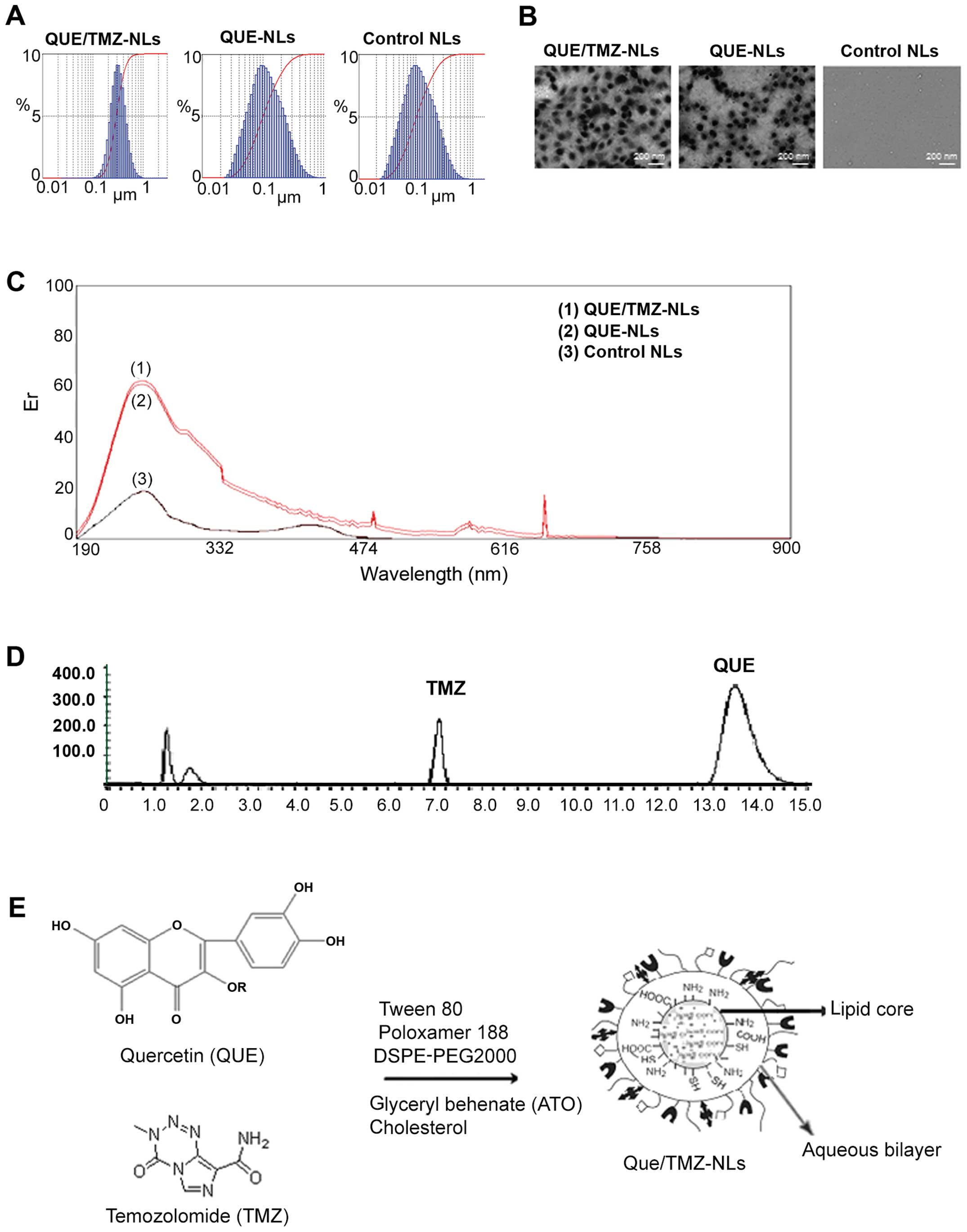 | Figure 1Characterization of the quercetin and
temozolomide-loaded nanoliposomes (QUE/TMZ-NLs). (A) A laser
particle analyzer was used to examine the size distribution of the
QUE/TMZ-NLs, QUE-NLs, and control NLs, the average diameters of
which were 196.5±47.3, 134.5±42.2 and 106.8±37.6 nm, respectively.
(B) Representative TEM images of the QUE-TMZ-NLs, QUE-NLs and the
control NLs. (C) The UV-VIS spectra show similar traces for the
QUE/TMZ-NLs, QUE-NLs and the control NLs, indicating that their
cores were identical. (D) Stacked HPLC traces of QUE and TMZ. (E)
Schematic illustration of the possible packing arrangement of the
PEGylated-coated QUE/TMZ-NLs consisting of an aqueous bilayer and a
lipid core. QUE, quercetin; TMZ, temozolomide; NLs, nanoliposomes;
TEM, transmission electron microscope. |
UV-VIS spectroscopy revealed similar spectra of the
QUE/TMZ-NLs and QUE-NLs, indicating that, as expected, the core of
the QUE/TMZ-NLs remained unaltered following the addition of TMZ
(Fig. 1C), compared to that of
the QUE-NLs, which was measured by the absorbance readings of the
liposome cores. HPLC spectroscopy further revealed that the ratio
of QUE/TMZ remained almost unaltered following the attachment of
DSPE-PEG2000 (Fig. 1D). The
schematic illustration in Fig. 1E
presents the possible packing arrangement of the PEGylated-coated
QUE/TMZ0-NLs consisting of an aqueous bilayer and a lipid core. The
hydrophobic components in the aqueous bilayer and QUE and TMZ were
located in the middle of the lipid core.
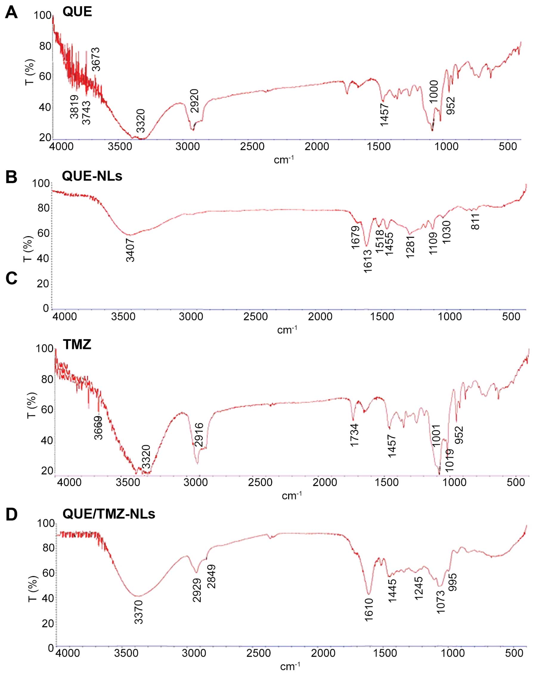
To further examine the drug/carrier interaction, we
analyzed the FTIR spectra of QUE, QUE-NLs, TMZ and QUE/TMZ-NLs. The
characteristic peaks associated with specific structural features
of the drug molecules were observed in QUE, QUE-NLs, TMZ and
QUE/TMZ-NLs (Fig. 2), indicating
that there was no substantial chemical interaction between the
drugs and the carriers. The lack of drug/carrier interaction was
possibly due to the high EE of QUE/TMZ in the nanoparticles in the
presence of ATO and the overlapping with the -NH groups in QUE/TMZ.
In addition, there were a large number of functional groups in QUE
and TMZ, providing possible masking effects on the infrared peaks,
which were otherwise visible.
EE and drug loading
The loading content and the EE of QUE or TMZ in the
NLs were calculated (Table I).
The EE of QUE in the QUE/TMZ-NLs varied in the range of 69.42 to
78.37%. The EE of TMZ ranged from 53.58 to 66.25%. Compared with
the EE of the QUE-NLs (85.72%), the addition of TMZ had no
significant effect on the EE of QUE in the liposomes.
 | Table IThe loading content, EE, particle
size, PI and ζ-potential of lipid formulations of QUE/TMZ. |
Table I
The loading content, EE, particle
size, PI and ζ-potential of lipid formulations of QUE/TMZ.
| Lipid
formulations | Size (nm) | PI | ζ-potential | Loading content
(%) | EE (%) |
|---|
| QUE-NLs | 134.6±62.2 | 0.26±0.06 | 21.8±5.4 | 26.23±2.34 | 85.72±9.34 |
| QUE/TMZ-NLs | 196.5±47.3 | 0.32±0.09 | 30.5±6.9 | – | – |
| QUE/TMZ-NLs
(QUE) | – | – | – | 23.42±2.17 | 69.42–78.37 |
| QUE/TMZ-NLs
(TMZ) | – | – | – | 15.87±1.96 | 53.58–66.25 |
| Control
liposomes | 106.8±37.6 | 0.17±0.04 | 16.6±3.8 | – | – |
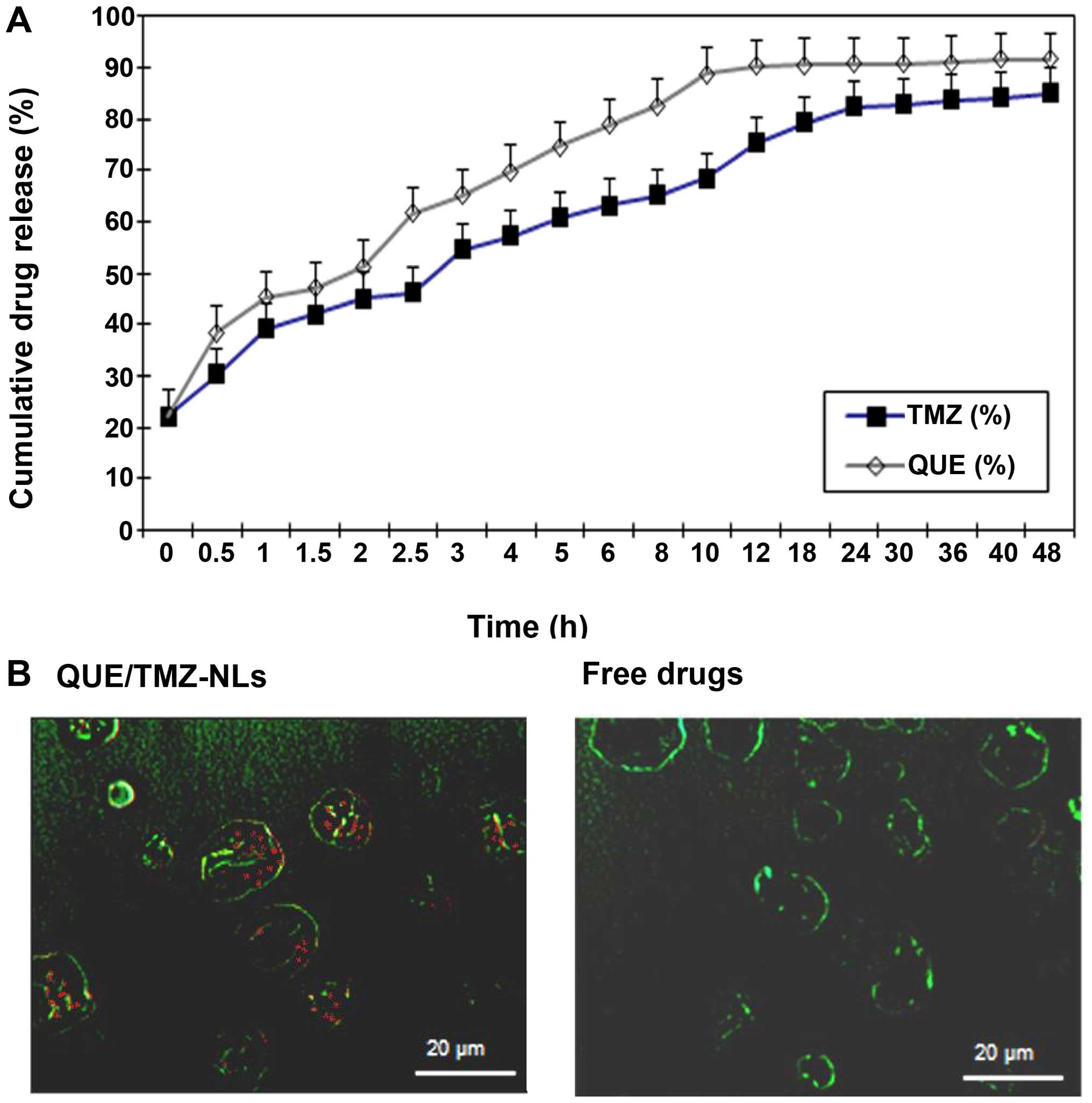
In vitro drug release profiles and uptake
of the QUE/TMZ-NLs by human U87 glioma cells
The release profile of QUE and TMZ from the
QUE/TMZ-NLs revealed sustained drug release at 37°C (Fig. 3A). Within 12 h, 90% of QUE was
released from the QUE/TMZ-NLs, whereas approximately 75% of TMZ was
released. Approximately 90% of TMZ was released from the
QUE/TMZ-NLs after 24 h.
We further examined the uptake of the QUE/TMZ-NLs by
the U87 cells. The QUE/TMZ-FITC-NLs were incubated with the U87
cells and the internalization of the QUE/TMZ-FITC-NLs was analyzed
using confocal fluorescence microscopy. The uptake of free QUE or
TMZ dissolved in DMSO was compared at the same concentrations. The
uptake of the QUE/TMZ-NLs in the cytoplasm appeared diffuse rather
than punctate (Fig. 3B),
suggesting that the internalized drugs in the QUE/TMZ-NLs had
escaped from the endosomes. By contrast, the uptake of the free
drugs was substantially lower, due to the limited solubility of QUE
and TMZ in the cell culture media.
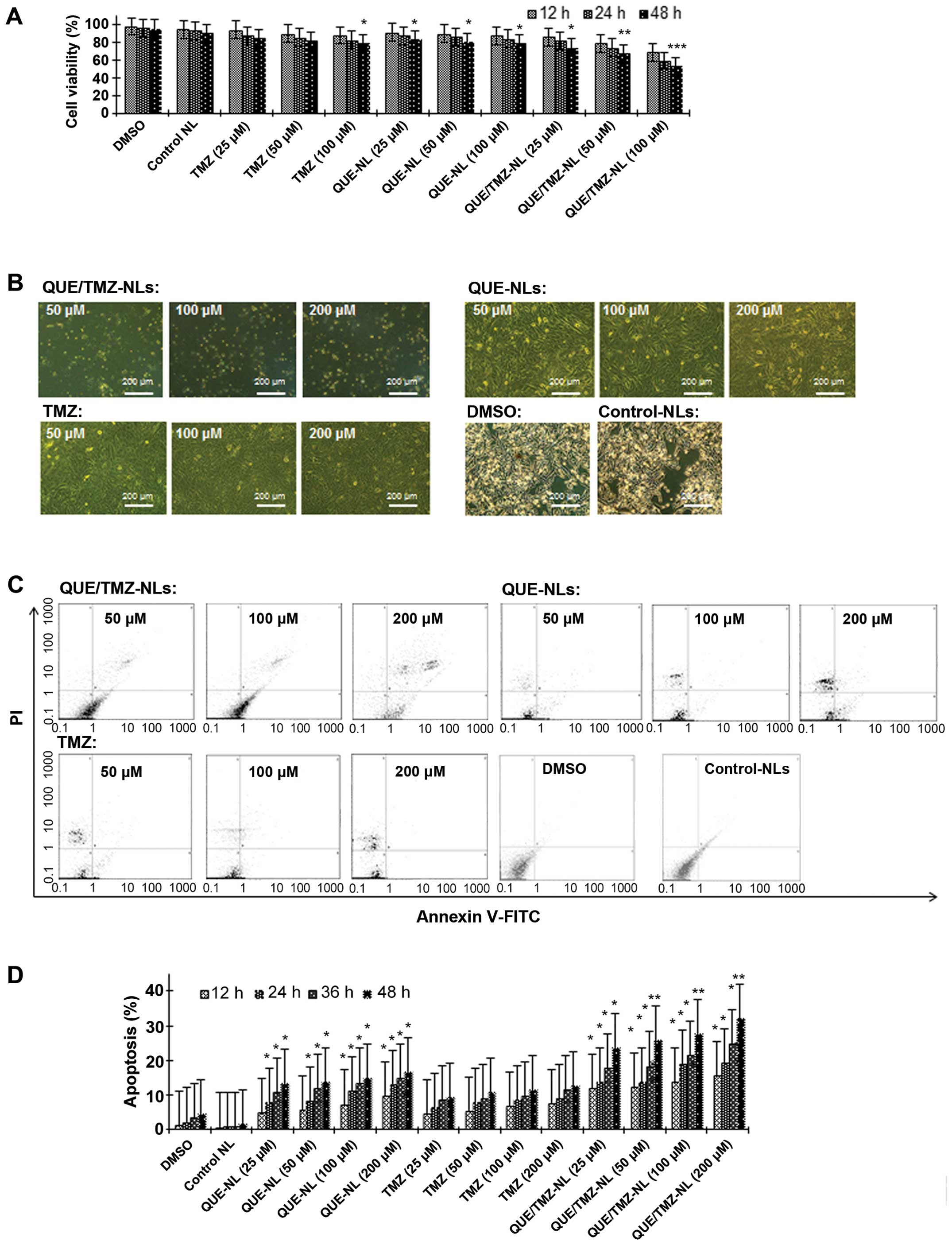
QUE/TMZ-NLs exerts cytotoxic effects on
the U87 glioma cells in vitro
As shown by MTT assay, the QUE/TMZ-NLs significantly
decreased the viability of the U87 cells compared with free TMZ or
the QUE-NLs (Fig. 4A). While free
TMZ did not show any noticeable cytotoxicity towards the U87 glioma
cells at the concentrations tested and the QUE-NLs caused only
limited cytotoxicity, the QUE/TMZ-NLs induced significant cell
death at the same concentrations, inducing a 1.5-fold increase in
cytotoxicity compared with QUE at 25–100 µM and a 1.8-fold
increase in cytotoxicity compared with free TMZ. In addition, the
QUE/TMZ-NLs induced marked morphological changes and cell death
(apoptotic cells were characterized by condensed or fragmented
nuclei) at 100 µM following 24 h of incubation with the U87
cells (Fig. 4B), whereas QUE or
TMZ alone had minimal effects on cell morphology. We further
demonstrated that the QUE/TMZ-NL-induced cell death was due to
apoptosis. The percentage of the Annexin V+ cells
markedly increased, and the effects of the QUE/TMZ-NLs were more
prominent than those of QUE or TMZ alone, while the percentage of
PI− cells was very low (Fig. 4C); these effects occurred in a
time- and dose-dependent manner (Fig.
4D).
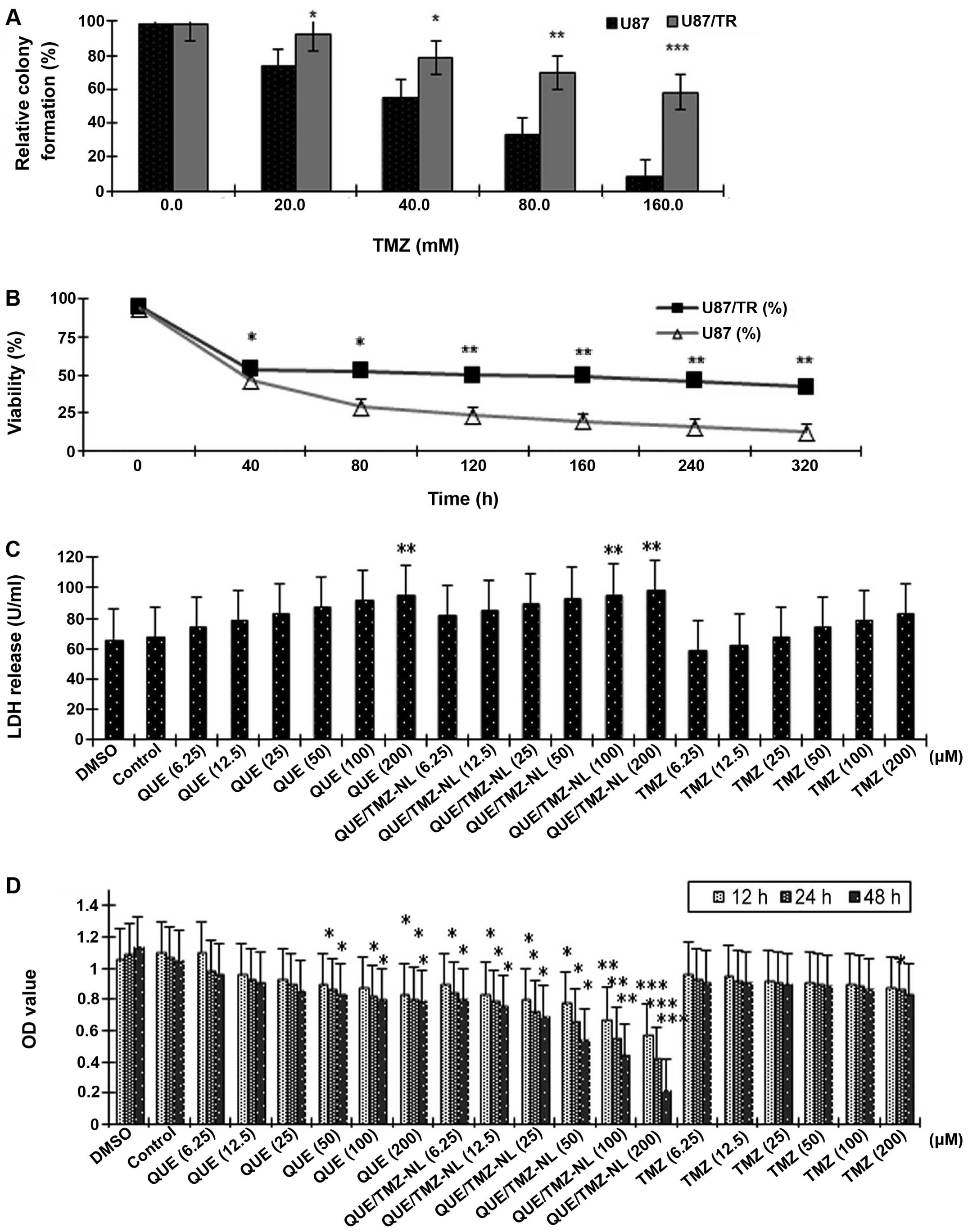
QUE/TMZ-NLs exert cytotoxic effects on
the TMZ-resistant U87/TR cells in vitro
Drug resistance is a major issue in the treatment of
brain tumors (26). Thus, in the
present study, we established a TMZ-resistant brain tumor cell
line, U87/TR, in order to examine the cytotoxic effects of the
QUE/TMZ-NLs. Compared with the parental U87 cells, the U87/TR cells
had a significantly better ability in forming colonies in the
presence of TMZ (p<0.05), with resistance indexes in a range of
1.5–8.0 at different TMZ concentrations (Fig. 5A). As shown by MTT assay, the IC50
value of TMZ was 9.24 mM in the U87 cells, and was 38.65 mM in the
U87/TR cells, with a resistance index of 4.18 (Fig. 5B).
Cell death was further assessed by measuring the
release of LDH from the U87/TR cells treated with or without the
QUE/TMZ-NLs. The QUE/TMZ-NLs significantly upregulated the release
of LDH by the U87/TR cells (p<0.01), compared with the control
NLs and the DMSO control. TMZ alone had no significant effect on
the release of LDH by the U87/TR cells (Fig. 5C). These results indicate that the
QUE/TMZ-NLs have superior anticancer effects on TMZ-resistant
glioma cells.
In addition, the OD value, measured by MTT assay,
which indicates the number of viable cells, was significantly
decreased by the QUE/TMZ-NLs in a time- and dose-dependent manner.
Compared with the same concentrations of QUE or TMZ, treatment with
the QUE/TMZ-NLs resulted in significantly lower OD values (Fig. 5D).
Biodistribution of the QUE/TMZ-NLs in
vivo
To determine the biodistribution of the QUE/TMZ-NLs,
QUE-NLs and free TMZ in rats with in situ glioma, we
administered these NLs or free drugs intragastrically, and
collected brain, liver, heart, kidney, spleen and lung samples
after 2, 4, 8, 12, 24 or 48 h for HPLC analysis (Fig. 6A). Compared with the QUE-NLs or
free TMZ, the QUE/TMZ-NLs significantly accumulated in the brain,
indicating that the QUE/TMZ-NLs would achieve improved therapeutic
efficacy and potency for the treatment of glioma. Free TMZ and the
QUE-NLs were accumulated in the kidneys and heart. The QUE-NLs and
the QUE/TMZ-NLs were accumulated in the liver, but the content of
QUE/TMZ-NLs in the brain was higher than that in the liver.
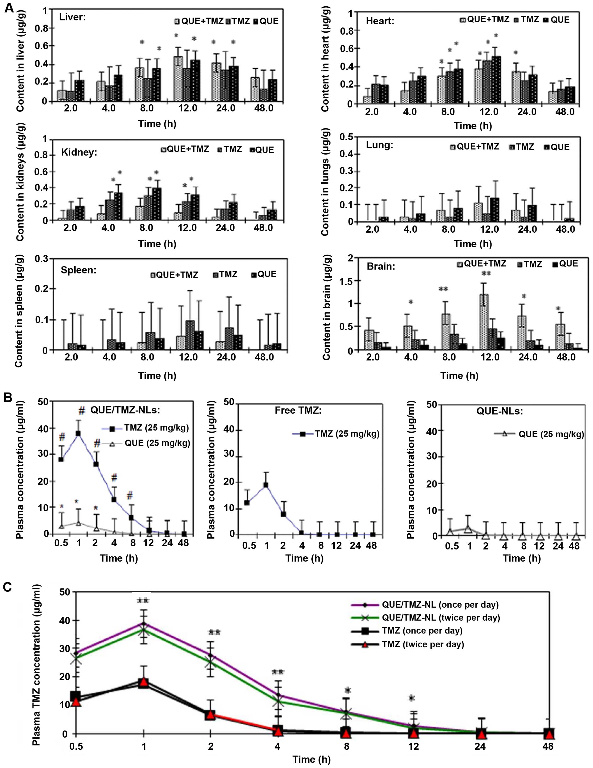 | Figure 6Biodistribution and clearance of the
quercetin and temozolomide-loaded nanoliposomes (QUE/TMZ-NLs)
compared with the QUE-NLs or free TMZ. (A) The biodistribution of
QUE and TMZ in the QUE/TMZ-NL, QUE-NLs and free TMZ in the brain,
liver, heart, kidneys, lungs and spleen of glioma-bearing rats. The
y-axis in all images was normalized to the total content of QUE and
TMZ divided by the weight of the organ. This value is then divided
by the initial QUE or TMZ content administered in each rat. ANOVA
analysis showed statistical significance (*p<0.05 and
**p<0.01) compared with the QUE-NLs and free TMZ. (B)
Plasma drug concentrations were shown at the indicated time points
after the administration of a single dose of TMZ at 25 mg/kg,
QUE-NLs at 25 mg/kg, and 2-in-1 QUE/TMZ-NL with QUE at 25 mg/kg and
TMZ at 25 mg/kg. #p<0.05 for the TMZ concentration
difference between free TMZ and 2-in-1 QUE/TMZ-NLs.
*p<0.05 for the QUE concentration difference between
QUE-NLs and QUE/TMZ-NLs (means ± SEM, n=6). (C) Blood clearance of
QUE and TMZ in glioma-bearing rats after a single administration or
dual administrations of QUE/TMZ-NLs. The plasma TMZ concentration
decreased significantly at 1, 2 and 4 h after the repeated
administration of QUE/TMZ-NLs. ANOVA revealed statistical
significance (*p<0.05 and **p<0.01)
compared with the QUE-NLs and free TMZ. QUE, quercetin; TMZ,
temozolomide; NLs, nanoliposomes. |
The plasma concentrations of the QUE/TMZ-NLs, free
QUE and TMZ were determined after they were administered to the
glioma-bearing rats (Fig. 6B). In
the form of QUE/TMZ-NLs, both QUE and TMZ exhibited higher plasma
concentrations compared with the free drugs. The drug clearance of
QUE/TMZ-NLs was also delayed. As regards the accelerated blood
clearance (ABC) phenomenon with the conventional DSPE-PEG2000
modified liposomes (27–29), we further compared the plasma
clearance rates of single and dual administrations. There was a
slight increase in the plasma clearance rate following the repeated
administration of the QUE/TMZ-NLs (Fig. 6C), whereas the plasma
concentration of TMZ administered as a free drug did not
significantly decrease after repeated administrations.
Discussion
Nanotechnology used in conjunction with existing
therapies has been shown to improve the reversal of drug
resistance. The related mechanisms include specific drug targeting,
enhanced cellular uptake and improved bioavailability of drugs with
poor physicochemical characteristics (30,31). Multidrug resistance is a common
issue that has been linked with the failure of chemotherapy for
brain tumors. To overcome drug resistance, multiple brain
tumor-targeted drug delivery systems have been developed with
decreased off-target toxicity and improved
pharmacokinetics/harmacodynamics compared with conventional
formulations (32). Lipid
nanoparticles, such as solid NLs and nanostructured lipid carriers
(NLCs), have been shown to provide a favorable means for efficient
drug delivery to tumor sites, while minimizing their side-effects
(33,34). In this study, we loaded QUE and
TMZ into a PEGylated liposomal carrier, characterized its
physicochemical and pharmacokinetic properties, and the antitumor
potency in vitro and in vivo.
Recently, anticancer drugs with methylating
properties, including TMZ, have been investigated, particularly for
the therapy of malignant glioblastomas (35). Although TMZ can be administered
orally and pass the blood brain barrier, its half-life is <2 h
and thus, it does not reach the tumor site at effective therapeutic
concentrations. In addition, the off-target toxicities of TMZ, such
as neurotoxicity and reproductive toxicity, further limit its
therapeutic efficacy in the treatment of glioma.
QUE has been identified as a potential agent for
cancer prevention due to its ability to suppress cancer initiation
and promote programmed cell death (36). QUE is also a free
radical-scavenging antioxidant owing to the abundancy of hydroxyl
groups and conjugated p orbitals which donate either electrons or
hydrogen, and scavenge H2O2 and superoxide
anion (14). It has been reported
that in low glutathione (GSH) environments, oxidized QUE reacts
with thiol groups in proteins, exerting cytotoxicity towards cells
(37,38). On the other hand, long-term
exposure to high concentrations of QUE has been demonstrated to
decrease the GSH content, suggesting the inability of QUE to
decrease reactive oxygen species (ROS) production for that period.
As a consequence, the pro-oxidant effect of QUE may prevail over
its antioxidant effect, thus resulting in cell death by damaging
the cellular compartments (39).
Recent findings have increased our understanding of
the pharmacological mechanisms of TMZ (40). One mechanism through which TMZ
exerts cytotoxic effects through its inhibitory effect on
phosphoinositide 3-kinase (PI3K), which is often overexpressed and
activated in glioblastoma. It was previoulsy reported that a PI3K
inhibitor sensitized glioblastoma cells to TMZ-induced apoptosis
in vitro through an unknown mechanism (41,42). In addition, QUE has been shown to
inhibit the activity of PI3K, suggesting that the inhibition of
PI3K is one of the mechanisms through which QUE enhances the
therapeutic efficiency of TMZ (43). Further studies on drugs that
enhance the potency of TMZ without significant off-target toxicity
are warranted, and this would help to improve the therapeutic
benefits and prolong survival.
The biodistribution and the antitumor activity of
the QUE/TMZ-NLs were evaluated in the present study. Of note, the
QUE/TMZ-NLs were not only more soluble in water and prolonged the
circulation times of QUE in the blood, but also exerted enhanced
antitumor effects by killing both drug-sensitive and drug-resistant
glioma cells, which was possibly due to the high intracellular drug
concentration. Other studies have also demonstrated that loading
hydrophobic anticancer agents into polymersomes has the potential
to reduce their systemic toxicity and enhance their antitumor
effects in animal models (44–46). Phospholipid NLs represent a
classic example of fully degradable drug delivery systems (47–49). PEG coating has been developed to
improve the circulation properties of carriers in the bloodstream.
For example, a previous study demonstrated that the PEGylation of
liposomes increases their half-life from <30 min to
approximately 5 h in mice (50).
Other benefits have also been found. For example, due to their
dense PEG surface brush, polymersomes and filomicelles are
compatible with blood (51), as
they: i) remain suspended and flexible in the plasma; ii) do not
adhere to red blood cells and leukocytes in the blood; iii) do not
fix opsonins or activate complement; and iv) do not cause
hemolysis.
In the present study, a slight ABC phenomenon was
observed when repeated administrations of the QUE/TMZ-NLs were
administered to the rats. The ABC phenomenon is important for the
development of drug delivery systems, particularly in the case of
repeated administrations of nanoparticles. Researchers have used
different animal models, including rats, mice, rhesus monkeys and
rabbit, to investigate the mechanism underlyingof the ABC
phenomenon. Dams et al reported that rhesus monkeys and rats
presented with the ABC phenomenon after repeated administrations of
PEGylated liposomes, whereas mice did not (52). Ishida et al examined the
liposomal characteristics that affected the ABC phenomenon and
demonstrated that an intense accelerated clearance may be induced
in mice (53). Although the
observations of the ABC phenomenon from animal experiments and from
clinical treatment may not be fully consistent, it is necessary to
carefully examine the distribution in the body and the
pharmacokinetics of the drugs when repeated administrations are
required.
In conclusion, in this study, a novel liposomal
formulation of QUE/TMZ-NLs was designed and evaluated. We
demonstrated that NLs serve as an effective drug delivery platform,
both in vitro and in vivo, when loaded with TMZ and
QUE. Importantly, the QUE/TMZ liposomal formulation proved to be
more effective at killing glioma cells than free TMZ or QUE.
Biodistribution assays demonstrated that although the QUE/TMZ-NLs
reached the tissues, they primarily accumulated in the brain and
the liver. The ABC phenomenon was induced by the prolonged
administration of PEGylated liposomes to rats and was accompanied
by a substantial increase in liver uptake. Taken together, these
findings indicate that this formulation possesses characteristics,
such as a high drug encapsulation ratio, a low in vitro
release rate, slow drug clearance and prolonged circulation time
in vivo.
Acknowledgments
The authors would like to thank Dr R.M. Li for
providing the cell lines, the Hubei Provincial Key Laboratory of
Embryo Stem Cells for providing the facilities for the experiments,
Mr. Z.Q. Liu and Mr. J.B. Feng for their assistance with the flow
cytometry experiments, and Dr D.S. Li for the helpful discussions
regarding chemistry. This study was supported by funding from the
project of the Hubei Provincial Education Department.
References
|
1
|
Allen TM and Cullis PR: Drug delivery
systems: entering the mainstream. Science. 303:1818–1822. 2004.
View Article : Google Scholar : PubMed/NCBI
|
|
2
|
Wang AZ, Gu F, Zhang L, Chan JM,
Radovic-Moreno A, Shaikh MR and Farokhzad OC: Biofunctionalized
targeted nanoparticles for therapeutic applications. Expert Opin
Biol Ther. 8:1063–1070. 2008. View Article : Google Scholar : PubMed/NCBI
|
|
3
|
Zhang L, Gu FX, Chan JM, Wang AZ, Langer
RS and Farokhzad OC: Nanoparticles in medicine: therapeutic
applications and developments. Clin Pharmacol Ther. 83:761–769.
2008. View Article : Google Scholar
|
|
4
|
Ji JL, Huang XF and Zhu HL: Curcumin and
its formulations: potential anti-cancer agents. Anticancer Agents
Med Chem. 12:210–218. 2012. View Article : Google Scholar
|
|
5
|
Yezhelyev MV, Gao X, Xing Y, Al-Hajj A,
Nie S and O'Regan RM: Emerging use of nanoparticles in diagnosis
and treatment of breast cancer. Lancet Oncol. 7:657–667. 2006.
View Article : Google Scholar : PubMed/NCBI
|
|
6
|
Papahadjopoulos D, Allen TM, Gabizon A,
Mayhew E, Matthay K, Huang SK, Lee KD, Woodle MC, Lasic DD and
Redemann C: Sterically stabilized liposomes: improvements in
pharmacokinetics and antitumor therapeutic efficacy. Proc Natl Acad
Sci USA. 88:11460–11464. 1991. View Article : Google Scholar : PubMed/NCBI
|
|
7
|
Gabizon A and Martin F: Polyethylene
glycol-coated (pegylated) liposomal doxorubicin. Rationale for use
in solid tumours. Drugs. 54(Suppl 4): 15–21. 1997. View Article : Google Scholar : PubMed/NCBI
|
|
8
|
Haran G, Cohen R, Bar LK and Barenholz Y:
Transmembrane ammonium sulfate gradients in liposomes produce
efficient and stable entrapment of amphipathic weak bases. Biochim
Biophys Acta. 1151:201–215. 1993. View Article : Google Scholar : PubMed/NCBI
|
|
9
|
Gang W, Jie WJ, Ping ZL, Ming S, Ying LJ,
Lei W and Fang Y: Liposomal quercetin: evaluating drug delivery in
vitro and biodistribution in vivo. Expert Opin Drug Deliv.
9:599–613. 2012. View Article : Google Scholar : PubMed/NCBI
|
|
10
|
Wang G, Wang J, Luo J, Wang L, Chen X,
Zhang L and Jiang S: PEG2000-DPSE-coated quercetin nanoparticles
remarkably enhanced anticancer effects through induced programed
cell death on C6 glioma cells. J Biomed Mater Res A. 101:3076–3085.
2013.PubMed/NCBI
|
|
11
|
Xavier CP, Lima CF, Rohde M and
Pereira-Wilson C: Quercetin enhances 5-fluorouracil-induced
apoptosis in MSI colorectal cancer cells through p53 modulation.
Cancer Chemother Pharmacol. 68:1449–1457. 2011. View Article : Google Scholar : PubMed/NCBI
|
|
12
|
Siegelin MD, Reuss DE, Habel A, Rami A and
von Deimling A: Quercetin promotes degradation of survivin and
thereby enhances death-receptor-mediated apoptosis in glioma cells.
Neurooncol. 11:122–131. 2009.
|
|
13
|
Kim EJ, Choi CH, Park JY, Kang SK and Kim
YK: Underlying mechanism of quercetin-induced cell death in human
glioma cells. Neurochem Res. 33:971–979. 2008. View Article : Google Scholar : PubMed/NCBI
|
|
14
|
Heijnen CG, Haenen GR, van Acker FA, van
der Vijgh WJ and Bast A: Flavonoids as peroxynitrite scavengers:
the role of the hydroxyl groups. Toxicol In Vitro. 15:3–6. 2001.
View Article : Google Scholar : PubMed/NCBI
|
|
15
|
Ong CS, Tran E, Nguyen TT, Ong CK, Lee SK,
Lee JJ, Ng CP, Leong C and Huynh H: Quercetin-induced growth
inhibition and cell death in nasopharyngeal carcinoma cells are
associated with increase in Bad and hypophosphorylated
retinoblastoma expressions. Oncol Rep. 11:727–733. 2004.PubMed/NCBI
|
|
16
|
Gupta K and Panda D: Perturbation of
microtubule polymerization by quercetin through tubulin binding: a
novel mechanism of its antiproliferative activity. Biochemistry.
41:13029–13038. 2002. View Article : Google Scholar : PubMed/NCBI
|
|
17
|
Wang IK, Lin-Shiau SY and Lin JK:
Induction of apoptosis by apigenin and related flavonoids through
cytochrome c release and activation of caspase-9 and caspase-3 in
leukaemia HL-60 cells. Eur J Cancer. 35:1517–1525. 1999. View Article : Google Scholar
|
|
18
|
Jakubowicz-Gil J, Langner E and Rzeski W:
Kinetic studies of the effects of Temodal and quercetin on
astrocytoma cells. Pharmacol Rep. 63:403–416. 2011. View Article : Google Scholar : PubMed/NCBI
|
|
19
|
Yang CY, Tsay SY and Tsiang RC:
Encapsulating aspirin into a surfactant-free ethyl cellulose
microsphere using non-toxic solvents by emulsion
solvent-evaporation technique. J Microencapsul. 18:223–236. 2001.
View Article : Google Scholar : PubMed/NCBI
|
|
20
|
Saha S, Reddy ChV, Xu S, Sankar S, Neamati
N and Patro B: Synthesis and SAR studies of marine natural products
ma'edamines A, B and their analogues. Bioorg Med Chem Lett.
23:5135–5139. 2013. View Article : Google Scholar : PubMed/NCBI
|
|
21
|
Zeng LF, Wang Y, Kazemi R, Xu S, Xu ZL,
Sanchez TW, Yang LM, Debnath B, Odde S, Xie H, et al: Repositioning
HIV-1 integrase inhibitors for cancer therapeutics:
1,6-naphthyridine-7-carboxamide as a promising scaffold with
drug-like properties. J Med Chem. 55:9492–9509. 2012. View Article : Google Scholar : PubMed/NCBI
|
|
22
|
Xu S, Oshima T, Imada T, Masuda M, Debnath
B, Grande F, Garofalo A and Neamati N: Stabilization of MDA-7/IL-24
for colon cancer therapy. Cancer Lett. 335:421–430. 2013.
View Article : Google Scholar : PubMed/NCBI
|
|
23
|
Xu S, Butkevich AN, Yamada R, Zhou Y,
Debnath B, Duncan R, Zandi E, Petasis NA and Neamati N: Discovery
of an orally active small-molecule irreversible inhibitor of
protein disulfide isomerase for ovarian cancer treatment. Proc Natl
Acad Sci USA. 109:16348–16353. 2012. View Article : Google Scholar : PubMed/NCBI
|
|
24
|
Yamada R, Kostova MB, Anchoori RK, Xu S,
Neamati N and Khan SR: Biological evaluation of paclitaxel-peptide
conjugates as a model for MMP2-targeted drug delivery. Cancer Biol
Ther. 9:192–203. 2010. View Article : Google Scholar
|
|
25
|
Xu S, Grande F, Garofalo A and Neamati N:
Discovery of a novel orally active small-molecule gp130 inhibitor
for the treatment of ovarian cancer. Mol Cancer Ther. 12:937–949.
2013. View Article : Google Scholar : PubMed/NCBI
|
|
26
|
Thirant C, Gavard J, Junier MP and
Chneiweiss H: Critical multiple angiogenic factors secreted by
glioblastoma stem-like cells underline the need for combinatorial
anti-angiogenic therapeutic strategies. Proteomics Clin Appl.
7:79–90. 2013. View Article : Google Scholar
|
|
27
|
Koide H, Asai T, Hatanaka K, Akai S, Ishii
T, Kenjo E, Ishida T, Kiwada H, Tsukada H and Oku N: T
cell-independent B cell response is responsible for ABC phenomenon
induced by repeated injection of PEGylated liposomes. Int J Pharm.
392:218–223. 2010. View Article : Google Scholar : PubMed/NCBI
|
|
28
|
Ishida T, Atobe K, Wang X and Kiwada H:
Accelerated blood clearance of PEGylated liposomes upon repeated
injections: effect of doxorubicin-encapsulation and high-dose first
injection. J Control Release. 115:251–258. 2006. View Article : Google Scholar : PubMed/NCBI
|
|
29
|
Ishihara T, Takeda M, Sakamoto H, Kimoto
A, Kobayashi C, Takasaki N, Yuki K, Tanaka K, Takenaga M, Igarashi
R, et al: Accelerated blood clearance phenomenon upon repeated
injection of PEG-modified PLA-nanoparticles. Pharm Res.
26:2270–2279. 2009. View Article : Google Scholar : PubMed/NCBI
|
|
30
|
Liu Y and Lu W: Recent advances in brain
tumor-targeted nano-drug delivery systems. Expert Opin Drug Deliv.
9:671–686. 2012. View Article : Google Scholar : PubMed/NCBI
|
|
31
|
Lin X, Gao R, Zhang Y, Qi N, Zhang Y,
Zhang K, He H and Tang X: Lipid nanoparticles for chemotherapeutic
applications: strategies to improve anticancer efficacy. Expert
Opin Drug Deliv. 9:767–781. 2012. View Article : Google Scholar : PubMed/NCBI
|
|
32
|
Fang JY and Al-Suwayeh SA: Nanoparticles
as delivery carriers for anticancer prodrugs. Expert Opin Drug
Deliv. 9:657–669. 2012. View Article : Google Scholar : PubMed/NCBI
|
|
33
|
Battaglia L and Gallarate M: Lipid
nanoparticles: state of the art, new preparation methods and
challenges in drug delivery. Expert Opin Drug Deliv. 9:497–508.
2012. View Article : Google Scholar : PubMed/NCBI
|
|
34
|
Hofheinz RD, Gnad-Vogt SU, Beyer U and
Hochhaus A: Liposomal encapsulated anti-cancer drugs. Anticancer
Drugs. 16:691–707. 2005. View Article : Google Scholar : PubMed/NCBI
|
|
35
|
Agnihotri S, Gajadhar AS, Ternamian C,
Gorlia T, Diefes KL, Mischel PS, Kelly J, McGown G, Thorncroft M,
Carlson BL, et al: Alkylpurine-DNA-N-glycosylase confers resistance
to temozolomide in xenograft models of glioblastoma multiforme and
is associated with poor survival in patients. J Clin Invest.
122:253–266. 2012. View Article : Google Scholar :
|
|
36
|
Russo M, Spagnuolo C, Tedesco I, Bilotto S
and Russo GL: The flavonoid quercetin in disease prevention and
therapy: facts and fancies. Biochem Pharmacol. 83:6–15. 2012.
View Article : Google Scholar
|
|
37
|
Boots AW, Balk JM, Bast A and Haenen GR:
The reversibility of the glutathionyl-quercetin adduct spreads
oxidized quercetin-induced toxicity. Biochem Biophys Res Commun.
338:923–929. 2005. View Article : Google Scholar : PubMed/NCBI
|
|
38
|
Boots AW, Li H, Schins RP, Duffin R,
Heemskerk JW, Bast A and Haenen GR: the quercetin paradox. Toxicol
Appl Pharmacol. 222:89–96. 2007. View Article : Google Scholar : PubMed/NCBI
|
|
39
|
Ferraresi R, Troiano L, Roat E, Lugli E,
Nemes E, Nasi M, Pinti M, Fernandez MI, Cooper EL and Cossarizza A:
Essential requirement of reduced glutathione (GSH) for the
anti-oxidant effect of the flavonoid quercetin. Free Radic Res.
39:1249–1258. 2005. View Article : Google Scholar : PubMed/NCBI
|
|
40
|
Lee SW, Kim HK, Lee NH, Yi HY, Kim HS,
Hong SH, Hong YK and Joe YA: The synergistic effect of combination
temozolomide and chloroquine treatment is dependent on autophagy
formation and p53 status in glioma cells. Cancer Lett. 360:195–204.
2015. View Article : Google Scholar : PubMed/NCBI
|
|
41
|
Zheng H, Ying H, Yan H, Kimmelman AC,
Hiller DJ, Chen AJ, Perry SR, Tonon G, Chu GC, Ding Z, et al: p53
and Pten control neural and glioma stem/progenitor cell renewal and
differentiation. Nature. 455:1129–1133. 2008. View Article : Google Scholar : PubMed/NCBI
|
|
42
|
Lino MM and Merlo A: PI3Kinase signaling
in glioblastoma. J Neurooncol. 103:417–427. 2011. View Article : Google Scholar :
|
|
43
|
Fan QW, Cheng C, Hackett C, Feldman M,
Houseman BT, Nicolaides T, Haas-Kogan D, James CD, Oakes SA,
Debnath J, et al: Akt and autophagy cooperate to promote survival
of drug-resistant glioma. Sci Signal. 3:ra812010.PubMed/NCBI
|
|
44
|
Ahmed F and Discher DE: Self-porating
polymersomes of PEG-PLA and PEG-PCL: Hydrolysis-triggered
controlled release vesicles. J Control Release. 96:37–53. 2004.
View Article : Google Scholar : PubMed/NCBI
|
|
45
|
Cai S, Vijayan K, Cheng D, Lima EM and
Discher DE: Micelles of different morphologies - advantages of
worm-like filomicelles of PEO-PCL in paclitaxel delivery. Pharm
Res. 24:2099–2109. 2007. View Article : Google Scholar : PubMed/NCBI
|
|
46
|
Ahmed F, Pakunlu RI, Srinivas G, Brannan
A, Bates F, Klein ML, Minko T and Discher DE: Shrinkage of a
rapidly growing tumor by drug-loaded polymersomes: pH-triggered
release through copolymer degradation. Mol Pharm. 3:340–350. 2006.
View Article : Google Scholar : PubMed/NCBI
|
|
47
|
Kumar N, Ravikumar MN and Domb AJ:
Biodegradable block copolymers. Adv Drug Deliv Rev. 53:23–44. 2001.
View Article : Google Scholar : PubMed/NCBI
|
|
48
|
Shive MS and Anderson JM: Biodegradation
and biocompatibility of PLA and PLGA microspheres. Adv Drug Deliv
Rev. 28:5–24. 1997. View Article : Google Scholar
|
|
49
|
Siepmann J and Göpferich A: Mathematical
modeling of bioerodible, polymeric drug delivery systems. Adv Drug
Deliv Rev. 48:229–247. 2001. View Article : Google Scholar : PubMed/NCBI
|
|
50
|
Chow TH, Lin YY, Hwang JJ, Wang HE, Tseng
YL, Wang SJ, Liu RS, Lin WJ, Yang CS and Ting G: Improvement of
biodistribution and therapeutic index via increase of polyethylene
glycol on drug-carrying liposomes in an HT-29/luc xenografted mouse
model. Anticancer Res. 29:2111–2120. 2009.PubMed/NCBI
|
|
51
|
Lee JS, Ankone M, Pieters E, Schiffelers
RM, Hennink WE and Feijen J: Circulation kinetics and
biodistribution of dual-labeled polymersomes with modulated surface
charge in tumor-bearing mice: comparison with stealth liposomes. J
Control Release. 155:282–288. 2011. View Article : Google Scholar : PubMed/NCBI
|
|
52
|
Dams ET, Laverman P, Oyen WJ, Storm G,
Scherphof GL, van Der Meer JW, Corstens FH and Boerman OC:
Accelerated blood clearance and altered biodistribution of repeated
injections of sterically stabilized liposomes. J Pharmacol Exp
Ther. 292:1071–1079. 2000.PubMed/NCBI
|
|
53
|
Ishida T, Ichikawa T, Ichihara M, Sadzuka
Y and Kiwada H: Effect of the physicochemical properties of
initially injected liposomes on the clearance of subsequently
injected PEGylated liposomes in mice. J Control Release.
95:403–412. 2004. View Article : Google Scholar : PubMed/NCBI
|