|
1
|
Siebert E, Prüss H, Klingebiel R, Failli
V, Einhäupl KM and Schwab JM: Lumbar spinal stenosis: syndrome,
diagnostics and treatment. Nat Rev Neurol. 5:392–403. 2009.
View Article : Google Scholar : PubMed/NCBI
|
|
2
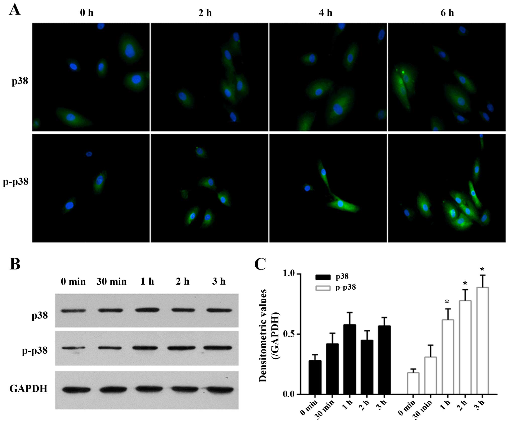
|
Botwin KP and Gruber RD: Lumbar spinal
stenosis: anatomy and pathogenesis. Phys Med Rehabil Clin N Am.
14:1–15. 2003. View Article : Google Scholar : PubMed/NCBI
|
|
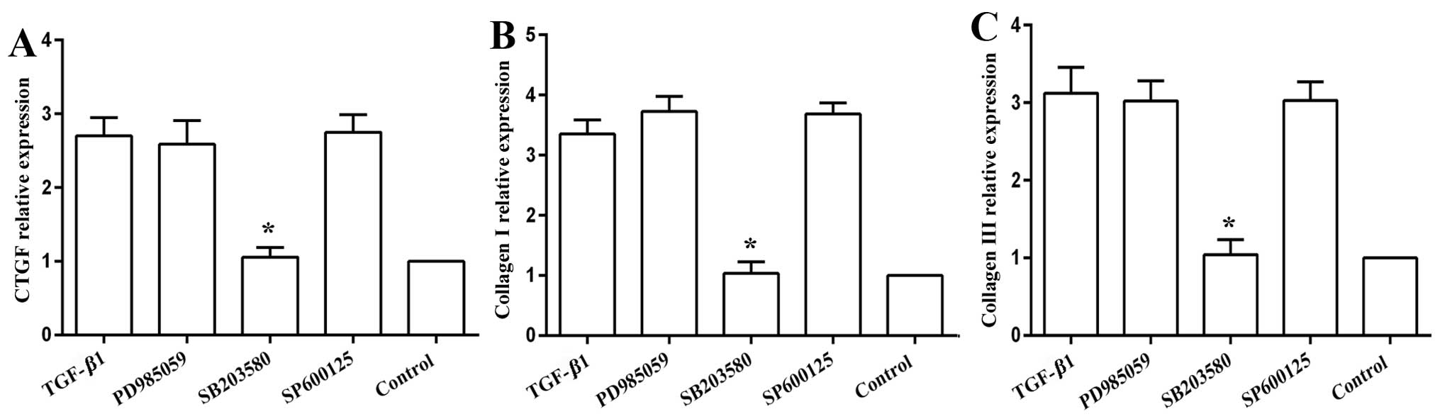
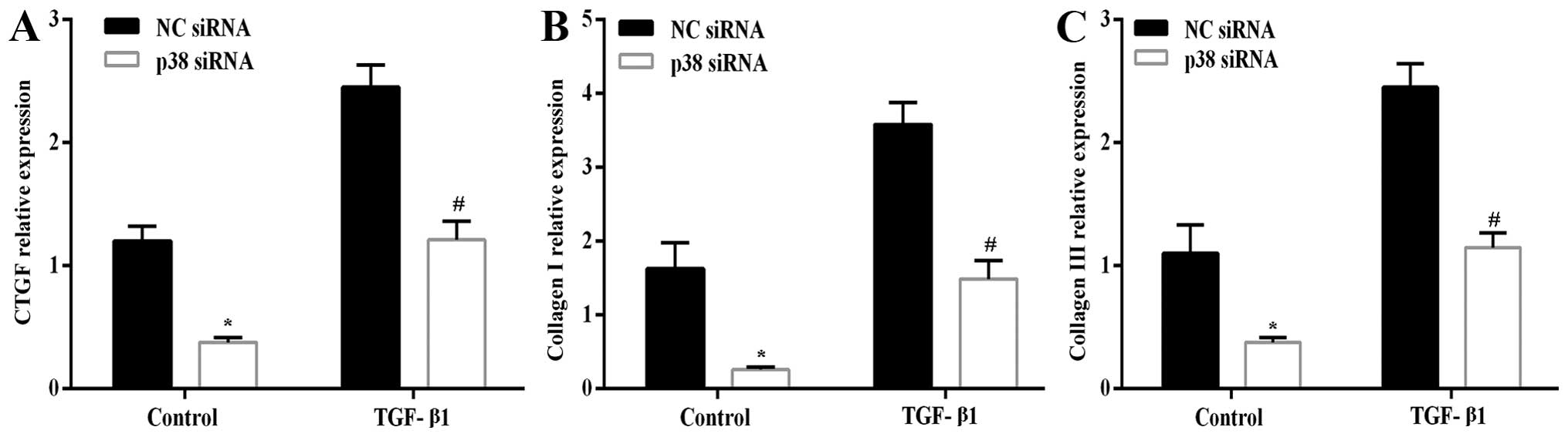
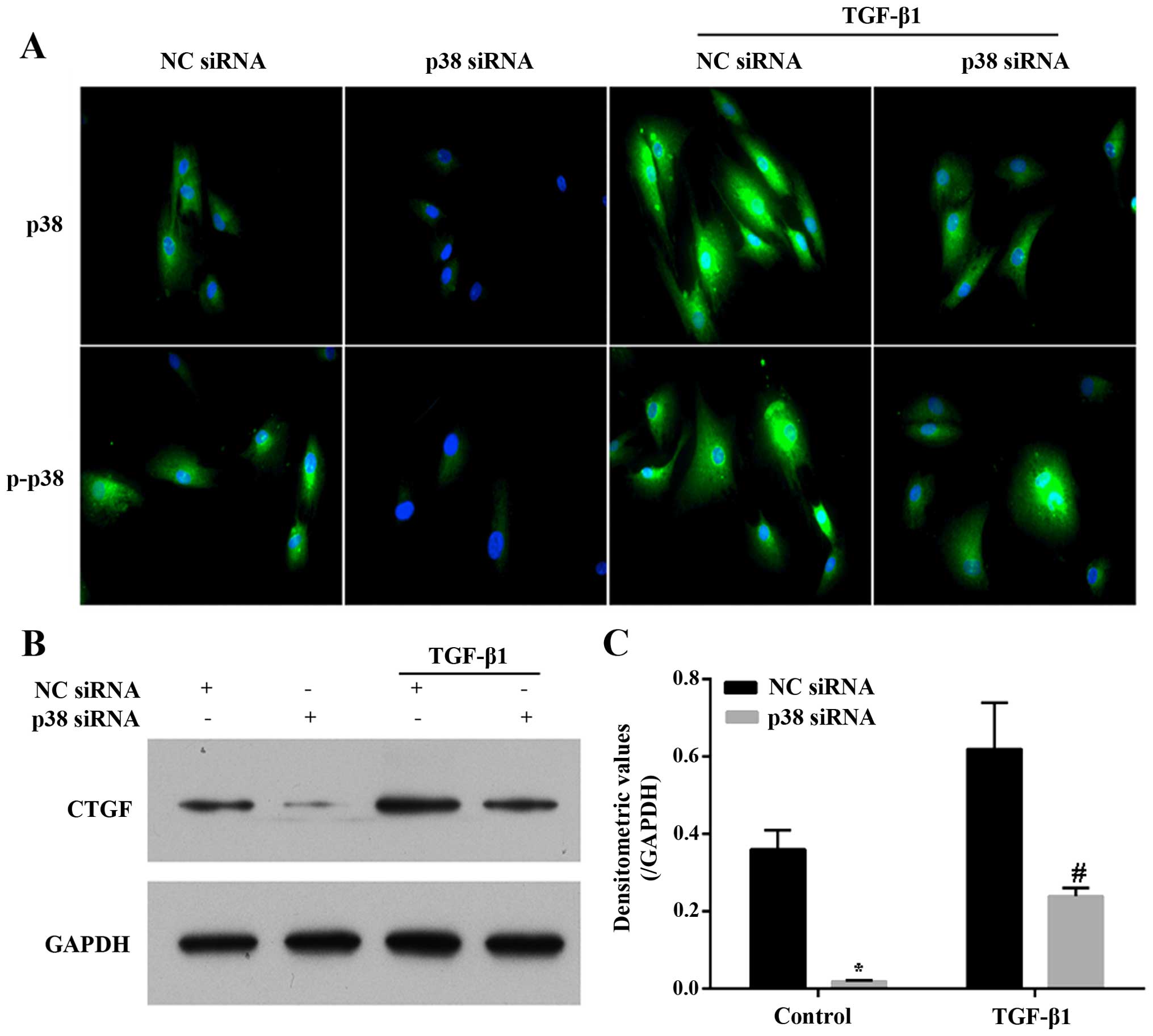
3
|
Fukuyama S, Nakamura T, Ikeda T and Takagi
K: The effect of mechanical stress on hypertrophy of the lumbar
ligamentum flavum. J Spinal Disord. 8:126–130. 1995. View Article : Google Scholar : PubMed/NCBI
|
|
4
|
Behm B, Babilas P, Landthaler M and
Schreml S: Cytokines, chemokines and growth factors in wound
healing. J Eur Acad Dermatol Venereol. 26:812–820. 2012. View Article : Google Scholar : PubMed/NCBI
|
|
5
|
Matsumoto Y, Fujiwara T, Imamura R, Okada
Y, Harimaya K, Doi T, Kawaguchi K, Okada S, Yamada Y, Oda Y and
Iwamoto Y: Hematoma of the ligamentum flavum in the thoracic spine:
report of two cases and possible role of the transforming growth
factor beta-vascular endothelial growth factor signaling axis in
its pathogenesis. J Orthop Sci. 18:347–354. 2013. View Article : Google Scholar
|
|
6
|
Maezawa Y, Baba H, Uchida K, Kokubo Y,
Kubota C and Noriki S: Ligamentum flavum hematoma in the thoracic
spine. Clin Imaging. 25:265–267. 2001. View Article : Google Scholar : PubMed/NCBI
|
|
7
|
Sairyo K, Biyani A, Goel VK, Leaman DW,
Booth R Jr, Thomas J, Ebraheim NA, Cowgill IA and Mohan SE: Lumbar
ligamentum flavum hypertrophy is due to accumulation of
inflammation-related scar tissue. Spine. 32:E340–E347. 2007.
View Article : Google Scholar : PubMed/NCBI
|
|
8
|
Kosaka H, Sairyo K, Biyani A, Leaman D,
Yeasting R, Higashino K, Sakai T, Katoh S, Sano T, Goel VK and
Yasui N: Pathomechanism of loss of elasticity and hypertrophy of
lumbar ligamentum flavum in elderly patients with lumbar spinal
canal stenosis. Spine. 32:2805–2811. 2007. View Article : Google Scholar
|
|
9
|
Nakamura T, Okada T, Endo M, Kadomatsu T,
Taniwaki T, Sei A, Odagiri H, Masuda T, Fujimoto T, Nakamura T, et
al: Angiopoietin-like protein 2 induced by mechanical stress
accelerates degeneration and hypertrophy of the ligamentum flavum
in lumbar spinal canal stenosis. PLoS One. 9:e855422014. View Article : Google Scholar : PubMed/NCBI
|
|
10
|
Nakatani T, Marui T, Hitora T, Doita M,
Nishida K and Kurosaka M: Mechanical stretching force promotes
collagen synthesis by cultured cells from human ligamentum flavum
via transforming growth factor-beta1. J Orthop Res. 20:1380–1386.
2002. View Article : Google Scholar : PubMed/NCBI
|
|
11
|
Ariel A and Timor O: Hanging in the
balance: endogenous anti-inflammatory mechanisms in tissue repair
and fibrosis. J Pathol. 229:250–263. 2013. View Article : Google Scholar
|
|
12
|
Seko Y, Seko Y, Takahashi N, Shibuya M and
Yazaki Y: Pulsatile stretch stimulates vascular endothelial growth
factor (VEGF) secretion by cultured rat cardiac myocytes. Biochem
Biophys Res Commun. 254:462–465. 1999. View Article : Google Scholar : PubMed/NCBI
|
|
13
|
Löhr M, Hampl JA, Lee JY, Ernestus RI,
Deckert M and Stenzel W: Hypertrophy of the lumbar ligamentum
flavum is associated with inflammation-related TGF-β expression.
Acta Neurochir (Wien). 153:134–141. 2011. View Article : Google Scholar
|
|
14
|
Park JB, Chang H and Lee JK: Quantitative
analysis of transforming growth factor-beta 1 in ligamentum flavum
of lumbar spinal stenosis and disc herniation. Spine. 26:E492–E495.
2001. View Article : Google Scholar : PubMed/NCBI
|
|
15
|
Chen YT, Wei JD, Wang JP, Lee HH, Chiang
ER, Lai HC, Chen LL, Lee YT, Tsai CC, Liu CL, et al: Isolation of
mesenchymal stem cells from human ligamentum flavum: implicating
etiology of ligamentum flavum hypertrophy. Spine. 36:E1193–E1200.
2011. View Article : Google Scholar : PubMed/NCBI
|
|
16
|
Zhong ZM, Zha DS, Xiao WD, Wu SH, Wu Q,
Zhang Y, Liu FQ and Chen JT: Hypertrophy of ligamentum flavum in
lumbar spine stenosis associated with the increased expression of
connective tissue growth factor. J Orthop Res. 29:1592–1597. 2011.
View Article : Google Scholar : PubMed/NCBI
|
|
17
|
Brigstock DR: The connective tissue growth
factor/cysteine-rich 61/nephroblastoma overexpressed (CCN) family.
Endocr Rev. 20:189–206. 1999.PubMed/NCBI
|
|
18
|
Taipale J, Miyazono K, Heldin CH and
Keski-Oja J: Latent transforming growth factor-beta 1 associates to
fibroblast extracellular matrix via latent TGF-beta binding
protein. J Cell Biol. 124:171–181. 1994. View Article : Google Scholar : PubMed/NCBI
|
|
19
|
Igarashi A, Okochi H, Bradham DM and
Grotendorst GR: Regulation of connective tissue growth factor gene
expression in human skin fibroblasts and during wound repair. Mol
Biol Cell. 4:637–645. 1993. View Article : Google Scholar : PubMed/NCBI
|
|
20
|
Abreu JG, Ketpura NI, Reversade B and De
Robertis EM: Connective-tissue growth factor (CTGF) modulates cell
signalling by BMP and TGF-beta. Nat Cell Biol. 4:599–604.
2002.PubMed/NCBI
|
|
21
|
Chen Z, Gibson TB, Robinson F, Silvestro
L, Pearson G, Xu B, Wright A, Vanderbilt C and Cobb MH: MAP
kinases. Chem Rev. 101:2449–2476. 2001. View Article : Google Scholar : PubMed/NCBI
|
|
22
|
Gu J, Liu X, Wang QX, Tan HW, Guo M, Jiang
WF and Zhou L: Angiotensin II increases CTGF expression via
MAPKs/TGF-β1/TRAF6 pathway in atrial fibroblasts. Exp Cell Res.
318:2105–2115. 2012. View Article : Google Scholar : PubMed/NCBI
|
|
23
|
Specchia N, Pagnotta A, Gigante A,
Logroscino G and Toesca A: Characterization of cultured human
ligamentum flavum cells in lumbar spine stenosis. J Orthop Res.
19:294–300. 2001. View Article : Google Scholar : PubMed/NCBI
|
|
24
|
Zhong ZM and Chen JT: Phenotypic
characterization of ligamentum flavum cells from patients with
ossification of ligamentum flavum. Yonsei Med J. 50:375–379. 2009.
View Article : Google Scholar : PubMed/NCBI
|
|
25
|
Mosmann T: Rapid colorimetric assay for
cellular growth and survival: application to proliferation and
cytotoxicity assays. J Immunol Methods. 65:55–63. 1983. View Article : Google Scholar : PubMed/NCBI
|
|
26
|
Zeng Y, Adamson RH, Curry FR and Tarbell
JM: Sphingosine-1-phosphate protects endothelial glycocalyx by
inhibiting syndecan-1 shedding. Am J Physiol Heart Circ Physiol.
306:H363–H372. 2014. View Article : Google Scholar :
|
|
27
|
Leask A and Abraham DJ: TGF-beta signaling
and the fibrotic response. FASEB J. 18:816–827. 2004. View Article : Google Scholar : PubMed/NCBI
|
|
28
|
Yoshida M, Shima K, Taniguchi Y, Tamaki T
and Tanaka T: Hypertrophied ligamentum flavum in lumbar spinal
canal stenosis. Pathogenesis and morphologic and
immunohistochemical observation. Spine. 17:1353–1360. 1992.
View Article : Google Scholar : PubMed/NCBI
|
|
29
|
Feng XH and Derynck R: Specificity and
versatility in tgf-beta signaling through Smads. Annu Rev Cell Dev
Biol. 21:659–693. 2005. View Article : Google Scholar : PubMed/NCBI
|
|
30
|
Massagué J: How cells read TGF-beta
signals. Nat Rev Mol Cell Biol. 1:169–178. 2000. View Article : Google Scholar
|
|
31
|
Strand DW, Liang YY, Yang F, Barron DA,
Ressler SJ, Schauer IG, Feng XH and Rowley DR: TGF-β induction of
FGF-2 expression in stromal cells requires integrated smad3 and
MAPK pathways. Am J Clin Exp Urol. 2:239–248. 2014.
|
|
32
|
Demagny H, Araki T and De Robertis EM: The
tumor suppressor Smad4/DPC4 is regulated by phosphorylations that
integrate FGF, Wnt, and TGF-β signaling. Cell Reports. 9:688–700.
2014. View Article : Google Scholar
|
|
33
|
Derynck R and Zhang YE: Smad-dependent and
Smad-independent pathways in TGF-beta family signalling. Nature.
425:577–584. 2003. View Article : Google Scholar : PubMed/NCBI
|
|
34
|
Javelaud D and Mauviel A: Crosstalk
mechanisms between the mitogen-activated protein kinase pathways
and Smad signaling downstream of TGF-beta: implications for
carcinogenesis. Oncogene. 24:5742–5750. 2005. View Article : Google Scholar : PubMed/NCBI
|
|
35
|
Binder DK, Schmidt MH and Weinstein PR:
Lumbar spinal stenosis. Semin Neurol. 22:157–166. 2002. View Article : Google Scholar
|
|
36
|
Omidi-Kashani F, Hasankhani EG and
Ashjazadeh A: Lumbar spinal stenosis: who should be fused? an
updated review. Asian Spine J. 8:521–530. 2014. View Article : Google Scholar : PubMed/NCBI
|