Introduction
Dental pulp participates in the reparative
regeneration of the tooth tissues (1). Dental pulp tissue contains multiple
cells that possess plasticity and multipotency, including
fibroblasts, inflammatory and immune cells, odontoblasts and
undifferentiated mesenchymal cells (1–3).
Fibroblasts and odontoblasts are the main cell types in dental pulp
(4). In response to stimuli or
injuries, dental pulp cells (DPCs) differentiate into odontoblasts
to replace the necrotic cells and then generate reparative dentin
(5).
Diabetes mellitus (DM) is a severe chronic disease
that markedly affects the health and life quality of individuals
(6). DM is a multi-organ and
multi-factorial metabolic disease that is characterized by absolute
or relative deficiency in insulin secretion, insulin resistance and
β-cell dysfunction, which may ultimately lead to hyperglycemia
(7–9). DM is a major cause of morbidity and
mortality worldwide (10).
Approximately 87 million adults suffered from the disease in 2014.
The number of individuals with DM is expected to increase to almost
592 million by the year 2035 (11). DM may lead to numerous
complications, including cardiovascular diseases, microvascular
diseases (such as diabetic retinopathy), osteoporosis and diabetic
nephropathy (12,13). Additionally, DM also affects the
functions of dental pulp and periapical tissues (14,15). A previous study indicated that DM
can result in the necrosis of dental pulp and in the development of
periapical lesions in diabetic rats (16). Long-term DM has been demonstrated
to increase the basement membranes thickness of dental pulp vessels
and to contribute to the occurrence and development of angiopathy
(17). It also been demonstrated
that rats with streptozotocin (STZ)-induced diabetes exhibit a
significant reduction in pulpal blood flow (18). Moreover, hyperglycemia inhibits
dentin bridge formation and enhances inflammatory cell infiltration
in diabetic rats (19).
Insulin-like growth factor-1 (IGF-1), a member of
the insulin-like peptide family, plays a vital role in the
survival, apoptosis and differentiation of cells within various
organs, including teeth (20–22). Joseph et al found that
secretory ameloblasts, secretory odontoblasts and mature
ameloblasts express high levels of IGF-1 in the development of the
rat incisor (23). It has also
been demonstrated that IGF-1 promotes human dental pulp stem cell
proliferation and osteogenic differentiation by increasing the
expression of differentiation markers through the mammalian target
of rapamycin (mTOR) signaling pathway (24). IGF-1 has also been reported to
protect DPCs against the cytotoxicity of composite materials via
endogenous antioxidant mechanisms (25).
In the present study, we cultured primary human DPCs
in order to investigate the effects of high glucose (GLU) on the
proliferation, apoptosis and differentiation/mineralization of
DPCs. We further evaluated the protective effects of IGF-1 under
high GLU conditions. Our study provides insight into possible
treatment options to counteract the oral complications associated
with DM.
Materials and methods
Isolation of human DPCs
Human premolars extracted for orthodontic purposes
and third molars from patients without dental carious and
periodontal problems (a total of 80 adults; age: 18–25 years old)
were obtained from China Medical University School and Hospital of
Stomatology. Dental pulp tissues were obtained from teeth and
washed with phosphate-buffered saline (PBS). All study protocols
were approved by the Ethics Committee of China Medical University.
Written informed consent was written from all patients. The tissues
were then cut into sections and digested with 0.3% type I
collagenase and 0.4% dispase at 37°C for 1 h. The isolated human
DPCs were cultured in Dulbecco's modified Eagle's medium (DMEM)
(Gibco, Grand Island, NY, USA) supplemented with 10% fetal bovine
serum (FBS) (HyClone Laboratories, Inc., Logan, UT, USA) at 37°C in
a 5% CO2 atmosphere.
Odontoblastic differentiation
The cells at passage 3 were seeded on culture
dishes. After being grown to 70–80% confluence, the cells were
cultured in odontoblastic induction medium (OM) for 1, 3, 7 and 14
days, composed of DMEM, 50 µg/ml ascorbic acid and 10 mM
β-glycerophosphate sodium (both from Sigma-Aldrich, St. Louis, MO,
USA).
Cell proliferation assay
The DPCs were seeded into 96-well plates at a
density of 2×10³ cells/well and cultured in DMEM
containing various concentrations of GLU (10, 25 and 50 mM)
(Sigma-Aldrich). The cells were cultured for 24, 48 and 72 h. Cell
Counting Kit-8 (CCK-8) agent (10 µl) (Beyotime Institute of
Biotechnology, Inc., Haimen, China) was then added to each well and
the cells were incubated for 1 h at 37°C. The absorbance was
measured at 450 nm using a microplate reader (ELX-800; BioTek
Instruments, Inc., Winooski, VT, USA). The optimum concentration of
GLU was determined.
In addition, the cells were plated in 96-well plates
(2×10³ cells/well) and maintained in DMEM containing
high GLU (25 mM). The dose of GLU (25 mM) used was the minimum
effective dose. The cells were then treated with various
concentrations of IGF-1 (10, 50 and 100 ng/ml) for 24, 48 and 72 h
(Peprotech Inc., Rocky Hill, NJ, USA). Cell proliferation was
evaluated by CCK-8 assay (Beyotime Institute of Biotechnology,
Inc.) according to the manufacturer's instructions.
Cell apoptosis assay
The cells were harvested after the corresponding
treatments and washed twice with PBS. Following centrifugation at
800 rpm for 5 min, the cells were resuspended in 500 µl
binding buffer gently. Subsequently, 5 µl Annexin V-FITC and
5 µl propidium iodide (PI) (KeyGen Biotech Co., Nanjing,
China) were added to the suspension and mixed immediately. The cell
suspension was incubated at room temperature for 15 min in the dark
and analyzed by flow cytometry (Model C6; BD Biosciences, San Jose,
CA, USA).
Alkaline phosphatase (ALP) activity
assay
The cells were resuspended in 100 µl PBS and
subjected to repeated freeze-thawing cycles. The supernatant was
obtained by centrifugation at 12,000 rpm for 10 min and quantified
by BCA (Wanleibio, Shenyang, China). The activity of ALP was
measured using an ALP assay kit (Nanjing Jiancheng Bioengineering
Institute, Nanjing, China) according to the manufacturer's
instructions and expressed as U/g protein.
von Kossa staining
The fixed cells were washed 3 times with deionized
water. Subsequently, the cells were stained with 1% silver nitrate
(Jizhun, Shanghai, China) and exposed to ultraviolet light for 20
min. After washing with deionized water, the cells were incubated
for 5 min with 5% sodium thiosulfate and counterstained for 30 sec
with 0.1% nuclear fast red (both from Sinopharm Chemical Reagent
Co., Ltd., Beijing, China). The coverslips were dehydrated in
ethanol (75, 85, 95 and 100%) and captured under a microscope
(DP73; Olympus, Tokyo, Japan).
Western blot analysis
Total proteins were extracted from the cells using
the total protein extraction kit purchased from Wanleibio and
quantified. Total proteins (40 µg in each lane) were
subjected to 8, 10 or 15% SDS-PAGE (Wanleibio), followed by
transfer onto PVDF membranes (EMD Millipore, Bedford, MA, USA). The
membranes were blocked with non-fat milk and then incubated at 4°C
overnight with antibodies against osteocalcin (OCN) (1:200;
sc-376835), osteonectin (ON) (1:200; sc-398419), dentin matrix
protein-1 (DMP-1) (1:200; sc-73633) (all from Santa Cruz
Biotechnology, Inc., Santa Cruz, CA, USA), osteopontin (OPN)
(1:500; D121078; Sangon Biotech Co., Ltd., Shanghai, China), dentin
sialoprotein (DSP) (1:500; bs-8557R; BIOSS, Beijing, China), IGF-1
(1:400; BA0939), IGF-1 receptor (IGF-1R) (1:400; BA0498),
IGF-binding protein 1 (IGFBP1) (1:400; BA1749) and IGFBP3 (1:400;
BA3641) (all from Boster Biological Technology, Ltd., Wuhan,
China). The membranes were washed and incubated at 37°C for 45 min
with goat anti-rabbit/mouse horseradish peroxidase (HRP)-conjugated
secondary antibody (1:5,000; WLA023/WLA024; Wanleibio). The protein
bands were visualized by using enhanced chemiluminescence (ECL)
substrate (Wanleibio) and quantified using Gel-Pro Analyzer 4.0
(Media Cybernetics, Inc., Rockville, MD, USA).
Reverse transcription-quantitative
polymerase chain reaction (RT-qPCR)
Total RNA was extracted using the RNApure total RNA
extraction kit (BioTeke Corp., Beijing, China) and 1 µg
total RNA was reverse transcribed into cDNA using M-MLV reverse
transcriptase ((BioTeke Corp.) according to the manufacturer's
instructions. The primers used in this study were synthesized by
Sangon Biotech Co., Ltd. and were as follows: IGF-1 forward,
5′-ACAAGCCCACAGGGTATG-3′ and IGF-1 reverse,
5′-CACTCCCTCTACTTGCGTTCT-3′; IGF-1R forward,
5′-TGCTGTATGCCTCTGTGAACC-3′ and IGF-1R reverse,
5′-AGACCATCCCAAACGACCC-3′; IGFBP1 forward,
5′-CCTGCCAAACTGCAACAAG-3′ and IGFBP1 reverse,
5′-CCCATTCCAAGGGTAGACG-3′; IGFBP3 forward,
5′-TAAGGTGGAGTCCTACTTGTTT-3′ and IGFBP3 reverse,
5′-ACTTGTGATGCCTCTGAATG-3′; and β-actin forward,
5′-CTTAGTTGCGTTACACCCTTTCTTG-3′ and β-actin reverse,
5′-CTGTCACCTTCACCGTTCCAGTTT-3′. qPCR amplification (95°C for 10
min, followed by 40 cycles of 95°C for 10 sec, 60°C for 20 sec and
72°C for 30 sec) was performed on an Exicycler™ 96 quantitative
fluorescence analyzer (Bioneer Co., Daejeon, Korea) using
SYBR-Green (Solarbio, Beijing, China). Gene expression levels were
normalized to β-actin levels and calculated using 2−ΔΔCt
method (26).
Statistical analysis
Data are expressed as the meand ± SD. All data were
analyzed by one-way analysis of variance (ANOVA) and Bonferroni's
multiple comparison test. A P-value <0.05 was considered to
indicate a statistically significant difference.
Results
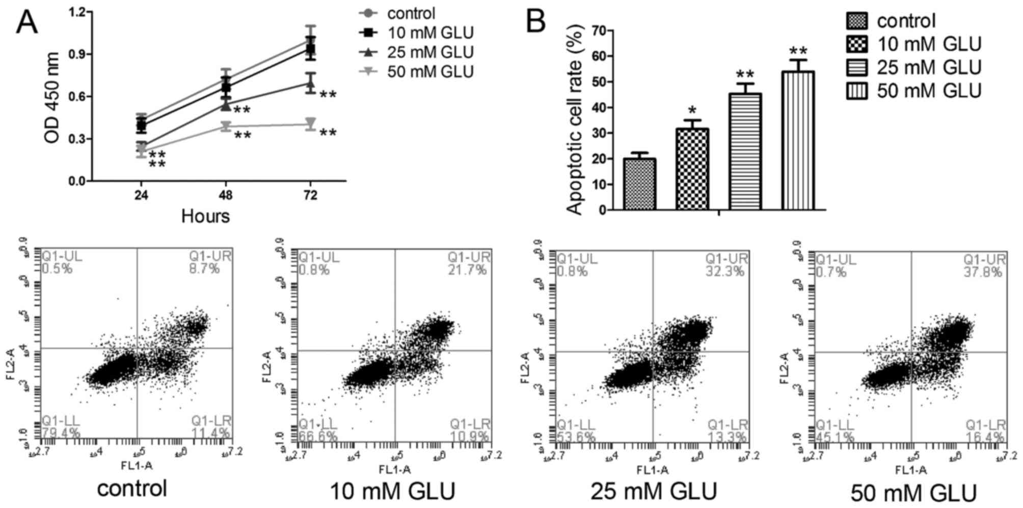
High GLU inhibits the proliferation and
promotes the apoptosis of human DPCs
In this study, CCK-8 assay was performed to evaluate
the effects of high GLU on the proliferation of DPCs. As shown in
Fig. 1A, exposure to high GLU (at
concentrations of 25 and 50 mM) for 24, 48 and 72 h significantly
decreased the viability of the DPCs compared with the control
group. The effects of high GLU on cell apoptosis were also
investigated by Annexin V-FITC/PI staining. As shown in Fig. 1B, exposure to GLU (10, 25 and 50
mM) markedly promoted the apoptosis of the DPCs compared with the
control group, as evidenced by the increased apoptotic rate.
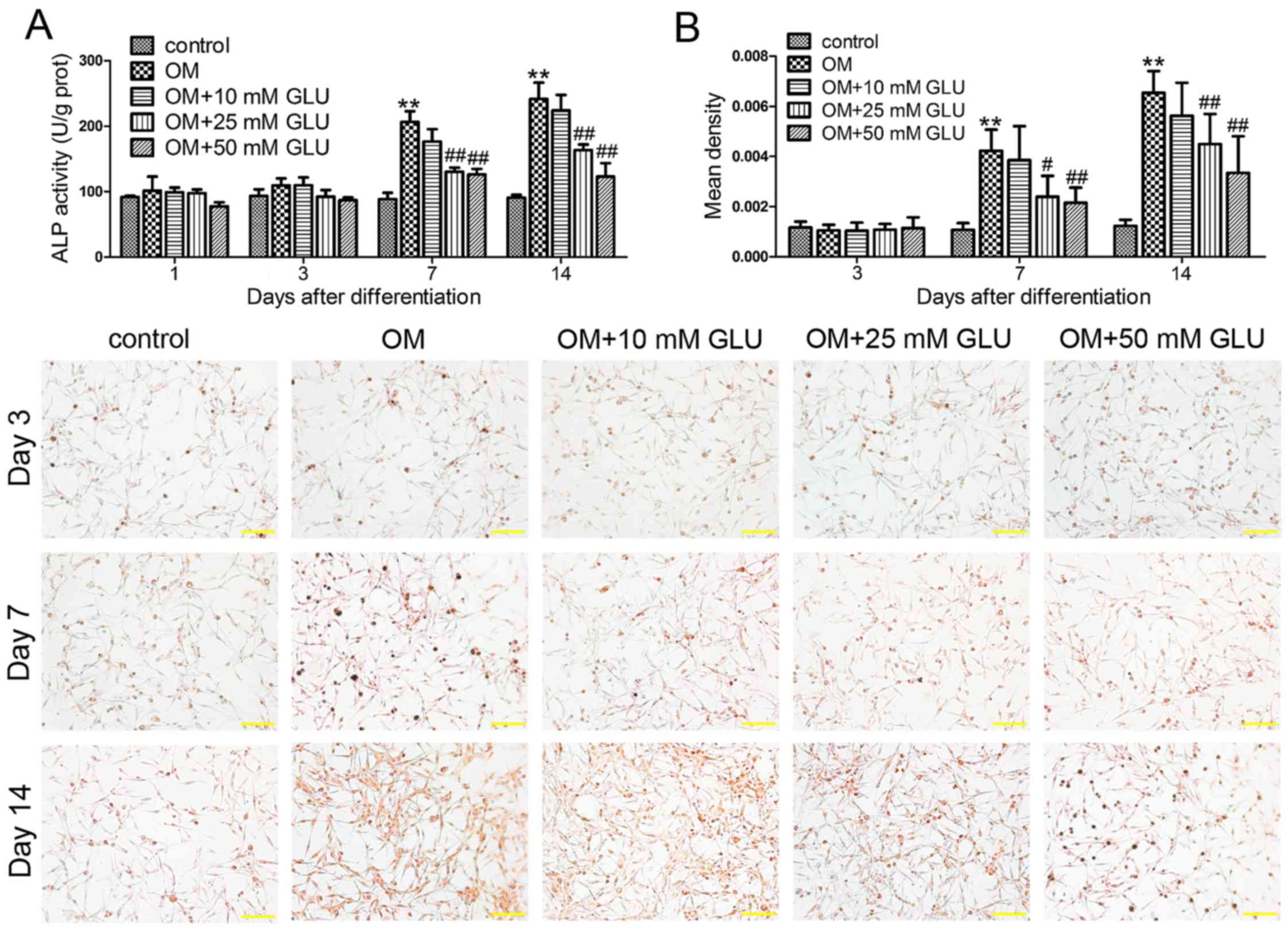
High GLU inhibits the odontoblastic
differentiation of and mineralization in DPCs
No statistically significant differences were
observed in the activity of ALP among the groups on days 1 and 3
(Fig. 2A). ALP activity in the
cells cultured in OM was significantly higher than that of the
untreated control cells on days 7 and 14. Exposure to hight GLU (25
and 50 mM) significantly reduced this increase from day 7 after
differentiation. Mineralization in DPCs was assessed by von Kossa
staining. Our data indicated that mineralized matrix formation in
the cells cultured in OM was markedly enhanced compared with the
control group on days 7 and 14 (Fig.
2B). However, exposure to high GLU (25 and 50 mM) significantly
inhibited odontoblastic mineralization.
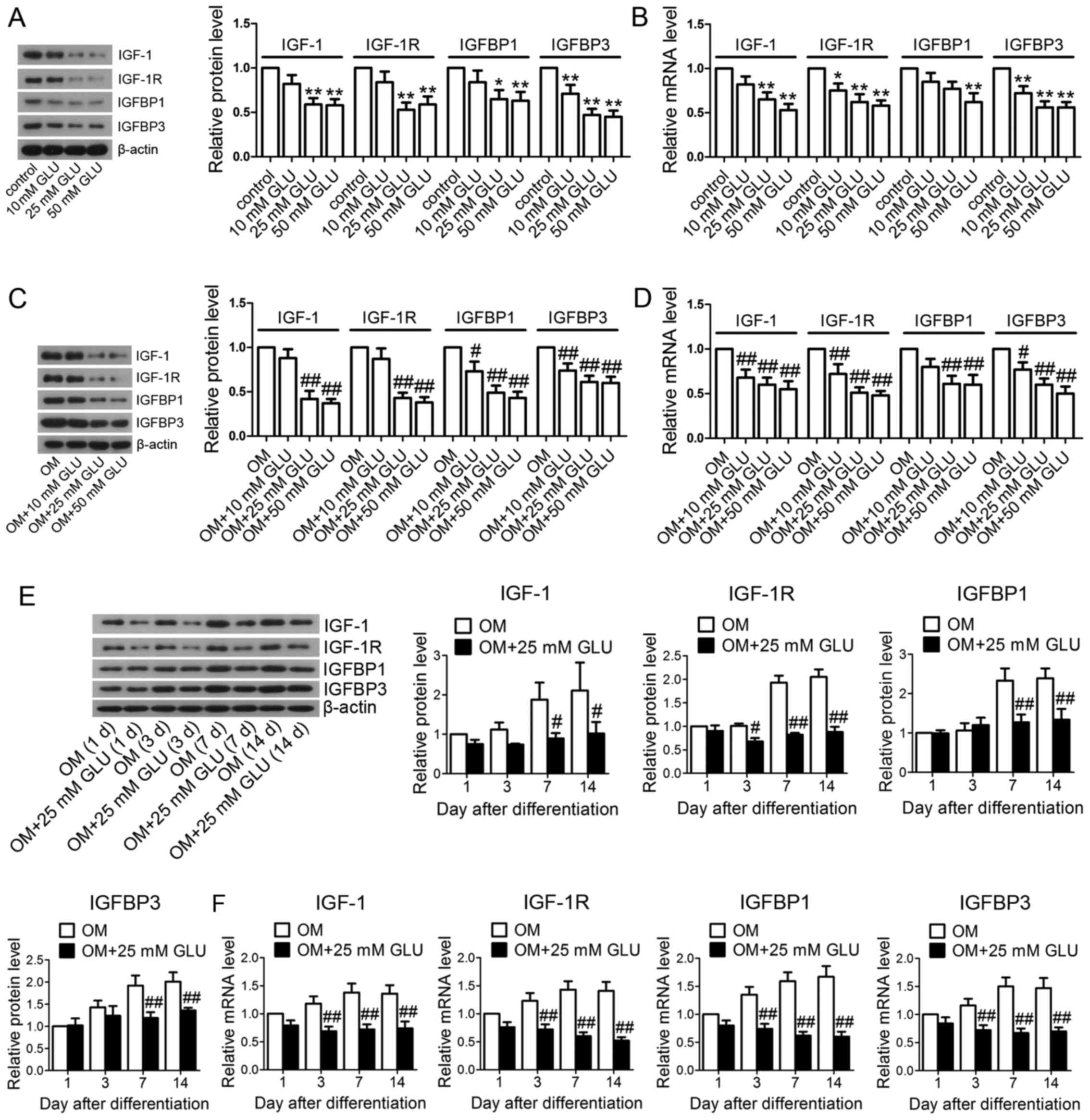
High GLU reduces the expression levels of
IGF family members in DPCs
A previous study reported that IGF-1 can promote the
proliferation and osteogenic differentiation of human dental pulp
stem cells (24). Therefore, we
examined the expression levels of several IGF family members in the
DPCs after the indicated treatments. The cells were cultured in
normal DMEM and exposed to increasing concentrations of GLU for 24
h. Compared with the control group, there was a significant
reduction in the IGF-1, IGF-1R, IGFBP1 and IGFBP3 protein levels in
the GLU-exposed cells (Fig. 3A).
Consistent with the results of western blot analysis, the
corresponding decreases were confirmed by RT-qPCR (Fig. 3B).
Furthermore, the cells were maintained in
differentiation medium. After 7 days of differentiation, the mRNA
and protein levels of IGF-1, IGF-1R, IGFBP1 and IGFBP3 in the OM +
GLU groups were markedly decreased in comparison with the OM group,
as evaluated by western blot analysis (Fig. 3C) and RT-qPCR (Fig. 3D). Several days after
differentiation, the protein levels of IGF-1 (days 7 and 14),
IGF-1R (days 3, 7 and 14), IGFBP1 (days 7 and 14) and IGFBP3 (days
7 and 14) in the OM + 25 mM GLU group were significantly lower than
those in the OM group (Fig. 3E).
Markedly decreased mRNA levels were firstly observed on day 3
(Fig. 3F).
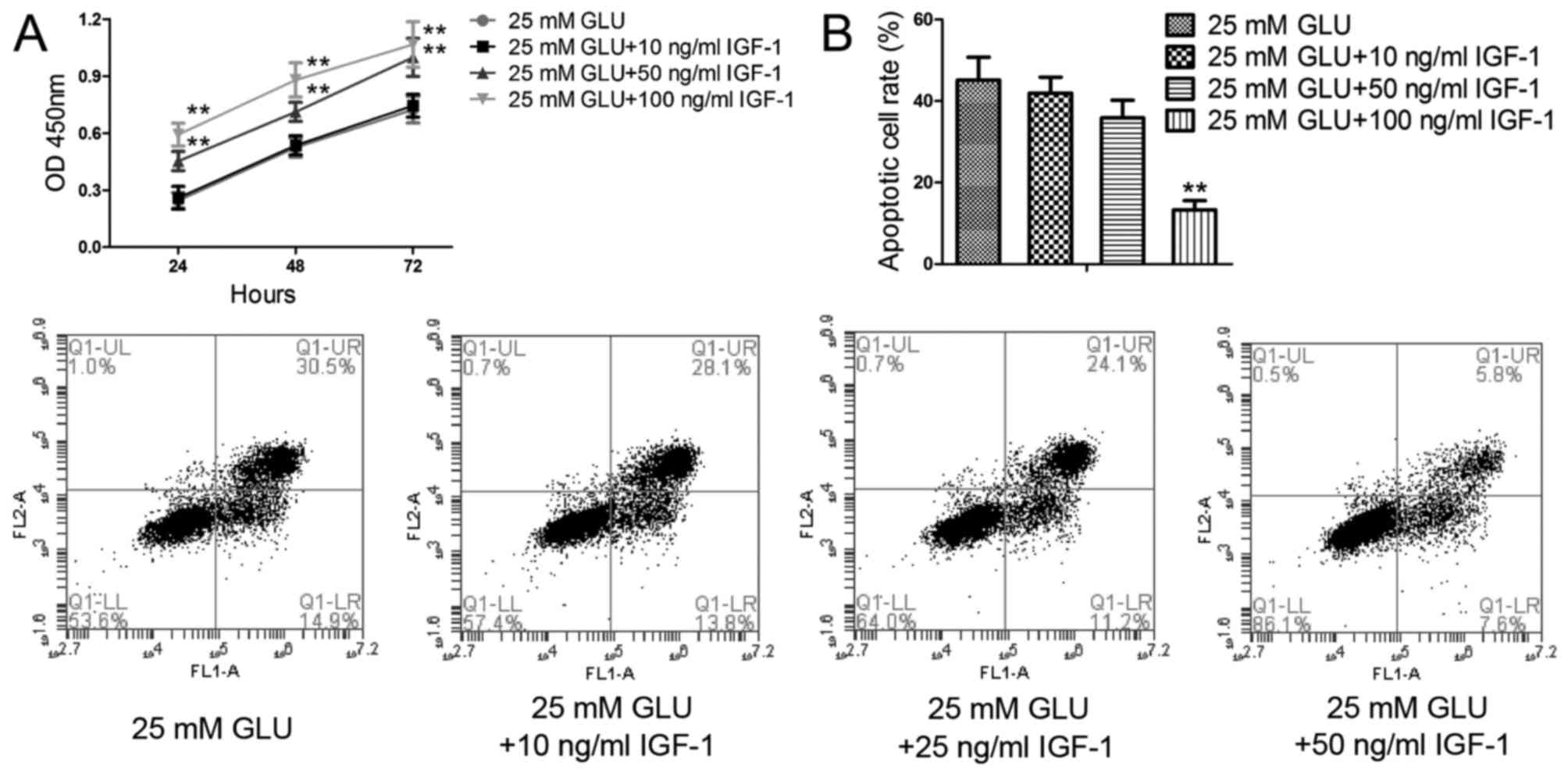
IGF-1 abolishes the effect of high GLU on
the proliferation and apoptosis of DPCs
The cells were then exposed to 25 mM GLU and various
concentrations of IGF-1. As shown in Fig. 4A, in the presence of IGF-1 at
concentrations of 50 and 100 ng/ml, cell viability was
significantly increased compared with the 25 mM GLU group. As
expected, the pro-apoptotic effects of high GLU were markedly
suppressed by treatment with 100 ng/ml IGF-1 (Fig. 4B).
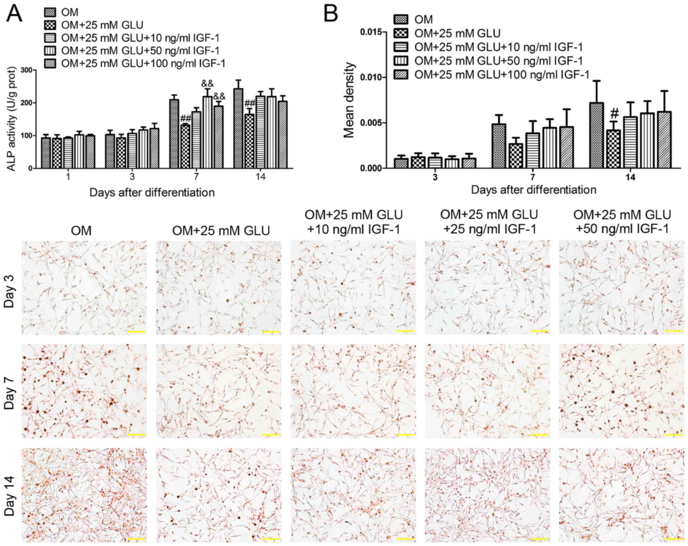
IGF-1 attenuates the effect of high GLU
on the odontoblastic differentiation of and mineralization in
DPCs
Subsquently, the cells were cultured for different
periods of time in OM with 25 mM GLU and IGF-1. The results
demonstrated that the inhibitory effects of high GLU on ALP
activity (Fig. 5A) were markedly
abolished by IGF-1 (50 and 100 ng/ml) on day 7 after odontoblastic
induction. Treatment with various concentrations of IGF-1 reversed
the effects of high GLU on mineralization in DPCs. However, the
difference was not statistically significant (Fig. 5B).
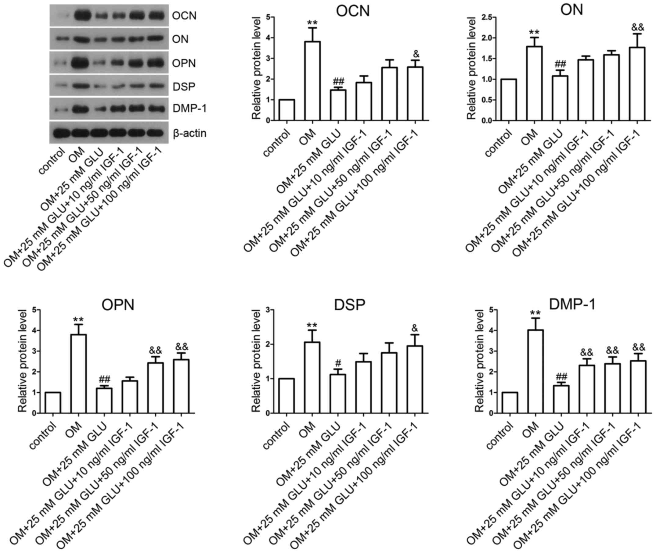
IGF-1 restores the decreased levels of
mineralization-related proteins induced by high GLU
Furthermore, western blot analysis was used to
measure the expression levels of mineralization-related proteins.
Our results revealed that the OCN, ON, OPN, DSP and DMP-1 levels in
the OM group were significantly higher than those in the control
group (Fig. 6). In the presence
of 25 mM GLU, the levels of these mineralization-related proteins
in the OM + 25 mM GLU group were markedly decreased compared with
the OM group. However, IGF-1 treatment significantly restored the
high GLU-induced decrease in the levels of mineralization-related
proteins, including OCN, ON, OPN, DSP and DMP-1.
Discussion
The process of dentinal regeneration involves the
proliferation and differentiation of DPCs into odontoblasts, dental
pulp healing and reparative dentin formation (27). A high sucrose diet affects the
dentin-pulp complex and reduces dentin formation, which contributes
to caries in dentin (28). The
effect of high GLU on the proliferation, apoptosis and
differentiation of DPCs warrants further investigation. In the
present study, we demonstrated that high GLU suppressed the
proliferation, induced the apoptosis and inhibited the
differentiation of human DPCs, accompanied by reduced levels of
IGF-1 family members, including IGF-1, IGF-1R, IGFBP1 and IGFBP3.
We then evaluated the effects of IGF-1 on the biological properties
of DPCs under high GLU conditions. We found that IGF-1 treatment
reversed the effects of high GLU on DPCs.
It has been reported that hyperglycemia inhibits
pulp repair (29). A previous
study demonstrated that a high concentration of D-GLU (30 mM)
markedly inhibited the proliferation of MD10-F2 pulp cells compared
with the controls in vitro (30). Furthermore, previous studies have
also demonstrated that high GLU (25 mM) reduces the proliferative
capability of human cavernous endothelial cells (HCECs) and induces
cell apoptosis in vitro (31,32). Our experiments consistently
demonstrated that high GLU, particularly at the concentrations of
25 and 50 mM, markedly suppressed human DPC proliferation and
promoted cell apoptosis. ALP is a marker of odontoblastic
differentiation and its activity is enhanced during odontoblastic
induction (33,34). We found that high GLU inhibited
ALP activity and mineralization in DPCs, suggesting that high GLU
inhibited the proliferation and differentiation and induced the
apoptosis of DPCs.
IGF-1 is an ubiquitous peptide hormone and an
important anti-apoptotic factor (35). IGFBPs constitute multiple proteins
that bind to IGFs and modulate the interaction of IGFs with their
receptors (36). Yu et al
reported that high GLU leads to cardiomyocyte H9c2 cell apoptosis
by decreasing IGF-1R expression (37). Therefore, we hypothesized that
high GLU would also affect the expression of IGF-1 family members
in human DPCs that underwent apoptosis and differentiation. Our
results revealed that 25 and 50 mM GLU markedly decreased the
expression levels of IGF-1, IGF-1R, IGFBP1 and IGFBP3 in DPCs that
were cultured in normal DMEM for 24 h or in OM after 7 days of
differentiation. Additionally, we found that 25 mM GLU
significantly decreased the IGF-1, IGF-1R, IGFBP1 and IGFBP3 levels
from day 3 or 7 after differentiation.
IGF-1 binds to and activates the receptor IGF-1R and
thus promotes cell proliferation and survival (38). Zhang et al reported that
IGF-1 attenuated high fat diet-induced mitochondrial damage,
myocardial contraction dysfunction and cardiomyocyte apoptosis
(39). As expected, our results
revealed that IGF-1 reversed the effects of high GLU on cell
proliferation and protected the DPCs against apoptosis, suggesting
the protective effects of IGF-1 in DPCs under conditions of high
GLU.
It is well known that high GLU inhibits the
osteoblast differentiation of MC3T3-E1 cells (40). IGF-1 has been demonstrated to
promote the proliferation and differentiation of osteoblasts and
chondrocytes in vitro (41). However, the effects of IGF-1 on
the differentiation of human DPCs under high GLU conditions remain
unknown. The process of odontoblastic differentiation involves
multiple proteins (1). OCN is an
important differentiation marker that is found in odontoblasts,
dentine matrix and bone matrix. It is associated with
mineralization and matrix deposition (42). ON is a major non-collagenous
protein that is responsible for bone and dentin mineralization
(43). OPN, a secreted
glycophosphoprotein, is an odontoblastic marker for early
differentiation (44). DSPP is a
collagenous extracellular matrix (ECM) protein that participates in
dentin mineralization. It can be cleaved into DSP and dentin
phosphoprotein (DPP) (45). DMP-1
is also correlated with dentin formation and mineralization
(46). In our study, the
differentiation of DPCs into odontoblastic cells was evaluated by
measuring ALP activity, mineralization and the levels of
mineralization-associated proteins (OCN, ON, OPN, DSP and DMP-1).
We provide similar findings that 25 mM GLU markedly suppressed the
activity of ALP and decreased the mineralized matrix deposition in
primary human DPCs that underwent differentiation. Furthermore, 25
mM GLU significantly decreased OCN, ON, OPN, DSP and DMP-1
expression in DPCs during differentiation. These results indicate
that high GLU inhibits the odontoblastic differentiation of DPCs.
However, IGF-1 restored ALP activity and mineralization in the
DPCs, suggesting that IGF-1 attenuates the effects of high GLU and
promotes the odontoblastic differentiation of DPCs under high GLU
conditions by increasing the expression levels of
mineralization-related proteins.
In conclusion, the findings of the present study
suggest that IGF-1 promotes the survival and odontoblastic
differentiation of DPCs, and protects the cells against apoptosis
in a high GLU environment.
References
|
1
|
Wang YL, Hu YJ and Zhang FH: Effects of
GPNMB on proliferation and odontoblastic differentiation of human
dental pulp cells. Int J Clin Exp Pathol. 8:6498–6504.
2015.PubMed/NCBI
|
|
2
|
Wu Q, Qi S, Ma J, Chen F, Chen J, Li J,
Zhang X, Xu Y, Pan Q and Wang R: The Effect of NRAGE on cell cycle
and apoptosis of human dental pulp cells and MDPC-23. Int J Clin
Exp Med. 8:10657–10667. 2015.PubMed/NCBI
|
|
3
|
Cooper PR, Takahashi Y, Graham LW, Simon
S, Imazato S and Smith AJ: Inflammation-regeneration interplay in
the dentine-pulp complex. J Dent. 38:687–697. 2010. View Article : Google Scholar : PubMed/NCBI
|
|
4
|
Jiang W, Lv H, Wang H, Wang D, Sun S, Jia
Q, Wang P, Song B and Ni L: Activation of the NLRP3/caspase-1
inflammasome in human dental pulp tissue and human dental pulp
fibroblasts. Cell Tissue Res. 361:541–555. 2015. View Article : Google Scholar : PubMed/NCBI
|
|
5
|
Han N, Zheng Y, Li R, Li X, Zhou M, Niu Y
and Zhang Q: β-catenin enhances odontoblastic differentiation of
dental pulp cells through activation of Runx2. PLoS One.
9:e888902014. View Article : Google Scholar
|
|
6
|
Saleh F, Ara F, Mumu SJ and Hafez MA:
Assessment of health-related quality of life of Bangladeshi
patients with type 2 diabetes using the EQ-5D: a cross-sectional
study. BMC Res Notes. 8:4972015. View Article : Google Scholar : PubMed/NCBI
|
|
7
|
Stumvoll M, Goldstein BJ and van Haeften
TW: Type 2 diabetes: principles of pathogenesis and therapy.
Lancet. 365:1333–1346. 2005. View Article : Google Scholar : PubMed/NCBI
|
|
8
|
Al Ghamdi AA, Badr G, Hozzein WN, Allam A,
Al-Waili NS, Al-Wadaan MA and Garraud O: Oral supplementation of
diabetic mice with propolis restores the proliferation capacity and
chemotaxis of B and T lymphocytes towards CCL21 and CXCL12 by
modulating the lipid profile, the pro-inflammatory cytokine levels
and oxidative stress. BMC Immunol. 16:542015. View Article : Google Scholar : PubMed/NCBI
|
|
9
|
Peng H, Li J, Chen X, Zhou X, Zhu W and Li
F: Genetic variants of PTPN2 gene in chinese children with type 1
diabetes mellitus. Med Sci Monit. 21:2653–2658. 2015. View Article : Google Scholar : PubMed/NCBI
|
|
10
|
Ranasinghe P, Pigera S, Galappatthy P,
Katulanda P and Constantine GR: Zinc and diabetes mellitus:
understanding molecular mechanisms and clinical implications. Daru.
23:442015. View Article : Google Scholar : PubMed/NCBI
|
|
11
|
Shi Y and Hu FB: The global implications
of diabetes and cancer. Lancet. 383:1947–1948. 2014. View Article : Google Scholar : PubMed/NCBI
|
|
12
|
Fung CS, Wan EY, Jiao F and Lam CL:
Five-year change of clinical and complications profile of diabetic
patients under primary care: a population-based longitudinal study
on 127,977 diabetic patients. Diabetol Metab Syndr. 7:792015.
View Article : Google Scholar : PubMed/NCBI
|
|
13
|
Elabd S and Sabry I: Two birds with one
stone: possible dual-role of oxytocin in the treatment of diabetes
and osteoporosis. Front Endocrinol (Lausanne). 6:1212015.
|
|
14
|
Ferreira MM, Carrilho E and Carrilho F:
Diabetes mellitus and its influence on the success of endodontic
treatment: a retrospective clinical study. Acta Med Port. 27:15–22.
2014.In Portuguese. View Article : Google Scholar : PubMed/NCBI
|
|
15
|
Madani ZS, Haddadi A, Mesgarani A,
Seyedmajidi M, Mostafazadeh A, Bijani A and Ashraphpour M:
Histopathologic responses of the dental pulp to calcium-enriched
mixture (CEM) and mineral trioxide aggregate (MTA) in diabetic and
non-diabetic rats. Int J Mol Cell Med. 3:263–271. 2014.
|
|
16
|
Claudino M, Nunes IS, Gennaro G, Cestari
TM, Spadella CT, Garlet GP and de Assis GF: Diabetes triggers the
loss of tooth structure associated to radiographical and
histological dental changes and its evolution to progressive pulp
and periapical lesions in rats. Arch Oral Biol. 60:1690–1698. 2015.
View Article : Google Scholar : PubMed/NCBI
|
|
17
|
Catanzaro O, Dziubecki D, Lauria LC, Ceron
CM and Rodriguez RR: Diabetes and its effects on dental pulp. J
Oral Sci. 48:195–199. 2006. View Article : Google Scholar
|
|
18
|
Amatyakul S, Chakraphan D, Chotpaibulpan S
and Patumraj S: The effect of long-term supplementation of vitamin
C on pulpal blood flow in streptozotocin-induced diabetic rats.
Clin Hemorheol Microcirc. 29:313–319. 2003.
|
|
19
|
Garber SE, Shabahang S, Escher AP and
Torabinejad M: The effect of hyperglycemia on pulpal healing in
rats. J Endod. 35:60–62. 2009. View Article : Google Scholar
|
|
20
|
Wolfe A, Divall S and Wu S: The regulation
of reproductive neuroendocrine function by insulin and insulin-like
growth factor-1 (IGF-1). Front Neuroendocrinol. 35:558–572. 2014.
View Article : Google Scholar : PubMed/NCBI
|
|
21
|
Limesand KH, Chibly AM and Fribley A:
Impact of targeting insulin-like growth factor signaling in head
and neck cancers. Growth Horm IGF Res. 23:135–140. 2013. View Article : Google Scholar : PubMed/NCBI
|
|
22
|
Catón J, Bringas P Jr and Zeichner-David
M: IGFs increase enamel formation by inducing expression of enamel
mineralizing specific genes. Arch Oral Biol. 50:123–129. 2005.
View Article : Google Scholar : PubMed/NCBI
|
|
23
|
Joseph BK, Savage NW, Young WG, Gupta GS,
Breier BH and Waters MJ: Expression and regulation of insulin-like
growth factor-I in the rat incisor. Growth Factors. 8:267–275.
1993. View Article : Google Scholar : PubMed/NCBI
|
|
24
|
Feng X, Huang D, Lu X, Feng G, Xing J, Lu
J, Xu K, Xia W, Meng Y, Tao T, et al: Insulin-like growth factor 1
can promote proliferation and osteogenic differentiation of human
dental pulp stem cells via mTOR pathway. Dev Growth Differ.
56:615–624. 2014. View Article : Google Scholar : PubMed/NCBI
|
|
25
|
Pauly K, Fritz K, Furey A and Lobner D:
Insulin-like growth factor 1 and transforming growth factor-β
stimulate cystine/glutamate exchange activity in dental pulp cells.
J Endod. 37:943–947. 2011. View Article : Google Scholar : PubMed/NCBI
|
|
26
|
Livak KJ and Schmittgen TD: Analysis of
relative gene expression data using real-time quantitative PCR and
the 2(−Delta Delta C(T)) Method. Methods. 25:402–408. 2001.
View Article : Google Scholar
|
|
27
|
Woo SM, Lim HS, Jeong KY, Kim SM, Kim WJ
and Jung JY: Vitamin D promotes odontogenic differentiation of
human dental pulp cells via ERK activation. Mol Cells. 38:604–609.
2015. View Article : Google Scholar : PubMed/NCBI
|
|
28
|
Välikangas L, Pekkala E, Larmas M, Risteli
J, Salo T and Tjäderhane L: The effects of high levels of glucose
and insulin on type I collagen synthesis in mature human
odontoblasts and pulp tissue in vitro. Adv Dent Res. 15:72–75.
2001. View Article : Google Scholar
|
|
29
|
Ryan ME, Carnu O and Kamer A: The
influence of diabetes on the periodontal tissues. J Am Dent Assoc.
134:34S–40S. 2003. View Article : Google Scholar
|
|
30
|
Yeh CK, Harris SE, Mohan S, Horn D,
Fajardo R, Chun YH, Jorgensen J, Macdougall M and Abboud-Werner S:
Hyperglycemia and xerostomia are key determinants of tooth decay in
type 1 diabetic mice. Lab Invest. 92:868–882. 2012. View Article : Google Scholar : PubMed/NCBI
|
|
31
|
Li H, Xu Y, Guan R, Matheu M, Lei H, Tian
W, Gao Z, Lin G, Guo Y, Xin Z, et al: Icariside II prevents
high-glucose-induced injury on human cavernous endothelial cells
through Akt-eNOS signaling pathway. Andrology. 3:408–416. 2015.
View Article : Google Scholar : PubMed/NCBI
|
|
32
|
Ning H, Qiu X, Baine L, Lin G, Lue TF and
Lin CS: Effects of high glucose on human cavernous endothelial
cells. Urology. 80:1162.e7–e11. 2012. View Article : Google Scholar
|
|
33
|
Qi S, Wu Q, Ma J, Li J, Chen F, Xu Y, Pan
Q and Wang R: Effects of neurotrophin receptor-mediated MAGE
homology on proliferation and odontoblastic differentiation of
mouse dental pulp cells. Cell Prolif. 48:221–230. 2015. View Article : Google Scholar : PubMed/NCBI
|
|
34
|
Lee DH, Lim BS, Lee YK and Yang HC:
Effects of hydrogen peroxide (H2O2) on
alkaline phosphatase activity and matrix mineralization of
odontoblast and osteoblast cell lines. Cell Biol Toxicol. 22:39–46.
2006. View Article : Google Scholar : PubMed/NCBI
|
|
35
|
Zhang GW, Gu TX, Guan XY, Sun XJ, Qi X, Li
XY, Wang XB, Lv F, Yu L, Jiang DQ, et al: HGF and IGF-1 promote
protective effects of allogeneic BMSC transplantation in rabbit
model of acute myocardial infarction. Cell Prolif. 48:661–670.
2015. View Article : Google Scholar : PubMed/NCBI
|
|
36
|
Song SE, Kim YW, Kim JY, Lee DH, Kim JR
and Park SY: IGFBP5 mediates high glucose-induced cardiac
fibroblast activation. J Mol Endocrinol. 50:291–303. 2013.
View Article : Google Scholar : PubMed/NCBI
|
|
37
|
Yu XY, Geng YJ, Liang JL, Lin QX, Lin SG,
Zhang S and Li Y: High levels of glucose induce apoptosis in
cardiomyocyte via epigenetic regulation of the insulin-like growth
factor receptor. Exp Cell Res. 316:2903–2909. 2010. View Article : Google Scholar : PubMed/NCBI
|
|
38
|
Chen C, Xu Y and Song Y: IGF-1
gene-modified muscle-derived stem cells are resistant to oxidative
stress via enhanced activation of IGF-1R/PI3K/AKT signaling and
secretion of VEGF. Mol Cell Biochem. 386:167–175. 2014. View Article : Google Scholar
|
|
39
|
Zhang Y, Yuan M, Bradley KM, Dong F,
Anversa P and Ren J: Insulin-like growth factor 1 alleviates
high-fat diet-induced myocardial contractile dysfunction: role of
insulin signaling and mitochondrial function. Hypertension.
59:680–693. 2012. View Article : Google Scholar : PubMed/NCBI
|
|
40
|
You L, Gu W, Chen L, Pan L, Chen J and
Peng Y: miR-378 overexpression attenuates high glucose-suppressed
osteogenic differentiation through targeting CASP3 and activating
PI3K/Akt signaling pathway. Int J Clin Exp Pathol. 7:7249–7261.
2014.PubMed/NCBI
|
|
41
|
Onishi T, Kinoshita S, Shintani S, Sobue S
and Ooshima T: Stimulation of proliferation and differentiation of
dog dental pulp cells in serum-free culture medium by insulin-like
growth factor. Arch Oral Biol. 44:361–371. 1999. View Article : Google Scholar : PubMed/NCBI
|
|
42
|
Ito K, Matsuoka K, Matsuzaka K, Morinaga K
and Inoue T: Hypoxic condition promotes differentiation and
mineralization of dental pulp cells in vivo. Int Endod J.
48:115–123. 2015. View Article : Google Scholar
|
|
43
|
Karanxha L, Park SJ, Son WJ, Nör JE and
Min KS: Combined effects of simvastatin and enamel matrix
derivative on odontoblastic differentiation of human dental pulp
cells. J Endod. 39:76–82. 2013. View Article : Google Scholar
|
|
44
|
Liu M, Wang Q, Tang R, Cao R and Wang X:
Nel-like molecule 1 contributes to the odontoblastic
differentiation of human dental pulp cells. J Endod. 42:95–100.
2016. View Article : Google Scholar
|
|
45
|
Paula-Silva FW, Ghosh A, Silva LA and
Kapila YL: TNF-alpha promotes an odontoblastic phenotype in dental
pulp cells. J Dent Res. 88:339–344. 2009. View Article : Google Scholar : PubMed/NCBI
|
|
46
|
Qi SC, Cui C, Yan YH, Sun GH and Zhu SR:
Effects of high-mobility group box 1 on the proliferation and
odontoblastic differentiation of human dental pulp cells. Int Endod
J. 46:1153–1163. 2013. View Article : Google Scholar : PubMed/NCBI
|