Introduction
Cognitive impairment and decline, particularly the
impairment of learning and memory, in patients with stroke during
the acute and chronic phases, markedly affect the rehabilitation
programs of physical ability and the activities of daily living
(1,2). Electroacupuncture (EA) treatment,
originating from acupuncture in ancient China, delivers electrical
stimulation to acupoints through acupuncture needles, which is a
simple, convenient and cost-effective treatment that has been
widely used for treating cognitive impairment following cerebral
ischemia (3,4). However, the functional mechanisms of
EA have not been fully elucidated.
Functional imaging studies have been used to
identify the brain regions that are associated with cognitive
behavioral alterations. Some of these regions, such as the
hippocampus (HPC) and prefrontal cortex (PFC), are crucial for
regulating learning and memory behaviors, such as spatial
exploration and motor learning (5,6).
Moreover, it is becoming increasingly clear that certain
neurochemical and metabolic changes occur in the brain after
learning and training, which are correlated with the relative
specificity of brain regions, biochemical substances and behaviors
(7). However, it has been
previously demonstrated that focal brain ischemia caused
neurochemical and metabolic changes in the brain, including
creatine (Cr), 1actic acid, N-acetylaspartate (NAA),
γ-aminobutyric acid, glutamate (Glu), glutamine and myoinositol
alterations (8). During early
acute cerebral ischemia, the excitatory neurotoxicity induced by
excessive secretion of certain neurochemicals may lead to neuronal
damage; by contrast, during the chronic phase, these neurochemicals
act as a neurotransmitters or neuromodulators that may improve
nervous system activity (9,10).
It has been reported that EA may improve cognitive behavior via
regulation of neuromodulator signaling in HPC following focal
cerebral ischemia (11). Our
previous reports have demonstrated that EA at the GV 20 and DU 24
acupoints may improve learning and memory ability and alleviate the
histopathological lesions of HPC in a rat model of ischemic stroke
(12). However, the association
between the improved cognitive function by EA at the GV 20 and DU
24 acupoints and the neurochemical changes in HPC and FPC following
an ischemic stroke remains unknown.
Proton magnetic resonance spectroscopy
(1H-MRS) is a novel approach to non-invasive detection
of metabolites through recording the chemical biology wave
frequency in the brain, with generation of visual images (13). The aim of the present study was to
elucidate whether EA at the GV 20 and DU 24 acupoints improved the
learning and memory impairment via neurochemical biomarker
detection with 1H-MRS in the HPC and PFC of rats with
ischemia and reperfusion (I/R) injury.
Materials and methods
Ethics statement
A total of 36 male Sprague-Dawley rats (2 months
old; weighing 260±20 g) was obtained from the Shanghai SLAC
Laboratory Animal Co., Ltd. (Shanghai, China). All experiments were
performed strictly in accordance with the International Ethical
Guidelines and the National Institutes of Health Guide for the Care
and Use of Laboratory Animals, and were approved by the Ethics
Committee of Fujian University of Traditional Chinese Medicine
(protocol no. FUTCM-2014019). For euthanasia, 3% sodium
pentobarbital (40 mg/kg body weight, i.p.) was used. The middle
cerebral artery occlusion (MCAO) surgery was peformed under general
anesthesia (1.5% isoflurane in 68.5% N2O and 30%
O2). All efforts were made to minimize animal
suffering.
Grouping and model of ischemic
stroke
The animals were randomly divided into three groups
(n=12 per group) as follows: i) The sham operation group (sham),
ii) the MCAO and reperfusion group (MCAO) and iii) the MCAO and EA
treatment group (MCAO + EA). The MCAO model of ischemic stroke was
established as previously reported (14,15). Briefly, a 18–22-mm nylon
monofilament (Jialing-Bio, Guangzhou, China) with a rounded tip was
inserted into the left common external carotid artery, and was
advanced through the internal carotid artery until the origin of
the middle cerebral artery (MCA) was blocked. Ischemia was
monitored using transcranial temporal laser Doppler (BIOPAC
Systems, Inc., Goleta, CA, USA) and an 80% decrease in blood flow
after the occlusion was noted. After 2 h of occlusion, reperfusion
was achieved by extracting the filament to restore blood flow. The
sham-operated rats underwent the same procedure, but arterial
occlusion was not performed.
EA treatment
The rats in the MCAO + EA group received EA
treatment at the Baihui (GV 20, located in the center of the
parietal bone) and Shenting (DU 24, located in the anterior median
line) acupoints using an EA apparatus (model G6805; SMIF, Shanghai,
China). The stimulation parameters were as follows: Dilatational
wave of 1–20 Hz (adjusted to the muscle twitch threshold), peak
voltage of 6 V, 1 mA intensity for 30 min/day for 7 consecutive
days.
Assessment of neurological deficit
scores
Neurological deficit scores were assessed to confirm
successful MCAO and the therapeutic efficacy of EA. The
neurological deficit scores were assessed in each animal at 2 h, 24
h, 3, 5 and 7 days following I/R in a blinded manner, according to
a well-established four-point neurological scale (16): Score 0, no apparent deficits; 1,
failure to fully extend the right forepaw; 2, circling to the
right; 3, falling or leaning over to the right; 4, no spontaneous
walking and depressed level of consciousness. Rats subjected to
MCAO with neurological deficit scores of 1–3 were used in
subsequent experiments.
Morris water maze (MWM) test
Cognitive function was tested with the MWM test
(17,18), which was conducted in a circular
pool with a diameter of 150 cm and a height of 60 cm. The pool was
filled to a depth of 30 cm with water (22±1°C) and divided into
four equal quadrants. A circular escape platform (10 cm in
diameter) was placed at the midpoint of the target quadrant and
submerged ~1.5 cm below the surface of the water. For the place
navigation trials, the rats were trained for 4 days. Each trial was
started by placing the rats in one of the four quadrants. The
animals were allowed to swim in the pool for 90 sec to find the
hidden platform. If an animal did not find the platform within this
period, it was manually guided to the platform by the researchers.
The rats rested for 10 sec between two consecutive trials.
Post-training probe trial tests were conducted 1 day after the
final training session. The hidden platform was removed, and rats
were allowed to swim freely for 90 sec. The occupancy and crossing
of animals in the proximity of the target quadrant (the quadrant
including the hidden platform during training trials), were then
recorded.
Magnetic resonance spectroscopy (MRS)
scans
After EA treatment for 7 days and the MWM test,
single-voxel 1H-MRS experiments were performed on a 7.0
T MRI scanner (BioSpec 70/20USR; Bruker BioSpin MRI GmbH,
Ettlingen, Germany) using a 38-mm birdcage rat brain quadrature
resonator for radiofrequency transmission and reception. The
animals were anesthetized with isoflurane/O2 (with 3%
induction for 5 min and 1.2–1.5% to maintain the rats in a state of
deep anesthesia) and kept body temperature with a hot-water
circuit. After anesthesia, the rats were placed in a prone position
on a custom-made holder to minimize head movements, with a set head
position and real-time monitoring of the breathing rate at 40
breaths/min. The rats' temperature was maintained at 37±2°C during
scanning in the holder to minimize head movements while respiration
was maintained.
T2-weighted imaging (T2WI) in three planes with a
fast spin echo pulse sequence was first acquired to control rat
head positioning. T2WI scans were acquired using rapid acquisition
with relaxation enhancement pulse sequence with the following
parameters: Field of view, 32 × 32 mm; matrix size, 256×256;
repetition time (TR), 4200 msec; echo time (TE), 55 msec; slice
thickness, 1.0 mm; slice gap, 0 mm; and acquisition time, 4 min and
51 sec.
For single-voxel 1H-MRS, the volume of
interest (20×20×20 mm3) was placed at the HPC and PFC
regions in the coronal T2W image centre (bregma value −2.40 and
+3.24 mm of the standard rat brain atlas). Point resolved selective
spectroscopy sequence was used for signal acquisition, with TR,
1,500 msec; TE, 16.168 msec, and scan duration for each side, 9
min. In addition, the order of acquisition of the right and left
HPC and PFC spectra alternated between scanned animals, to minimize
the introduction of artifactual hemispheric differences.
Measurement of cerebral infarct
volume
ImageJ analysis and processing was applied for T2W
images. The volume of each brain cerebral infarct was calculated as
follows: Percentage of cerebral infarct volume = (cerebral infarct
volume/whole brain volume) ×100.
1H-MRS spectral
processing
Spectral image analysis and data processing were
performed using automatic analysis software to analyze spectrum
signal with MR (19). The
metabolite area under the peak was quantified using a quantum
estimation method with a subtraction approach for background
modeling. A simulated basis set was used to estimate peak areas. To
reduce systematic variations between animals, a relative
quantification method using the Cr peak as the internal spectral
reference was applied. NAA/Cr, Cho/Cr and Glu/Cr were statistically
evaluated. Each MRS metabolite was identified by its part per
million (ppm) position of the nuclear spectrum (20), including NAA 2.02 ppm, Cr 3.05
ppm, choline compounds (Cho) 3.20 ppm and Glu 2.2 ppm.
Statistical analysis
All data were analyzed using SPSS 18.0 software (IBM
SPSS, Armonk, NY, USA). Statistical data analysis was performed
with the unpaired Student's t-test or analysis of variance (ANOVA).
Considering that the acquisition trials of neurological deficit
scores and MWM test were carried out on 4 time points,
repeated-measures ANOVA was performed. All data were presented as
mean ± standard deviation (SD). P<0.05 was considered to
indicate a statistically significant difference.
Results
EA attenuates neurological deficits and
infarct volume in rats with I/R injury
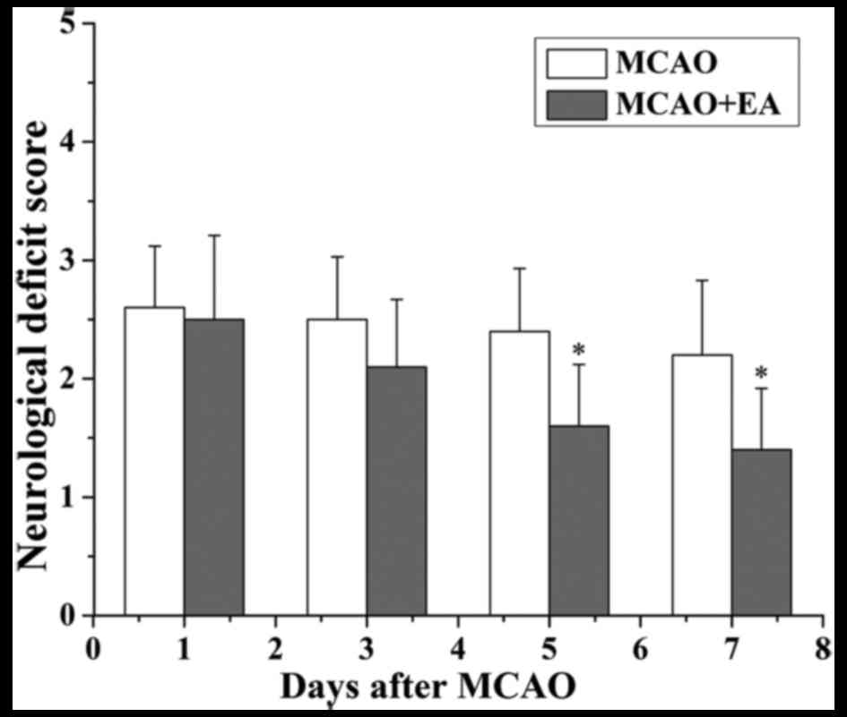
In the present study, the neurological scores were
used to evaluate whether EA at the GV 20 and DU 24 acupoints can
improve neurological function at 24 h, 3, 5 and 7 days after I/R
injury. The rats in the sham group did not display any signs of
neurological deficits. However, the rats in the MCAO and the MCAO +
EA groups exhibited different degrees of neurological deficits
(Fig. 1). At 24 h and 3 days
following I/R injury, the difference in the neurological scores
between the MCAO and the MCAO + EA groups was not statistically
significant (P>0.05). However, at 7 days after I/R injury, the
neurological score of the MCAO + EA group was significantly reduced
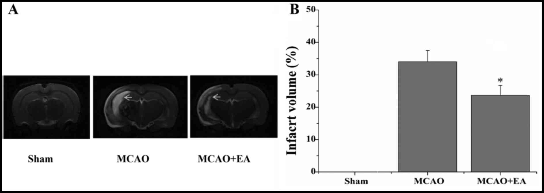
compared with the MCAO group (P<0.01). The infarct volume was
measured using T2WI, and the infarct region displayed high signal
intensity. The infarct volume in the MCAO + EA group was
significantly lower compared with the MCAO group (P<0.05;
Fig. 2).
EA improves learning and memory in rats
with I/R injury
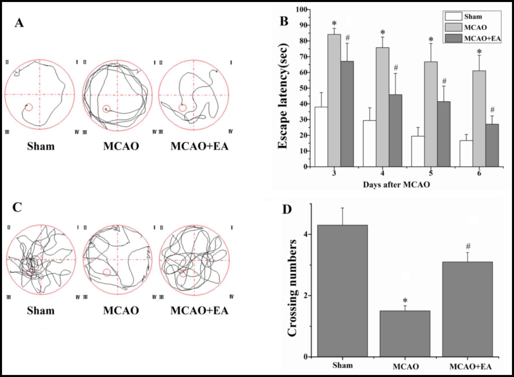
The MWM experiment was performed to evaluate the
effects of EA at the GV 20 and DU 24 acupoints on learning and
memory in rats with I/R injury. In the place navigation test, the
escape latency in rats of the MCAO group was longer compared with
the sham group (P<0.01), and that of rats of the MCAO + EA group
was shorter compared with the MCAO group (P<0.01; Fig. 3). In addition, in the spatial
probe test, the swim time of the rats of the MCAO group in the
platform quadrant was significantly lower compared with the sham
group (P<0.01). However, the swim time of the rats in the MCAO +
EA group was significantly longer compared with the MCAO group
(P<0.01; Fig. 3).
Effect of EA on NAA/Cr, Cho/Cr and Glu/Cr
in HPC and PFC of rats with I/R injury
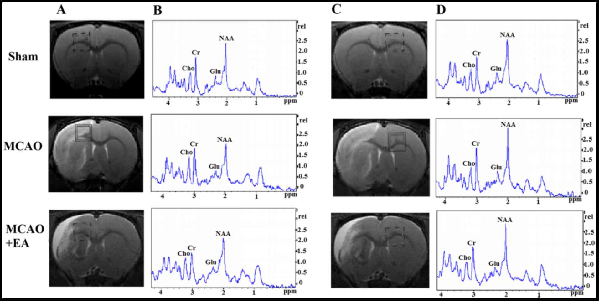
The analysis of neurochemicals in the brain at 7
days after EA treatment: As can be seen in the green box area
(Fig. 4, left), T2-weight imaging
was measured with shimming and detected for MRS. Compared with the
sham group, the ratio of NAA/Cr in the left HPC of the MCAO group
was decreased (P<0.05), whereas the Cho/Cr and Glu/Cr ratios did
not change significantly (P>0.05). In addition, there was no
statistically significant difference in the NAA/Cr, Cho/Cr and
Glu/Cr ratios in the right HPC between the sham and the MCAO groups
(P>0.05). However, a significant decrease in NAA/Cr and Cho/Cr
ratios was observed in the left/right HPC of the MCAO group
compared with the sham group (P<0.05); the left/right HPC Glu/Cr
ratio did not differ significantly between the MCAO and sham groups
(P>0.05; Fig. 4 and Table I).
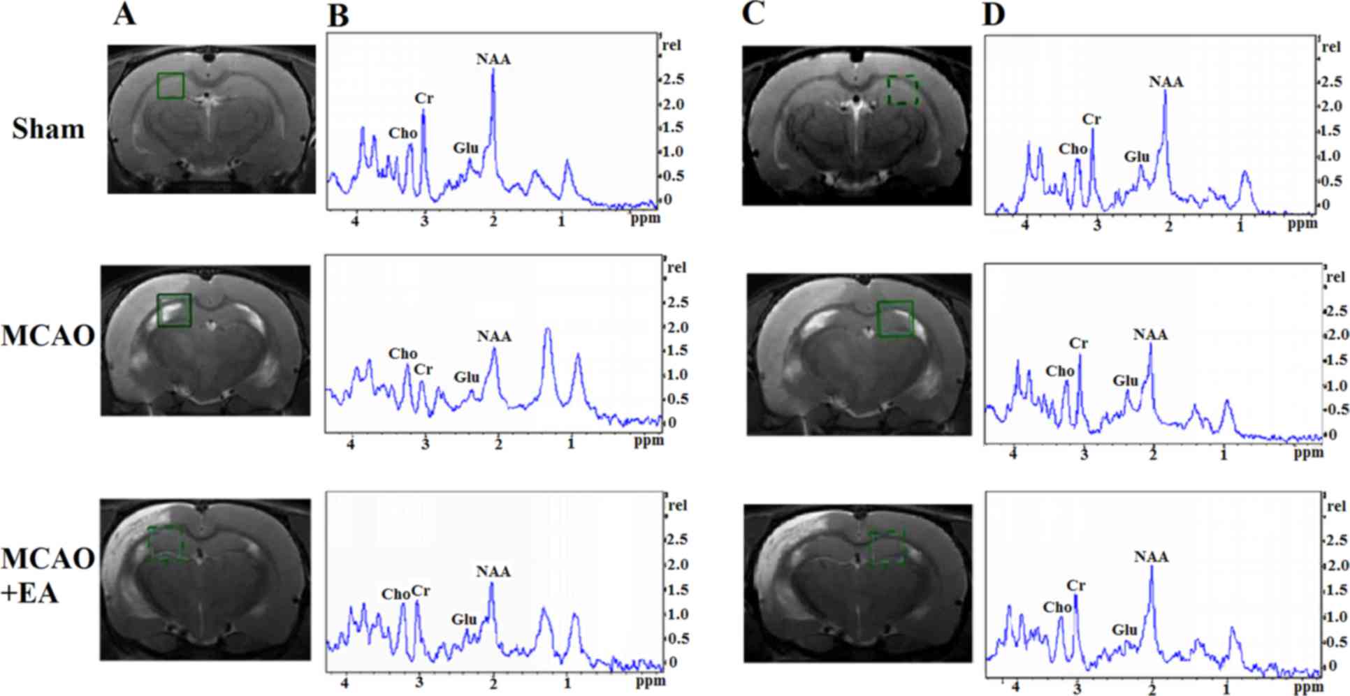 | Figure 4Changes in brain metabolites in HPC by
MRS in rats with cerebral I/R injury. (A and C) Localization of the
left and right HPC VOI on the T2-weighted scan, which is an MRS
shimming region. (B and D) 1H-MRS exhibited the NAA peak
at 2.02 ppm, the Glu peak at 2.2 ppm, the Cho peak at 3.20 ppm, and
the Cr peak at 3.05 ppm. HPC, hippocampus; MRS, magnetic resonance
spectroscopy; I/R, ischemia and reperfusion; VOI, volume of
interest; NAA, N-acetylaspartate; Glu, glutamate; Cho,
choline; Cr, creatine. |
 | Table IMetabolite ratios in the right and
left HPC regions. |
Table I
Metabolite ratios in the right and
left HPC regions.
| Laterality | Metabolites | Sham | MCAO | MCAO + EA |
|---|
| Left | NAA/Cr | 1.44±0.29 | 1.08±0.25a | 1.31±0.28b |
| Glu/Cr | 0.56±0.07 | 0.56±0.16 | 0.55±0.10 |
| Cho/Cr | 0.65±0.12 | 0.62±0.10 | 0.67±0.15 |
| Right | NAA/Cr | 1.39±0.11 | 1.37±0.13 | 1.37±0.20 |
| Glu/Cr | 0.57±0.09 | 0.55±0.17 | 0.58±0.11 |
| Cho/Cr | 0.70±0.13 | 0.63±0.14 | 0.67±0.09 |
| Left/right | NAA/Cr | 0.95±0.10 | 0.76±0.12a | 0.84±0.20b |
| Glu/Cr | 0.85±0.13 | 0.87±0.16 | 0.83±0.23 |
| Cho/Cr | 0.93±0.25 | 0.81±0.10a | 0.90±0.25 |
Compared with the MCAO group, EA reduced the NAA/Cr
ratio in the left HPC (P<0.05), whereas the Cho/Cr and Glu/Cr
ratios exhibited no significant changes (P>0.05). In the right
HPC, the NAA/Cr, Cho/Cr and Glu/Cr ratios exhibited no differences
following EA treatment compared with the MCAO group (P>0.05). As
regards the ratios of left̸right neurochemicals in HPC after EA
treatment, the NAA/Cr ratio in the MCAO + EA group was improved
compared with the MCAO group (P<0.05). Although there was no
difference in the Cho/Cr ratio between the MCAO and the MCAO + EA
groups, Cho/Cr exhibited an increasing tendency following EA
treatment. However, the difference in the Glu/Cr ratio between the
two groups was not statistically significant (P>0.05; Fig. 4 and Table I).
In the MCAO group, the NAA/Cr and Glu/Cr ratios in
the left PFC were decreased (P<0.05), whereas the Cho/Cr ratio
exhibited no significant changes compared with the sham group
(P>0.05). The differences in the NAA/Cr, Cho/Cr and Glu/Cr
ratios of the right PFC between the MCAO and the MCAO + EA groups
were not statistically significant (P>0.05). However, the
NAA/Cr, Cho/Cr and Glu/Cr ratios in left/right PFC were decreased
in the MCAO group compared with the sham group (P<0.05; Fig. 5 and Table II).
 | Figure 5Changes in brain metabolites in TPC by
MRS detection in rats rats with cerebral I/R injury. (A and C)
Localization of the left and right TPC VOI on the T2-weighted
scans, which is an MRS shimming region. (B and D) 1H-MRS
exhibited the NAA peak at 2.02 ppm, the Glu peak at 2.2 ppm, the
Cho peak at 3.20 ppm, and the Cr peak at 3.05 ppm. MRS, magnetic
resonance spectroscopy; I/R, ischemia and reperfusion; VOI, volume
of interest; NAA, N-acetylaspartate; Glu, glutamate; Cho,
choline; Cr, creatine. |
| Table IIMetabolite ratios in the right and
left TPC regions. |
Table II
Metabolite ratios in the right and
left TPC regions.
| Laterality | Metabolites | Sham | MCAO | MCAO + EA |
|---|
| Left | NAA/Cr | 1.47±0.10 | 0.92±0.15a | 1.27±0.20b |
| Glu/Cr | 0.69±0.10 | 0.37±0.08a | 0.57±0.11b |
| Cho/Cr | 0.74±0.17 | 0.68±0.18 | 0.70±0.15 |
| Right | NAA/Cr | 1.45±0.20 | 1.34±0.18 | 1.38±0.19 |
| Glu/Cr | 0.63±0.06 | 0.60±0.11 | 0.57±0.07 |
| Cho/Cr | 0.77±0.13 | 0.72±0.11 | 0.73±0.20 |
| Left/right | NAA/Cr | 0.97±0.24 | 0.68±0.17a | 0.84±0.16b |
| Glu/Cr | 0.83±0.28 | 0.69±0.24a | 0.79±0.12b |
| Cho/Cr | 0.98±0.16 | 0.81±0.11a | 0.89±0.11b |
Compared with the MCAO group, the NAA/Cr and Glu/Cr
ratios in the PFC of the EA+MCAO group were increased (P<0.05),
whereas Cho/Cr exhibited no obvious changes (P>0.05). The
NAA/Cr, Cho/Cr and Glu/Cr ratios in the right PFC did not exhibit
statistically significant differences (P>0.05). However, the
NAA/Cr, Cho/Cr and Glu/Cr ratios in the left/right PFC increased
following EA treatment (P<0.05; Fig. 5 and Table II).
Discussion
MRS is a novel technique for detecting brain
metabolites in vivo non-invasively and non-radioactively,
according to different nuclei and compounds forming different
magnetic resonance phenomena and chemical shifts. It was previously
confirmed that the HPC and PFC regions play key roles in learning
and memory through their specific structure, location and
interconnection with other brain regions (21). Thus, in the present study,
1H MRS was used to monitor the neurochemical alterations
in the HPC and PFC regions associated with learning and memory
changes following cerebral I/R injury.
NAA is a type of specific amino acid, which is
mainly present in neurons and axons. NAA also is a neuronal marker
and its concentration may be sensitive to the density of neurons.
When NAA declines in the brain, nervous functional impairment may
develop (22,23). Previous studies demonstrated that
the NAA level is positively correlated with learning and memory in
different diseases (24,25), whereas a higher ratio of NAA/Cr is
accompanied with better Mini-Mental State Examination scores in
Alzheimer's disease (26).
Moreover, Cho is an important neurotransmitter precursor compound
of acetylcholine, which is related to memory, recognition and
emotional behavior (27). It has
been reported that it is crucial for learning and memory to
maintain a stable level of acetylcholine in the frontal cortex and
HPC (28).
The results of the present study revealed that the
left/right NAA/Cr and Cho/Cr ratios were obviously decreased in HPC
as well as PFC at 7 days following cerebral I/R injury accompanied
with learning and memory impairment as indicated by the MWM test.
These results are consistent with those of other studies reporting
that HPC neurochemicals and spatial learning and memory are closely
correlated (29). It has been
reported that NAA rapidly decreased in the ischemic core within 6
h, and declined to 0 at 7 days after acute cerebral infarction;
however, there was no obvious change in the ischemic penumbra
within 48 h, after which time it gradually declined to 20–40%
(30,31). In the present study, we observed
that the NAA ratio of left/right HPC and PFC was decreased to
20–40% at the 7 days following cerebral I/R injury.
EA is an effective novel treatment based on the
combination of traditional Chinese acupuncture with modern
electrotherapy, exhibiting confirmed clinical efficacy in the
treatment of stroke and cognitive impairment (32). In the present study, an MWM test
was performed to assess the effect of EA on learning and memory
following cerebral I/R injury. The results demonstrated that EA at
the GV 20 and DU 24 acupoints improved learning and memory ability,
along with ameliorated neurological deficits and reduced cerebral
infarct volume, which were consistent with our previous findings
(33).
In the HPC and PFC regions, we observed that EA at
GV 20 and DU 24 increased the metabolism of left/right NAA and Cho
at 7 days after EA treatment. These findings are similar to those
of a previous study, which demonstrated that EA at the bilateral
Hegu (LI 4) and Taichong (LR 3) acupoints may increase the levels
of NAA in the ischemic penumbra and improve the memory scale score
(34). In addition, it has been
reported that the dynamic changes of Glu in the cerebral cortex are
associated with cognitive function. The excessive secretion of
extracellular Glu following cerebral ischemia induced excitatory
toxic effects or oxidative stress, which may damage neurons and
cause cognitive impairment (35).
In the present study, it was demonstrated that EA at the GV 20 and
DU 24 acupoints increased the left/right Glu level in PFC at 7 days
after EA treatment. A possible explanation for this finding is that
EA at the GV 20 and DU 24 acupoints may promote Glu-mediated
synaptic transmission; this was observed found in PFC but not in
HPC, which requires further investigation.
In conclusion, the results of the present study
demonstrated that EA at the GV 20 and DU 24 acupoints may alleviate
neurological deficits, reduce infarct volume and improve the
learning and memory ability following cerebral I/R injury, possibly
via enhancing the neurochemical metabolism of NAA and Cho in the
HPC and FPC regions.
Acknowledgments
The present study was supported by the National
Natural Science Foundation of China (grant no. 81403450).
References
|
1
|
Renjen PN, Gauba C and Chaudhari D:
Cognitive impairment after stroke. Cureus. 7:e3352015.PubMed/NCBI
|
|
2
|
Edwards JD, Jacova C, Sepehry AA, Pratt B
and Benavente OR: A quantitative systematic review of
domain-specific cognitive impairment in lacunar stroke. Neurology.
80:315–322. 2013. View Article : Google Scholar : PubMed/NCBI
|
|
3
|
Liu W, Wang X, Zheng Y, Shang G, Huang J,
Tao J and Chen L: Electroacupuncture inhibits inflammatory injury
by targeting the miR-9-mediated NF-κB signaling pathway following
ischemic stroke. Mol Med Rep. 13:1618–1626. 2016. View Article : Google Scholar : PubMed/NCBI
|
|
4
|
Li X, Guo F, Zhang Q, Huo T, Liu L, Wei H,
Xiong L and Wang Q: Electroacupuncture decreases cognitive
impairment and promotes neurogenesis in the APP/S1 transgenic mice.
BMC Complement Altern Med. 14:372014. View Article : Google Scholar
|
|
5
|
Ding X, Li CY, Wang QS, Du FZ, Ke ZW, Peng
F, Wang J and Chen L: Patterns in default-mode network connectivity
for determining outcomes in cognitive function in acute stroke
patients. Neuroscience. 277:637–646. 2014. View Article : Google Scholar : PubMed/NCBI
|
|
6
|
Witte AV, Kerti L, Margulies DS and Flöel
A: Effects of resveratrol on memory performance, hippocampal
functional connectivity, and glucose metabolism in healthy older
adults. J Neurosci. 34:7862–7870. 2014. View Article : Google Scholar : PubMed/NCBI
|
|
7
|
Rosenberg T, Gal-Ben-Ari S, Dieterich DC,
Kreutz MR, Ziv NE, Gundelfinger ED and Rosenblum K: The roles of
protein expression in synaptic plasticity and memory consolidation.
Front Mol Neurosci. 7:862014. View Article : Google Scholar : PubMed/NCBI
|
|
8
|
Yang M, Wang S, Hao F, Li Y, Tang H and
Shi X: NMR analysis of the rat neurochemical changes induced by
middle cerebral artery occlusion. Talanta. 88:136–144. 2012.
View Article : Google Scholar : PubMed/NCBI
|
|
9
|
Mattfeld AT and Stark CEL: Functional
contributions and interactions between the human hippocampus and
subregions of the striatum during arbitrary associative learning
and memory. Hippocampus. 25:900–911. 2015. View Article : Google Scholar : PubMed/NCBI
|
|
10
|
Shi L, Pu J, Xu L, Malaguit J, Zhang J and
Chen S: The efficacy and safety of cilostazol for the secondary
prevention of ischemic stroke in acute and chronic phases in Asian
population - an updated meta-analysis. BMC Neurol. 14:2512014.
View Article : Google Scholar
|
|
11
|
Liu ZY, Guo H, Zhang XL, Liu J, Qu HY,
Peng W, Bao YM, Yin LL and Song YX: Impacts of electroacupuncture
on left hippocampus NAA/Cr for patients of Uygur and Han
nationality with mild cognitive impairment. Zhongguo Zhen Jiu.
31:773–777. 2011.In Chinese. PubMed/NCBI
|
|
12
|
Feng X, Yang S, Liu J, Huang J, Peng J,
Lin J, Tao J and Chen L: Electroacupuncture ameliorates cognitive
impairment through inhibition of NF-κB-mediated neuronal cell
apoptosis in cerebral ischemia-reperfusion injured rats. Mol Med
Rep. 7:1516–1522. 2013. View Article : Google Scholar : PubMed/NCBI
|
|
13
|
Haga KK, Khor YP, Farrall A and Wardlaw
JM: A systematic review of brain metabolite changes, measured with
1H magnetic resonance spectroscopy, in healthy aging.
Neurobiol Aging. 30:353–363. 2009. View Article : Google Scholar
|
|
14
|
Longa EZ, Weinstein PR, Carlson S and
Cummins R: Reversible middle cerebral artery occlusion without
craniectomy in rats. Stroke. 20:84–91. 1989. View Article : Google Scholar : PubMed/NCBI
|
|
15
|
Tao J, Xue XH, Chen LD, Yang SL, Jiang SM,
Gao YL and Wang XB: Electroacupuncture improves neurological
deficits and enhances proliferation and differentiation of
endogenous nerve stem cells in rats with focal cerebral ischemia.
Neurol Res. 32:198–204. 2010. View Article : Google Scholar
|
|
16
|
Lan L, Tao J, Chen A, Xie G, Huang J, Lin
J, Peng J and Chen L: Electroacupuncture exerts anti-inflammatory
effects in cerebral ischemia-reperfusion injured rats via
suppression of the TLR4/NF-κB pathway. Int J Mol Med. 31:75–80.
2013. View Article : Google Scholar
|
|
17
|
Pouzet B, Zhang WN, Feldon J and Rawlins
JN: Hippocampal lesioned rats are able to learn a spatial position
using non-spatial strategies. Behav Brain Res. 133:279–291. 2002.
View Article : Google Scholar : PubMed/NCBI
|
|
18
|
Veng LM, Granholm AC and Rose GM:
Age-related sex differences in spatial learning and basal forebrain
cholinergic neurons in F344 rats. Physiol Behav. 80:27–36. 2003.
View Article : Google Scholar : PubMed/NCBI
|
|
19
|
Hui Xi G, Zhang J, Liu Z, Zhang S, Teng X,
Chan G, Wu KC, Nie EX, Shan BB, et al: Learning and memory
alterations are associated with hippocampal N-acetylaspartate in a
rat model of depression as measured by 1H-MRS. PLoS One.
6:e286862011. View Article : Google Scholar : PubMed/NCBI
|
|
20
|
Zhou IY, Chan RW, Ho LC and Wu EX:
Longitudinal metabolic changes in the hippocampus and thalamus of
the maternal brain revealed by proton magnetic resonance
spectroscopy. Neurosci Lett. 553:170–175. 2013. View Article : Google Scholar : PubMed/NCBI
|
|
21
|
Milner B and Klein D: Loss of recent
memory after bilateral hippocampal lesions: Memory and
memories-looking back and looking forward. J Neurol Neurosurg
Psychiatry. 87:2302016. View Article : Google Scholar
|
|
22
|
Jones RS and Waldman AD: 1H-MRS evaluation
of metabolism in Alzheimer's disease and vascular dementia. Neurol
Res. 26:488–495. 2004. View Article : Google Scholar : PubMed/NCBI
|
|
23
|
Bertolino A, Frye M, Callicott JH, Mattay
VS, Rakow R, Shelton-Repella J, Post R and Weinberger DR: Neuronal
pathology in the hippocampal area of patients with bipolar
disorder: A study with proton magnetic resonance spectroscopic
imaging. Biol Psychiatry. 53:906–913. 2003. View Article : Google Scholar : PubMed/NCBI
|
|
24
|
Jayaweera HK, Lagopoulos J, Duffy SL,
Lewis SJ, Hermens DF, Norrie L, Hickie IB and Naismith SL:
Spectroscopic markers of memory impairment, symptom severity and
age of onset in older people with lifetime depression: Discrete
roles of N-acetylaspartate and glutamate. J Affect Disord.
183:31–38. 2015. View Article : Google Scholar : PubMed/NCBI
|
|
25
|
Zhou IY, Ding AY, Li Q, McAlonan GM and Wu
EX: Magnetic resonance spectroscopy reveals N-acetylaspartate
reduction in hippocampus and cingulate cortex after fear
conditioning. Psychiatry Res. 204:178–183. 2012. View Article : Google Scholar : PubMed/NCBI
|
|
26
|
Penner J, Wells JL, Borrie MJ,
Woolmore-Goodwin SM and Bartha R: Reduced N-acetylaspartate to
creatine ratio in the posterior cingulate correlates with cognition
in Alzheimer's disease following four months of rivastigmine
treatment. Dement Geriatr Cogn Disord. 39:68–80. 2015. View Article : Google Scholar
|
|
27
|
Liu H and Wang X: Correlation of iron
deposition and change of gliocyte metabolism in the basal ganglia
region evaluated using magnetic resonance imaging techniques: An in
vivo study. Arch Med Sci. 12:163–171. 2016. View Article : Google Scholar : PubMed/NCBI
|
|
28
|
Stoiljkovic M, Leventhal L, Chen A, Chen
T, Driscoll R, Flood D, Hodgdon H, Hurst R, Nagy D, Piser T, et al:
Concentration-response relationship of the α7 nicotinic
acetyl-choline receptor agonist FRM-17874 across multiple in vitro
and in vivo assays. Biochem Pharmacol. 97:576–589. 2015. View Article : Google Scholar : PubMed/NCBI
|
|
29
|
Caldwell KK, Goggin SL, Tyler CR and Allan
AM: Prenatal alcohol exposure is associated with altered
subcellular distribution of glucocorticoid and mineralocorticoid
receptors in the adolescent mouse hippocampal formation. Alcohol
Clin Exp Res. 38:392–400. 2014. View Article : Google Scholar
|
|
30
|
Demougeot C, Marie C, Giroud M and Beley
A: N-acetylaspartate: A literature review of animal research on
brain ischaemia. J Neurochem. 90:776–783. 2004. View Article : Google Scholar : PubMed/NCBI
|
|
31
|
Aoki Y, Inokuchi R and Suwa H: Reduced
N-acetylaspartate in the hippocampus in patients with fibromyalgia:
A meta-analysis. Psychiatry Res. 213:242–248. 2013. View Article : Google Scholar : PubMed/NCBI
|
|
32
|
Lin R, Wu Y, Tao J, Chen B, Chen J, Zhao
C, Yu K, Li X and Chen LD: Electroacupuncture improves cognitive
function through Rho GTPases and enhances dendritic spine
plasticity in rats with cerebral ischemia-reperfusion. Mol Med Rep.
13:2655–2660. 2016. View Article : Google Scholar : PubMed/NCBI
|
|
33
|
Lin R, Lin Y, Tao J, Chen B, Yu K, Chen J,
Li X and Chen LD: Electroacupuncture ameliorates learning and
memory in rats with cerebral ischemia-reperfusion injury by
inhibiting oxidative stress and promoting p-CREB expression in the
hippocampus. Mol Med Rep. 12:6807–6814. 2015. View Article : Google Scholar : PubMed/NCBI
|
|
34
|
Zhang J and Shen Y: Magnetic resonance
spectroscopic study of memory impairment after cerebral infarction
treated with electroacupuncture. Zhongguo Zhen Jiu. 35:657–660.
2015.In Chinese. PubMed/NCBI
|
|
35
|
Zhao L, Zhang H, Zheng Z and Huang J:
Electroacupuncture on the head points for improving gnosia in
patients with vascular dementia. J Tradit Chin Med. 29:29–34. 2009.
View Article : Google Scholar : PubMed/NCBI
|