Introduction
Diabetes mellitus (DM) is a severe metabolic
disorder, which is associated with a high rate of hospitalization
and mortality, due to diverse micro- and macrovascular
complications (1). The
coexistence of DM in patients with coronary artery disease (CAD)
results in increased severity of vascular lumen stenosis and plaque
burden (2,3). Furthermore, the risk of restenosis
following percutaneous coronary intervention is increased in these
patients (4,5). In healthy individuals, physiological
concentrations of insulin facilitate vasodilatation and maintain
blood circulation. However, in patients with type 2 DM,
hyperinsulinemia, secondary to insulin resistance, is a prominent
feature; therefore, the physiological functions of insulin are
impaired. Previous clinical and epidemiological studies have
indicated that hyperinsulinemia serves a critical role in the
pathogenesis of DM-associated vascular diseases (6–8).
In addition, high serum levels of insulin are regarded as an
independent risk factor and poor prognostic indicator of CAD
(9).
Vascular smooth muscle cells (VSMCs) have an impo
rtant role in the pathogenesis of vascular occlusive diseases
(10–12). VSMC proliferation, and migration
of VSMCs from the media to the intima, is a critical step in
atherosclerosis-associated intimal thickening and post-angioplasty
restenosis (10,13). As well as hyperglycemic stress in
type 2 DM, hyperinsulinemia may also induce VSMC dysfunction. It
has previously been reported that abnormal serum insulin levels are
capable of promoting the progression of atherosclerosis (14). In addition, increased
proliferation and migration of VSMCs, as well as increased
oxidative stress and inflammation, may be closely linked (15). Further attention should therefore
be paid to elucidate the molecular mechanisms underlying
hyperinsulinemia-induced VSMC dysfunction, in order to identify
potential targets and novel therapeutic approaches for the
treatment of DM-associated cardiovascular complications.
Lysine (K)-specific demethylase 3A (KDM3A), also
known as Jumonji domain-containing 1A, contains a zinc finger
structural domain and possesses histone demethylase activity
(16). Previous studies have
indicated that KDM3A participates in numerous pathophysiological
processes, through the modulation of cellular proliferation,
senility, metabolism and tumor occurrence (17–19). However, its potential functions in
DM-associated cardiovascular complications have yet to be fully
elaborated. Our previous study demonstrated that KDM3A expression
was upregulated in type 2 diabetic vascular and heart tissue,
whereas downregulation of KDM3A markedly promoted rehabilitation of
diabetic vascular complications and attenuated diabetic myocardial
injury; KDM3A inhibition also significantly alleviated high
glucose-induced VSMC dysfunction in vitro (20). However, to the best of our
knowledge, the effects of KDM3A on hyperinsulinemia-induced VSMC
dysfunction remain unknown. The present study aimed to explore the
potential protective effects and underlying mechanisms of KDM3A
inhibition on VSMC proliferation and migration, as well as
oxidative stress, inflammation and apoptosis in response to a high
concentration of insulin in vitro.
Materials and methods
Primary VSMC isolation and culture
Primary VSMCs were isolated from the thoracic aorta
of male Sprague-Dawley (SD) rats (weight, 150–180 g) according to
the previously described tissue explant method (21). Ten male SD wild-type rats (SPF
grade, 4–5 week) were obtained from Wuhan University Experiment
Animal Center. Rats were given free access to standard rat chow and
water until the time of the experiment and were housed in a
temperature- and light-controlled environment (24°C; 12/12
h-light/dark cycle). In order to obtain the primary VSMCs, the SD
rats were sacrificed. Cells were cultured in Dulbecco’s modified
Eagle’s medium (DMEM; Hyclone; GE Healthcare Life Sciences, Logan,
UT, USA) supplemented with 10% fetal bovine serum (FBS; Gibco;
Thermo Fisher Scientific, Inc., Waltham, MA, USA) and 1%
penicillin/streptomycin (Hyclone; GE Healthcare Life Sciences) at
37°C in an atmosphere containing 95% air and 5% CO2.
VSMCs were identified by morphology and immunoreactivity to
α-smooth muscle actin, as previously described (21). VSMCs at the 3rd and 5th passages
were used for the subsequent experiments. The present study was
approved by the Animal Care and Use Committee of Wuhan University
(Wuhan, China), and experiments were conducted in accordance with
Institutional Guidelines and the Guide for the Care and Use of
Laboratory Animals (NIH publication no. 85–23, revised 1996).
Small interfering (si)RNA transduction
and experimental designs
siRNA specific to the rat KDM3A gene was constructed
and purchased from Shanghai GeneChem Co., Ltd. (Shanghai, China).
Our preliminary study indicated that a lentivirus encoding
KDM3A-siRNA exhibited optimal infection efficiency with 40
multiplicity of infection (MOI) in rat VSMCs (20). In the present study, equal amounts
of lenti-KDM3A-siRNA or lenti-control-siRNA (as a negative control;
siRNA sequence, GCAGGTGTCACTAGGCTTA; Shanghai GeneChem Co., Ltd.)
at 40 MOI were transduced into VSMCs once they reached 40–50%
confluence. The lentiviruses were constructed by Shanghai GeneChem
Co., Ltd. After 12 h, the lentivirus-containing serum-free medium
was replaced with complete culture medium. A total of 72 h
post-transduction, enhanced green fluorescent protein expression
was observed under a fluorescent microscopy to confirm successful
transduction of the lentiviruses. In addition, western blotting was
used to directly detect KDM3A expression, and to thereby indicate
the efficiency of lentivirus transduction.
To determine whether KDM3A participated in high
insulin-induced VSMC dysfunction, VSMCs were initially divided into
two groups: i) Control group, in which VSMCs were cultured in
normal medium for 5 days and ii) high concentration insulin
stimulation group (HI), in which VSMCs were cultured in medium
containing 100 nM insulin (to imitate the hyperinsulinemic state)
for 5 days. Secondly, to further determine the precise roles and
underlying mechanism of KDM3A in VSMCs undergoing treatment with a
high concentration of insulin, VSMCs were randomly divided into
three groups: i) Normal medium + control siRNA group (N + C-siRNA),
in which VSMCs incubated with lenti-control-siRNA for 12 h, and
incubated in culture medium until 72 h post-tranfection, and were
then cultured with normal medium for a further 5 days; ii) high
concentration insulin stimulation + control siRNA group (HI +
C-siRNA), in which VSMCs transduced with lenti-control-siRNA for 72
h were cultured in medium containing 100 nM insulin for 5 days;
iii) high concentration insulin stimulation + KDM3A-siRNA group (HI
+ K-siRNA), in which VSMCs transduced with lenti-KDM3A-siRNA for 72
h were cultured in medium containing 100 nM insulin for 5 days.
Cell proliferation and migration
assays
To determine the proliferative ability of VSMCs in
the various groups, cells were digested and seeded in 96-well
plates at 6×10³ cells/well in 200 μl
corresponding culture medium. Following 24 h incubation, 20
μl Cell Counting kit-8 (CCK-8) reagent (Dojindo Molecular
Technologies, Inc., Kumamoto, Japan) was added to each well and the
cells were incubated for a further 4 h. Subsequently, the optical
density (OD) values of all wells were determined using a microplate
spectrophotometer (Bio-Rad Laboratories, Inc., Hercules, CA, USA)
at 450 nm.
A Transwell chamber assay (Corning Inc., Corning,
NY, USA) was performed to measure the migratory activity of VMSCs.
Briefly, VSMCs were digested and seeded in the upper chamber with
200 μl serum-free medium at 1×10⁵. Subsequently,
600 μl DMEM containing 10% FBS was added to the lower
chamber and was incubated for 12 h at 37°C. The non-migrating cells
on the upper side of the membrane were gently removed using a
cotton swab. The cells that had migrated to the underside of the
membrane were fixed with methanol for 30 min and stained with 0.5%
crystal violet in the dark for 30 min at 25°C. The number of
migrated cells was counted under a light microscope (Olympus
Corporation, Tokyo, Japan) at ×200 magnification in at least three
random fields.
Reactive oxygen species (ROS)
detection
Dihydroethidium (DHE) fluorescent probe (Beyotime
Institute of Biotechnology, Haimen, China) was used to detect ROS
generation in VSMCs. Briefly, VSMCs from each group were adjusted
to a density of 1.5×10⁵/well and were seeded into
24-well plates. After 24 h, the medium was removed and the DHE
fluorescent probe, dissolved in serum-free medium, was added to all
of the wells (10 μmol/l) for 30 min at 37°C. Subsequently,
DHE fluorescence intensity was immediately observed using a
fluorescence microscopy at an excitatory wavelength of 495 nm.
Images from at least three random fields were captured and the mean
fluorescence intensity was calculated with ImageJ software version
1.49 (National Institutes of Health, Bethesda, MD, USA).
Apoptosis assays
For the identification of apoptotic cells, Annexin
V-allophycocyanin (APC)/7-aminoactinomycin (7-AAD) double staining
was performed. Briefly, cells were harvested, washed twice with
ice-cold PBS and were then stained using an apoptosis detection kit
(Apoptosis Assay kit; Sigma-Aldrich; Merck KGaA, Darmstadt,
Germany) according to the manufacturer’s protocol. Cells were
resuspended in PBS and incubated with binding buffer containing 5
μl 7-AAD at room temperature for 15 min in the dark.
Subsequently, 1 μl Annexin V-APC was added to the cell
suspension and incubated for 15 min in the dark. Finally, cells
were analyzed by fluorescence-activated cell sorting within 1 h
using a FACSCalibur with CellQuest software version 5.1 (Bio-Rad
Laboratories, Inc.). Cells exhibiting Annexin V-APC-and
7-AAD-negative staining were considered viable. Cells that stained
positively for Annexin V-APC only were considered early apoptotic.
Cells exhibiting Annexin V-APC- and 7-AAD-positive staining were
considered late apoptotic or necrosis. The percentage of early
apoptotic plus late apoptotic cells was calculated following flow
cytometric analysis.
Reverse transcription-quantitative
polymerase chain reaction (RT-qPCR) assay
RT-qPCR was used to detect the mRNA expression
levels of interleukin (IL)-6 and monocyte chemotactic protein
(MCP)-1 in VSMCs. Total RNA was extracted from the samples and was
purified using a RNA isolation kit (TRIzol; Applied Biosystems;
Thermo Fisher Scientific, Inc.). Isolated RNA was reverse
transcribed into cDNA using a cDNA synthesis kit (First-Strand
Synthesis system; Invitrogen; Thermo Fisher Scientific, Inc.)
according to the manufacturer’s protocol. cDNA was then amplified
using an ABI Prism 7500 Sequence Detection system (Applied Biosy
stems; Thermo Fisher Scientific, Inc.) in the presence of the
appropriate proportions of primers and SYBR-Green Supermix (Bio-Rad
Laboratories, Inc.). PCR cycling conditions were as follows:
Predenaturation at 50°C for 120 sec, followed by 40 cycles of
denaturation at 95°C for 15 sec, annealing at 60°C for 30 sec and
extension at 60°C for 30 sec. The 4 types of deoxynucleo-tides in
the single chain were connected to the primers according to the
principles of Watson-Crick base pairing, and then extended to form
complementary DNA double strand. GAPDH expression was used as an
internal control. Gene expression levels were calculated using the
comparative quantification method (2−ΔΔCq) (22). The primer sequences used in the
present study were as follows: MCP-1, forward
5′-GTCGGCTGGAGAACTACAAGA-3′, reverse 5′-GTGCTGAAGTCCTTAGGGTTG-3′;
IL-6, forward 5′-GTTGCCTTCTTGGGACTGATG-3′, reverse
5′-TACTGGTCTGTTGTGGGTGGT-3′; and GAPDH, forward
5′-ACAGCAACAGGGTGGTGGAC-3′ and reverse
5′-TTTGAGGGTGCAGCGAACTT-3′.
Western blotting
VSMCs were lysed using a RIPA Lysis buffer (P0013B;
Beyotime Institute of Biotechnology), and nuclear and cytoplasmic
proteins were extracted according to the manufacturer’s protocol
(Beyotime Institute of Biotechnology). Following centrifugation at
14926 × g at 4°C for 5 min, the supernatant was collected and
protein concentration was detected using a bicinchoninic acid
protein assay (Beyotime Institute of Biotechnology). Protein
samples (concentration, 4.5 μg/μl) were separated by
10% SDS-PAGE and were electrophoretically transferred onto
polyvinylidene fluoride (PVDF) membranes (EMD Millipore, Billerica,
MA, USA). The nonspecific binding sites in the membranes were
blocked with 5% non-fat dried milk dissolved in Tris-buffered
saline containing 0.1% Tween-20 for 90 min at 25°C. Subsequently,
the PVDF membranes were incubated with primary antibodies
(dilution, 1:1,000) against KDM3A (ab106456), GADPH (ab181602),
B-cell lymphoma 2 (Bcl-2)-associated X protein (Bax; ab32503) and
Bcl-2 (ab32124) (all Abcam, Cambridge, MA, USA); extracellular
signal-regulated kinase (ERK)1/2 (9102) and phosphorylated
(p)-ERK1/2 (4370) (both Cell Signaling Technology, Inc., Danvers,
MA, USA); c-Jun NH2-terminal protein kinase (JNK)
(51151-1-AP; Proteintech Group, Inc., Chicago, IL, USA), p-JNK
(ab131499; Abcam), p38 (9212) and p-p38 (4511) (both Cell Signaling
Technology, Inc.), nuclear factor (NF)-κB/p65 (BS1257; Bioworld
Technology, Inc., St. Louis Park, MN, USA) overnight at 4°C,
followed by incubation with corresponding secondary antibodies
(HRP-labeled goat anti-rabbit; dilution 1:50,000; BA1054; Boster,
Wuhan, China) at room temperature for 60 min. The protein bands
were visualized using an enhanced chemiluminescence system (Thermo
Fisher Scientific, Inc.). GAPDH was used as the internal loading
control. The blots were semi-quantified using BandScan 4.30 (Glyko,
Novato, CA, USA).
Statistical analysis
Statistical analyses were conducted using SPSS
statistical software (version 19.0; IBM Corp., Armonk, NY, USA).
All continuous values are expressed as the mean ± standard
deviation. Student’s t-test was used for between-group comparisons.
One-way analysis of variance followed by Bonferroni post hoc test
was used for multiple comparisons. P<0.05 was considered to
indicate a statistically significant difference.
Results
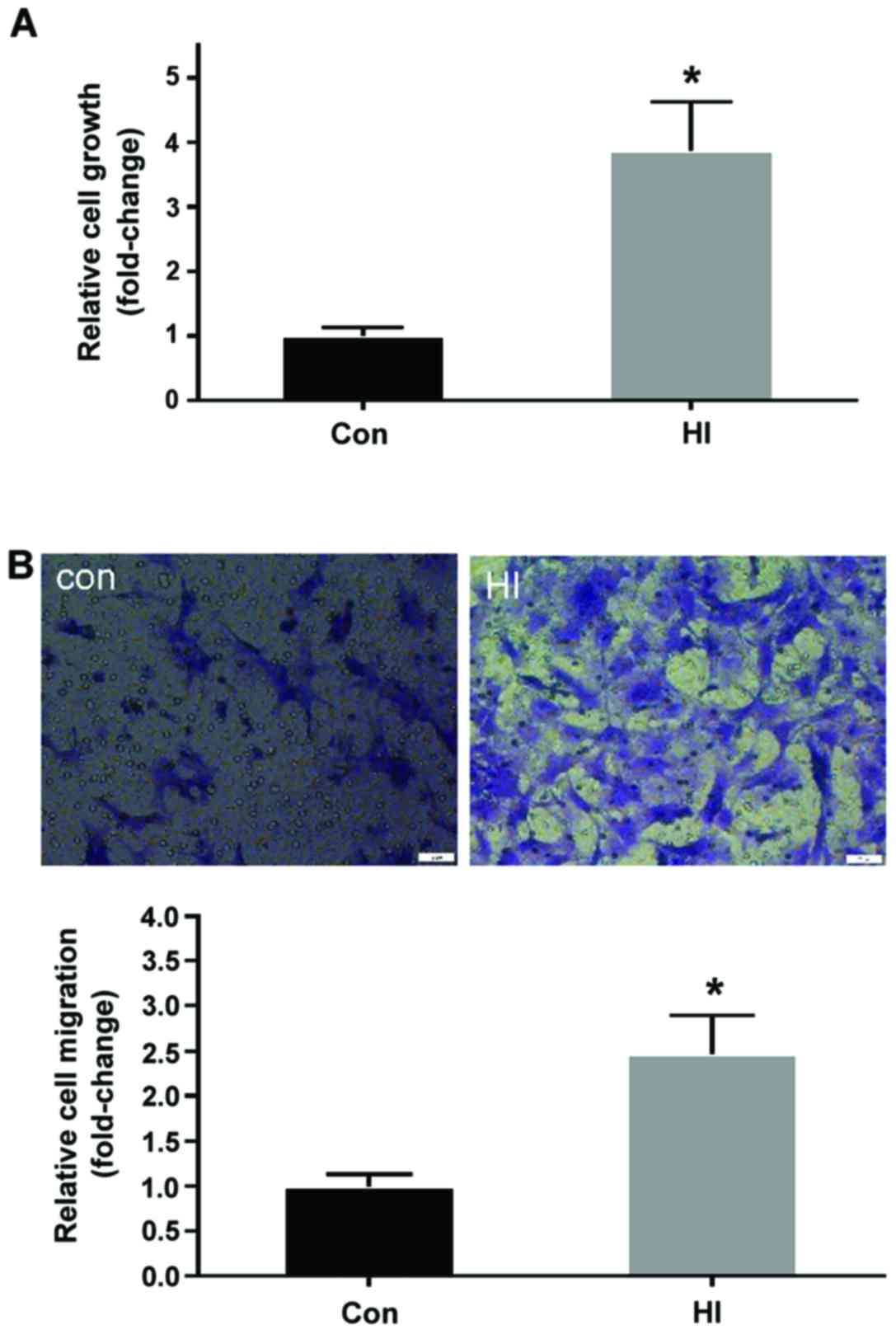
High insulin stimulation promotes the
proliferation and migration of VSMCs
The effects of high concentration insulin on the
proliferation and migration of VSMCs were determined using a CCK-8
assay kit and Transwell chamber assay, respectively. As shown in
Fig. 1A, VSMC proliferation was
significantly increased >2-fold in the HI group compared with in
the control group (P<0.05). Consistently, the migration of VSMCs
in the HI group was markedly elevated compared with in the control
group (P<0.05; Fig. 1B). These
findings indicated that high insulin concentration may induce VSMC
dysfunction by increasing proliferative and migratory
abilities.
High insulin stimulation induces ROS
generation, apoptosis and inflammation, accompanied by increased
KDM3A expression in VSMCs
ROS generation, apoptosis and inflammation are
considered the central contributors associated with the induction
of VSMC dysfunction. To further investigate the effects of high
insulin levels on ROS production in VSMCs, ROS concentrations were
evaluated by measuring the intensity of DHE fluorescence. As shown
in Fig. 2A, compared with in the
control group, the mean fluorescent density of ROS in the HI group
was significantly enhanced (P<0.05). The effects of insulin were
also observed on apoptosis and inflammation. As shown in Fig. 2B, the percentage of apoptotic
cells (2.32±0.46%) was very low in the control group, whereas the
percentage of apoptotic cells was increased to 16.20±0.30%
following treatment with 100 nM insulin (P<0.05). Furthermore,
the ratio between proapoptotic Bax and anti-apoptotic Bcl-2
expression was significantly altered in response to high insulin
levels. As shown in Fig. 2C, the
expression levels of Bax were markedly increased, whereas Bcl-2
expression was decreased, in the HI group compared with in the
control group (P<0.05). With regards to inflammation, the mRNA
expression levels of IL-6 and MCP-1 were significantly increased in
VSMCs following stimulation with high insulin levels compared with
in the control group (P<0.05; Fig.
2D).
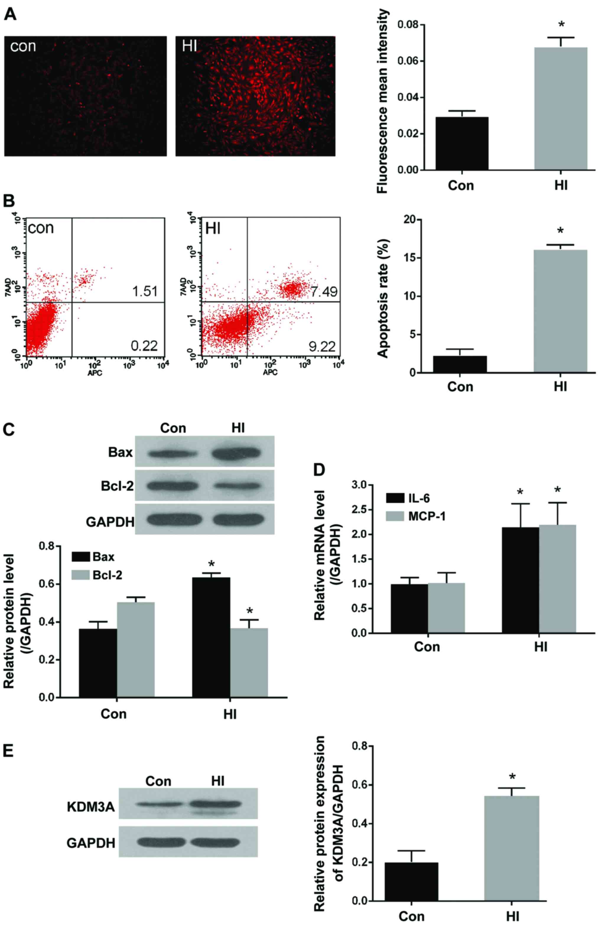 | Figure 2Treatment with a high insulin
concentration markedly increases ROS production, cell apoptosis and
inflammation, accompanied by increased KDM3A expression. (A)
Representative fluorescent images of dihydroethidium staining in
the control and HI groups (magnification, ×100) (left panel).
Fluorescent mean density was determined using ImageJ software
(right panel). (B) Representative flow cytometry plots of apoptotic
VSMCs following stimulation with 100 nM insulin (left panel).
Relative apoptotic rates of VSMCs were determined in the control
and HI groups (right panel). (C) Representative immunoblots for Bax
and Bcl-2 expression in VSMCs stimulated with 100 nM insulin (upper
panel). Densitometric analysis of western blotting (lower panel).
(D) Effects of high insulin stimulation on mRNA expression levels
of IL-6 and MCP-1 in VSMCs. (E) Effects of 100 nM insulin on KDM3A
expression, as determined by western blotting (left panel).
Densitometric analyses of western blotting (right panel). All
values are expressed as the mean ± standard deviation, n=3.
*P<0.05 vs. the Con group. Bax, Bcl-2-associated X
protein; Bcl-2, B-cell lymphoma 2; Con, control group; HI, high
insulin group; IL, interleukin; KDM3A, lysine (K)-specific
demethylase 3A; MCP-1, monocyte chemotactic protein-1; ROS,
reactive oxygen species; VSMCs, vascular smooth muscle cells |
The present study aimed to further determine whether
KDM3A is involved in high insulin-induced VSMC pathological injury.
Western blotting was performed to detect KDM3A expression. As shown
in Fig. 2E, KDM3A expression was
markedly upregulated in high insulin-stimulated VSMCs compared with
in the control group (P<0.05). Taken to gether these findings
suggested that KDM3A may participate in the numerous VMSC injuries
caused by high insulin levels. Subsequently, the precise effects of
KDM3A, and the potential underlying mechanisms, were further
explored.
KDM3A knockdown attenuates high
insulin-induced VSMC dysfunction
To determine the role of KDM3A in high
insulin-induced VSMC dysfunction, a lentivirus encoding KDM3A-siRNA
was transduced into VSMCs prior to high insulin treatment.
Fluorescent microscopy and western blotting were then performed to
confirm that the lentivirus had been successful transduced. As
shown in Fig. 3A, VSMCs
transduced with lentivirus emitted strong green fluorescence; the
transduction efficiency was ~90.1% at 40 MOI. Furthermore, the
elevated expression of KDM3A in the HI + C-siRNA group was
significantly abolished by lenti-KDM3A-siRNA transduction (Fig. 3B). These results suggested that
lenti-KDM3A-siRNA transduction could suppress KDM3A expression in
high insulin-induced VSMCs.
 | Figure 3Transduction with lentiviral vectors
containing K-siRNA suppresses KDM3A protein expression in VSMCs.
KDM3A knockdown attenuates high insulin-induced VSMC proliferation
and migration. (A) Representative images of the efficiency of
lentiviral transduction at 40 MOI in VSMCs. VSMCs were transduced
with lentivirus for 72 h and observed by fluorescent microscopy
(magnification, ×100). (B) Effects of lenti-K-siRNA on KDM3A
protein expression, as determined by western blotting (upper
panel). Densitometric analysis of western blotting (lower panel).
(C) Effects of KDM3A knockdown on VSMC proliferation (n=5). (D)
Representative images of the effects of KDM3A knockdown on VSMC
migration (magnification, ×200) (upper panel). Relative cell
migration following transduction with lentivirus (n=3) (lower
panel). Data are expressed as the mean ± standard deviation, n=3.
**P<0.05 vs. the HI + C-siRNA group; #P<0.05 vs.
the N + C-siRNA group. C-siRNA, control siRNA; HI, high
concentration insulin stimulation; KDM3A, lysine (K)-specific
demethylase 3A; K-siRNA, KDM3A siRNA; N, normal medium; siRNA,
small interfering RNA; VSMCs, vascular smooth muscle cells. |
The effects of KDM3A inhibition on VSMC dysfunction,
including the effects on proliferation and migration, following
treatment with high insulin levels were investigated. As shown in
Fig. 3C, the OD values at 450 nm
in the HI + C-siRNA group were increased ~2-fold compared with in
the N + C-siRNA group (P<0.05). However, KDM3A knockdown
resulted in a 45.7% decrease in OD values compared with in the HI +
C-siRNA group (P<0.05). In addition, the effects of KDM3A
inhibition on cell migration were similar (Fig. 3D). Briefly, the mean number of
migrated cells in the HI + C-siRNA group was significantly
increased compared with in the N + C-siRNA group (P<0.05),
whereas the number of migrated cells was reduced bŷ25.7% in
KDM3A-silenced VSMCs compared with in the HI+C-siRNA group
(P<0.05). These observations indicated that knockdown of KDM3A
expression may attenuate high insulin-induced proliferation and
migration of VSMCs .
KDM3A knockdown mitigates high
insulin-induced ROS generation, apoptosis and inflammation
The present study clearly demonstrated that ROS
generation, apoptosis and inflammation are crucial contributors to
high insulin-induced VSMC dysfunction. Therefore, the effects of
KDM3A knockdown on these pathological events were determined. As
presented in Fig. 4A, DHE
fluorescent density was markedly enhanced in the HI + C-siRNA group
compared with in the N + C-siRNA group (P<0.05). However,
transduction with lenti-KDM3A-siRNA led to a marked decrease in
mean fluorescent density compared with in the HI + C-siRNA group
(P<0.05). With regards to cellular apoptosis, the percentage of
apoptotic cells was decreased by 48.4% in lenti-KDM3A-siRNA
transduced VSMCs compared with in cells in the HI + C-siRNA group
(P<0.05; Fig. 4B).
Furthermore, KDM3A inhibition significantly downregulated Bax
expression and upregulated Bcl-2 expression compared with in the HI
+ C-siRNA group (P<0.05; Fig.
4C). IL-6 and MCP-1 are commonly regarded as the hallmarks of
inflammatory injury in VSMCs. The present study indicated that
lenti-KDM3A-siRNA transduction markedly decreased the mRNA
expression levels of IL-6 and MCP-1 by 32.2 and 34.3% compared with
in the HI + C-siRNA group, respectively (P<0.05; Fig. 4D). These results further indicated
that KDM3A knockdown may reverse excessive generation of ROS,
apoptosis and inflammation, and thereby attenuate VSMC dysfunction
caused by high insulin stimulation.
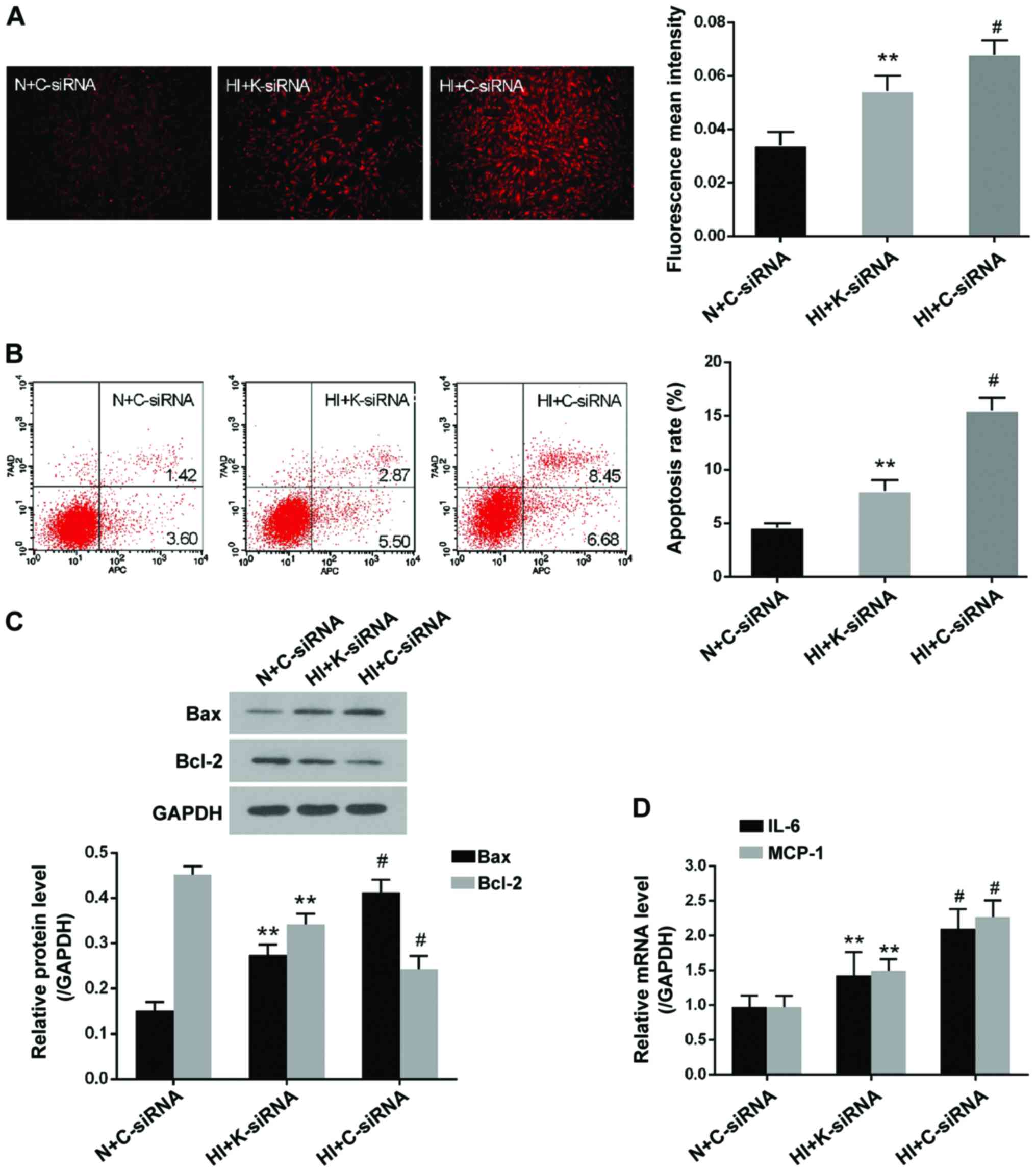 | Figure 4KDM3A knockdown decreases reactive
oxygen species generation, cell apoptosis and inflammation in high
insulin-treated VSMCs. (A) Representative fluorescent images of
dihydroethidium staining post-transduction with lenti-K-siRNA or
lenti-C-siRNA (magnification, ×100) (left panel). Fluorescent mean
density measured by ImageJ software (right panel). (B)
Representative flow cytometry plots of the effects of KDM3A
inhibition on high insulin-induced cellular apoptosis (left panel).
Relative apoptosis rates post-transduction with lentiviruses (right
panel). (C) Representative immunoblots for Bax and Bcl-2 expression
post-transduction with lenti-K-siRNA or lenti-C-siRNA (upper
panel). Densitometric analysis of western blotting (lower panel).
(D) Effects of KDM3A inhibition on the mRNA expression levels of
IL-6 and MCP-1 in VSMCs. Data are expressed as the mean ± standard
deviation, n=3. **P<0.05 vs. the HI + C-siRNA group;
#P<0.05 vs. the N + C-siRNA group. Bax,
Bcl-2-associated X protein; Bcl-2, B-cell lymphoma 2; C-siRNA,
control siRNA; HI, high concentration insulin stimulation; IL-6,
interleukin-6; KDM3A, lysine (K)-specific demethylase 3A; MCP-1,
monocyte chemotactic protein-1; K-siRNA, KDM3A siRNA; N, normal
medium; siRNA, small interfering RNA; VSMCs, vascular smooth muscle
cells |
KDM3A knockdown inactivates
mitogen-activated protein kinase (MAPK)/NF-κB signaling in high
insulin-treated VSMCs
Activation of MAPK/NF-κB signaling pathways serves a
key role in expediting VSMC dysfunction. To determine whether KDM3A
knockdown affects MAPK/NF-κB activity and impacts high
insulin-induced VSMCs pathology, western blotting was conducted to
detect ERK1/2, JNK, p38 MAPK and nuclear NF-κB/p65 protein
expression. As shown in Fig. 5,
high concentration insulin significantly upregulated the expression
of p-ERK1/2, p-JNK, p-p38 MAPK and nuclear NF-κB/p65 in VSMCs
compared with in the N + C-siRNA group (P<0.05), whereas
lenti-KDM3A-siRNA transduction into VSMCs markedly reduced the
expression levels of p-ERK1/2, p-JNK, p-p38 MAPK and NF-κB/p65 by
26.01, 26.08, 33.56 and 24.00% compared with in the HI + C-siRNA
group, respectively (P<0.05). The expression levels of total
ERK1/2, JNK and p38 MAPK were not signifi-cantly altered. These
findings indicated that the cytoprotecive effects of KDM3A
knockdown were in part associated with inactivation of MAPK/NF-κB
signaling pathways.
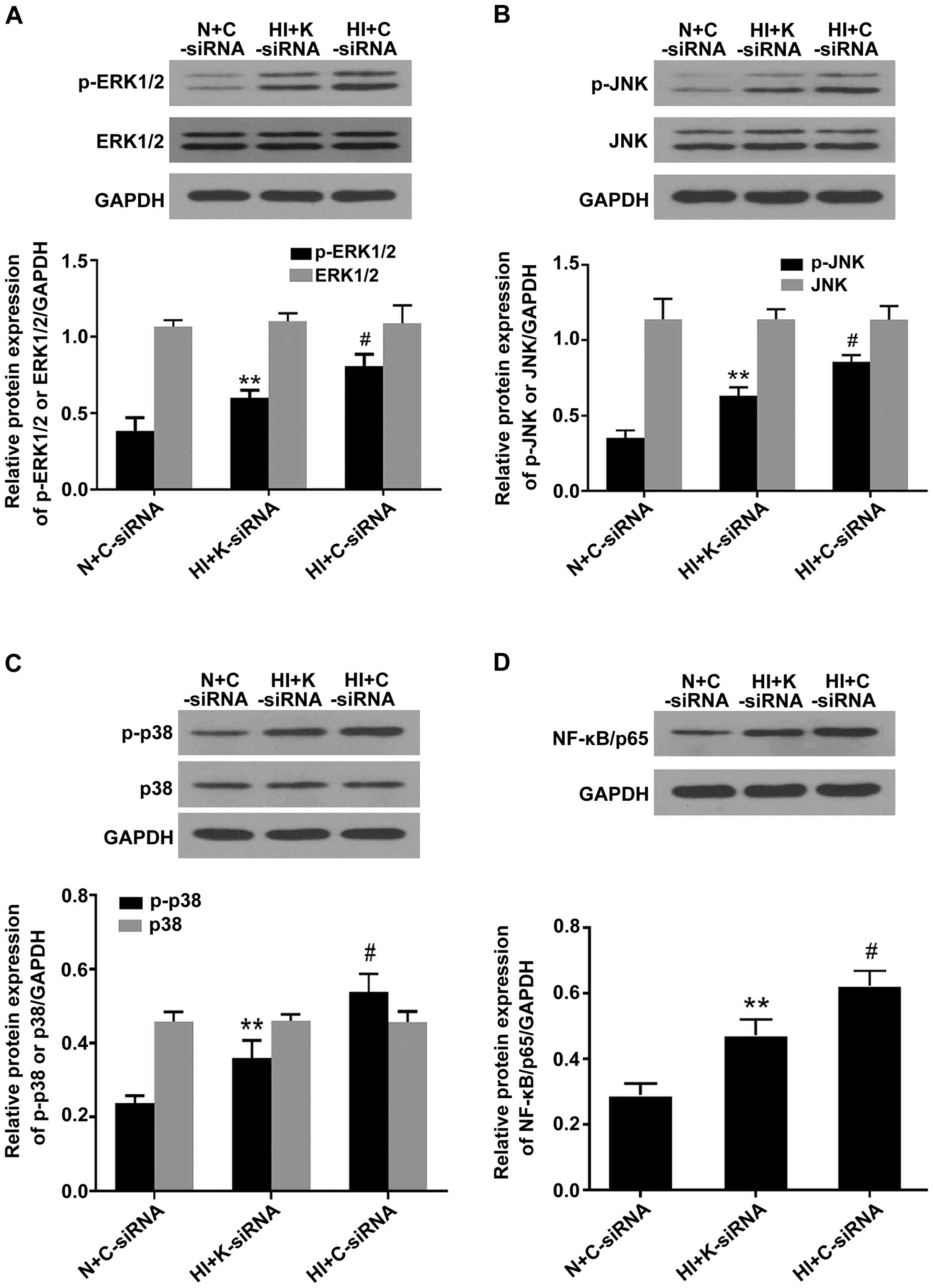 | Figure 5Knockdown of KDM3A suppresses the
expression levels of p-ERK1/2, p-JNK, p-p38 MAPK and NF-κB/p65 in
vascular smooth muscle cells following high concentration insulin
stimulation. (A) Representative immunoblots of p-ERK1/2, ERK1/2 and
GAPDH (upper panel). Densitometric analysis (lower panel). (B)
Representative immunoblots of p-JNK, JNK and GAPDH (upper panel).
Densitometric analysis (lower panel). (C) Representative
immunoblots of p-p38 MAPK, p38 MAPK and GAPDH (upper panel).
Densitometric analysis (lower panel). (D) Representative
immunoblots of nuclear NF-κB/p65 and GAPDH (upper panel).
Densitometric analysis (lower panel). Values are expressed as the
mean ± standard deviation, n=3. **P<0.05 vs. the HI +
C-siRNA group; #P<0.05 vs. the N + C-siRNA group.
ERK1/2, extracellular signal-regulated kinases 1/2; C-siRNA,
control siRNA; HI, high concentration insulin stimulation; JNK,
c-Jun NH2-terminal protein kinase; KDMA3, lysine
(K)-specific demethylase 3A; K-siRNA, KDM3A siRNA; MAPK,
mitogen-activated protein kinase; N, normal medium; NF-κB, nuclear
factor-κB; p, phosphorylated; siRNA, small interfering RNA. |
Discussion
Although it has previously been demonstrated that
KDM3A inhibition may exert protective effects against
hyperglycemia-induced VSMC lesions (20), the detailed involvement of KDM3A
in hyperinsulinemia-induced VSMC damage remains to be investigated.
The present study demonstrated that stimulation with a high
concentration of insulin significantly increased proliferation and
migration of VSMCs in vitro, alongside increased ROS
generation, apoptosis and inflammation. In addition, these outcomes
were associated with a marked upregulation of KDM3A. Therefore, the
present study hypothesized, and subsequently confirmed, that KDM3A
inhibition via lentivirus vectors could evidently ameliorate
cellular proliferation and migration, and limit inflammation,
oxidative stress and apoptotic cascade in high insulin-treated
VSMCs. Mechanistic studies indicated that KDM3A knockdown markedly
suppressed p-ERK1/2, p-JNK, p-p38 MAPK and NF-κB/p65 expression. To
the best of our knowledge, the present study is the first to
elaborate the effects of KDM3A on modulation of high
insulin-induced VSMC dysfunction. These results indicated that
KDM3A may be a potential target to attenuate
hyperinsulinemia-induced vascular injury.
It is well established that DM is closely associated
with adverse health outcomes, among which micro- and macrovascular
complications are considered the most severe manifestations
(23,24). Intensive treatment regimens that
normalize glucose levels were previously considered effective
therapeutic approaches; however, the risk of cardiovascular events
is not efficiently controlled. In addition, whether micro- and/or
macrovascular lesions may be attenuated by intensive
glucose-lowering therapy remains controversial (25,26). Type 2 DM is comprised of complex
metabolic disorders, including hyperglycemia, hyperlipidemia and
hyperinsulinemia (21).
Experimental studies have demonstrated that high concentrations of
insulin may confer direct or indirect effects on amplifying
cardiovascular stress (15).
Abhijit et al reported that VSMCs exposed to
hyperinsulinemia may exhibit increased ROS generation (27). In addition, Shiny et al and
Martínez-Hervás et al provided evidence to suggest that
hyperinsulinemia aggravates the inflammatory response and reduces
VSMC survival, respectively (28,29). It is well documented that ROS and
inflammation may induce VSMC proliferation and migration (15,27,28); however, the association between
apoptosis and VSMC dysfunction remains debatable; a study has
indicated that apoptosis may also trigger VSMC proliferation,
leading to neointimal hyperplasia (30); however, the underlying mechanisms
of this phenomenon are unclear (30). In line with previous studies, the
present study indicated that stimulation with high concentration
insulin promoted VSMC dysfunction and pathological injuries.
Notably, due to the controversial information regarding intensive
glucose-lowering therapy, as well as the roles of hyperinsulinemia
in DM-associated cardiovascular complications irrespective of
hyperglycemia (31), reversing
the hyperinsulinemia-induced VSMC dysfunction in type 2 DM may be
considered a promising therapeutic approach to protect against
vascular complications.
MAPK cascades are evolutionary conserved signaling
modules that consist of protein-serine/threonine kinase family
proteins, including ERK1/2, JNK and p38 MAPK (32). NF-κB serves as an important
downstream transcription factor of MAPK activation in VSMCs
(33). MAPK/NF-κB signaling
pathways may translate environmental information into appropriate
responses via phosphorylating their substrate proteins and thus
regulate various cellular processes, including proliferation,
differentiation, inflammation, survival and apoptosis (34). These signaling pathways also
participate in numerous pathological processes in VSMCs. Liu et
al previously demonstrated that insulin-mediated proliferation
of VSMCs could be suppressed by PD98059, a selective inhibitor of
the ERK1/2 signaling pathway, thus suggesting that MAPKs have a
regulatory impact on insulin-induced VSMC injuries (35). Oxidative stress and inflammation
are vital factors associated with VSMC dysfunction; therefore, any
factor that increases oxidative stress and inflammation may lead to
VSMC injuries (15). ERK, JNK,
p38 MAPK and NF-κB are all ROS-sensitive regulators (36). In addition, ROS generation may be
mediated by activation of MAPK/NF-κB signal pathways (31). Inhibition of ERK1/2, JNK or p38
MAPK by specific inhibitors or siRNAs has been reported to
attenuate ROS accumulation-induced VSMC injuries (36,37). Therefore, activation of MAPKs and
ROS production may augment each other under pathological
conditions. Furthermore, causal associations exist between
activation of MAPK/NF-κB signaling pathways and inflammation. Our
previous study indicated that ERK1/2 and NF-κB activities could
increase IL-6 and MCP-1 expression in VSMCs (36); these findings were concordant with
the findings published by Wronkowitz et al (38). In addition, close associations
exist between MAPK activation and VSMC apoptosis. Gomez et
al (30) indicated that p38
MAPK and JNK pathways initiated first apoptotic wave of VSMCs and
contributed to vascular inflammation; these results were highly
consistent with those of another study (39). The present study demonstrated that
high concentration insulin could significantly exacerbate oxidative
stress, inflammation and apoptosis of VSMCs. However, whether
ERK1/2, JNK and p38 MAPK were involved in high insulin-induced
inflammation, ROS accumulation and apoptosis in VSMCs remained
ambiguous. In addition, the effects of MAPK subgroups may be
divergent according to the species from which VSMCs were obtained
and the external stimulus acting upon them. In the present study,
stimulating rat VSMCs with 100 nM insulin led to VSMC dysfunction;
in addition, oxidative stress, inflammation and apoptosis were
concomitantly increased. Notably, mechanistic studies further
revealed that the expression levels of p-ERK1/2, p-JNK, p-p38 MAPK
and NF-κB/p65 were significantly upregulated. Therefore, the
present study suggested that activation of MAPK/NF-κB signaling
pathways may serve prominent roles in aggravating the deleterious
effects of hyperinsulinemia on VSMCs.
KDM3A is a member of the 2-oxygluta
rate/Fe(II)-dependent Jumonji C-domain-containing protein family,
which selectively removes mono- and di-methyl groups from histone
H3 lysine H3K9 (40). Although
the biochemical features of KDM3A, as a histone demethylase, have
been well characterized, its physiological significance remains
incompletely understood. KDM3A was originally reported to be
upregulated and serve an important role in the setting of hypoxia
(41). To date, it has been
revealed that KDM3A may increase cell proliferation, migration,
invasion and angiogenesis in various cancer types, and elevated
KDM3A may be a poor prognostic maker in patients with cancer
(17–19,42). Furthermore, KDM3A has also been
reported to be involved in stem cell renewal and VSMC
differentiation (43). However,
its potential functions in diabetic cardiovascular complications
have rarely been studied. Our previous findings demonstrated that
the expression of KDM3A was significantly increased in injured
blood vessels following carotid artery balloon treatment in rats
with type 2 DM. Furth ermore, downregulating KDM3A could markedly
inhibit vascular remodeling and efficiently promote renovation of
endothelium, even though blood sugar levels continued to rise. Our
previous study also indicated that VSMC proliferation and migration
were enhanced in the KDM3A-upregulated group under high glucose
conditions; these effects were blunted by KDM3A gene knockdown
(20). In another study using
diabetic db/db mice, it was demonstrated that
2,4-pyridinedicarboxylic acid, an inhibitor of KDM3A, could inhibit
myocardial fibrosis by increasing the density of cardiac
microcirculation (unpublished data). Therefore, it may be
hypothesized that KDM3A serves a vital role in diabetic
cardiovascular complications. Since our previous study mainly
focused on hyperglycemia, and since hyperinsulinemia is a prominent
feature of type 2 DM that may exert adverse effects on the
cardiovascular system, the present study aimed to determine whether
KDM3A participated in hyperinsulinemia-induced VSMC injuries. The
present study indicated that incubating VSMCs with 100 nM insulin
markedly contributed to cellular dysfunction, alongside KDM3A
upregulation. Furthermore, a high concentration of insulin
activated MAPK/NF-κB signaling pathways, whereas KDM3A knockdown
via its specific siRNA markedly reduced the expression levels of
p-ERK1/2, p-JNK, p-p38 MAPK and NF-κB/p65. In addition, KDM3A
knockdown attenuated oxidative stress, inflammation and apoptosis,
and limited proliferation and migration of VSMCs. These findings
indicated that KDM3A may act as an upstream regulatory factor of
MAPK/NF-κB signa ling pathways in VSMCs undergoing stimulation with
a high concentration of insulin, and its effects may be partly
mediated through MAPK/NF-κB signaling pathways.
In conclusion, the present study is the first, to
the best of our knowledge, to report the effects of KDM3A on
aggravating hyperinsulinemia-induced VSMC injuries. Notably, the
results indicated that silencing KDM3A expression could attenuate
VSMC damage caused by high insulin levels, at least in part,
through scavenging ROS, moderating inflammation and suppressing
apoptotic activation, in a MAPK/NF-κB pathway-dependent manner.
Therefore, KDM3A may be a promising therapeutic target to prevent
DM-associated cardiovascular diseases.
Acknowledgments
The present study was supported by the National
Natural Science Foundation of China (grant nos. 81200156 and
81570331).
References
|
1
|
Wegner M, Neddermann D,
Piorunska-Stolzmann M and Jagodzinski PP: Role of epigenetic
mechanisms in the development of chronic complications of diabetes.
Diabetes Res Clin Pract. 105:164–175. 2014. View Article : Google Scholar : PubMed/NCBI
|
|
2
|
van der Leeuw J, Beulens JW, van Dieren S,
Schalkwijk CG, Glatz JF, Hofker MH, Verschuren WM, Boer JM, van der
Graaf Y, Visseren FL, et al: Novel biomarkers to improve the
prediction of cardiovascular event risk in type 2 diabetes
mellitus. J Am Heart Assoc. 5:52016. View Article : Google Scholar
|
|
3
|
Ndrepepa G, Colleran R, Luttert A, Braun
S, Cassese S, Kufner S, Hieber J, Fusaro M, Laugwitz KL, Schunkert
H, et al: Prognostic value of gamma-glutamyl transferase in
patients with diabetes mellitus and coronary artery disease. Clin
Biochem. 49:1127–1132. 2016. View Article : Google Scholar : PubMed/NCBI
|
|
4
|
Minha S, Bental T, Assali A, Vaknin-Assa
H, Lev EI, Rechavia E, Battler A and Kornowski R: A comparative
analysis of major clinical outcomes using drug-eluting stents
versus bare metal stents in diabetic versus nondiabetic patients.
Catheter Cardiovasc Interv. 78:710–717. 2011. View Article : Google Scholar : PubMed/NCBI
|
|
5
|
Myers GR and Weintraub WS: Coronary artery
disease: revascularization strategies for patients with CAD and
diabetes. Nat Rev Cardiol. 7:364–366. 2010. View Article : Google Scholar : PubMed/NCBI
|
|
6
|
Beckman JA, Paneni F, Cosentino F and
Creager MA: Diabetes and vascular disease: pathophysiology,
clinical consequences, and medical therapy: part II. Eur Heart J.
34:2444–2452. 2013. View Article : Google Scholar : PubMed/NCBI
|
|
7
|
El-Atat FA, Stas SN, McFarlane SI and
Sowers JR: The relationship between hyperinsulinemia, hypertension
and progressive renal disease. J Am Soc Nephrol. 15:2816–2827.
2004. View Article : Google Scholar : PubMed/NCBI
|
|
8
|
Nakamura A, Monma Y, Kajitani S, Noda K,
Nakajima S, Endo H, Takahashi T and Nozaki E: Effect of glycemic
state on postprandial hyperlipidemia and hyperinsulinemia in
patients with coronary artery disease. Heart Vessels. 31:1446–1455.
2016. View Article : Google Scholar :
|
|
9
|
Lee KK, Fortmann SP, Fair JM, Iribarren C,
Rubin GD, Varady A, Go AS, Quertermous T and Hlatky MA: Insulin
resistance independently predicts the progression of coronary
artery calcification. Am Heart J. 157:939–945. 2009. View Article : Google Scholar : PubMed/NCBI
|
|
10
|
Sun HJ, Zhao MX, Ren XS, Liu TY, Chen Q,
Li YH, Kang YM, Wang JJ and Zhu GQ: Salusin-β promotes vascular
smooth muscle cell migration and intimal hyperplasia after vascular
injury via ROS/NFκB/MMP-9 pathway. Antioxid Redox Signal.
24:1045–1057. 2016. View Article : Google Scholar : PubMed/NCBI
|
|
11
|
Xu Z, Han Y, Liu J, Jiang F, Hu H, Wang Y,
Liu Q, Gong Y and Li X: miR-135b5p and miR-499a-3p promote cell
proliferation and migration in atherosclerosis by directly
targeting MEF2C. Sci Rep. 5:122762015. View Article : Google Scholar
|
|
12
|
Chen J, Zhang J, Xu L, Xu C, Chen S, Yang
J and Jiang H: Inhibition of neointimal hyperplasia in the rat
carotid artery injury model by a HMGB1 inhibitor. Atherosclerosis.
224:332–339. 2012. View Article : Google Scholar : PubMed/NCBI
|
|
13
|
Wu G, Cai J, Han Y, Chen J, Huang ZP, Chen
C, Cai Y, Huang H, Yang Y, Liu Y, et al: LincRNA-p21 regulates
neointima formation, vascular smooth muscle cell proliferation,
apoptosis, and atherosclerosis by enhancing p53 activity.
Circulation. 130:1452–1465. 2014. View Article : Google Scholar : PubMed/NCBI
|
|
14
|
Caccamo G, Bonura F, Bonura F, Vitale G,
Novo G, Evola S, Evola G, Grisanti MR and Novo S: Insulin
resistance and acute coronary syndrome. Atherosclerosis.
211:672–675. 2010. View Article : Google Scholar : PubMed/NCBI
|
|
15
|
Wang X, Yu C, Zhang B and Wang Y: The
injurious effects of hyperinsulinism on blood vessels. Cell Biochem
Biophys. 69:213–218. 2014. View Article : Google Scholar
|
|
16
|
Dubin SB: Characterization of amniotic
fluid lamellar bodies by resistive-pulse counting: relationship to
measures of fetal lung maturity. Clin Chem. 35:612–616.
1989.PubMed/NCBI
|
|
17
|
Tateishi K, Okada Y, Kallin EM and Zhang
Y: Role of Jhdm2a in regulating metabolic gene expression and
obesity resistance. Nature. 458:757–761. 2009. View Article : Google Scholar : PubMed/NCBI
|
|
18
|
Osawa T, Tsuchida R, Muramatsu M,
Shimamura T, Wang F, Suehiro J, Kanki Y, Wada Y, Yuasa Y, Aburatani
H, et al: Inhibition of histone demethylase JMJD1A improves
anti-angiogenic therapy and reduces tumor-associated macrophages.
Cancer Res. 73:3019–3028. 2013. View Article : Google Scholar : PubMed/NCBI
|
|
19
|
Parrish JK, Sechler M, Winn RA and
Jedlicka P: The histone demethylase KDM3A is a
microRNA-22-regulated tumor promoter in Ewing sarcoma. Oncogene.
34:257–262. 2015. View Article : Google Scholar :
|
|
20
|
Jing C, Xu L, Zhang J, Hu X, Chen S, Hu Q,
Xu C and Jiang H: GW24-e2386 down-regulation of histone demethylase
KDM3A attenuates balloon injury-induced neointimal hyperplasia in
diabetic rats through modulation of epigenetic histone lysine 9
di-methylation. Heart. 2013:A9–A10. 2013.
|
|
21
|
Qi H, Jing Z, Xiaolin W, Changwu X,
Xiaorong H, Jian Y, Jing C and Hong J: Histone demethylase JMJD2A
inhibition attenuates neointimal hyperplasia in the carotid
arteries of balloon-injured diabetic rats via transcriptional
silencing: inflammatory gene expression in vascular smooth muscle
cells. Cell Physiol Biochem. 37:719–734. 2015. View Article : Google Scholar : PubMed/NCBI
|
|
22
|
Livak KJ and Schmittgen TD: Analysis of
relative gene expression data using real-time quantitative PCR and
the 2(−Delta Delta C(T)) Method. Methods. 25:402–408. 2001.
View Article : Google Scholar
|
|
23
|
Cavicchioli MG, Guerbali CC, Ochiai C,
Silva RM, Camara G and Petry TB: The contribution of diabetes
education in the treatment of people with type 2 diabetes and risk
of cardiovascular disease. Curr Atheroscler Rep. 18:442016.
View Article : Google Scholar : PubMed/NCBI
|
|
24
|
Forbes JM and Cooper ME: Mechanisms of
diabetic complications. Physiol Rev. 93:137–188. 2013. View Article : Google Scholar : PubMed/NCBI
|
|
25
|
Patel A, MacMahon S, Chalmers J, Neal B,
Billot L, Woodward M, Marre M, Cooper M, Glasziou P, Grobbee D, et
al ADVANCE Collaborative Group: Intensive blood glucose control and
vascular outcomes in patients with type 2 diabetes. N Engl J Med.
358:2560–2572. 2008. View Article : Google Scholar : PubMed/NCBI
|
|
26
|
Duckworth W, Abraira C, Moritz T, Reda D,
Emanuele N, Reaven PD, Zieve FJ, Marks J, Davis SN, Hayward R, et
al VADT Investigators: Glucose control and vascular complications
in veterans with type 2 diabetes. N Engl J Med. 360:129–139. 2009.
View Article : Google Scholar
|
|
27
|
Abhijit S, Bhaskaran R, Narayanasamy A,
Chakroborty A, Manickam N, Dixit M, Mohan V and Balasubramanyam M:
Hyperinsulinemia-induced vascular smooth muscle cell (VSMC)
migration and proliferation is mediated by converging mechanisms of
mitochondrial dysfunction and oxidative stress. Mol Cell Biochem.
373:95–105. 2013. View Article : Google Scholar
|
|
28
|
Shiny A, Regin B, Mohan V and
Balasubramanyam M: Coordinated augmentation of NFAT and NOD
signaling mediates proliferative VSMC phenotype switch under
hyperinsulinemia. Atherosclerosis. 246:257–266. 2016. View Article : Google Scholar : PubMed/NCBI
|
|
29
|
Martínez-Hervás S, Vinué A, Núñez L,
Andrés-Blasco I, Piqueras L, Real JT, Ascaso JF, Burks DJ, Sanz MJ
and González-Navarro H: Insulin resistance aggravates
atherosclerosis by reducing vascular smooth muscle cell survival
and increasing CX3CL1/CX3CR1 axis. Cardiovasc Res. 103:324–336.
2014. View Article : Google Scholar : PubMed/NCBI
|
|
30
|
Gomez C, Martinez L, Mesa A, Duque JC,
Escobar LA, Pham SM and Vazquez-Padron RI: Oxidative stress induces
early-onset apoptosis of vascular smooth muscle cells and neointima
formation in response to injury. Biosci Rep. 35:352015. View Article : Google Scholar
|
|
31
|
Ceriello A, Ihnat MA and Thorpe JE:
Clinical review 2: the ‘metabolic memory’: is more than just tight
glucose control necessary to prevent diabetic complications? J Clin
Endocrinol Metab. 94:410–415. 2009. View Article : Google Scholar
|
|
32
|
Lv H, Yu Z, Zheng Y, Wang L, Qin X, Cheng
G and Ci X: Isovitexin exerts anti-inflammatory and anti-oxidant
activities on lipopolysaccharide-induced acute lung injury by
inhibiting MAPK and NF-κB and activating HO-1/Nrf2 pathways. Int J
Biol Sci. 12:72–86. 2016. View Article : Google Scholar :
|
|
33
|
Zhang X, Liu J, Pang X, Zhao J, Wang S and
Wu D: Aldosterone induces C-reactive protein expression via
MR-ROS-MAPK-NF-κB signal pathway in rat vascular smooth muscle
cells. Mol Cell Endocrinol. 395:61–68. 2014. View Article : Google Scholar : PubMed/NCBI
|
|
34
|
Chen J, Xu J, Li J, Du L, Chen T, Liu P,
Peng S, Wang M and Song H: Epigallocatechin-3-gallate attenuates
lipopoly-saccharide-induced mastitis in rats via suppressing MAPK
mediated inflammatory responses and oxidative stress. Int
Immunopharmacol. 26:147–152. 2015. View Article : Google Scholar : PubMed/NCBI
|
|
35
|
Liu K, Zhao W, Gao X, Huang F, Kou J and
Liu B: Diosgenin ameliorates palmitate-induced endothelial
dysfunction and insulin resistance via blocking IKKβ and IRS-1
pathways. Atherosclerosis. 223:350–358. 2012. View Article : Google Scholar : PubMed/NCBI
|
|
36
|
Zhang J, Chen J, Yang J, Xu CW, Pu P, Ding
JW and Jiang H: Resveratrol attenuates oxidative stress induced by
balloon injury in the rat carotid artery through actions on the
ERK1/2 and NF-kappa B pathway. Cell Physiol Biochem. 31:230–241.
2013. View Article : Google Scholar : PubMed/NCBI
|
|
37
|
Zhang X, Wang X, Wu T, Li B, Liu T, Wang
R, Liu Q, Liu Z, Gong Y and Shao C: Isoliensinine induces apoptosis
in triple-negative human breast cancer cells through ROS generation
and p38 MAPK/JNK activation. Sci Rep. 5:125792015. View Article : Google Scholar : PubMed/NCBI
|
|
38
|
Wronkowitz N, Görgens SW, Romacho T,
Villalobos LA, Sánchez-Ferrer CF, Peiró C, Sell H and Eckel J:
Soluble DPP4 induces inflammation and proliferation of human smooth
muscle cells via protease-activated receptor 2. Biochim Biophys
Acta. 1842:1613–1621. 2014. View Article : Google Scholar : PubMed/NCBI
|
|
39
|
Perez-Vizcaino F, Bishop-Bailley D, Lodi
F, Duarte J, Cogolludo A, Moreno L, Bosca L, Mitchell JA and Warner
TD: The flavonoid quercetin induces apoptosis and inhibits JNK
activation in intimal vascular smooth muscle cells. Biochem Biophys
Res Commun. 346:919–925. 2006. View Article : Google Scholar : PubMed/NCBI
|
|
40
|
Yamane K, Toumazou C, Tsukada Y,
Erdjument-Bromage H, Tempst P, Wong J and Zhang Y: JHDM2A, a
JmjC-containing H3K9 demethylase, facilitates transcription
activation by androgen receptor. Cell. 125:483–495. 2006.
View Article : Google Scholar : PubMed/NCBI
|
|
41
|
Wellmann S, Bettkober M, Zelmer A, Seeger
K, Faigle M, Eltzschig HK and Bührer C: Hypoxia upregulates the
histone demethylase JMJD1A via HIF-1. Biochem Biophys Res Commun.
372:892–897. 2008. View Article : Google Scholar : PubMed/NCBI
|
|
42
|
Uemura M, Yamamoto H, Takemasa I, Mimori
K, Hemmi H, Mizushima T, Ikeda M, Sekimoto M, Matsuura N, Doki Y,
et al: Jumonji domain containing 1A is a novel prognostic marker
for colorectal cancer: in vivo identification from hypoxic tumor
cells. Clin Cancer Res. 16:4636–4646. 2010. View Article : Google Scholar : PubMed/NCBI
|
|
43
|
Lockman K, Taylor JM and Mack CP: The
histone demethylase, Jmjd1a, interacts with the myocardin factors
to regulate SMC differentiation marker gene expression. Circ Res.
101:e115–e123. 2007. View Article : Google Scholar : PubMed/NCBI
|