Introduction
Gastric cancer (GC) is a heterogeneous disease with
two distinct morphological subtypes: gastric intestinal type
adenocarcinoma and diffuse gastric adenocarcinoma. Intestinal and
diffuse type GC show variable environmental aetiologies, clinical
manifestation and genetic background (1,2).
Diffuse gastric adenocarcinoma is often seen in female and young
individuals, while the intestinal type adenocarcinoma is more often
associated with intestinal metaplasia and Helicobacter
pylori infection (3). Based
on previous studies we know that frequent inactivating mutations in
cell adhesion and chromatin remodelling genes exist in addition to
TP53 mutations (4,5). Although some essential factors for
resolution were identified in recent years, the clinical trials
still lack effective methods to treat the disease and reliable
biomarkers to monitor its progression (6–9).
Regenerating islet-derived 3 α (REG3A) is a member
of REG protein family and also named as human
hepatocarcinoma-intestine-pancreas (HIP) or human
pancreatitis-associated protein (PAP) (10–12). REG3A is a secreted
calcium-dependent lectin protein which is related with pancreatic
islet cell regeneration, pancreatic stellate cell activation (PSCs)
(13,14) and liver regeneration (15). It has been reported that REG3A
plays important roles in a number of human cancers, including GC
(16), pancreatic cancer
(17–19) and colorectal cancer (20). REG3A also regulates keratinocyte
proliferation and differentiation after skin injury (21). However, the exact function of
REG3A on GC and the details of the pathways has not been
demonstrated.
In this study, we showed that the expression of
REG3A was significantly downregulated in GC and closely related
with patient prognoses. REG3A could regulate the invasion,
proliferation and apoptosis of GC cells through
phosphatidylinositol 3 kinase (PI3K)/Akt-GSK3β signaling pathway
axis.
Materials and methods
Cell culture
Human GC cell lines, including AGS, BGC-823, HGC-27,
MGC-803, MKN-45 and SGC-7901 were purchased from Cell Bank of the
Chinese Academy of Sciences. Cells were cultured in RPMI-1640
medium supplemented with 10% (v/v) fetal calf serum and 1%
antibiotics at 37°C in a humidified incubator under 5%
CO2 condition.
Clinical samples
Human gastric tumor (19 cases) and normal tissues
(15 cases) were obtained from Department of Bone Tumor, Yantai
Mountain Hospital. All human materials were obtained with informed
consent, and protocols were approved by the ethics review committee
of the World Health Organization Collaborating Center for Research
in Human Production.
Data mining using TCGA and GEO
REG3A gene expression were analyzed using microarray
gene expression datasets deposited in GEO database. A combined
filter was applied to display the corresponding datasets. The
cancer type was defined as GC and data type was mRNA, and analysis
type was cancer versus normal analysis. The expression levels of
REG3A gene were read from the displayed bar chart and these data
were analyzed by Excel. Further, the gene expression data for GC
was downloaded from TCGA. The RNA-seq gene expression data contain
log2-transformed RNA-seq by expectation maximization
(RSEM) values summarized at gene level.
Quantitative real-time PCR
Total RNA was extracted using TRIzol reagent, and
reverse transcribed through PrimeScript RT-PCR kit (Takara, Dalian,
China) according to the protocol. Real-time PCR analyses were
performed with SYBR Premix Ex Taq (Takara) on a 7300 Real-time PCR
system (Applied Biosystems, Waltham, MA, USA) at the recommended
thermal cycling settings: one initial cycle at 95°C for 30 sec
followed by 40 cycles of 5 sec at 95°C and 31 sec at 60°C.
Western blotting
Cells were lysed in lysis buffer. Proteins were
separated by sodium dodecyl sulfate-polyacrylamide gel
electrophoresis (SDS-PAGE) under reducing condition, followed by
blocking in phosphate-buffered saline (PBS)/Tween-20 containing 1%
BSA. The membrane was incubated with antibodies for REG3A (Abcam,
Cambridge, MA, USA), phospho-Akt, total-Akt, phospho-GSK3β,
total-GSK3β (all from Cell Signaling Technology, Beverly, MA, USA),
glyceraldehyde 3-phosphate dehydrogenase (GAPDH) (Sigma, St. Louis,
MO, USA) and species-specific secondary antibodies. Bound secondary
antibodies were revealed by Odyssey imaging system (LI-COR
Biosciences, Lincoln, NE, USA).
Lentivirus production and cell
transduction
Virus packaging was performed in 293T cells after
cotransfection of pEZ-lv105 vector (GeneCopoeia, Guangzhou, China)
using lipofectamine 2000 (Invitrogen, Carlsbad, CA, USA). viruses
were harvested at 48 and 72 h after transfection, and virus titers
were determined. Target cells (1×10⁵), including MGC-803
and BGC-823 cells, were infected with 1×10⁶ recombinant
lentivirus-transducing units in the presence of 6 µg/ml
polybrene (Sigma).
siRNA transfection
Small interfering RNA duplexes for REG3A was
produced by GenePharma (Shanghai, China). Transfection steps
followed the manufacturer's protocols.
Invasion assays
MGC-803 and BGC-823 cells were detached and
resuspended in serum-free DMEM. Approximately 2×10⁴
cells in 0.1 ml were placed in Matrigel (BD Biosciences, Bedford,
MA, USA)-coated inserts (Millipore, Billerica, MA, USA) on the
24-well plate. DMEM containing 5% (v/v) fetal bovine serum (FBS)
was added to the bottom chamber. Cells were incubated at 37°C and
allowed to invade through Matrigel for 48 h. After incubation,
filters were fixed and stained with 0.1% (w/v) crystal violet.
Non-invading cells were removed using a cotton swab while invading
cells on the underside of the filter were counted under a
microscope at a magnification, ×200 or ×400. At least five grids
per field were counted and the experiments were repeated at least
twice.
Apoptosis assays
Per well 5×10⁵ cells were cultured on
12-well plates and serum starved for 48 h at 37°C in a 5%
CO2 atmosphere. After incubation, adherent cells were
detached with 0.25% trypsin/0.01% EDTA in 1X PBS. Detached and
suspended cells were harvested in complete DMEM medium and
centrifuged at 1,000 rpm for 5 min. Each of the cells were washed
with 1X PBS and stained with 100 µl binding buffer
containing 3.5 µl Annexin v and 3.5 µl propidium
iodide (PI). Cells were incubated at room temperature for 15 min
and analyzed by flow cytometry (BD Biosciences).
Statistical analysis
Data are presented as the means ± standard error of
the mean (SEM). Statistical analyses were performed using SPSS 16.0
for windows (IBM, Chicago, Il, USA). Cumulative survival time was
calculated by the Kaplan-Meier method and analyzed by the log-rank
test. The Chi-square test, and Student's t-test were used for
comparison between groups. values of P<0.05 were considered
statistically significant.
Results
REG3A expression is downregulated in GC
tissues and closely related with patient prognoses
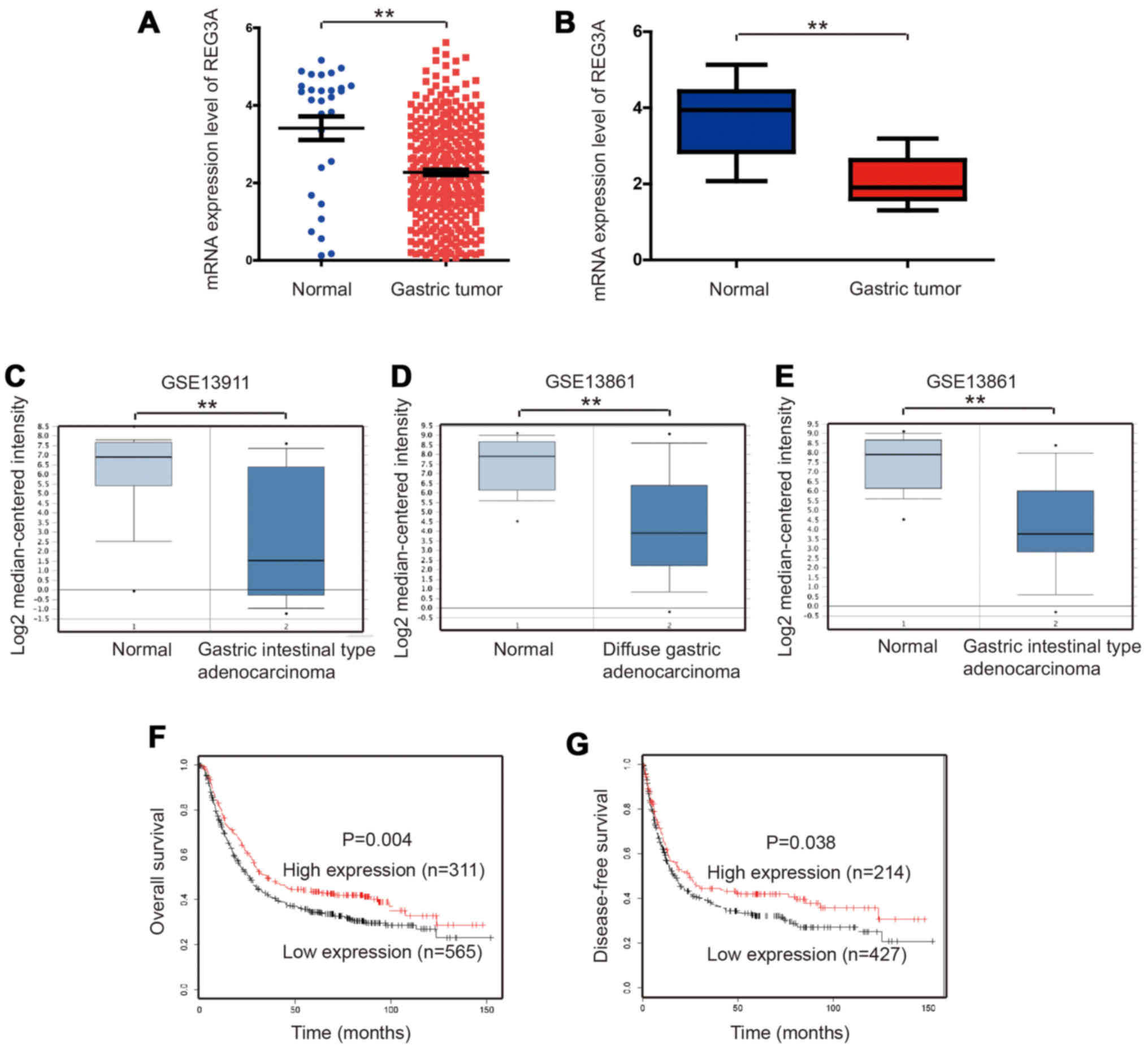
To determine the expression of REG3A in GC, we
analyzed the microarray data from TCGA or GEO datasets. The TCGA
dataset showed that the expression level of REG3A was significantly
downregulated in GC tissues, compared with normal tissues (Fig. 1A). Further, we collected 19 cases
of gastric tumor and 15 cases of normal tissues. By quantitative
real-time PCR, we found that the expression level of REG3A was
significantly downregulated in GC tissues (Fig. 1B).
It was further proven in GSE13911 and GSE13861
datasets, in which REG3A expression was downregulated in gastric
intestinal type adenocarcinoma or diffuse gastric adenocarcinoma
respectively (Fig. 1C–E). We also
analyzed the data from KMplot. REG3A expression was closely related
with patient prognoses. High REG3A expression was associated with
improved overall survival (OS) (P=0.004) or disease-free survival
(DFS) (P=0.038) (Fig. 1F and
G).
Overexpression of REG3A in GC cells
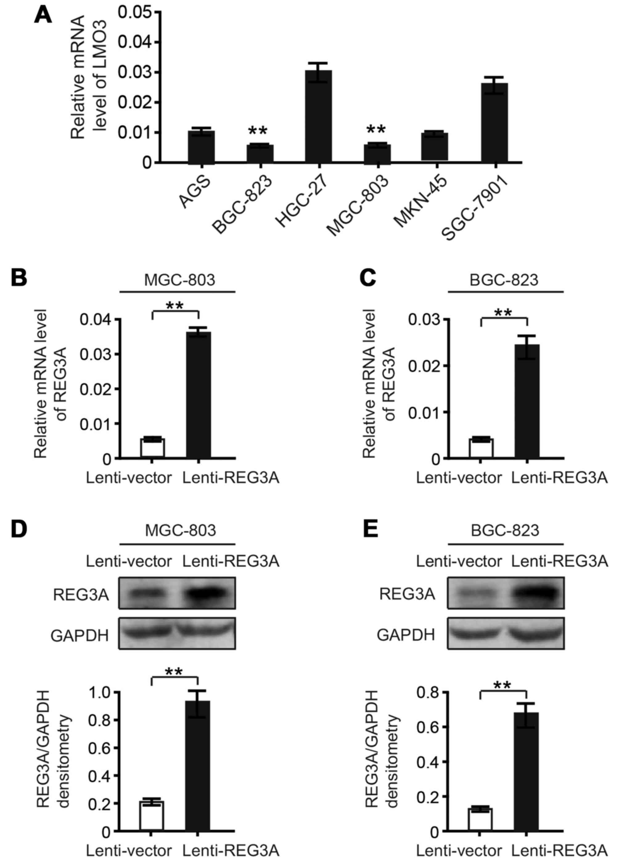
To further investigate the role of REG3A in GC, we
first detected the expression level of REG3A in six GC cell lines.
As shown in Fig. 2A, we found
that REG3A had low expression levels in MGC-803 and BGC-823 cells.
We established stable cell lines transduced by the lentivirus
carrying the REG3A gene, designated as Lenti-REG3A, in MGC-803 and
BGC-823 cells. The results from real-time PCR and western blotting
showed that REG3A was overexpressed in both MGC-803 (Fig. 2B and D) and BGC-823 cells
(Fig. 2C and E).
Overexpression of REG3A reduces the
invasion and proliferation of GC cells, while increases the
apoptosis of GC cells
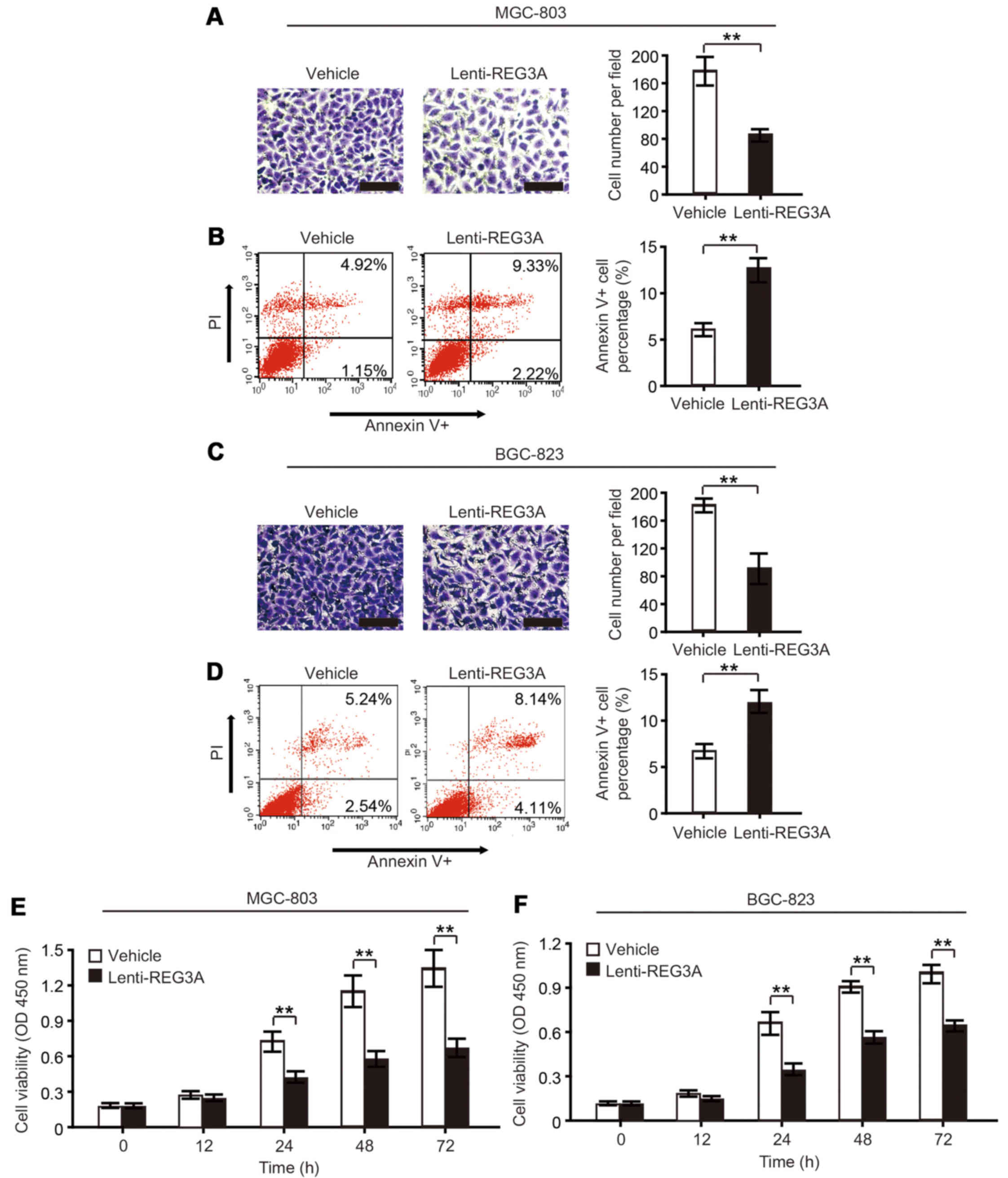
We first investigated the role of REG3A in the
invasion and apoptosis of GC cells. By Transwell Matrigel invasion
assay, we found that REG3A overexpression reduced invasiveness of
MGC-803 and BGC-823 cells after 48 h (Fig. 3A and C). Moreover, we found that
apoptosis of GC cells was affected by overexpression of REG3A.
Apoptosis of GC cells was determined by flow cytometry after the
cells were serum starved for 48 h. The results showed that the
apoptosis rate was increased by the overexpression of REG3A in
MGC-803 and BGC-823 cells (Fig. 3B
and D).
We further investigated the proliferation of GC
cells after REG3A overexpression by cell counting kit-8 (CCK-8)
proliferation assay. It was found that the proliferation of MGC-803
or BGC-823 cells was significantly reduced by REG3A overexpression
at 24, 48 and 72 h time-points (Fig.
3E and F).
Knockdown of REG3A promotes the invasion
and proliferation of GC cells, while suppresses apoptosis of GC
cells
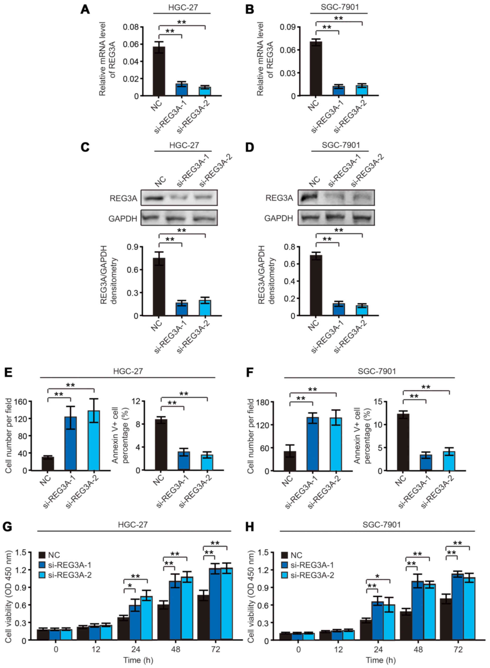
LMO3 had relative high expression levels in HGC-27
and SGC-7901 cells (Fig. 2A). So
we selected HGC-27 and SGC-7901 cells and knocked out REG3A by
using siRNA (labeled as si-REG3A-1 and si-REG3A-2). Through
real-time PCR and western blotting analysis we found that REG3A was
successfully silenced in HGC-27 (Fig.
4A and C) and SGC-7901 cells (Fig. 4B and D).
By Transwell Matrigel invasion assay and flow
cytometry analysis, we found that knockdown of REG3A promoted the
invasiveness and suppressed the apoptosis of HGC-27 (Fig. 4E) and SGC-7901 (Fig. 4F) cells after 48 h. Further, by
CCK-8 cell viability assay, we found that the cell viability of
HGC-27 and SGC-7901 cells was significantly increased by knockdown
of REG3A at 24, 48 and 72 h time-points respectively (Fig. 4G and H).
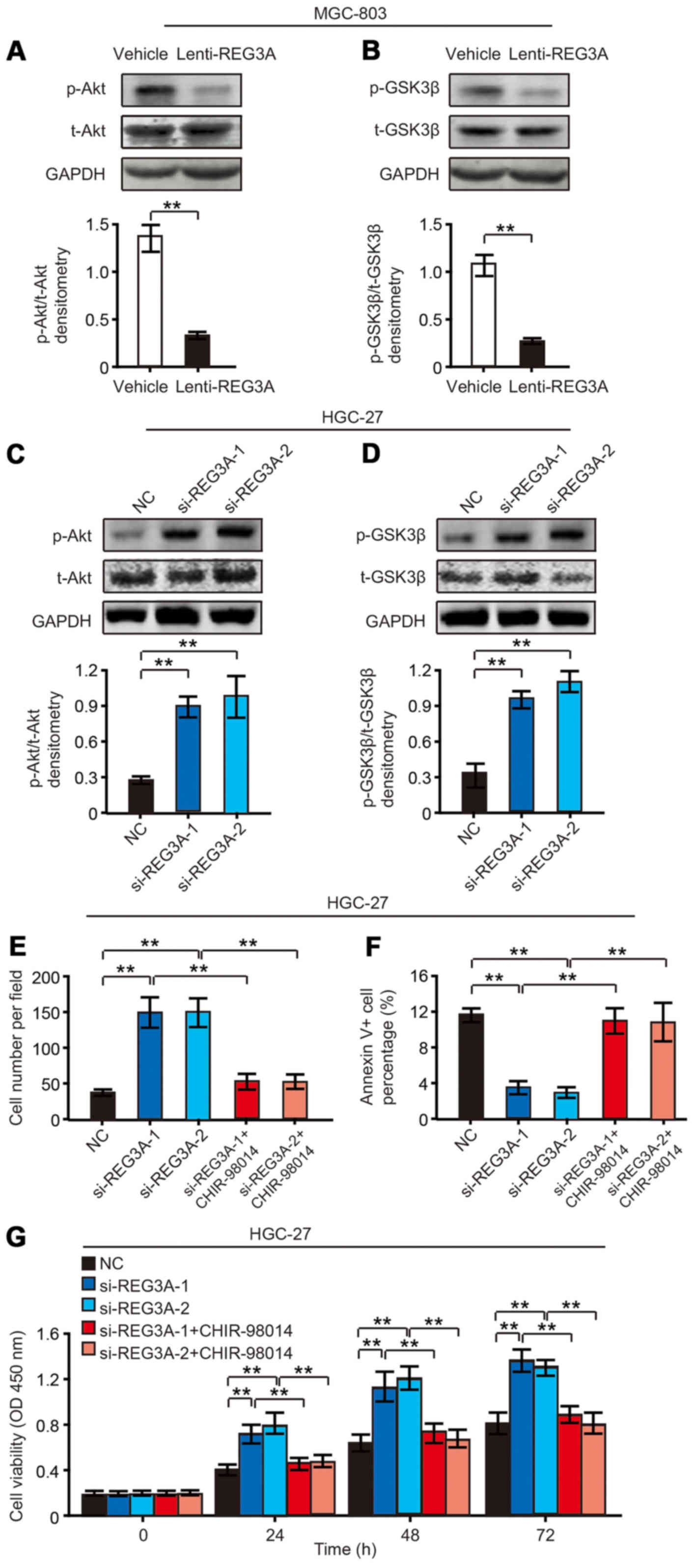
The effects of REG3A on GC cells are
dependent on PI3K/Akt and GSK3β signaling pathway
To uncover the molecular mechanism of REG3A in GC
cells, we performed western blotting to detect PI3K/Akt related
signaling pathway in REG3A overexpressed MGC-803 cells and control
cells. It was found that the phosphorylation of Akt was
significantly suppressed by the overexpression of REG3A (Fig. 5A). Further, we found that the
phosphorylation of GSK3β was also suppressed by REG3A
overexpression (Fig. 5B).
Further, we detected Akt-GSK3β signaling pathway in
REG3A silenced HGC-27 cells and control cells. It was found that
REG3A knockdown significantly increased the phosphorylation of Akt
(Fig. 5C). Furthermore, the
phosphorylation of GSK3β was also increased by silencing REG3A
(Fig. 5D). Then by using
CHIR-98014 (the inhibitor of GSK3β), it was found that CHIR-98014
could abrogate the effects of silenced REG3A on HGC-27 cells
(Fig. 5E–G).
These results indicated that REG3A suppressed GC
cell invasion, proliferation and promoted GC cell apoptosis
dependent on Akt-GSK3β signaling.
Discussion
REG3A belongs to REG protein family, which includes
REG1, REG3A and REG4. Previous study indicated that REG3A was
downregulated in most primary human GC cells (16). However, in the last nine years no
studies on REG3A in GC were reported. Its biological functions and
related mechanism remain unclear. In this study, we deeply
investigate the exact role of REG3A on GC. It was found that REG3A
expression was obviously downregulated in GC, and high REG3A
expression was associated with improved OS and DFS of patients.
Furthermore, we revealed the biological functions of
REG3A in GC. We found that the invasion, proliferation and
apoptosis of GC cells were regulated by REG3A. REG3A overexpression
was able to suppress the invasion, and proliferation promoting
apoptosis of GC cells. While REG3A knockdown had a reverse effect
on the invasion, proliferation and apoptosis of GC cells.
REG3A was previously identified as a secreted
protein induced by interleukin-17 (IL-17), then stimulating the
proliferation and inhibiting terminal differentiation of
keratinocytes during skin injury through PI3K/Akt pathway (21). It was also reported that Erk1/2
pathway lies downstream of REG signaling (22). Fibronectin 1 (FN1) was identified
as a potential interaction partner for REG3A (12). FN1 stimulated the growth of
non-small cell lung carcinoma cell via activating Akt signaling,
and stimulated lung carcinoma cell growth via the phosphorylation
of Erk (23,24). Here, we demonstrated that REG3A
overexpression suppressed the phosphorylation of Akt and downstream
GSK3β. While REG3A knockdown increased the phosphorylation of Akt
and GSK3β, and the inhibitor of GSK3β could abrogate these effects.
It was suggested that PI3K/Akt-GSK3β signaling played important
roles in REG3A-regulated GC cells invasion, proliferation and
apoptosis.
In conclusion, this study indicated that REG3A
overexpression suppresses GC cells invasion, proliferation and
promotes apoptosis, which are dependent on PI3K/Akt and GSK3β
signaling pathway. REG3A may be used as a promising therapeutic
strategy for GC in future.
Acknowledgments
The authors gratefully acknowledge the assistance of
the Department of General Surgery for their help in collecting
medical records.
Notes
[1]
Funding
The present study was supported by the National
Natural Science Foundation of China (grant no. 81201624).
[2] Availability
of data and material
The authors declare that all of the data and
material are freely available on reasonable request.
[3] Authors'
contributions
YSQ and GJL performed the experiments, analyzed the
data and wrote the paper. NNJ supervised the experiments and edited
the manuscript.
[4] Ethics
approval and consent to participate
All human materials were obtained following informed
consent, and protocols were approved by the ethical review
committee of the World Health Organization Collaborating Center for
Research in Human Production.
[5] Consent for
publication
Not applicable.
[6] Competing
interests
The authors declare that they have no competing
interests.
[7] Authors'
information
YSQ, Department of General Surgery, Yantai Shan
Hospital, 91 Jiefang Road, Zhifu District, Yantai 264000, China;
GJl and NNJ, Department of Bone Tumor, Yantai Shan Hospital, 91
Jiefang Road, Zhifu District, Yantai 264000, China, Tel:
+86-535-6602001.
References
|
1
|
Wong SS, Kim KM, Ting JC, Yu K, Fu J, Liu
S, Cristescu R, Nebozhyn M, Gong L, Yue YG, et al: Genomic
landscape and genetic heterogeneity in gastric adenocarcinoma
revealed by whole-genome sequencing. Nat Commun. 5:54772014.
View Article : Google Scholar : PubMed/NCBI
|
|
2
|
Kang G, Hwang WC, Do IG, Wang K, Kang SY,
Lee J, Park SH, Park JO, Kang WK, Jang J, et al: Exome sequencing
identifies early gastric carcinoma as an early stage of advanced
gastric cancer. PLoS One. 8:e827702013. View Article : Google Scholar :
|
|
3
|
Hu B, El Hajj N, Sittler S, Lammert N,
Barnes R and Meloni-Ehrig A: Gastric cancer: Classification,
histology and application of molecular pathology. J Gastrointest
Oncol. 3:251–261. 2012.PubMed/NCBI
|
|
4
|
Zang ZJ, Cutcutache I, Poon SL, Zhang SL,
McPherson JR, Tao J, Rajasegaran V, Heng HL, Deng N, Gan A, et al:
Exome sequencing of gastric adenocarcinoma identifies recurrent
somatic mutations in cell adhesion and chromatin remodeling genes.
Nat Genet. 44:570–574. 2012. View
Article : Google Scholar : PubMed/NCBI
|
|
5
|
Bass AJ, Thorsson V, Shmulevich I,
Reynolds SM, Miller M, Bernard B, Hinoue T, Laird PW, Curtis C,
Shen H, et al Cancer Genome Atlas Research Network: Comprehensive
molecular characterization of gastric adenocarcinoma. Nature.
513:202–209. 2014. View Article : Google Scholar :
|
|
6
|
Ding Y, Yang Q, Wang B, Ye G and Tong X:
The correlation of MGMT promoter methylation and
clinicopathological features in gastric cancer: A systematic review
and meta-analysis. PLoS One. 11:e01655092016. View Article : Google Scholar : PubMed/NCBI
|
|
7
|
Baroudi O and Benammar-Elgaaied A:
Involvement of genetic factors and lifestyle on the occurrence of
colorectal and gastric cancer. Crit Rev Oncol Hematol. 107:72–81.
2016. View Article : Google Scholar : PubMed/NCBI
|
|
8
|
Zhou J, Shen J, Seifer BJ, Jiang S, Wang
J, Xiong H, Xie L, Wang L and Sui X: Approaches and genetic
determinants in predicting response to neoadjuvant chemotherapy in
locally advanced gastric cancer. Oncotarget. 8:30477–30494.
2017.
|
|
9
|
Qi J, Zhang P, Wang Y, Chen H and Li Y:
Does total gastrectomy provide better outcomes than distal subtotal
gastrectomy for distal gastric cancer? A systematic review and
meta-analysis. PLoS One. 11:e01651792016. View Article : Google Scholar : PubMed/NCBI
|
|
10
|
Lasserre C, Christa L, Simon MT, Vernier P
and Bréchot C: A novel gene (HIP) activated in human primary liver
cancer. Cancer Res. 52:5089–5095. 1992.PubMed/NCBI
|
|
11
|
Dusetti NJ, Frigerio JM, Fox MF, Swallow
DM, Dagorn JC and Iovanna JL: Molecular cloning, genomic
organization, and chromosomal localization of the human
pancreatitis-associated protein (PAP) gene. Genomics. 19:108–114.
1994. View Article : Google Scholar : PubMed/NCBI
|
|
12
|
Christa L, Carnot F, Simon MT, Levavasseur
F, Stinnakre MG, Lasserre C, Thepot D, Clement B, Devinoy E and
Brechot C: HIP/PAP is an adhesive protein expressed in
hepatocarcinoma, normal Paneth, and pancreatic cells. Am J Physiol.
271:G993–G1002. 1996.PubMed/NCBI
|
|
13
|
Liu JL, Cui W, Li B and Lu Y: Possible
roles of reg family proteins in pancreatic islet cell growth.
Endocr Metab Immune Disord Drug Targets. 8:1–10. 2008. View Article : Google Scholar : PubMed/NCBI
|
|
14
|
Li L, Bachem MG, Zhou S, Sun Z, Chen J,
Siech M, Bimmler D and Graf R: Pancreatitis-associated protein
inhibits human pancreatic stellate cell MMP-1 and -2, TIMP-1 and -2
secretion and RECK expression. Pancreatology. 9:99–110. 2009.
View Article : Google Scholar
|
|
15
|
Lieu HT, Batteux F, Simon MT, Cortes A,
Nicco C, Zavala F, Pauloin A, Tralhao JG, Soubrane O, Weill B, et
al: HIP/PAP accelerates liver regeneration and protects against
acetaminophen injury in mice. Hepatology. 42:618–626. 2005.
View Article : Google Scholar : PubMed/NCBI
|
|
16
|
Choi B, Suh Y, Kim WH, Christa L, Park J
and Bae CD: Downregulation of regenerating islet-derived 3 alpha
(REG3A) in primary human gastric adenocarcinomas. Exp Mol Med.
39:796–804. 2007. View Article : Google Scholar : PubMed/NCBI
|
|
17
|
Xu Q, Fu R, Yin G, Liu X, Liu Y and Xiang
M: Microarray-based gene expression profiling reveals genes and
pathways involved in the oncogenic function of REG3A on pancreatic
cancer cells. Gene. 578:263–273. 2016. View Article : Google Scholar : PubMed/NCBI
|
|
18
|
Liu X, Wang J, Wang H, Yin G, Liu Y, Lei X
and Xiang M: REG3A accelerates pancreatic cancer cell growth under
IL-6-associated inflammatory condition: Involvement of a
REG3A-JAK2/STAT3 positive feedback loop. Cancer Lett. 362:45–60.
2015. View Article : Google Scholar : PubMed/NCBI
|
|
19
|
Wang J, Zhou H, Han Y, Liu X, Wang M, Wang
X, Yin G, Li X and Xiang M: SOCS3 methylation in synergy with Reg3A
overexpression promotes cell growth in pancreatic cancer. J Mol Med
(Berl). 92:1257–1269. 2014. View Article : Google Scholar
|
|
20
|
Ye Y, Xiao L, Wang SJ, Yue W, Yin QS, Sun
MY, Xia W, Shao ZY and Zhang H: Up-regulation of REG3A in
colorectal cancer cells confers proliferation and correlates with
colorectal cancer risk. Oncotarget. 7:3921–3933. 2016. View Article : Google Scholar :
|
|
21
|
Lai Y, Li D, Li C, Muehleisen B, Radek KA,
Park HJ, Jiang Z, Li Z, Lei H, Quan Y, et al: The antimicrobial
protein REG3A regulates keratinocyte proliferation and
differentiation after skin injury. Immunity. 37:74–84. 2012.
View Article : Google Scholar : PubMed/NCBI
|
|
22
|
Kadowaki Y, Ishihara S, Miyaoka Y, Rumi
MA, Sato H, Kazumori H, Adachi K, Takasawa S, Okamoto H, Chiba T,
et al: Reg protein is overexpressed in gastric cancer cells, where
it activates a signal transduction pathway that converges on ERK1/2
to stimulate growth. FEBS Lett. 530:59–64. 2002. View Article : Google Scholar : PubMed/NCBI
|
|
23
|
Han S, Khuri FR and Roman J: Fibronectin
stimulates non-small cell lung carcinoma cell growth through
activation of Akt/mammalian target of rapamycin/S6 kinase and
inactivation of lKB1/AMP-activated protein kinase signal pathways.
Cancer Res. 66:315–323. 2006. View Article : Google Scholar : PubMed/NCBI
|
|
24
|
Han S, Sidell N and Roman J: Fibronectin
stimulates human lung carcinoma cell proliferation by suppressing
p21 gene expression via signals involving Erk and Rho kinase.
Cancer Lett. 219:71–81. 2005. View Article : Google Scholar : PubMed/NCBI
|