|
1
|
Review Team; LaBrecque DR, Abbas Z, Anania
F, Ferenci P, Khan AG, Goh KL, Hamid SS, Isakov V, Lizarzabal M, et
al: World Gastroenterology Organisation global guidelines:
Nonalcoholic fatty liver disease and nonalcoholic steatohepatitis.
J Clin Gastroenterol. 48:467–473. 2014.PubMed/NCBI
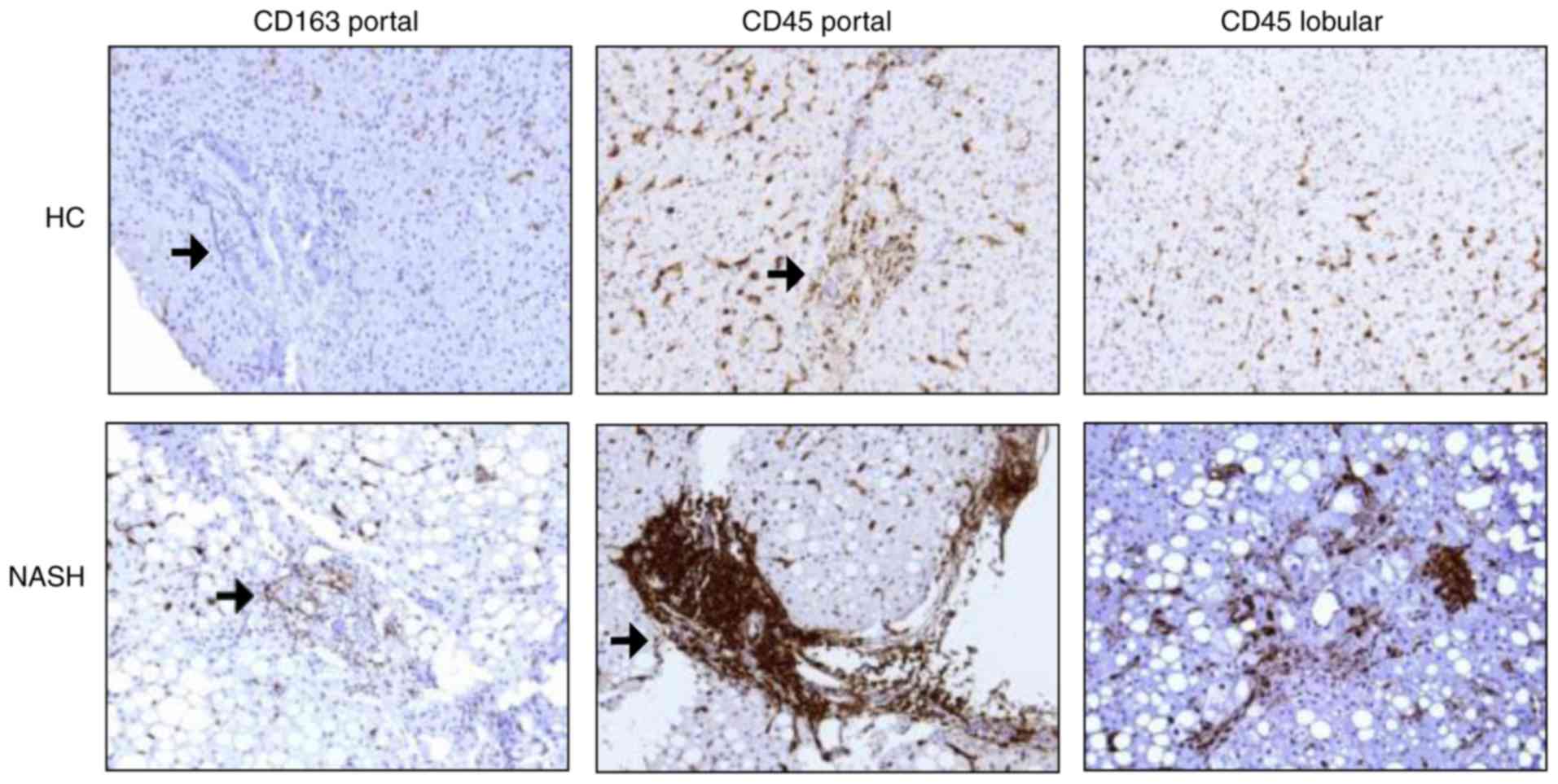
|
|
2
|
Zhu L, Baker SS, Gill C, Liu W, Alkhouri
R, Baker RD and Gill SR: Characterization of gut microbiomes in
nonalcoholic steatohepatitis (NASH) patients: A connection between
endogenous alcohol and NASH. Hepatology. 57:601–609. 2013.
View Article : Google Scholar
|
|
3
|
Wong VW, Tse CH, Lam TT, Wong GL, Chim AM,
Chu WC, Yeung DK, Law PT, Kwan HS, Yu J, et al: Molecular
characterization of the fecal microbiota in patients with
nonalcoholic steatohepatitis-a longitudinal study. PLoS One.
8:e628852013. View Article : Google Scholar
|
|
4
|
Spencer MD, Hamp TJ, Reid RW, Fischer LM,
Zeisel SH and Fodor AA: Association between composition of the
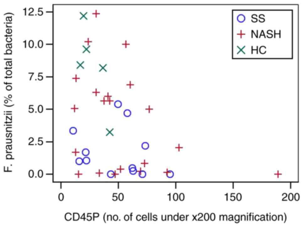
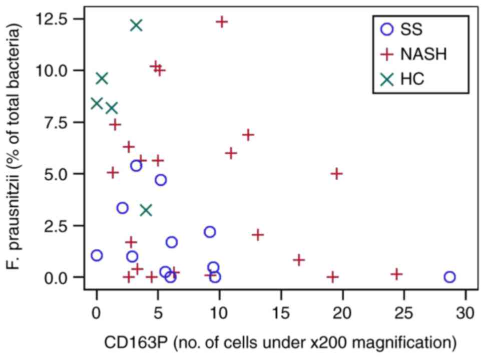
human gastrointestinal microbiome and development of fatty liver
with choline deficiency. Gastroenterology. 140:976–986. 2011.
View Article : Google Scholar :
|
|
5
|
Raman M, Ahmed I, Gillevet PM, Probert CS,
Ratcliffe NM, Smith S, Greenwood R, Sikaroodi M, Lam V, Crotty P,
et al: Fecal microbiome and volatile organic compound metabolome in
obese humans with nonalcoholic fatty liver disease. Clin
Gastroenterol Hepatol. 11:868–875. e1–e3. 2013. View Article : Google Scholar : PubMed/NCBI
|
|
6
|
Mouzaki M, Comelli EM, Arendt BM, Bonengel
J, Fung SK, Fischer SE, McGilvray ID and Allard JP: Intestinal
microbiota in patients with nonalcoholic fatty liver disease.
Hepatology. 58:120–127. 2013. View Article : Google Scholar : PubMed/NCBI
|
|
7
|
Nobili V, Cutrera R, Liccardo D, Pavone M,
Devito R, Giorgio V, Verrillo E, Baviera G and Musso G: Obstructive
sleep apnea syndrome affects liver histology and inflammatory cell
activation in pediatric nonalcoholic fatty liver disease,
regardless of obesity/insulin resistance. Am J Respir Crit Care
Med. 189:66–76. 2014.
|
|
8
|
Gadd VL, Skoien R, Powell EE, Fagan KJ,
Winterford C, Horsfall L, Irvine K and Clouston AD: The portal
inflammatory infiltrate and ductular reaction in human nonalcoholic
fatty liver disease. Hepatology. 59:1393–1405. 2014. View Article : Google Scholar
|
|
9
|
De Vito R, Alisi A, Masotti A, Ceccarelli
S, Panera N, Citti A, Salata M, Valenti L, Feldstein AE and Nobili
V: Markers of activated inflammatory cells correlate with severity
of liver damage in children with nonalcoholic fatty liver disease.
Int J Mol Med. 30:49–56. 2012.PubMed/NCBI
|
|
10
|
Torlakovic EE, Naresh K, Kremer M, van der
Walt J, Hyjek E and Porwit A: Call for a European programme in
external quality assurance for bone marrow immunohistochemistry;
report of a European Bone Marrow Working Group pilot study. J Clin
Pathol. 62:547–551. 2009. View Article : Google Scholar : PubMed/NCBI
|
|
11
|
Arrese M, Cabrera D, Kalergis AM and
Feldstein AE: Innate immunity and inflammation in NAFLD/NASH. Dig
Dis Sci. 61:1294–1303. 2016. View Article : Google Scholar : PubMed/NCBI
|
|
12
|
Caldeira PC, Oliveira e Silva KR, Vidigal
PV, Grossmann Sde M and do Carmo MA: Inflammatory cells in minor
salivary glands of patients with chronic hepatitis C:
Immunophenotype, pattern of distribution, and comparison with liver
samples. Hum Immunol. 75:422–427. 2014. View Article : Google Scholar : PubMed/NCBI
|
|
13
|
Jiang W, Wu N, Wang X, Chi Y, Zhang Y, Qiu
X, Hu Y, Li J and Liu Y: Dysbiosis gut microbiota associated with
inflammation and impaired mucosal immune function in intestine of
humans with non-alcoholic fatty liver disease. Sci Rep. 5:80962015.
View Article : Google Scholar : PubMed/NCBI
|
|
14
|
Kudo H, Takahara T, Yata Y, Kawai K, Zhang
W and Sugiyama T: Lipopolysaccharide triggered TNF-alpha-induced
hepatocyte apoptosis in a murine non-alcoholic steatohepatitis
model. J Hepatol. 51:168–175. 2009. View Article : Google Scholar : PubMed/NCBI
|
|
15
|
Thuy S, Ladurner R, Volynets V, Wagner S,
Strahl S, Königsrainer A, Maier KP, Bischoff SC and Bergheim I:
Nonalcoholic fatty liver disease in humans is associated with
increased plasma endotoxin and plasminogen activator inhibitor 1
concentrations and with fructose intake. The J Nutr. 138:1452–1455.
2008. View Article : Google Scholar : PubMed/NCBI
|
|
16
|
Alisi A, Manco M, Devito R, Piemonte F and
Nobili V: Endotoxin and plasminogen activator inhibitor-1 serum
levels associated with nonalcoholic steatohepatitis in children. J
Pediatr Gastroenterol Nutr. 50:645–649. 2010. View Article : Google Scholar : PubMed/NCBI
|
|
17
|
Ganz M and Szabo G: Immune and
inflammatory pathways in NASH. Hepatol Int. 7(Suppl 2): S771–S781.
2013. View Article : Google Scholar
|
|
18
|
Aron-Wisnewsky J, Gaborit B, Dutour A and
Clement K: Gut microbiota and non-alcoholic fatty liver disease:
New insights. Clin Microbiol Infect. 19:338–348. 2013. View Article : Google Scholar : PubMed/NCBI
|
|
19
|
Matthews DR, Hosker JP, Rudenski AS,
Naylor BA, Treacher DF and Turner RC: Homeostasis model assessment:
Insulin resistance and beta-cell function from fasting plasma
glucose and insulin concentrations in man. Diabetologia.
28:412–419. 1985. View Article : Google Scholar : PubMed/NCBI
|
|
20
|
Brunt EM, Janney CG, Di Bisceglie AM,
Neuschwander-Tetri BA and Bacon BR: Nonalcoholic steatohepatitis: A
proposal for grading and staging the histological lesions. Am J
Gastroenterol. 94:2467–2474. 1999. View Article : Google Scholar : PubMed/NCBI
|
|
21
|
Brunt EM, Kleiner DE, Wilson LA and Belt
P: Nonalcoholic fatty liver disease (NAFLD) activity score and the
histopathologic diagnosis in NAFLD: Distinct clinico-pathologic
meanings. Hepatology. 53:810–820. 2011. View Article : Google Scholar : PubMed/NCBI
|
|
22
|
Qin J, Li R, Raes J, Arumugam M, Burgdorf
KS, Manichanh C, Nielsen T, Pons N, Levenez F, Yamada T, et al: A
human gut microbial gene catalogue established by metagenomic
sequencing. Nature. 464:59–65. 2010. View Article : Google Scholar : PubMed/NCBI
|
|
23
|
Furet JP, Firmesse O, Gourmelon M,
Bridonneau C, Tap J, Mondot S, Doré J and Corthier G: Comparative
assessment of human and farm animal faecal microbiota using
real-time quantitative PCR. FEMS Microbiol Ecol. 68:351–362. 2009.
View Article : Google Scholar : PubMed/NCBI
|
|
24
|
Mariat D, Firmesse O, Levenez F, Guimarăes
V, Sokol H, Doré J, Corthier G and Furet JP: The
Firmicutes/Bacteroidetes ratio of the human microbiota changes with
age. BMC Microbiol. 9:1232009. View Article : Google Scholar : PubMed/NCBI
|
|
25
|
Da Silva HE, Teterina A, Comelli EM, Taibi
A, Arendt BM, Fischer SE, Lou W and Allard JP: Nonalcoholic fatty
liver disease is associated with dysbiosis independent of body mass
index and insulin resistance. Sci Rep. 8:14662018. View Article : Google Scholar : PubMed/NCBI
|
|
26
|
Boursier J, Mueller O, Barret M, Machado
M, Fizanne L, Araujo-Perez F, Guy CD, Seed PC, Rawls JF, David LA,
et al: The severity of nonalcoholic fatty liver disease is
associated with gut dysbiosis and shift in the metabolic function
of the gut microbiota. Hepatology. 63:764–775. 2016. View Article : Google Scholar :
|
|
27
|
Adler M, Taylor S, Okebugwu K, Yee H,
Fielding C, Fielding G and Poles M: Intrahepatic natural killer T
cell populations are increased in human hepatic steatosis. World J
Gastroenterol. 17:1725–1731. 2011. View Article : Google Scholar : PubMed/NCBI
|
|
28
|
Tajiri K, Shimizu Y, Tsuneyama K and
Sugiyama T: Role of liver-infiltrating
CD3+CD56+ natural killer T cells in the
pathogenesis of nonalcoholic fatty liver disease. Eur J
Gastroenterol Hepatol. 21:673–680. 2009. View Article : Google Scholar : PubMed/NCBI
|
|
29
|
Lotowska JM, Sobaniec-Lotowska ME and
Lebensztejn DM: The role of Kupffer cells in the morphogenesis of
nonalcoholic steatohepatitis-ultrastructural findings. The first
report in pediatric patients. Scand J Gastroenterol. 48:352–357.
2013. View Article : Google Scholar
|
|
30
|
Kazankov K, Tordjman J, Møller HJ,
Vilstrup H, Poitou C, Bedossa P, Bouillot JL, Clement K and
Grønbaek H: Macrophage activation marker soluble CD163 and
non-alcoholic fatty liver disease in morbidly obese patients
undergoing bariatric surgery. J Gastroenterol Hepatol.
30:1293–1300. 2015. View Article : Google Scholar : PubMed/NCBI
|
|
31
|
Cani PD and Delzenne NM: The role of the
gut microbiota in energy metabolism and metabolic disease. Curr
Pharm Des. 15:1546–1558. 2009. View Article : Google Scholar : PubMed/NCBI
|
|
32
|
Miquel S, Martín R, Rossi O,
Bermúdez-Humarán LG, Chatel JM, Sokol H, Thomas M, Wells JM and
Langella P: Faecalibacterium prausnitzii and human intestinal
health. Curr Opin Microbiol. 16:255–261. 2013. View Article : Google Scholar : PubMed/NCBI
|
|
33
|
Zhang M, Qiu X, Zhang H, Yang X, Hong N,
Yang Y, Chen H and Yu C: Faecalibacterium prausnitzii inhibits
interleukin-17 to ameliorate colorectal colitis in rats. PLoS One.
9:e1091462014. View Article : Google Scholar : PubMed/NCBI
|
|
34
|
Sokol H, Pigneur B, Watterlot L, Lakhdari
O, Bermúdez-Humarán LG, Gratadoux JJ, Blugeon S, Bridonneau C,
Furet JP, Corthier G, et al: Faecalibacterium prausnitzii is an
anti-inflammatory commensal bacterium identified by gut microbiota
analysis of Crohn disease patients. Proc Natl Acad Sci USA.
105:16731–16736. 2008. View Article : Google Scholar : PubMed/NCBI
|
|
35
|
Mattace Raso G, Simeoli R, Russo R, Iacono
A, Santoro A, Paciello O, Ferrante MC, Canani RB, Calignano A and
Meli R: Effects of sodium butyrate and its synthetic amide
derivative on liver inflammation and glucose tolerance in an animal
model of steatosis induced by high fat diet. PLoS One.
8:e686262013. View Article : Google Scholar : PubMed/NCBI
|
|
36
|
Kles KA and Chang EB: Short-chain fatty
acids impact on intestinal adaptation, inflammation, carcinoma, and
failure. Gastroenterology. 130(Suppl 1): S100–S105. 2006.
View Article : Google Scholar : PubMed/NCBI
|