Introduction
Cognitive impairment has been defined as a clinical
state with characteristics similar to those of normal aging and
mild dementia (1). The disorder
is commonly diagnosed in the aged population, particularly in those
suffering from Alzheimer’s disease (AD), Parkinson’s disease (PD),
vascular dementia and ischemic stroke. Furthermore, it is the major
clinical presentation in dementia with Lewy bodies (DLB) (2). Considering the aging rate of the
world’s population, cognitive disorders have become a critical
public issue as they affect the quality of life of patients, as
well as that of the caring family members (3,4).
Whereas a number of studies have been conducted to examine and
identify the individuals who are at a high risk of suffering from
cognitive dysfunction, the mechanisms of action and the
effectiveness of drugs and rehabilitation treatments remain
unclear. Given the poor effectiveness of modern medicine in
treating cognitive dysfunction, a number of patients have begun to
turn to alternative and complementary medicinal therapies for
assistance.
Amongst the different alternative medicine
therapies, acupuncture therapy is a commonly used treating modality
in China for thousands of years against diverse disorders. The
practice of acupuncture therapy encompasses a heterogeneous set of
interventions, which may take action through the induction of a
wide range of biological responses, either locally at the needle
sites or/and distally in the peripheral nerves (5). In clinical practice in China,
acupuncture has been used as an alternative therapy for patients
with AD and stroke-related dementia to improve the quality of life
and for the prevention of cognitive function decline in patients
(6-9). However, controversies on the
effectiveness of acupuncture still exist due to the lack of
evidence on the effectiveness and mechanisms of acupuncture
treatments on the nervous system. Thus, a comprehensive exploration
of the mechanisms driving the effectiveness of acupuncture on
cognitive dysfunction is imperative to promote the practical
application of acupuncture therapy.
Emerging evidence has indicated that oxidative
stress is closely related to aging and neurodegenerative diseases
(10). As previously reported by
Manczak et al (11),
during the onset of AD, the progression of dementia is associated
with neurofibrillary tangles and the overproduction of amyloid β
(Aβ) plaques. Generally, it is accepted that the progressing
accumulation of Aβ will initiate a cascade of cellular changes that
are lethal to the cells, including mitochondrial oxidative damage
(12-14). However, the mechanisms that
mediate Aβ in vivo are not yet fully understood. Recent
studies have indicated that translocase of outer mitochondrial
membrane 40 (TOMM40) regulates the influx of Aβ to the mitochondria
via the Tom40 outer membrane pore (15). Furthermore, according to Caselli
et al (16) and Roses
et al (17), TOMM40 also
influences the performance of age-related memory, which indicates
the potential of TOMM40 as a promising therapeutic target for
cognitive dysfunction (18).
Accordingly, the restoration of mitochondrial function is critical
to the successful management of nerve disorders, particularly
cognitive dysfunction. While the mechanisms involved are not yet
fully understood, various studies have reported the improving
effects of acupuncture on mitochondrial function (19,20). Based on these findings, the
current study aimed to investigate the mechanisms involved in the
interaction between acupuncture treatment and mitochondrial
function. By creating a rat model of brain ischemia induced by
middle cerebral artery occlusion (MCAO), the effects of ‘governor
vessel-unblocking and mind-regulating’ acupuncture therapy on
cognitive dysfunction in the experimental rats were examined.
Thereafter, the molecular mechanisms underlying the effects of
acupuncture treatment on mitochondrial function were examined by
focusing on the expression of TOMM40 and another translocase of
mitochondrial membrane, translocase of inner mitochondrial membrane
(TIMM17A). It was found that ‘governor vessel-unblocking and
mind-regulating’ acupuncture therapy suppressed the expression of
both indicators and inhibited the damage induced by Aβ on the
mitochondria, which could resulted in the amelioration of cognitive
dysfunction in rats.
Materials and methods
Chemicals and animals
Antibodies against TOMM40 (monoclonal; cat. no.
66658), TIMM17A (polyclonal; cat. no. 11189; Proteintech, Rosemont,
IL, USA), amyloid precursor protein (APP; monoclonal; cat. no.
ab32136), cyclooxygenase (COX, monoclonal; cat. no. ab109025), Aβ
(polyclonal; cat. no. ab2539) and Aβ oligomer were purchased from
Abcam (Cambridge, MA, USA). Antibody against GAPDH (RC-5G5) was
purchased from Aksomics Inc. (Shanghai, China). Secondary HRP goat
(BA1054) anti-rabbit and goat anti-mouse IgG (BA1051) antibodies
were purchased from Wuhan Boster Biological Technology, Ltd.
(Wuhan, China). Nimodipine (Nimotop; standard treatment for
ischemic stroke and AD) was purchased from Qilu Pharmaceutical Co.,
Ltd. (Jinan, China). A total of 24 60-day-old SPF Sprague-Dawley
rats (weighing 220±20 g, female) were obtained from Guangzhou
University of Chinese Medicine, Guangzhou, China, and housed at
room temperature (20-25°C) in a humidified chamber
(55±5%) supplemented with food and water ad libitum.
Establishment of cognitive dysfunction
model using the MCAO method
All the assays using the animals were approved by
the Institutional Animal Ethics Committee and Animal Care
Guidelines for the Care and Use of Guangdong Provincial Hospital.
In the current study, cognitive dysfunction was induced using the
MCAO method. Briefly, the rats were anesthetized using 100 mg/kg
ketamine plus 10 mg/kg xylazine administered via the intramuscular
route. The left common carotid artery (LCCA) of the rats was
exposed through a transverse neck incision, and a small incision
was then made on the LCCA through which a 0.28-mm nylon filament
was introduced into the distal left internal carotid artery for the
occlusion of left middle cerebral artery (LMCA), which would lead
to brain infarction of its supplying region. One hour after the
occlusion, the nylon filament was removed and the muscle and skin
were closed in layers. The rats in the sham-operated group
underwent the same surgical procedures but without the occlusion
treatment. The successful establishment of the model of MCAO was
assessed using the Longa score, as previously described (21) and the results are presented in
Table I. The score is explained
as follows: 0, no neurological deficit symptoms, activity is
completely normal; 1, mild neurological deficit, unable to fully
extend the opposite front paw; 2, moderate neurological deficit,
turning to the opposite side when crawling; 3, severe neurological
deficit, tilt towards the opposite side when crawling; 4, loss of
consciousness, inability to crawl; 5, death. Following wound
closure, the rats were housed for 10 days prior to treatment with
acupuncture. The rats were deeply anesthetized with an
intraperitoneal injection of pentobarbital sodium (100 mg/kg) and
the brains were removed for analysis.
 | Table IAssessment of the establishment of
the MCAO model using the Longa score. |
Table I
Assessment of the establishment of
the MCAO model using the Longa score.
| Group | Longa score
|
|---|
| Pre-surgery | Post-surgery |
|---|
| Sham | 0.0±0.0 | 0.0±0.0 |
| MCAO | 0.0±0.0 |
1.33±0.49a,b |
‘Governor vessel-unblocking and
mind-regulating’ acupuncture therapy and animal grouping
‘Governor vessel-unblocking and mind-regulating’
acupuncture therapy is based on the theory of acupoints on the Du
channel in Traditional Chinese Medicine (TCM). In the current
study, all the acupoints were recognized according to a previous
publication (22) and the
treatment was performed by a senior practitioner. Ten days after
the model of MCAO was induced, the rats were fastened in a
restrainer for a long period for acclimatization, which was
validated by the absence of struggling. The manual twist
acupunctures were then needled at the Baihui, Dazhui, Renzhong and
Fengfu acupuncture points for 20 min per day for 15 days.
To assess the effects of acupuncture treatment on
cognitive dysfunction, 40 rats were randomly divided into 4 groups
(10 in each group) as follows: i) The sham-operated group, in which
the rats underwent the same procedures as those in the surgery
group only without occlusion of the arteries; ii) the MCAO group,
in which the rats were subjected to MCAO for the induction of
cognitive dysfunction; iii) the MCAO + Acupuncture group, in which
the rats with cognitive dysfunction were treated with ‘governor
vessel-unblocking and mind-regulating’ acupuncture therapy for 15
days; and iv) the MCAO + Nimotop group, in which the rats with
cognitive dysfunction were treated with Nimotop (20 mg/kg body
weight) per day for 15 days. Upon completion of the culture, all
the rats were subjected to the Morris water maze (MWM) test for the
evaluation of their cognitive function. Two days after the MWM
test, all the rats were sacrificed to collect cortical layer and
hippocampus tissues, as well as mitochondria in brain tissues for
subsequent assays.
MWM test
The MWM was used to test the learning and memorizing
abilities of the rats. The assays were performed routinely as
reported previously (23,24) with two investigators blinded to
the experimental design. The test included a 1-day probe trial and
a 2-day visible platform trial. Briefly, for visible platform trail
in 60 sec, the rats were allowed to swim for 60 sec before getting
to the platform for 4 times the first day and 1 time the second
day. If the rats failed, the investigator would help the rats to
stay on the platform for 10 sec before another test. For probe
trial in 120 sec, the time through the quadrant of the former
platform position was measured.
H&E and Nissl staining
The histological changes in the sections of brain
tissues from the different groups were observed using H&E
staining. Briefly, the tissues were fixed in Bouin solution (4%
formaldehyde), dehydrated using alcohol and vitrified in
dimethylbenzene. The samples were then embedded, sectioned and
stained with hematoxylin at room temperature for 2 min and then
with eosin for 3-5 sec. The results were studied under a microscope
(CX41; Olympus Corp., Tokyo, Japan) at magnification, ×400.
Following H&E staining, the nuclei in tissue were stained blue
by hematoxylin and cytoplasms were stained red by eosin. The
effects of acupuncture treatment on neurons in brain tissues were
detected using Nissl staining following standard procedures.
Terminal-deoxynucleoitidyl transferase
mediated nick-end labeling (TUNEL) staining
Cell apoptotic rates were determined using TUNEL
staining. Briefly, the brain sections were permeabilized with 0.1%
Triton X-100 at room temperature for 8 min. The sections were then
washed with PBS buffer prior to incubation in 3%
H2O2 for 10 min at room temperature.
Following 3 5-min washes with PBS buffer, the sections were covered
with TUNEL reaction solution and incubated at 37°C for 1
h in a humidified chamber in the dark. The tissues were then washed
and stained with 4, 6-diamino-2-phenyl indole (DAPI) for 5 min at
room temperature and imaged using a fluorescence microscope (FV10i;
Olympus Corp.) at magnification, ×400.
Immunohistochemical detection
For immunohistochemical assay, the tissue slides
were placed in 60°C overnight prior to incubation with
dimethylbenzene for dewaxing. The slides from the different groups
were fixed using methanol solution with 3%
H2O2 and blocked with 1% BSA for 30 min at
37°C and incubated with primary antibodies against
TOMM40 (1:400), TIMM17A (1:200), Aβ (1:500) and COX (1:500) at
4°C overnight. Secondary antibodies (IgG HRP; 1:3,000;
cat. no. ab97051; Abcam) were added to the slides and placed at
37°C for 30 min before another 4 cycles of a PBS wash.
DAB was then added to the slides and allowed to react for 3-10 min
until the reaction was terminated by ddH2O. The slides
were re-stained using hematoxylin and dehydrated. The results were
recorded using a microscope (FV10i; Olympus Corp.) at
magnification, ×400.
Enzyme-linked immunosorbent assay
(ELISA)
The production of adenosine triphosphate (ATP),
nitric oxide (NO), inducible NO synthase (iNOS) and superoxide
dismutase (SOD) in the brain tissues of the different groups was
measured using respective ELISA kits (Wuhan Boster Biological
Technology, Ltd., China) according to the manufacturer’s
instructions.
Reverse transcription-quantitative PCR
(RT-qPCR)
Total RNA from the different samples was extracted
using the RNA Purified Total RNA Extraction kit according to the
manufacturer’s instructions (BioTeke, Wuxi, China). Total RNA was
reverse transcribed into cDNA templates using Super M-MLV reverse
transcriptase (BioTeke). The final RT-PCR reaction mixture of
volume 20 µl consisted of 10 µl of Bestar®
SUBR-Green qPCR master Mix, 0.5 µl of each primer (TOMM40
forward, 5′-CTT CCT CTT CAA AGG CTC TGT-3′ and reverse, 5′-ACT TAT
TCT TGC GGT GGT TC-3′; TIMM17A forward, 5′-CTG GCA GCA AGA AAT
GGA-3′ and reverse, 5′-AGG CAA ACC TGG TCA ACA-3′; APP forward,
5′-CCA CAT CGT GAT TCC TT ACC-3′ and reverse, 5′-CCA GAC ATC GGA
GTC GTC C-3′; COX forward, 5′-AGC CAT TTC TAC TTC GGT GTG-3′ and
reverse, 5′-ATT GGT GCC CTT GTT CAT CT-3′; and GAPDH forward,
5′-CCT CGT CTC ATA GAC AAG ATG GT-3′ and reverse, 5′-GGG TAG AGT
CAT ACT GGA ACA TG-3′), 2 µl of the cDNA template and 7
µl of Rnase free H2O. The amplifi-cation
parameters were set as follows: Denaturation at 94°C for 2 min,
followed by 40 cycles at 94°C for 20 sec, 58°C for 20 sec, and 72°C
for 20 sec. The relative mRNA expression levels were calculated
with ExicyclerTM 96 (Bioneer Corporation, Daejeon, Korea) according
to the expression of 2−ΔΔcq (25).
Western blot analysis
Total protein product from the different groups was
extracted using the Total Protein Extraction kit according to the
manufacturer’s instructions (Wanleibio, Beijing, China) and protein
concentrations were determined using the BCA method. A total of 20
µl of protein (40 µg) was subjected to 10% sodium
dodecylsulfate polyacrylamide gel electrophoresis (SDS-PAGE) and
transferred onto polyvinyli-dene difluoride (PVDF) membranes. The
membranes were then washed with TTBS for 5 min prior to incubation
in skim milk powder solution for 1 h. Primary antibodies against
TOMM40 (1:2,000), TIMM17A (1:1,000), Aβ (1:800), Aβ oligomer
(1:100), COX (1:800), and GAPDH (1:10,000) were incubated with the
membranes at 4°C overnight and secondary HRP goat anti-rabbit
antibodies (1:20,000) were incubated with the membranes for 45 min
at 37°C. Following additional 6 washes using TTBS, the blots were
developed using Beyo ECL Plus reagent and the results were detected
using the Gel Imaging System. The relative protein expression
levels were calculated with Gel-Pro-Analyzer (Media Cybernetics,
Rockville, MD, USA).
Flow cytometric analysis
Mitochondria were isolated from the rat hippocampal
tissue using a Tissue Mitochondria Isolation kit (#C3606; Beyotime
Institute of Biotechnology, Jiangsu, China). The isolated
mitochondria of the hippocampus were stained with a cationic
mitochondrion-specific dye, JC-1 (Beyotime Institute of
Biotechnology) for 15 min at 37°C, and washed twice with PBS. The
fluorescence was analyzed using a flow cytometer (FACSCalibur; BD
Biosciences, San Jose, CA, USA).
Reactive oxygen species (ROS) production in the rat
hippocampal tissues was assayed using the fluorescent probe, DHE
(Vigorous Biotechnology, Beijing, China). The rat hippocampal
tissues obtained from the 4 experimental groups were washed, cut
into sections and homogenized. The tissue homogenate was incubated
with the fluorescent probe, DHE, for 20 min at 37°C. Finally, the
fluorescence was analyzed using a flow cytometer (FACSCalibur; BD
Biosciences) according to the manufacturer’s instructions.
Statistical analysis
All the data are expressed as the means ± standard
deviation. For the behavior assay, each group was represented by 10
replicates. For the histological, biochemical and molecular
detections, each group was represented by 6 replicates. One-way
ANOVA and the post hoc LSD test was performed using the general
liner model. P<0.05 was considered to indicate a statistically
significant difference. All statistical analyses were conducted
using SPSS version 19.0 (IBM, Armonk, NY, USA).
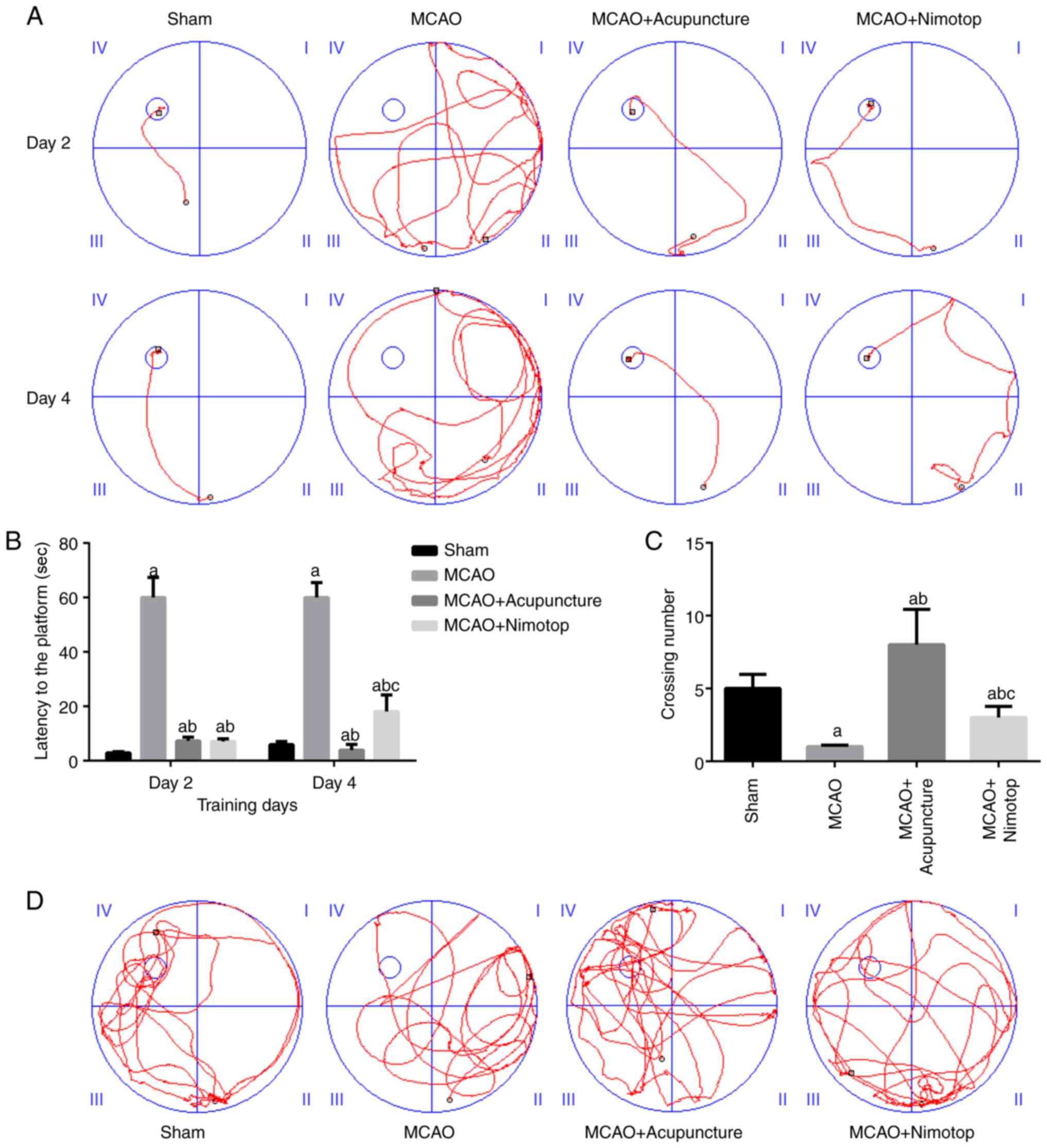
Results
‘Governor vessel-unblocking and
mind-regulating’ acupunc- ture therapy improves the learning and
memorizing ability of rats subjected to MCAO
The results of the MWM test revealed that
acupuncture treatment improved the learning and memorizing ability
of the rats with cognitive dysfunction. For the visible platform
trial, grouping acted as an independent factor that influenced the
latency of the rats. As shown in Fig.
1A and B, the latency time of the rats in the MCAO +
Acupuncture group was lower than that of the rats in the MCAO
group, and the difference was statistically significant
(P<0.05), thereby representing the restoration of the cognitive
function of the rats. The results of the probe trail confirmed the
conclusion of the visible platform trial, as the rats in the MCAO +
Acupuncture group had a significantly higher crossing number than
the rats in the other 3 groups (P<0.05) (Fig. 1C and D). Thus, ‘governor
vessel-unblocking and mind-regulating’ acupuncture therapy
alleviated the cognitive dysfunction of the model rats.
‘Governor vessel-unblocking and
mind-regulating’ acupuncture therapy improves histological changes
in brain tissues of MCAO rats
The histological changes in the brain tissues were
detected following the induction of ischemia and acupuncture
treatment. As shown in Fig. 2,
the induction of cognitive dysfunction was associated with aging
(cytoplasm stained dark pink by H&E staining and stained blue
by Nissl staining) and the deterioration of the regular structure
of the cells when compared with the sham-operated group. Treatment
with both ‘governor vessel-unblocking and mind-regulating’
acupuncture and Nimotop alleviated the negative effects of ischemia
on the brain cells, with more cells retaining their normal shape
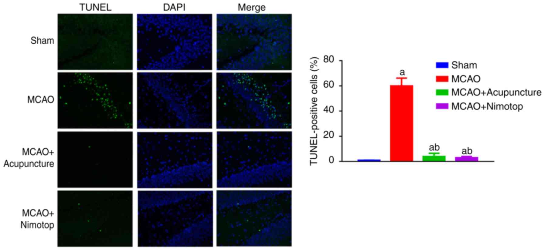
and structure (Fig. 2). In
addition, as illustrated by TUNEL staining, a significantly high
number of apoptotic cells (stained green) was detected in the MCAO
group, and the impairments on brain tissues due to ischemia were
alleviated by acupuncture treatment (Fig. 3).
‘Governor vessel-unblocking and
mind-regulating’ acupuncture therapy suppresses oxidative stress in
the brain tissues of rats subjected to MCAO
Based on the results of ELISA, the level of ATP in
the brain tissues of the model rats was suppressed after the
induction of cognitive dysfunction; however, ‘governor
vessel-unblocking and mind-regulating’ acupuncture therapy improved
the synthesis of ATP (Fig. 4A).
Moreover, the decreased production of SOD was also restored by
‘governor vessel-unblocking and mind-regulating’ acupuncture
therapy, and the difference between the MCAO group and MCAO +
Acupuncture group was statistically significant (P<0.05;
Fig. 4B). The levels of factors
which are upregulated during ischemia and which contribute to the
pathogenesis of neurodegenerative disorders, including NO and iNOS,
were suppressed by ‘governor vessel-unblocking and mind-regulating’
acupuncture therapy (Fig. 4C and
D). Taken together, the above-mentioned results suggest the
potential of ‘governor vessel-unblocking and mind-regulating’
acupuncture therapy to relieve brain tissues from chronic stress
due to ischemia.
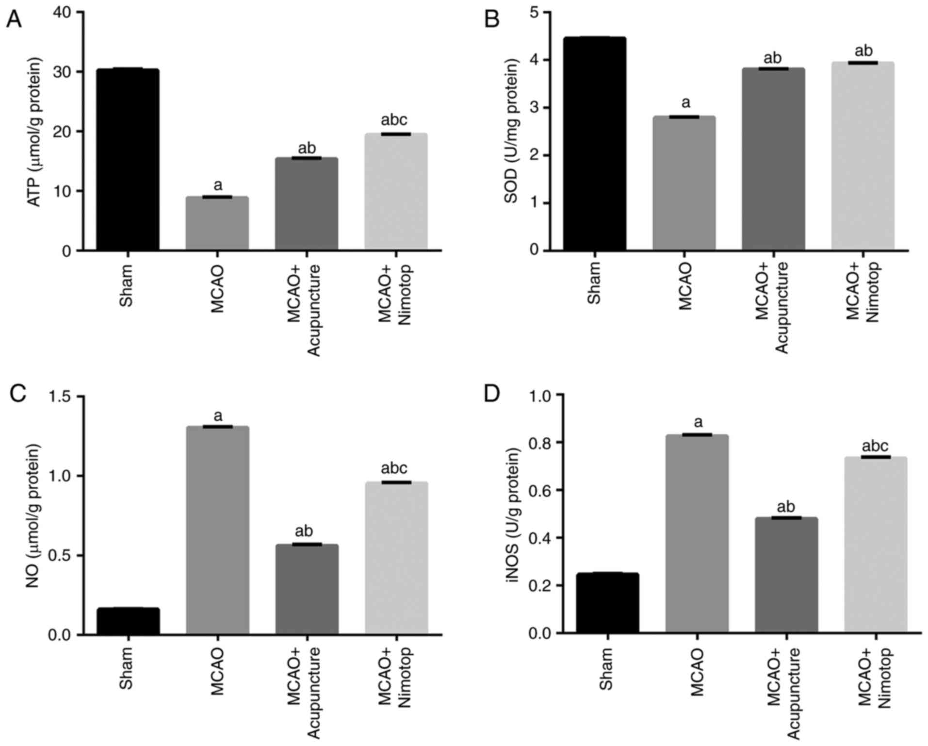 | Figure 4‘Governor vessel-unblocking and
mind-regulating’ acupuncture therapy suppresses oxidative stress
and restores energy production in brain tissues. (A-D) Quantitative
analysis results of the production of ATP, SOD, NO and iNOS. The
production of ATP and SOD was inhibited by ischemia and restored by
‘governor vessel-unblocking and mind-regulating’ acupuncture
therapy, while the production of NO and iNOS was induced by
ischemia and suppressed by ‘governor vessel-unblocking and
mind-regulating’ acupuncture therapy. aP<0.05,
significantly different from the sham-operated (sham) group;
bP<0.05, significantly different from the MCAO group;
cP<0.05, significantly different from the MCAO +
Acupuncture group. Each group is represented by 10 replicates.
Error bars stand for standard deviation. MCAO, middle cerebral
artery occlusion; ATP, adenosine triphosphate; SOD, superoxide
dismutase; NO, nitric oxide; iNOS, and inducible nitric oxide
synthase. |
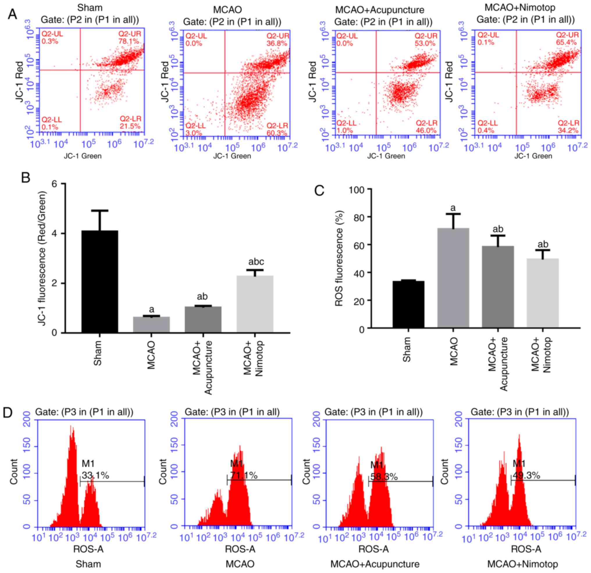
‘Governor vessel-unblocking and
mind-regulating’ acupuncture therapy improves membrane potential
and oxidative stress in the mitochondria in brain tissues of rats
subjected to MCAO
The induction of cognitive disorders is closely
associated with the dysfunction of the mitochondria in brain cells.
In this study, the results from flow cytometric analysis revealed
that the induction of cognitive dysfunction initiated membrane
depolarization in the brain mitochondria (Fig. 5A and B): Low levels of JC-1 were
measured in the MCAO group. Following ‘governor vessel-unblocking
and mind-regulating’ acupuncture therapy, the membrane potential
returned to a relatively normal level and the effect was more
potent compared to treatment with Nimotop. Furthermore, the
oxidative stress induced by ischemia was also alleviated by
‘governor vessel-unblocking and mind-regulating’ acupuncture
therapy, with the increased levels of ROS being inhibited in the
MCAO + Acupuncture group (Fig. 5C and
D).
‘governor vessel-unblocking and
mind-regulating’ acupunc- ture therapy downregulates the expression
of TOMM40 and TIMM17A
A previous study demonstrated that the afferent
impulses induced by acupuncture are mainly transmitted by Aβ and Aδ
fibres (26). In the current
study, the theory was taken one step further by focusing on the
mediators of Aβ, including TOMM40 and TIMM17A. As illustrated by
immunochemical detection, following the establishment of the
cognitive dysfunction model, the production and distribution of
TOMM40, TIMM17A and Aβ were all increased in the rat brain tissues
(Fig. 6A-C), while the expression
of COX was downregulated (Fig.
6D). As an indicator, positive cells were stained brown. With
‘governor vessel-unblocking and mind-regulating’ acupuncture
therapy, the expression levels of these indicators were reversed.
The change patterns of these indicators were synchronized with the
behavioral improvement of models and function restoration of brain
mitochondrion. Furthermore, the mechanisms involved in the effects
of ‘governor vessel-unblocking and mind-regulating’ acupuncture
therapy were validated by RT-qPCR (Fig. 7) and western blot analysis
(Fig. 8). Apart from the
quantification of the expression of TOMM40, TIMM17A and COX, the
following two assays also detected the precursor of Aβ and APP. It
was observed that the enhanced expression of Aβ led to the
downregulation of APP, which was inhibited by ‘governor
vessel-unblocking and mind-regulating’ acupuncture therapy. Given
the effects of ‘governor vessel-unblocking and mind-regulating’
acupuncture therapy on TOMM40 and TIMM17A expression, representing
its potential for modulating the influx of Aβ into the
mitochondria, the effects of ‘governor vessel-unblocking and
mind-regulating’ acupuncture therapy on the transition between Aβ
and APP may also infer the function of ‘governor vessel-unblocking
and mind-regulating’ acupuncture therapy to restrict the synthesis
of Aβ as well. Moreover, the administration of acupuncture to the
normal rats had no effect on the expression of COX (data not
shown). Combined with the results of the behavioral tests, it can
be concluded that ‘governor vessel-unblocking and mind-regulating’
acupuncture therapy had minial side-effects on the normal
biological functions of the brain tissues of rats.
 | Figure 6‘Governor vessel-unblocking and
mind-regulating’ acupuncture therapy inhibits the expression of
TOMM40, TIMM17A and Aβ, while it increases the expression of COX.
(A-D) Immunochemical detection (magnification, ×400) of TOMM40,
TIMM17A and Aβ and COX levels and quantification of these levels.
aP<0.05, significantly different from the
sham-operated (sham) group; bP<0.05, significantly
different from the MCAO group; cP<0.05, significantly
different from the MCAO + Acupuncture group. Each group is
represented by 10 replicates. Error bars stand for standard
deviation. MCAO, middle cerebral artery occlusion; TOMM40,
translocase of outer mitochondrial membrane 40; TIMM17A,
translocase of inner mitochondrial membrane 17A; Aβ, amyloid β;
COX, cyclooxygenase. |
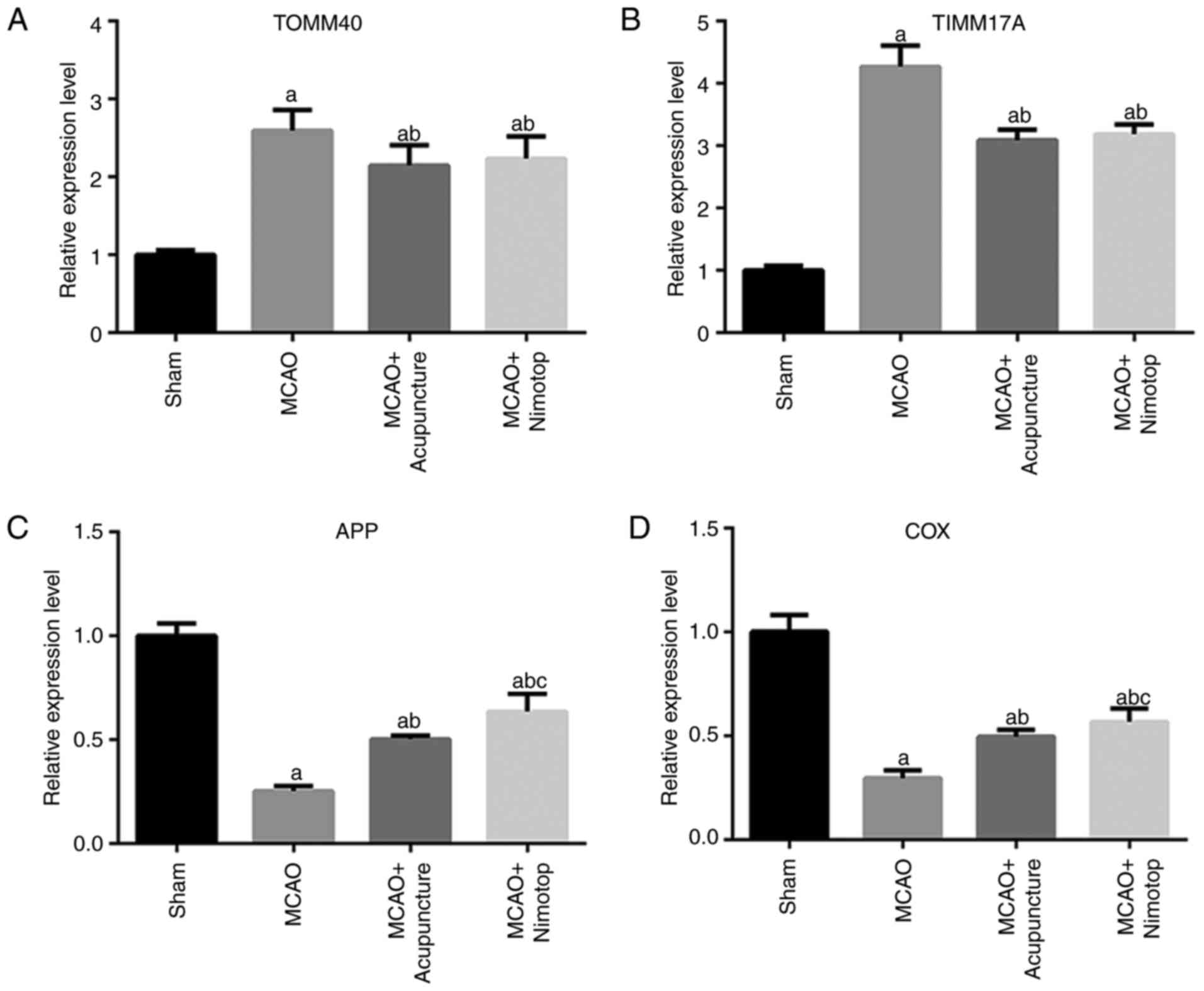 | Figure 7‘Governor vessel-unblocking and
mind-regulating’ acupuncture therapy exerts its effects on
cognitive dysfunction by downregulating the expression of TOMM40
and TIMM17A. (A-D) Quantitative analysis results of RT-qPCR
validation of TOMM40, TIMM17A, Aβ and COX expression, respectively.
The induction of ischemia increased the levels of TOMM40 and
TIMM17A, and decreased the levels of APP and COX. ‘Governor
vessel-unblocking and mind-regulating’ acupuncture therapy reversed
the expression patterns of all the indicators.
aP<0.05, significantly different from the
sham-operated (sham) group; bP<0.05, significantly
different from the MCAO group; cP<0.05, significantly
different from the MCAO + Acupuncture group. Each group is
represented by 10 replicates. Error bars stand for standard
deviation. MCAO, middle cerebral artery occlusion; TOMM40,
translocase of outer mitochondrial membrane 40; TIMM17A,
translocase of inner mitochondrial membrane 17A; APP, amyloid
precursor protein; COX, cyclooxygenase. |
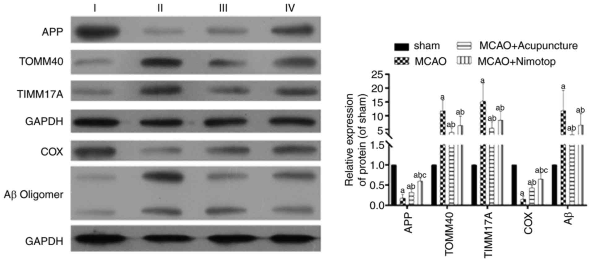 | Figure 8Representative images of western blot
analysis detections of TOMM40, TIMM17A, APP, Aβ and COX. The
induction of ischemia increased the level of TOMM40, TIMM17A and Aβ
and decreased the level of APP and COX. ‘Governor vessel-unblocking
and mind-regulating’ acupuncture therapy reversed the expression
patterns of all the indicators. The lanes in the blots are labeled
as follows: I, sham-operated (sham) group; II, MCAO group; III,
MCAO + Acupuncture group; IV, MCAO + Nimotop group.
aP<0.05, significantly different from the
sham-operated (sham) group; bP<0.05, significantly
different from the MCAO group; cP<0.05, significantly
different from the MCAO + Acupuncture group. Each group is
represented by 10 replicates. Error bars stand for standard
deviation. MCAO, middle cerebral artery occlusion; TOMM40,
translocase of outer mitochondrial membrane 40; TIMM17A,
translocase of inner mitochondrial membrane 17A; APP, amyloid
precursor protein; COX, cyclooxygenase. |
Discussion
As a traditional and potent therapeutic strategy,
acupuncture has been used in the treatment of various diseases in
China for centuries (27). As
regards brain disorders, numerous studies have been conducted to
validate its potential for improvement of not only the function of
brain cells, but also the cognitive function of patients (1,2,28).
Being recognized as the one of the most severe impairments of brain
disorders, cognitive dysfunction severely affects the quality of
life of patients with AD, PD and vascular dementia, and poses a
great threat to public health worldwide. Fortunately, the
effectiveness of acupuncture treatment on cognitive dysfunction in
recent years (28-30) offers an inspirable hint that the
treatment can improve the prognosis of patients with brain
disorders. However, due to the lack of a scientific explanation
that meets the criteria of natural science and evidence-based
medicine, acupuncture treatment has been classified as an
alternative medicine, the effect of which is attributed to a
placebo (31). Moreover, recent
studies on acupuncture treatment have focused on the description of
the behavioral changes of patients or model animals instead of
exploring the underlying molecular mechanisms associated with the
treatment (29,30). To promote the application of the
therapy in clinical treatment and its acceptance by modern
medicine, comprehensive investigations on the pathways through
which acupuncture treatment might take its action is in demand.
Therefore, in the current study, the effect of ‘governor
vessel-unblocking and mind-regulating’ acupuncture therapy on the
expression levels of indicators related to mitochondrial function
in brain tissues were examined. The results revealed that the
treatment inhibited the expression levels of TOMM40 and TIMM17A, as
well as the accumulation of Aβ in the brain mitochondria.
In the theory of TCM, cognitive dysfunction results
from the deficiency and/or dysfunction of ‘Yang Qi’, the traveling
of which in the human body depends on the clear passage in the ‘Du’
channel (32). Therefore,
therapies that can improve the function of the ‘Du’ channel may
contribute to the amelioration of cognitive dysfunction.
Accordingly, ‘governor vessel-unblocking and mind-regulating’
acupuncture therapy was developed and employed in the current study
for cognitive improvement in a rat model. Theoretically, the method
stimulates the points belonging to the ‘Du’ channel in the brain
using acupuncture, opening up the ‘Du’ channel and restoring neural
function.
The effects of ‘governor vessel-unblocking and
mind-regulating’ acupuncture therapy were firstly validated in a
behavioral test. The MWM is a powerful tool for assessing spatial
learning and memory in rats. In the current study, rats in the MCAO
group exhibited cognitive impairments in the MWM test, i.e., they
reached the platform with a longer latency and remained in the
quadrant where the platform had been previously located with less
time, indicating the deficiency in learning and memorizing ability.
Following acupuncture treatment, the rats in the MCAO + Acupuncture
group exhibited an improvement in the acquisition in the visible
platform trial and retention in the probe trial: They reached the
platform with lower latency and crossed over former platform
position more frequently. Apart from the MWM test, the effects of
‘governor vessel-unblocking and mind-regulating’ acupuncture
therapy were also verified by H&E staining, in which the brain
cells of rats in the MCAO + Acupuncture group retained their normal
structure compared with those of the rats in the MCAO group. It was
more inspirable to record that the rats in MCAO + Acupuncture group
even performed better in the MWM test than those in the MCAO +
Nimotop group, indicating a potent treatment effect of ‘governor
vessel-unblocking and mind-regulating’ acupuncture therapy for
cognitive dysfunction.
In addition to the ameliorating effects on the brain
cell structure and brain function of model rats, ‘governor
vessel-unblocking and mind-regulating’ acupuncture therapy
suppressed oxidative stress in the brain tissues of ischemic rats.
Following treatment with ‘governor vessel-unblocking and
mind-regulating’ acupuncture, the production of ATP and SOD was
augmented, while the production of NO and iNOS was decreased.
Oxidative stress plays a vital role in neuronal damage and in
cognitive deficits in the elderly (33) and is frequently recorded along
with symptoms in patients with AD and PD (34). As the major source of free
radicals in cells, the mitochondria are easily affected by the
oxidative stress associated with brain disorders (35,36). In the current study, the membrane
potential of the mitochondria was depolarized following the
induction of cognitive dysfunction. Furthermore, the production of
ROS in the mitochondria was increased, confirming the functional
dysregulation of the mitochondria induced by ischemia. The
abnormality of mitochondrial function was accompanied by
impairments in energy production (less ATP production) in the brain
tissues of the model rats, which was particularly important in view
of the significant mental retardation characteristic of patients
with AD, PD and vascular dementia (29). Following treatment with ‘governor
vessel-unblocking and mind-regulating’ acupuncture, the membrane
potential and ROS production in the mitochondria were restored to
their normal levels. Such a modulating effect of acupuncture on
mitochondrial function is vital for targeted interventions on the
mitochondria in delaying AD progression in elderly individuals
(11,37,38). The possible interaction between
acupuncture treatments and mitochondrial functions was also
reported by Lu. They showed that acupuncture can induce
afferent impulses transmitted by Aβ fibres (39).
Aβ is generated by the abnormal processing of APP,
which constitutes a major component of neurotic plaques or amyloid
deposits found in brains affected by AD and PD (40,41). The accumulation of Aβ in the
mitochondria suppresses the activity of COX and impairs
mitochondrial metabolism (11).
The results were verified in the current study by detections at
molecular levels. The expression of COX was decreased both at the
mRNA and protein levels following the induction of cognitive
dysfunction, whereas the expression of Aβ increased. We also
demonstrated the mechanisms involved in the effects of acupuncture
on cognitive dysfunction. It has been hypothesized that the
mitochondria exert neurotoxicity by allowing the influx of Aβ to
cells via the Tom40 import pore (18). The pore is governed by TOMM40 and
is essential for mitochondrial survival (42,43). Consequently, the expression of
TOMM40 was detected in the current study. In addition, for the
exploration of novel biomarkers associated with the dysregulation
of Aβ, the expression of TIMM17A was also primarily assessed. It
was found that both indicators were upregulated in the rats with
cognitive dysfunction. Following treatment with ‘governor
vessel-unblocking and mind-regulating’ acupuncture, the levels of
TOMM40 and TIMM17A were suppressed with the decrease in the Aβ
levels. Previous studies have indicated that TOMM40 influences the
Aβ influx in an ApoE-dependent manner (44-46). However, no study to date has yet
reported the association between Aβ and TIMM17A, at least to the
best of our knowledge. The indicator is only proven to be
associated with the adverse pathological and clinical outcomes in
human breast cancer (47). Based
the results of the current study, TIMM17A may also participate in
the effects of acupuncture on cognitive dysfunction, although the
detail mechanisms warrant further investigation.
Conclusively, in this study, we demonstrated that
treatment with ‘governor vessel-unblocking and mind-regulating’
acupuncture contributed to the amelioration of cognitive
dysfunction in rats subjected to MCAO. Following the administration
of acupuncture, the accumulation of Aβ in the brain mitochondria
was inhibited. The process was mediated through the suppression on
TOMM40 and TIMM17A by acupuncture stimulation. The findings
outlined in this study provide insight into the molecular
mechanisms associated with the treatment of acupuncture. ‘governor
vessel-unblocking and mind-regulating’ acupuncture therapy not only
influenced the behavior of the rats subjected to MCAO, but also
modulated the signaling pathway involved in the pathogenesis of
cognitive dysfunction. Our results may prove to be an inspiration
for the scientific explanation of effects of acupuncture treatment.
Further comprehensive studies are required to promote the
application of this treatment modality in clinical practice in the
future.
Acknowledgments
Not applicable.
Funding
The present study was supported by the Guangdong
Province Enterprise Technology R&D and appreciation
transformation special fund project plan (2013B021800211).
Availability of data and materials
All data generated or analyzed during this study are
included in this published article or are available from the
corresponding author on reasonable request.
Authors’ contributions
XS designed the research, collected the data, and
wrote the draft. ZW collected and analyzed the data. FM collected
the data. ZF performed the data analysis. SD designed the
experiment and revised the draft. HQ designed the research. JZ
wrote the draft and designed the research. All authors have read
and approved the final manuscript.
Ethics approval and consent to
participate
All the assays using the animals were approved by
the Institutional Animal Ethics Committee and Animal Care
Guidelines for the Care and Use of Guangdong Provincial
Hospital.
Patient consent for publication
Not applicable.
Competing interests
The authors declare that they have no competing
interests.
References
|
1
|
Lu X, Hongcai S, Jiaying W, Jing H and Jun
X: Assessing the quality of reports about randomized controlled
trials of acupuncture treatment on mild cognitive impairment. PLoS
One. 6:e169222011. View Article : Google Scholar : PubMed/NCBI
|
|
2
|
Leung MC, Yip KK, Ho YS, Siu FK, Li WC and
Garner B: Mechanisms underlying the effect of acupuncture on
cognitive improvement: A systematic review of animal studies. J
Neuroimmune Pharmacol. 9:492–507. 2014. View Article : Google Scholar : PubMed/NCBI
|
|
3
|
Aggarwal NT, Tripathi M, Dodge HH, Alladi
S and Anstey KJ: Trends in Alzheimer’s disease and dementia in the
Asian-pacific region. Int J Alzheimers Dis. 2012.171327:2012.
|
|
4
|
World Health Organization and Alzheimer’s
Disease International Dementia: A public health priority. World
Health Organization; Geneva: 2012
|
|
5
|
Rabinstein AA and Shulman LM: Acupuncture
in clinical neurology. Neurologist. 9:137–148. 2003. View Article : Google Scholar : PubMed/NCBI
|
|
6
|
Yu J, Zhang X, Liu C, Meng Y and Han J:
Effect of acupuncture treatment on vascular dementia. Neurol Res.
28:97–103. 2006. View Article : Google Scholar : PubMed/NCBI
|
|
7
|
Chou P, Chu H and Lin JG: Effects of
electroacupuncture treatment on impaired cognition and quality of
life in Taiwanese stroke patients. J Altern Complement Med.
15:1067–1073. 2009.
|
|
8
|
Shen PF, Kong L, Ni LW, Guo HL, Yang S,
Zhang LL, Zhang ZL, Guo JK, Xiong J, Zhen Z and Shi XM: Acupuncture
intervention in ischemic stroke: A randomized controlled
prospective study. Am J Chin Med. 40:685–693. 2012. View Article : Google Scholar : PubMed/NCBI
|
|
9
|
Shi GX, Liu CZ, Li QQ, Zhu H and Wang LP:
Influence of acupuncture on cognitive function and markers of
oxidative DNA damage in patients with vascular dementia. J
Traditional Chin Med. 32:199–202. 2012. View Article : Google Scholar
|
|
10
|
Wang J, Markesbery WR and Lovell MA:
Increased oxidative damage in nuclear and mitochondrial DNA in mild
cognitive impairment. J Neurochem. 96:825–832. 2006. View Article : Google Scholar : PubMed/NCBI
|
|
11
|
Manczak M, Anekonda TS, Henson E, Park BS,
Quinn J and Reddy PH: Mitochondria are a direct site of Aβ
accumulation in Alzheimer’s disease neurons: Implications for free
radical generation and oxidative damage in disease progression. Hum
Mol Genetics. 15:1437–1449. 2006. View Article : Google Scholar
|
|
12
|
Pappolla MA, Omar RA, Kim KS and Robakis
NK: Immunohistochemical evidence of oxidative [corrected] stress in
Alzheimer’s disease. Am J Pathol. 140:621–628. 1992.PubMed/NCBI
|
|
13
|
Bozner P, Grishko V, LeDoux SP, Wilson GL,
Chyan YC and Pappolla MA: The amyloid beta protein induces
oxidative damage of mitochondrial DNA. J Neuropathol Exp Neurol.
56:1356–1362. 1997. View Article : Google Scholar : PubMed/NCBI
|
|
14
|
Pappolla MA, Chyan YJ, Omar RA, Hsiao K,
Perry G, Smith MA and Bozner P: Evidence of oxidative stress and in
vivo neurotoxicity of beta-amyloid in a transgenic mouse model of
Alzheimer’s disease: A chronic oxidative paradigm for testing
antioxidant therapies in vivo. Am J Pathol. 152:871–877.
1998.PubMed/NCBI
|
|
15
|
Yu CE, Seltman H, Peskind ER, Galloway N,
Zhou PX, Rosenthal E, Wijsman EM, Tsuang DW, Devlin B and
Schellenberg GD: Comprehensive analysis of APOE and selected
proximate markers for late-onset Alzheimer’s disease: Patterns of
linkage disequilibrium and disease/marker association. Genomics.
89:655–665. 2007. View Article : Google Scholar : PubMed/NCBI
|
|
16
|
Caselli RJ, Dueck AC, Huentelman MJ, Lutz
MW, Saunders AM, Reiman EM and Roses AD: Longitudinal modeling of
cognitive aging and the TOMM40 effect. Alzheimers Dement.
8:490–495. 2012. View Article : Google Scholar : PubMed/NCBI
|
|
17
|
Roses AD, Lutz MW, Crenshaw DG, Grossman
I, Saunders AM and Gottschalk WK: TOMM40 and APOE: Requirements for
replication studies of association with age of disease onset and
enrichment of a clinical trial. Alzheimers Dement. 9:132–136. 2013.
View Article : Google Scholar : PubMed/NCBI
|
|
18
|
Ferencz B, Karlsson S and Kalpouzos G:
Promising genetic biomarkers of preclinical Alzheimer’s disease:
The influence of APOE and TOMM40 on brain integrity. Int J
Alzheimers Dis. 2012.421452:2012.
|
|
19
|
Zhang X, Wu B, Nie K, Jia Y and Yu J:
Effects of acupuncture on declined cerebral blood flow, impaired
mitochondrial respiratory function and oxidative stress in
multi-infarct dementia rats. Neurochem Int. 65:23–29. 2014.
View Article : Google Scholar
|
|
20
|
Peng J, Zeng F, He YH, Tang Y, Yin HY and
Yu SG: Observation on the protective effect of electroacupuncture
on hippocampal neuronal mitochondria in SAMP 8 mice. Acupuncture
Res. 32:3642007.In Chinese.
|
|
21
|
Li RQ, Wan MY, Shi J, Wang HL, Liu FL, Liu
CM, Huang J, Liu RC, Ma L and Feng XD: Catgut implantation at
acupoints increases the expression of glutamate aspartate
transporter and glial glutamate transporter-1 in the brain of rats
with spasticity after stroke. Neural Regen Res. 13:1013–1018. 2018.
View Article : Google Scholar : PubMed/NCBI
|
|
22
|
Hua XB: The development of the rat
acupuncture point. Exper Anim Anim Exper. 1:1–6. 1991.
|
|
23
|
Chu Q, Yu J and Han J: Improvement of
acupuncture on cognitive function in senescence accelerated mouse
P8. Chin J Behav Med Sci. 14:964–965. 2005.
|
|
24
|
Liu CZ, Yu JC, Cheng HY, Jiang ZG, Li T,
Zhang XZ, Zhang LL and Han JX: Spatial memory performance and
hippocampal neuron number in osteoporotic SAMP6 mice. Exp Neurol.
201:452–460. 2006. View Article : Google Scholar : PubMed/NCBI
|
|
25
|
Livak KJ and Schmittgen TD: Analysis of
relative gene expression data using real-time quantitative PCR and
the 2(-Delta Delta C(T)) method. Methods. 25:402–408. 2001.
View Article : Google Scholar
|
|
26
|
Kim SA, Lee BH, Bae JH, Kim KJ, Steffensen
SC, Ryu YH, Leem JW, Yang CH and Kim HY: Peripheral afferent
mechanisms underlying acupuncture inhibition of cocaine behavioral
effects in rats. PLoS One. 8:e810182013. View Article : Google Scholar : PubMed/NCBI
|
|
27
|
Patil S, Sen S, Bral M, Reddy S, Bradley
KK, Cornett EM, Fox CJ and Kaye AD: The role of acupuncture in pain
management. Curr Pain Headache Rep. 20:222016. View Article : Google Scholar : PubMed/NCBI
|
|
28
|
Johnston MF, Yang C, Hui KK, Xiao B, Li XS
and Rusiewicz A: Acupuncture for chemotherapy-associated cognitive
dysfunction: A hypothesis-generating literature review to inform
clinical advice. Integr Cancer Ther. 6:36–41. 2007. View Article : Google Scholar : PubMed/NCBI
|
|
29
|
Yu J, Liu C, Zhang X and Han J:
Acupuncture improved cognitive impairment caused by multi-infarct
dementia in rats. Physiol Behav. 86:434–441. 2005. View Article : Google Scholar : PubMed/NCBI
|
|
30
|
Liu CZ, Yu JC, Zhang XZ, Fu WW, Wang T and
Han JX: Acupuncture prevents cognitive deficits and oxidative
stress in cerebral multi-infarction rats. Neurosci Lett. 393:45–50.
2006. View Article : Google Scholar
|
|
31
|
Madsen MV, Gøtzsche PC and Hróbjartsson A:
Acupuncture treatment for pain: Systematic review of randomised
clinical trials with acupuncture, placebo acupuncture, and no
acupuncture groups. BMJ. 338:a31152009. View Article : Google Scholar : PubMed/NCBI
|
|
32
|
Zeng F and Yu SG: The mechanism involved
in the treatment of senile dementia via regulation fo Du Channel. J
Sichuan Tradit Chin Med. 22:19–21. 2004.In Chinese.
|
|
33
|
Cantuti-Castelvetri I, Shukitt-Hale B and
Joseph JA: Neurobehavioral aspects of antioxidants in aging. Int J
Dev Neurosci. 18:367–381. 2000. View Article : Google Scholar : PubMed/NCBI
|
|
34
|
Ryglewicz D, Rodo M, Kunicki PK,
Bednarska-Makaruk M, Graban A, Lojkowska W and Wehr H: Plasma
antioxidant activity and vascular dementia. J Neurol Sci.
203:195–197. 2002. View Article : Google Scholar : PubMed/NCBI
|
|
35
|
Wallace DC: Mitochondrial genetics: A
paradigm for aging and degenerative diseases. Science. 256:628–632.
1992. View Article : Google Scholar : PubMed/NCBI
|
|
36
|
Wallace DC: Mitochondrial diseases in man
and mouse. Science. 283:1482–1488. 1999. View Article : Google Scholar : PubMed/NCBI
|
|
37
|
Reddy PH and Beal MF: Amyloid beta,
mitochondrial dysfunction and synaptic damage: Implications for
cognitive decline in aging and Alzheimer’s disease. Trends Mol Med.
14:45–53. 2008. View Article : Google Scholar : PubMed/NCBI
|
|
38
|
Sultana R and Butterfield DA: Oxidatively
modified, mitochondria-relevant brain proteins in subjects with
Alzheimer disease and mild cognitive impairment. J Bioenerg
Biomembr. 41:441–446. 2009. View Article : Google Scholar : PubMed/NCBI
|
|
39
|
Lu G, Liang R and Xie JQ: Characteristics
of afferent fiber innervation on acupoint Zusanli (Chin). Sci Sin.
22:495–503. 1979.
|
|
40
|
Glenner GG and Wong CW: Alzheimer’s
disease: Initial report of the purification and characterization of
a novel cerebrovascular amyloid protein. Biochem Biophys Res
Commun. 120:885–890. 1984. View Article : Google Scholar : PubMed/NCBI
|
|
41
|
Tabner BJ, Turnbull S, El-Agnaf OM and
Allsop D: Formation of hydrogen peroxide and hydroxyl radicals from
A(beta) and alpha-synuclein as a possible mechanism of cell death
in Alzheimer’s disease and Parkinson’s disease 1, 2. Free Radic
Biol Med. 32:1076–1083. 2002. View Article : Google Scholar : PubMed/NCBI
|
|
42
|
McBride HM, Neuspiel M and Wasiak S:
Mitochondria: More than just a powerhouse. Curr Biol. 16:R551–R560.
2006. View Article : Google Scholar : PubMed/NCBI
|
|
43
|
Humphries AD, Streimann IC, Stojanovski D,
Johnston AJ, Yano M, Hoogenraad NJ and Ryan MT: Dissection of the
mitochondrial import and assembly pathway for human Tom40. J Biol
Chem. 280:11535–11543. 2005. View Article : Google Scholar : PubMed/NCBI
|
|
44
|
Strittmatter WJ, Weisgraber KH, Huang DY,
Dong LM, Salvesen GS, Pericak-Vance M, Schmechel D, Saunders AM,
Goldgaber D and Roses AD: Binding of human apolipoprotein E to
synthetic amyloid beta peptide: Isoform-specific effects and
implications for late-onset Alzheimer disease. Proc Natl Acad Sci
USA. 90:8098–8102. 1993. View Article : Google Scholar : PubMed/NCBI
|
|
45
|
Sanan DA, Weisgraber KH, Russell SJ,
Mahley RW, Huang D, Saunders A, Schmechel D, Wisniewski T,
Frangione B and Roses AD: Apolipoprotein E associates with beta
amyloid peptide of Alzheimer’s disease to form novel monofibrils.
Isoform apoE4 associates more efficiently than apoE3. J Clin
Invest. 94:860–869. 1994. View Article : Google Scholar : PubMed/NCBI
|
|
46
|
Ye S, Huang Y, Müllendorff K, Dong L,
Giedt G, Meng EC, Cohen FE, Kuntz ID, Weisgraber KH and Mahley RW:
Apolipoprotein (apo) E4 enhances amyloid β peptide production in
cultured neuronal cells: ApoE structure as a potential therapeutic
target. Proc Natl Acad Sci USA. 102:18700–18705. 2005. View Article : Google Scholar
|
|
47
|
Salhab M, Patani N, Jiang W and Mokbel K:
High TIMM17A expression is associated with adverse pathological and
clinical outcomes in human breast cancer. Breast Cancer.
19:153–160. 2012. View Article : Google Scholar
|