Introduction
Hepatic malignant tumors can be divided into primary
and secondary: Primary hepatic malignant tumors originate from the
epithelium or mesenchymal tissue of the liver; the etiology and
exact molecular mechanisms of primary liver cancer are not
completely clear (1,2). The pathogenesis of liver cancer is
considered to be a complex multifactor process of multiple steps,
which is affected by both environmental and dietary factors
(3).
Long non-coding RNAs (lncRNAs) have been reported to
serve important roles in regulating a number of biological
processes, especially cancer development (4-6).
LncRNA breast cancer anti-estrogen resistance 4 (BCAR4) has been
demonstrated to regulate the progression of various types of
cancer, including colon, breast and non-small cell lung cancer.
Ouyang et al (7) have
reported that lncRNA BCAR4 can activate the Wnt/β-catenin signaling
pathway to promote colon cancer progression. In addition, Xing
et al (8) have
demonstrated that lncRNA BCAR4 participates in the regulation of
breast cancer development. Furthermore, Li et al (9) reported that lncRNA BCAR4 regulated
the progression of non-small cell lung cancer through
epithelial-mesenchymal transition. However, the function of lncRNA
BCAR4 in regulating liver cancer remains unknown.
MicroRNAs (miRNAs) have been reported to participate
in the regulation of multiple types of cancer by binding to lncRNAs
(10-13). MicroRNA (miR)-1261 was first
reported to promote invasion and migration of prostate cancer cells
by binding to lncRNA prostate cancer antigen 3 (14). In addition, miR-1261 has been
demonstrated to regulate the development of papillary thyroid
cancer and glioma (15,16). However, the role of miR-1261 in
regulating liver cancer progression remains unclear. Thus, the
present study aimed to examine the role of miR-1261 in the
regulation of liver cancer.
Anaphase-promoting complex subunit 11 (ANAPC11) has
been reported to mediate the degradation of cell cycle (17). In addition, overexpression of
ANAPC11 was associated with chromosomal instability in colorectal
cancer, lymphovascular invasion and residual tumors (18). However, the role of ANAPC11 in the
regulation of liver cancer remains unknown. This study aimed to
determine the function of ANAPC11 in regulating liver cancer
progression.
Materials and methods
Samples and cell lines
Human liver cancer samples and adjacent healthy
liver tissues were obtained from 30 patients (14 male and 16
female; mean age, 54 years; age range, 47-72 years). Patients were
diagnosed with liver cancer and underwent surgery at Taiyuan Second
People's Hospital between July 2016 and September 2019. The matched
adjacent non-cancer tissue was collected 2 cm away from the edge of
cancer tissue. Samples were obtained from patients during surgery,
and experienced pathologists confirmed the diagnosis. All
procedures were performed in accordance with the Helsinki
Declaration. Exclusion criteria included radiotherapy or
chemotherapy prior to surgical treatment, prior history of cancer
and a lack of the written informed consent. All samples were kept
in liquid nitrogen before use. This study was approved by the
Ethics Committee of Taiyuan Second People's Hospital. Written
informed consent was obtained from all patients.
The expression levels of BCAR4 were examined by
reverse transcription-quantitative PRC (RT-qPCR) and categorized as
low or high according to the median value.
Huh7 cell lines were obtained from the Japanese
Cancer Research Resources Bank and cultured in DMEM supplied with
10% FBS (Thermo Fisher Scientific, Inc.) with 5% CO2 at
37°C (19,20).
RT-qPCR
Total RNA form tissues and cells was extracted using
TRIzol® regent (Invitrogen; Thermo Fisher Scientific,
Inc.) and reverse-transcribed into cDNA by PrimeScript RT Reagent
kit (Promega Corporation) according to the manufacturer's
instructions. QPCR was performed using SYBR® Green PCR
Master Mix (Takara Bio, Inc.) on a 7300 Real-Time PCR System
(Applied Biosystems; Thermo Fisher Scientific, Inc.) using the
following thermocycling conditions: 95°C for 10 min, followed by 45
cycles of 95°C for 15 sec and 60°C for 40 sec (21). The primers used were as follows:
18S forward, 5′-GTA ACC CGT TGA ACC CCA TT-3′ and reverse, 5′-CCA
TCC AAT CGG TAG TAG CG-3′; lncRNA BCAR4 forward, 5′-GTG GGG ACA TTC
AAG TGA AC-3′ and reverse, 5′-GAT GAG CAA CAA GCT GCT GTG-3′;
ANAPC11 forward, 5′-GGA TGG CAT TTA ACG GAT G-3′ and reverse,
5′-GTC TGG GGA CCT AGA AGA CTC-3′; miR-1261 forward, 5′-TGC TAT GGA
TAA GGC TTT G-3′ and reverse, 5′-GCT GCT ATG GAG AAA GTT TC-3′.
Bioinformatics analysis
The target miRNAs of lncRNA BCAR4 were predicted by
bioinformatics analysis using the miRDB tool (http://mirdb.org). The potential target genes
regulated by miR-1261 were predicted by TargetScan 7.2 (http://www.targetscan.org/vert_72/).
Plasmid construction and
transfection
Full-length ANAPC11 coding region (ANAPC11 forward,
5′-ATG AAG GTG AAG ATT AAG TGC TGG AAC G-3′ and reverse, 5′-TCA GGA
TGC CCC TCC AGC GAG AG-3′) was cloned into pMy vectors (Addgene,
Inc.) to overexpress ANAPC11. An empty vector was used as a
negative control. miR-1261 mimic (5′-ACU AUG UUG ACA CUU UUA UCC
AA-3′), miR-1261 inhibitor (5′-UGA UAC AAC YGA AAA UAG GUU-3′),
mimic control (5′-ACA UCU GCG UAA GAU UCG AGU CUA-3′), control
inhibitor (5′-UAA CUA AUA CAU CGG AUU-3′), short hairpin (sh)RNA
targeting lncRNA BCAR4 (1 mg; shBCAR4-1; 5′-GCU GCG AGG GUA GAC AUC
U-3′ and shBCAR4-2; 5′-GUG AUU GCC AAA CGC UCC C-3′), shANAPC11
(5′-UCC CAG GAC AGG CAC AGG C-3′) and scramble control shRNA (1 mg;
5′-UAA GGC UAU GAA GAG AUA C-3′) were purchased from Shanghai
GenePharma Co, Ltd. and cloned into a PLKO.1 puro vector (cat. no.
8453; Addgene, Inc.). Huh7 cells were counted and seeded in a
6-well plate at 1×10⁶ cells/well. When the confluence
reached 90%, the cells were transfected with 50 nM miRNA mimic, 50
nM miRNA inhibitor or 4.0 µg corresponding plasmids using
Lipofectamine® 2000 (Invitrogen; Thermo Fisher
Scientific, Inc.) according to the manufacturer's protocol. Cells
were cultured at 37°C in a 5% CO2 incubator for 48 h and
harvested for subsequent experimentation.
Luciferase reporter assay
Luciferase assay was performed using the
Dual-Luciferase Reporter Assay System (Promega Corporation)
according to the manufacturer's instructions. Cells
(1×10⁶ cells/well) were seeded into 24-well plates 1 day
prior to transfection. Cells were transfected with a wild-type
(wt)-BCAR4 luciferase reporter gene vector, a mutant (mut)-BCAR4
vector containing a 6-bp mutation on the predicted miR-1261 binding
site within BCAR4, a wt-ANAPC11 3′UTR vector, or a mut-ANAPC11
3′UTR vector (all from Shanghai GenePharma Co., Ltd.) containing a
mutation in the predicted miR-1261 binding site in the 3′UTR of
ANAPC11, along with the miR-1261 mimic or mimic NC using
Lipofectamine® 2000 into Huh7 cells. Cells were cultured
at 37°C in a 5% CO2 incubator for 48 h. Then, luciferase
activity was measured. Firefly luciferase activity was normalized
to Renilla luciferase activity.
Cell Counting Kit-8 (CCK8) assay
CCK8 assay was performed using CCK8 (7Sea Biotech
Co., Ltd.) according to the manufacturer's instructions.
Colony formation assay
Huh7 cells were cultured to the logarithmic growth
phase and digested with 0.25% trypsin into single cells. The wells
of the control or shBACR4 group were inoculated with 1,000 cells in
DMEM with 10% FBS for about two weeks. When the clones were
visible, the supernatant was discarded, and the cells were fixed
with 4% paraformaldehyde for 15 min at room temperature. After
removing the fixative solution, cells were stained with Giemsa
solution for 30 min at room temperature. Subsequently, the staining
solution was washed out with running water, and the cells were
dried. Finally, images were acquired under an optical microscope
with x10 magnification (Olympus Corporation).
Transwell assay
Matrigel was used on the upper chamber of Transwell
chamber for the invasion assay (37°C for 30 min). Huh7 cells were
seeded (4×10⁴ cells/well) into Transwell chambers with
8-µm pores (BD Biosciences). Serum-free DMEM was added to
the upper chamber, and DMEM containing 10% FBS was added to the
lower chamber. After 24 h, the cells on the upper side of membrane
were removed using a cotton swab; the cells on the lower side of
the membrane were fixed with 4% paraformaldehyde for 30 min at room
temperature and stained with 0.1% crystal violet for 30 min at room
temperature. Finally, images were acquired using a light microscope
(Olympus Corporation) at x100 magnification. Each sample was
analyzed in three random fields.
Statistical analysis
Data are presented as the mean ± SD. GraphPad Prism
6 software (GraphPad Software, Inc.) was used to analyze the data.
Unpaired Student's t-test was used to analyze the differences
between two groups. One-way ANOVA followed by a Tukey's post hoc
test was used for multiple comparisons. The overall survival was
analyzed by Kaplan-Meier analysis and a log-rank test. P<0.05
was considered to indicate a statistically significant
difference.
Results
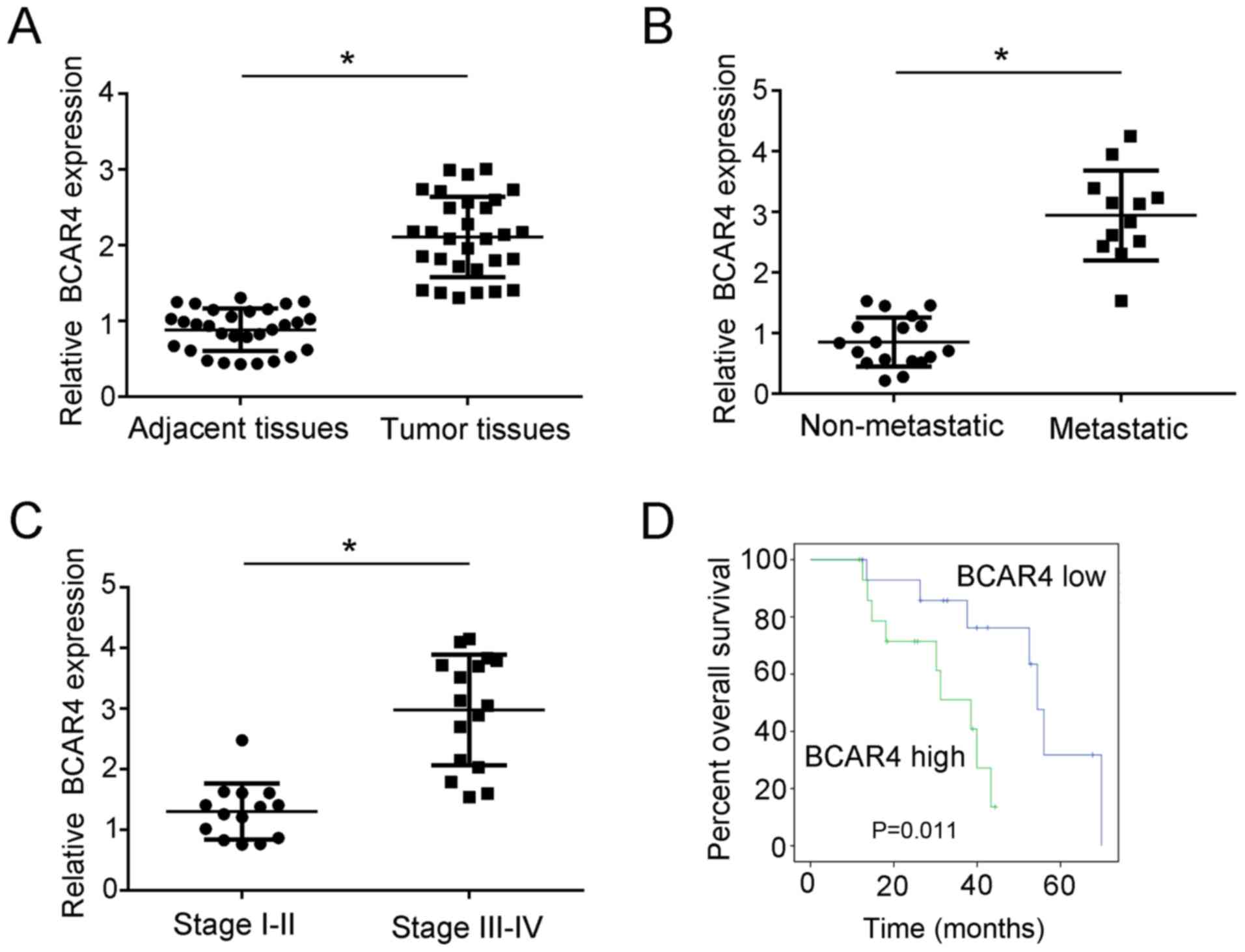
LncRNA BCAR4 is expressed in liver cancer
cells and associated with liver cancer progression
To explore the expression of lncRNA BCAR4 in liver
cancer tissues, RT-qPCR assay was performed. The results
demonstrated that the expression of lncRNA BCAR4 in liver cancer
tissues was significantly higher compared with that in paired
healthy liver tissues (Fig. 1A).
In addition, the association between liver cancer metastasis and
lncRNA BCAR4 expression was analyzed; lncRNA BCAR4 expression was
higher in the metastatic liver cancer group compared with that in
the non-metastatic group (Fig.
1B). The expression level of lncRNA BCAR4 was also higher in
liver cancer stages III and IV compared with that in stages I and
II (Fig. 1C). To examine the
overall survival rate of patients with liver cancer with high
lncRNA BCAR4 expression, Kaplan-Meier analysis was performed.
Patients with liver cancer were divided into two groups based on
the median expression of lncRNA BCAR4, and the results demonstrated
that the high lncRNA BCAR4 expression group exhibited a lower
survival rate compared with the low expression group (Fig. 1D). Taken together, these data
demonstrated that the expression of lncRNA BCAR4 was associated
with liver cancer progression.
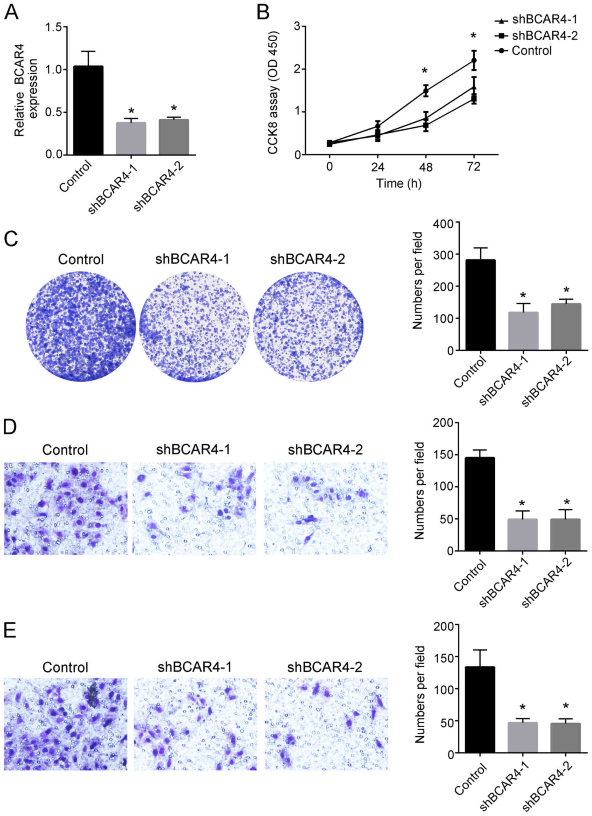
Knockdown of BCAR4 significantly
decreases the proliferative, migratory and invasive abilities of
liver cancer cells
To analyze the functions of lncRNA BCAR4 in
regulating liver cancer, shRNAs targeting the lncRNA BCAR4 sequence
were constructed and transfected into Huh7 liver cancer cells
(Fig. 2A). CCK8 assay results
demonstrated that knockdown of lncRNA BCAR4 decreased the
proliferative ability of liver cancer cells compared with that of
the control group (Fig. 2B). In
addition, colony formation assay was performed to further examine
the proliferative abilities of Huh7 cells. The results revealed
that the colony numbers were decreased after knockdown of lncRNA
BCAR4 in Huh7 cells compared with those in the control group
(Fig. 2C). Transwell assay
results also demonstrated that knockdown of lncRNA BCAR4
significantly decreased the migration and invasion of Huh7 cells
compared with the control group (Fig.
2D and E). Collectively, these results suggested that knockdown
of lncRNA BCAR4 inhibited liver cancer cell abilities associated
with cancer progression.
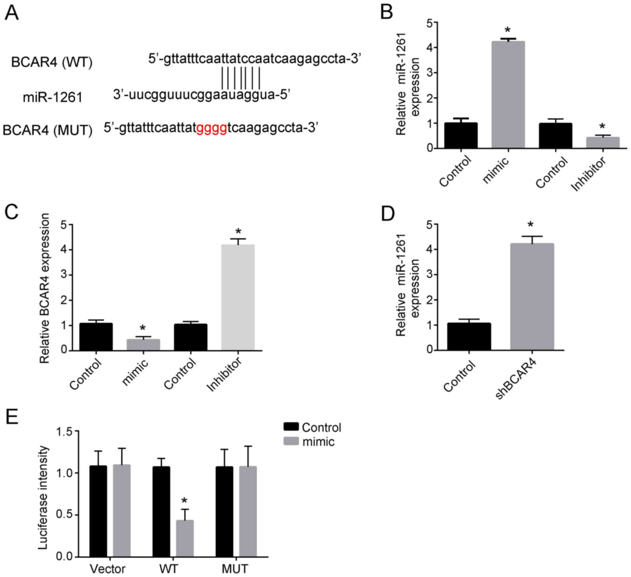
miR-1261 directly binds to lncRNA BCAR4
and affects its expression
As the aforementioned results demonstrated that
lncRNA BCAR4 was associated with liver cancer proliferation,
migration and invasion, bioinformatics analysis was performed to
identify the potential mechanism by which lncRNA BCAR4 regulated
liver cancer progression. The results revealed that lncRNA BCAR4
could directly bind miR-1261 (Fig.
3A). To explore the relationship between lncRNA BCAR4 and
miR-1261, miR-1261 mimic plasmid and inhibitor were used (Fig. 3B). miR-1261 mimics significantly
inhibited the expression of lncRNA BCAR4, whereas inhibition of
miR-1261 promoted BCAR4 expression compared with the corresponding
control groups (Fig. 3C).
Additionally, knockdown of lncRNA BCAR4 decreased the expression of
miR-1261 compared with the control group (Fig. 3D). Luciferase assays were also
performed, and the results confirmed that BCAR4 bound to miR-1261
directly (Fig. 3E). Collectively,
these results demonstrated that miR-1261 directly bound to BCAR4
and affected the expression of lncRNA BCAR4.
ANAPC11 is the downstream target of
miR-1261 and lncRNA BCAR4
To identify the potential target gene for lncRNA
BCAR4 and miR-1261, bioinformatics analysis was performed. The
results revealed that ANAPC11 was the potential target of miR-1261
(Fig. 4A). miR-1261 mimics
notably inhibited ANAPC11 expression, whereas inhibition of
miR-1261 increased the expression of ANAPC11 compared with the
corresponding control groups (Fig.
4B). Luciferase assay results revealed that knockdown of BCAR4
resulted in a significant decrease the luciferase activity of
wild-type ANPC11 (Fig. 4C). In
addition, knockdown of lncRNA BCAR4 decreased the expression of
ANAPC11 compared with that in the control group, whereas inhibition
of miR-1261 rescued the decreased expression of ANAPC11 caused by
knockdown of lncRNA BCAR4 (Fig.
4D). Consistently, overexpression of miR-1261, knockdown of
lncRNA BCAR4 or knockdown of ANAPC11 significantly decreased the
migration of Huh7 cells compared with that in the control group
(Fig. 4E). Additionally,
inhibition of miR-1261 rescued the reduced migration of Huh7 cells
caused by knockdown of ANAPC11, whereas overexpression of ANAPC11
rescued the decreased migration cells caused by knockdown of lncRNA
BCAR4 (Fig. 4E). These results
suggested that ANAPC11 may be a potential target of lncRNA BCAR4
and miR-1261.
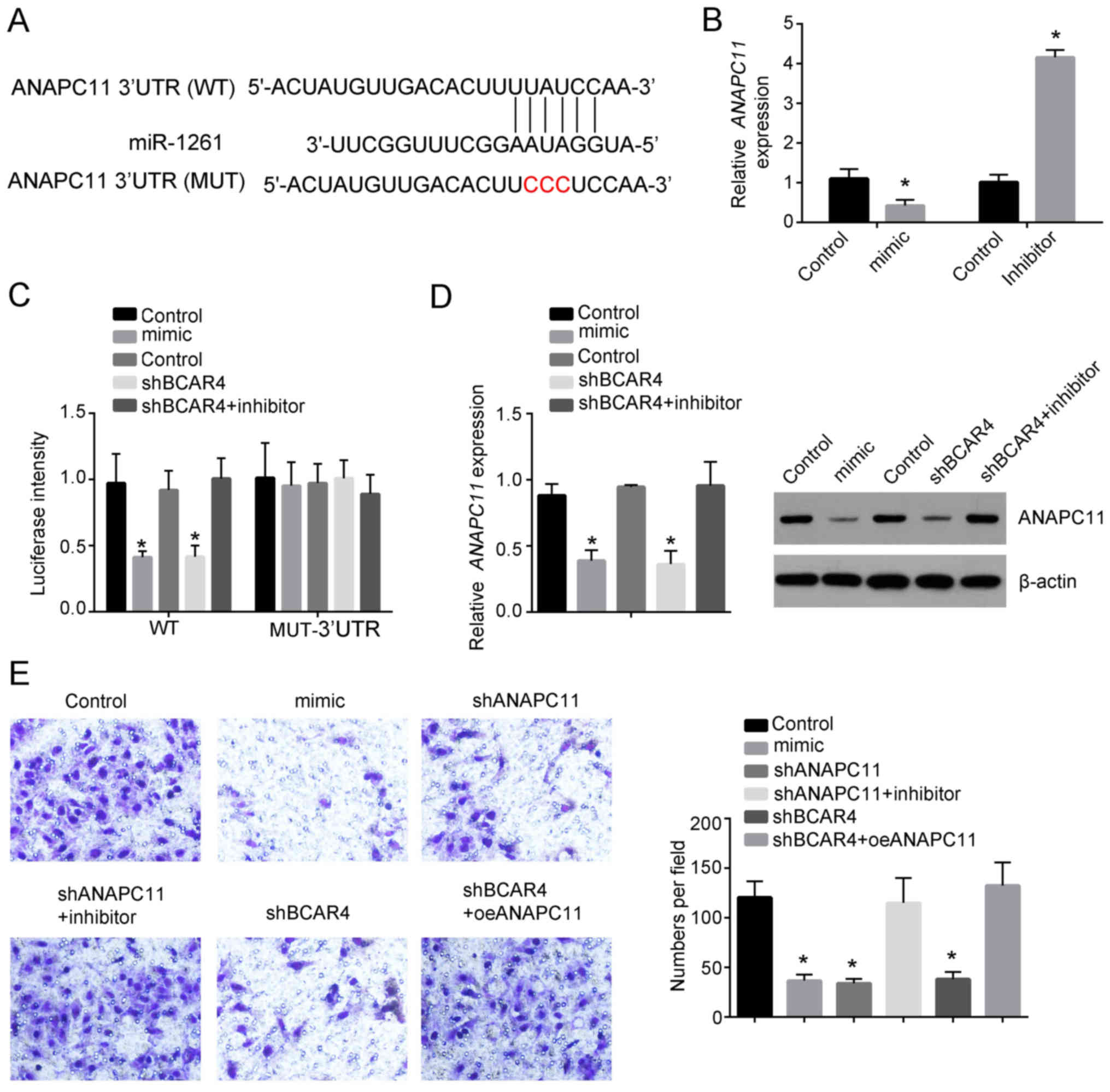 | Figure 4ANAPC11 is targeted by lncRNA BCAR4
and miR-1261. (A) The binding sites between miR-1261 and ANAPC11.
(B) Relative expression of ANAPC11 was examined by RT-qPCR after
transfecting miR-1261 overexpression plasmid or miR-1261 inhibitor
in Huh7 cells. The expression was normalized to that of 18S. (C)
Luciferase assay was performed to detect the luciferase intensity
after transfection with miR-1261 mimic, miR-1261 inhibitor, shBCAR4
or shBCAR4 together with the miR-1261 inhibitor or control plasmids
in Huh7 cells. (D) Relative expression of ANAPC11 was examined by
RT-qPCR after transfection with the miR-1261 mimic, miR-1261
inhibitor, shBCAR4 or shBCAR4 together with the miR-1261 inhibitor
or control in Huh7 cells. The expression was normalized to that of
18S. (E) Transwell assay was performed to determine the migratory
ability of Huh7 cells following transfection with the miR-1261 the
mimic, shANAPC11 or shANAPC11 together with the miR-1261 inhibitor,
shBCAR4, shBCAR4, oeANAPC11 or control plasmids. Plates were imaged
at x4 magnification. All experiments were repeated independently
three times. *P<0.05. ANAPC11, anaphase-promoting
complex subunit 11; miR, microRNA; lncRNA, long non-coding RNA;
BCAR4, breast cancer anti-estrogen resistance 4; RT-qPCR, reverse
transcription-quantitative PCR; sh, short hairpin; WT, wild-type;
MUT, mutant; oe, overexpression. |
Discussion
As a malignant tumor with high incidence, liver
cancer is a serious threat to human health (22,23). The incidence of liver cancer is
associated with hepatitis B and C virus infection, aflatoxin,
drinking water pollution, alcohol, liver cirrhosis, sex hormones,
nitrosamines and trace elements (24-26). However, the pathogenesis and
treatment of liver cancer is still unclear (27,28). Therefore, it is of great
significance to study the regulatory mechanism of liver cancer
tumorigenesis.
A number of lncRNAs have been reported to regulate
liver cancer progression. Mao et al (29) have reported that lncRNA LALR1
upregulates small nucleolar RNA C/D box 72 to promote the
proliferation and invasion of liver cancer cells. In addition,
lncRNA HAND2 antisense RNA 1 has been demonstrated to inhibit the
proliferation and migration of liver cancer cells (30). According to previous studies,
lncRNAs can promote or inhibit the progression of liver cancer;
therefore, it is important to explore the roles of different
lncRNAs in the regulation of liver cancer. LncRNA BCAR4 has been
reported to participate in the regulation of the progression of
multiple types of cancer (31-33). However, the role of lncRNA BCAR4
in liver cancer remains unknown. The results of the present study
demonstrated that the expression of lncRNA BCAR4 was significantly
higher in liver cancer compared with adjacent non-tumor tissues. In
addition, the proliferation, migration and invasion of Huh7 cells
were significantly decreased after knockdown of lncRNA BCAR4
compared with those in the control group.
In previous studies, lncRNAs were demonstrated to
regulate the progression of liver cancer cells through binding
miRNAs. For example, Wei et al (34) demonstrated that lncRNA MFI2
antisense RNA 1 promoted HCC progression by binding miR-134. In the
present study, lncRNA BCAR4 was demonstrated to bind miR-1261 and
target ANAPC11. He et al (14) have reported that miR-1261 can
promote the progression of prostate cancer. Wei et al
(15) have demonstrated that
miR-1261 was involved in the regulation of papillary thyroid cancer
progression. In addition, Zhang et al (16) reported that miR-1261 was involved
in glioma progression. However, the function of miR-1261 in the
regulation of other types of cancer remain unclear. ANAPC11 has
been reported to regulate the cell cycle distribution of 293T cells
(17). In addition, Drouet et
al (18) and Moyret-Lalle
et al (35) have
demonstrated that ANAPC11 is involved in the regulation of
colorectal cancer. However, the role of ANAPC11 in the regulation
of liver cancer remains unclear. The results of the present study
demonstrated that lncRNA BCAR4 promoted the expression of ANAPC11
by inhibiting miR-1261. Additionally, knockdown of ANAPC11
significantly inhibited the migration of liver cancer cells
compared with the control, whereas overexpression of ANAPC11
rescued the decreased migration of liver cancer cells caused by
knockdown of BCAR4.
Funding
No funding was received.
Availability of data and materials
The datasets generated and/or used during the
present study are available from the corresponding author on
reasonable request.
Authors' contributions
YZ and HZ conceived and designed the present study.
YZ performed the experiments, analyzed and interpreted the results.
HZ wrote the manuscript. Both authors read and approved the final
manuscript.
Ethics approval and consent to
participate
This study was approved by the Ethics Committee of
Taiyuan Second People's Hospital. Written informed consent was
obtained from all patients.
Patient consent for publication
Not applicable.
Competing interests
The authors declare that they have no competing
interests.
Acknowledgments
Not applicable.
References
|
1
|
Lazăr DC, Avram MF, Romoșan I, Văcariu V,
Goldiș A and Cornianu M: Malignant hepatic vascular tumors in
adults: Characteristics, diagnostic difficulties and current
management. World J Clin Oncol. 10:110–135. 2019. View Article : Google Scholar
|
|
2
|
Turley EA, Veiseh M, Radisky DC and
Bissell MJ: Mechanisms of disease: Epithelial-mesenchymal
transition-does cellular plasticity fuel neoplastic progression?
Nat Clin Pract Oncol. 5:280–290. 2008. View Article : Google Scholar : PubMed/NCBI
|
|
3
|
van Tienderen GS, Groot Koerkamp B,
IJzermans JNM, van der Laan LJW and Verstegen MMA: Recreating
tumour complexity in a dish: Organoid models to study liver cancer
cells and their extracellular environment. Cancers (Basel). 11:pii:
E1706. 2019. View Article : Google Scholar
|
|
4
|
Kang M, Sang Y, Gu H, Zheng L, Wang L, Liu
C, Shi Y, Shao A, Ding G, Chen S, et al: Long noncoding RNAs POLR2E
rs3787016 C/T and HULC rs7763881 A/C polymorphisms are associated
with decreased risk of esophageal cancer. Tumour Biol.
36:6401–6408. 2015. View Article : Google Scholar : PubMed/NCBI
|
|
5
|
Klec C, Gutschner T, Panzitt K and Pichler
M: Involvement of long non-coding RNA HULC (highly up-regulated in
liver cancer) in pathogenesis and implications for therapeutic
intervention. Expert Opin Ther Targets. 23:177–186. 2019.
View Article : Google Scholar : PubMed/NCBI
|
|
6
|
Kong X, Duan Y, Sang Y, Li Y, Zhang H,
Liang Y, Liu Y, Zhang N and Yang Q: LncRNA-CDC6 promotes breast
cancer progression and function as ceRNA to target CDC6 by sponging
microRNA-215. J Cell Physiol. 234:9105–9117. 2019. View Article : Google Scholar
|
|
7
|
Ouyang S, Zheng X, Zhou X, Chen Z, Yang X
and Xie M: LncRNA BCAR4 promotes colon cancer progression via
activating Wnt/β-catenin signaling. Oncotarget. 8:92815–92826.
2017. View Article : Google Scholar : PubMed/NCBI
|
|
8
|
Xing Z, Park PK, Lin C and Yang L: LncRNA
BCAR4 wires up signaling transduction in breast cancer. RNA Biol.
12:681–689. 2015. View Article : Google Scholar : PubMed/NCBI
|
|
9
|
Li N, Gao WJ and Liu NS: LncRNA BCAR4
promotes proliferation, invasion and metastasis of non-small cell
lung cancer cells by affecting epithelial-mesenchymal transition.
Eur Rev Med Pharmacol Sci. 21:2075–2086. 2017.PubMed/NCBI
|
|
10
|
Yin XH, Jin YH, Cao Y, Wong Y, Weng H, Sun
C, Deng JH and Zeng XT: Development of a 21-miRNA signature
associated with the prognosis of patients with bladder cancer.
Front Oncol. 9:7292019. View Article : Google Scholar : PubMed/NCBI
|
|
11
|
Hu Z, Gao S, Lindberg D, Panja D,
Wakabayashi Y, Li K, Kleinman JE, Zhu J and Li Z: Temporal dynamics
of miRNAs in human DLPFC and its association with miRNA
dysregulation in schizophrenia. Transl Psychiatry. 9:1962019.
View Article : Google Scholar : PubMed/NCBI
|
|
12
|
Angius A, Uva P, Pira G, Muroni MR, Sotgiu
G, Saderi L, Uleri E, Caocci M, Ibba G, Cesaraccio MR, et al:
Integrated analysis of miRNA and mRNA endorses a twenty miRNAs
signature for colorectal carcinoma. Int J Mol Sci. 20:pii: E4067.
2019. View Article : Google Scholar
|
|
13
|
Yen MC, Yeh IJ, Liu KT, Jian SF, Lin CJ,
Tsai MJ and Kuo PL: Next-generation sequencing predicts interaction
network between miRNA and target genes in lipoteichoic
acid-stimulated human neutrophils. Int J Mol Med. 44:1436–1446.
2019.PubMed/NCBI
|
|
14
|
He JH, Li BX, Han ZP, Zou MX, Wang L, Lv
YB, Zhou JB, Cao MR, Li YG and Zhang JZ: Snail-activated long
non-coding RNA PCA3 up-regulates PRKD3 expression by miR-1261
sponging, thereby promotes invasion and migration of prostate cance
cells. Tumour Biol. Oct 14–2016.Epub ahead of print. View Article : Google Scholar
|
|
15
|
Wei H, Pan L, Tao D and Li R: Circular RNA
circZFR contributes to papillary thyroid cancer cell proliferation
and invasion by sponging miR-1261 and facilitating C8orf4
expression. Biochem Biophys Res Commun. 503:56–61. 2018. View Article : Google Scholar : PubMed/NCBI
|
|
16
|
Zhang F, Mai SR, Cao FP, Cao CX and Zhang
L: MiR-1261/circ-PTPRZ1/PAK1 pathway regulates glioma cell growth
and invasion. Human Cell. 32:540–547. 2019. View Article : Google Scholar : PubMed/NCBI
|
|
17
|
Shi YJ and Huo KK: Knockdown expression of
Apc11 leads to cell-cycle distribution reduction in G2/M phase.
Genet Mol Res. 11:2814–2822. 2012. View Article : Google Scholar : PubMed/NCBI
|
|
18
|
Drouet Y, Treilleux I, Viari A, Léon S,
Devouassoux-Shisheboran M, Voirin N, de la Fouchardière C, Manship
B, Puisieux A, Lasset C and Moyret-Lalle C: Integrated analysis
highlights APC11 protein expression as a likely new independent
predictive marker for colorectal cancer. Sci Rep. 8:73862018.
View Article : Google Scholar : PubMed/NCBI
|
|
19
|
Wang G, Fang X, Han M, Wang X and Huang Q:
MicroRNA-493-5p promotes apoptosis and suppresses proliferation and
invasion in liver cancer cells by targeting VAMP2. Int J Mol Med.
41:1740–1748. 2018.PubMed/NCBI
|
|
20
|
Xin X, Wu M, Meng Q, Wang C, Lu Y, Yang Y,
Li X, Zheng Q, Pu H, Gui X, et al: Long noncoding RNA HULC
accelerates liver cancer by inhibiting PTEN via autophagy
cooperation to miR15a. Mol Cancer. 17:942018. View Article : Google Scholar : PubMed/NCBI
|
|
21
|
Li R, Zhu H, Yang D, Xia J and Zheng Z:
Long noncoding RNA lncBRM promotes proliferation and invasion of
colorectal cancer by sponging miR-204-3p and upregulating TPT1.
Biochem Biophys Res Commun. 508:1259–1263. 2019. View Article : Google Scholar
|
|
22
|
Francica G and Borzio M: Status of, and
strategies for improving, adherence to HCC screening and
surveillance. J Hepatocell Carcinoma. 6:131–141. 2019. View Article : Google Scholar : PubMed/NCBI
|
|
23
|
Viveiros P, Riaz A, Lewandowski RJ and
Mahalingam D: Current state of liver-directed therapies and
combinatory approaches with systemic therapy in hepatocellular
carcinoma (HCC). Cancers (Basel). 11:pii: E1085. 2019. View Article : Google Scholar
|
|
24
|
Gao Q, Zhang G, Zheng Y, Yang Y, Chen C,
Xia J, Liang L, Lei C, Hu Y, Cai X, et al: SLC27A5 deficiency
activates NRF2/TXNRD1 pathway by increased lipid peroxidation in
HCC. Cell Death Differ. 27:1086–1104. 2020. View Article : Google Scholar
|
|
25
|
Feld J: Update on the risk of primary and
recurrent HCC With the use of DAA therapy for HCV infection.
Gastroenterol Hepatol (N Y). 15:303–306. 2019.
|
|
26
|
Fang G, Zhang P, Liu J, Zhang X, Zhu X, Li
R and Wang H: Inhibition of GSK-3β activity suppresses HCC
malignant phenotype by inhibiting glycolysis via activating
AMPK/mTOR signaling. Cancer Lett. 463:11–26. 2019. View Article : Google Scholar : PubMed/NCBI
|
|
27
|
Shigeta K, Datta M, Hato T, Kitahara S,
Chen IX, Matsui A, Kikuchi H, Mamessier E, Aoki S, Ramjiawan RR, et
al: Dual programmed death receptor-1 and vascular endothelial
growth factor receptor-2 blockade promotes vascular normalization
and enhances antitumor immune responses in hepatocellular
carcinoma. Hepatology. Aug 5–2019.Epub ahead of print. PubMed/NCBI
|
|
28
|
Ju JX, Zeng QJ, Xu EJ, He XQ, Tan L, Huang
QN, Li K and Zheng RQ: Intraprocedural contrast-enhanced
ultrasound-CT/MR fusion imaging assessment in HCC thermal ablation
to reduce local tumor progression: Compared with routine
contrast-enhanced ultrasound. Int J Hyperthermia. 36:785–793. 2019.
View Article : Google Scholar : PubMed/NCBI
|
|
29
|
Mao LH, Chen SY, Li XQ, Xu F, Lei J, Wang
QL, Luo LY, Cao HY, Ge X, Ran T, et al: LncRNA-LALR1 upregulates
small nucleolar RNA SNORD72 to promote growth and invasion of
hepatocellular carcinoma. Aging (Albany NY). 12:4527–4546. 2020.
View Article : Google Scholar
|
|
30
|
Yan D, Jin F and Lin Y: lncRNA HAND2-AS1
inhibits liver cancer cell proliferation and migration by
upregulating SOCS5 to inactivate the JAK-STAT pathway. Cancer
Biother Radiopharm. 35:143–152. 2020. View Article : Google Scholar : PubMed/NCBI
|
|
31
|
Ouyang S, Zhou X, Chen Z, Wang M, Zheng X
and Xie M: LncRNA BCAR4, targeting to miR-665/STAT3 signaling,
maintains cancer stem cells stemness and promotes tumorigenicity in
colorectal cancer. Cancer Cell Int. 19:722019. View Article : Google Scholar : PubMed/NCBI
|
|
32
|
Godinho MF, Wulfkuhle JD, Look MP,
Sieuwerts AM, Sleijfer S, Foekens JA, Petricoin EF III, Dorssers LC
and van Agthoven T: BCAR4 induces antioestrogen resistance but
sensitises breast cancer to lapatinib. Br J Cancer. 107:947–955.
2012. View Article : Google Scholar : PubMed/NCBI
|
|
33
|
Yang H, Yan L, Sun K, Sun X, Zhang X, Cai
K and Song T: lncRNA BCAR4 increases viability, invasion, and
migration of non-small cell lung cancer cells by targeting
glioma-associated oncogene 2 (GLI2). Oncol Res. 27:359–369. 2019.
View Article : Google Scholar
|
|
34
|
Wei Y, Wang Z, Zong Y, Deng D, Chen P and
Lu J: LncRNA MFI2-AS1 promotes HCC progression and metastasis by
acting as a competing endogenous RNA of miR-134 to upregulate FOXM1
expression. Biomed Pharmacother. 125:1098902020. View Article : Google Scholar : PubMed/NCBI
|
|
35
|
Moyret-Lalle C, Drouet Y and Puisieux A:
The catalytic subunit of the anaphase-promoting complex, APC11, is
involved in CIN+ CRC progression. Med Sci (Paris).
35:118–122. 2019.In French. View Article : Google Scholar
|