Introduction
Diabetic nephropathy (DN) is a hard microvascular
complication of diabetes (1).
Histologically, mesangial cell proliferation and podocyte damage
are the major pathological features in the early stage of DN
(2). DN is structurally
characterized by the thickening of glomerular and tubular basement
membranes, which have been attributed to extracellular matrix (ECM)
synthesis (3-5). Deposition of ECM proteins such as
collagens and fibronectin in the tubulointerstitium and glomerular
mesangium contribute to tubulointerstitial fibrosis and
glomerulosclerosis, eventually resulting in renal fibrosis
(6,7). Pathologically, the progression of
renal fibrosis is one of the hallmarks of DN and further predicts
the deterioration of renal function (8,9).
Considering that tubulointerstitial fibrosis is the key underlying
pathology, understanding the regulatory mechanism underlying
fibrogenesis in the interstitium is key to developing therapeutic
targets for DN (10). However, to
date, the option to target renal fibrosis is still not available in
the clinic. Thus, more molecular pathways should be identified for
new therapeutic strategies for DN.
Transforming growth factor-β (TGFβ) is a vital
cytokine that promotes sclerosis and has been well documented in
fibrosis development in DN (11).
Highly expressed TGFβ occurs universally in chronic kidney diseases
in both animal models and humans. For example, animal models of
spontaneous diabetes have demonstrated increased TGFβ1 mRNA
expression at the onset of hyperglycemia (12,13); in addition, TGFβ receptors have
been described to be upregulated in renal disease models including
DN. A previous study revealed that TGFβ1 was increased in several
types of cells in the diabetic kidney, including mesangial cells,
thus contributing to fibrotic events and cell survival (10). Therefore, TGFβ has been evaluated
as a major target for DN treatment. In addition, evidence indicates
a primary role for TGFβ and its downstream signaling cascades in
the progression of renal diseases (14). TGFβ regulates numerous cell
behaviors including cell proliferation, differentiation, adhesion
and apoptosis (15,16). In addition, several intracellular
signaling cascades have been identified for TGFβ-induced renal
fibrosis, such as the mothers against decapentaplegic homologs
(Smads), mitogen activated protein kinase (MAPK) and Jagged/Notch
signaling pathway (10). Thus, it
is important to further understand the molecular mechanisms of
TGFβ-induced fibrotic events, which may lead to more effective
approaches for DN treatment.
MicroRNAs (miRNAs/miRs) are highly conserved small
non-coding RNAs (~22 nucleotides) with no protein coding capacity.
Generally, miRNAs act as a mediator of functional gene expression
by interacting with the 3′ untranslated region (3′UTR) of the
target gene. It has been proposed that miRNAs modulate the effects
of TGFβ1 in renal fibrosis, such as miRNA (miR)-433 (17). Cumulative evidence has
demonstrated the close association between dysregulation of miRNAs
and the progression of diabetes and diabetic complications,
including DN (18,19). A number of miRNAs have been
identified as early biomarkers in various chronic kidney diseases
due to their consistency and reproducibility in human peripheral
blood (20). In addition,
integrated serum miRNAs expression profiling may be used in DN for
identification of novel miRNAs (21). Among them, miR-135a-5p was
significantly upregulated in the serum and renal tissues from
patients with DN compared with healthy controls (21). However, the role and mechanism of
miR-135a-5p in DN hasn't been fully illuminated.
Sirtuin 1 (SIRT1) belongs to a highly conserved
family of NAD+-dependent deacetylase and serves as a
therapeutic target for DN (22).
The present study aimed to investigate the expression of
miR-135a-5p in peripheral blood of patients with DN, as well as the
role and mechanism of miR-135a-5p in TGFβ1-induced cell model of
renal fibrosis.
Materials and methods
Clinical specimens
Patients were diagnosed with DN confirmed by renal
biopsy, with nodular sclerosis (Kinnelstiel-Wilson lesion) in the
glomerulus between January 2015 and December 2017. Peripheral
venous blood samples were collected from 30 patients with DN (age,
43-73 years; male, 46.7%) and 30 patients with diabetes without DN
(age, 38-68 years; male, 50.0%) after informed consent was provided
by each individual. The clinical characteristics of the
participants are presented in Table
I. The blood samples were snap-frozen in liquid nitrogen
(-79°C). All protocols involving human subjects were approved by
the Ethics Committee of the Zhongnan Hospital of Wuhan University.
The renal tissue sections were obtained from 10 of 30 renal biopsy
specimens, and the control tissues were adjacent normal renal
tissue sections from patients with renal carcinoma with normal
kidney function (data not shown).
 | Table IClinical characteristics of patients
with DN or healthy controls (Control). |
Table I
Clinical characteristics of patients
with DN or healthy controls (Control).
| Clinical
features | DN (n=30) | Control (n=30) |
|---|
| Age, mean
years | 60.23 | 62.45 |
| Sex,
male/female | 16/14 | 15/15 |
| UAER,
µg/min | 248.75 | 14.35 |
| Scr,
µmol/l | 120.35 | 63.27 |
| BUN, mmol/l | 12.87 | 5.35 |
Cells and cell culture
Human proximal tubule cell lines (HK-2, cat. no.
CRL-2190) were purchased from the American Type Culture Collection,
and a human renal mesangial cell line (HMC, cat. no. 4200) was
obtained from ScienCell Research Laboratories, Inc. HK-2 and HMCs
were cultured and passaged in growth culture medium containing 90%
Dulbecco's modified Eagle medium (DMEM; Gibco; Thermo Fisher
Scientific, Inc.), 10% fetal bovine serum (FBS; Gibco; Thermo
Fisher Scientific, Inc.) and 1% antibiotics (100 U/ml penicillin
and 100 µg/ml streptomycin; Invitrogen; Thermo Fisher
Scientific, Inc.) at 37°C in a humidified environment containing 5%
CO2 for indicated times.
TGFβ1 treatment and high glucose
treatments
HK-2 and HMCs at 60% confluency were seeded into
6-well plates (Corning, Inc.) overnight. For TGFβ1 treatment, the
cells were made quiescent by incubation with serum-free medium for
16 h when grown to 80-90% confluence. A recombinant human TGFβ1
(cat. no. P01137; R&D Systems, Inc.) at a concentration of 10
ng/ml was added to the cell growth culture for 24 h to detect
changes in the expression of miR-135a-5p and SIRT1, fibrosis
response and TGFβ1/Smad3 signaling. For high glucose treatment,
different concentrations of D-glucose (5-30 mmol/l) were added into
DMEM, and HMC and HK-2 cells were incubated for 48 h before
detection of miR-135a-5p expression.
Cell transfection
HK-2 and HMCs at 60% confluency were seeded into
6-well plates (Corning, Inc.) and incubated overnight. miR-135a-5p
mimic (5′-UAU GGC UUU UUA UUC CUA UGU GA-3′), miR-135a-5p inhibitor
(anti-miR-135a-5p; 5′-UCA CAU AGG AAU AAA AAG CCA UA-3′), specific
small interfering (si)RNA against SIRT1 (siSIRT1; sense, 5′-GAU GAA
GUU GAC CUC CUC ATT-3′ and antisense, 5′-UGA GGA GGU CAA CUU CAU
CTT-3′) and the corresponding negative controls miR-NC mimic
(5′-GUC CAG UGA AUU CCC AG-3′), anti-NC (5′-TCA CAA CCT CCT AGA AAG
AGT AGA-3′) and siNC (sense, 5′-TTC TCC GAA CGT GTC ACG T-3′ and
anti-sense, 5′-ACG UGA CAC GUU CGG AGA A-3′) were acquired from
Shanghai GenePharma Co., Ltd. The overexpression of SIRT1 was
performed using a pcDNA3.1 plasmid (Invitrogen; Thermo Fisher
Scientific, Inc.). All transfections of oligo-nucleotides (30 nM)
or vectors (2 µg) were performed using
Lipofectamine® 2000 (Invitrogen; Thermo Fisher
Scientific, Inc.), and the transfected cells were collected for
further analysis after transfection for 36 h.
Reverse transcription-quantitative PCR
(RT-qPCR)
For examination of the expression of SIRT1 mRNA and
miR-135a-5p, total RNA was extracted from blood and treated cells
using TRIzol reagent (Invitrogen; Thermo Fisher Scientific, Inc.).
The first-strand cDNA was synthesized at 42°C for 15 min using
Fastking RT kit (Tiangen Biotech Co., Ltd.) and miRNA First-Strand
cDNA Synthesis kit (Tiangen Biotech Co., Ltd.). qPCR was performed
with SuperReal PreMix Plus (SYBR Green; Tiangen Biotech Co., Ltd.)
on the ABI PRISM 7500 Fast Real-time PCR System (Applied
Biosystems; Thermo Fisher Scientific, Inc.). The thermocycling
conditions were 40 cycles of 95°C for 15 sec, 60°C for 60 sec and
95°C for 15 sec. Relative gene expression was normalized to GAPDH
(for mRNAs) and U6 small nuclear RNA (U6; for miR-135a-5p). The
reactions were performed in at least three independent runs using
the following primers: SIRT1 isoform 2 forward, 5′-TGT GTC ATA GGT
TAG GTG GTG A-3′ and reverse, 5′-AGC CAA TTC TTT TTG TGT TCG TG-3′;
GAPDH forward, 5′-CTG GGC TAC ACT GAG CAC C-3′ and reverse, 5′-AAG
TGG TCG TTG AGG GCA ATG-3′; miR-135a-5p forward, 5′-TTG GTC TTG TTT
CCC GGT CC-3′ and reverse, 5′-TCA CAG CTC CAC AGG CT A AC-3′; U6
forward, 5′-CTC GCT TCG GCA GCA CA-3′ and reverse, 5′-AAC GCT TCA
CGA ATT TGC GT-3′. Expression levels were normalized to the
respective internal controls and calculated using the
2−ΔΔCq method (23).
Western blotting
To examine the expression of collagen I, α-smooth
muscle actin (α-SMA), fibronectin (FN) and epithelial (E)-cadherin,
total protein was extracted from blood and treated cells using RIPA
lysis buffer (Beyotime Institute of Biotechnology) supplemented
with the protease inhibitor phenylmethanesulfonyl fluoride
(MedChemExpress). A total of 20 µg of protein determined by
Bradford protein assay (Bio-Rad Laboratories, Inc.) was loaded for
the standard procedures of western blotting. The proteins were
transferred to PVDF membranes and incubated in a blocking buffer
(3% BSA; R&D Systems, Inc.) for 1 h at 25°C and with primary
antibodies overnight at 4°C. The primary antibodies against SIRT1
(cat. no. 8469; 1:1,000), collagen1A1 (cat. no. 84336; 1:1,000),
α-SMA (cat. no. 14968; 1:1,000), E-cadherin (cat. no. 14472;
1:1,000), Smad3 (cat. no. 9513; 1:1,000), phosphorylated (p)-Smad3
(cat. no. 9520; 1:1,000) and GAPDH (cat. no. 97166; 1:1,000) were
purchased from Cell Signaling Technology, Inc. The antibody against
FN (cat. no. 8422; 1:200) was provided by Santa Cruz Biotechnology,
Inc. After that, the membranes were washed with Tris-buffered
saline containing 0.1% Tween-20, and then incubated with
horseradish peroxidase-conjugated secondary mouse (cat. no. 7076;
1:2,500) or rabbit (cat. no. 7074; 1:2,500) antibody from Cell
Signaling Technology, Inc. at 25°C for 1.5 h. Protein bands were
finally detected by using an enhanced chemiluminescent substrate
(Pierce; Thermo Fisher Scientific, Inc.). The relative protein
expression was normalized to GAPDH according to the gray intensity
determined on Image-Pro Plus 6.0 software (Media Cybernetics,
Inc.).
Luciferase reporter assay and RNA
immunoprecipitation (RIP)
According to bioinformatics algorithms, human SIRT1
3′ untranslated region (UTR) contained a potential target site of
hsa-miR-135a-5p. Then, the wild type of SIRT1 3′ UTR fragment
(SIRT1-wt) containing AAA AAG CCA U was cloned by PCR methods into
pGL4 vector (Promega Corporation), as well as the mutated SIRT1 3′
UTR sequence (SIRT1-mut) containing UUU UUC GGU A. HK-2 and HMCs
were transfected according to the following groups: SIRT1-wt +
miR-NC mimic, SIRT1-wt + miR-135a-5p mimic, SIRT1-wut + miR-NC
mimic, SIRT1-wut + miR-135a-5p mimic. After incubation for 24 h,
cells were collected to measure Firefly and Renilla
luciferase activities using the Dual-Luciferase Reporter assay
system (Promega Corporation).
Magna RIP™ RNA-binding protein immunoprecipitation
kit (EMD Millipore) was used according to the manufacturer's
instructions. Briefly, HK-2 and HMC cells transfected with
miR-135a-5p mimic or miR-NC mimic were extracted in RIP lysis
buffer. Then, the cell extract was incubated with protein A/G
magnetic beads pre-coated with argonaute 2 or IgG antibody and
diluted with Salt solution II. The immunoprecipitated contents were
treated with Proteinase K and incubated in TRIzol®
reagent (Invitrogen) to detect the expression of SIRT1 mRNA with
RT-qPCR.
Statistical analysis
Data are presented as the mean ± standard error of
the mean from three independent experiments and were analyzed using
SPSS 19.0 software (SPSS, Inc.). The P-values were evaluated using
one-way analysis of variance followed by Tukey's post hoc test.
Spearman's rank correlation analysis was performed to confirm the
correlation between miR-135a-5p and SIRT1 in DN patients. P<0.05
was considered to indicate a statistically significant
difference.
Results
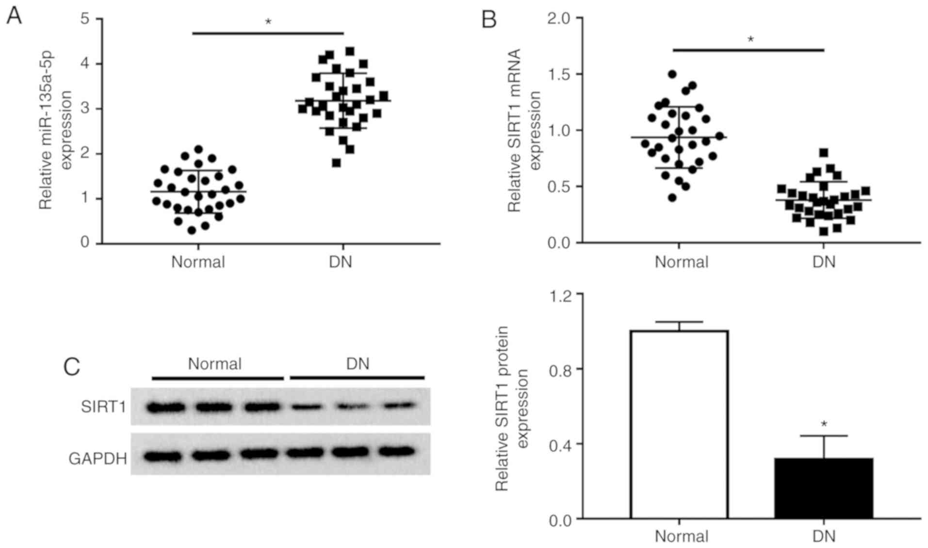
miR-135a-5p is upregulated and SIRT1 is
downregulated in patients with DN
Previous studies suggested the enhancement of
miR-135a-5p expression in renal fibrosis and the important role of
SIRT1 in mesangial cells and renal fibrosis (12,18). Consistently, the present study
observed a significantly increased expression of miR-135a-5p
(Fig. 1A) and a lower expression
of SIRT1 (Fig. 1B and C) in the
serum of patients with DN (n=30, Table I) as measured by RT-qPCR and
western blotting. Moreover, the expression of miR-135a-5p and SIRT1
in the renal tissues was also detected. As presented in Fig. S1A and B, miR-135a-5p was
upregulated, whereas SIRT1 was downregulated in the 10/30 renal
biopsy specimens compared with normal renal tissues. In addition,
there was a negative correlation between miR-135a-5p and SIRT1
expression in renal tissues of DN patients, according to Spearman's
rank correlation analysis (Fig.
S1C). These data suggested that miR-135a-5p and SIRT1 were
involved in renal fibrosis.
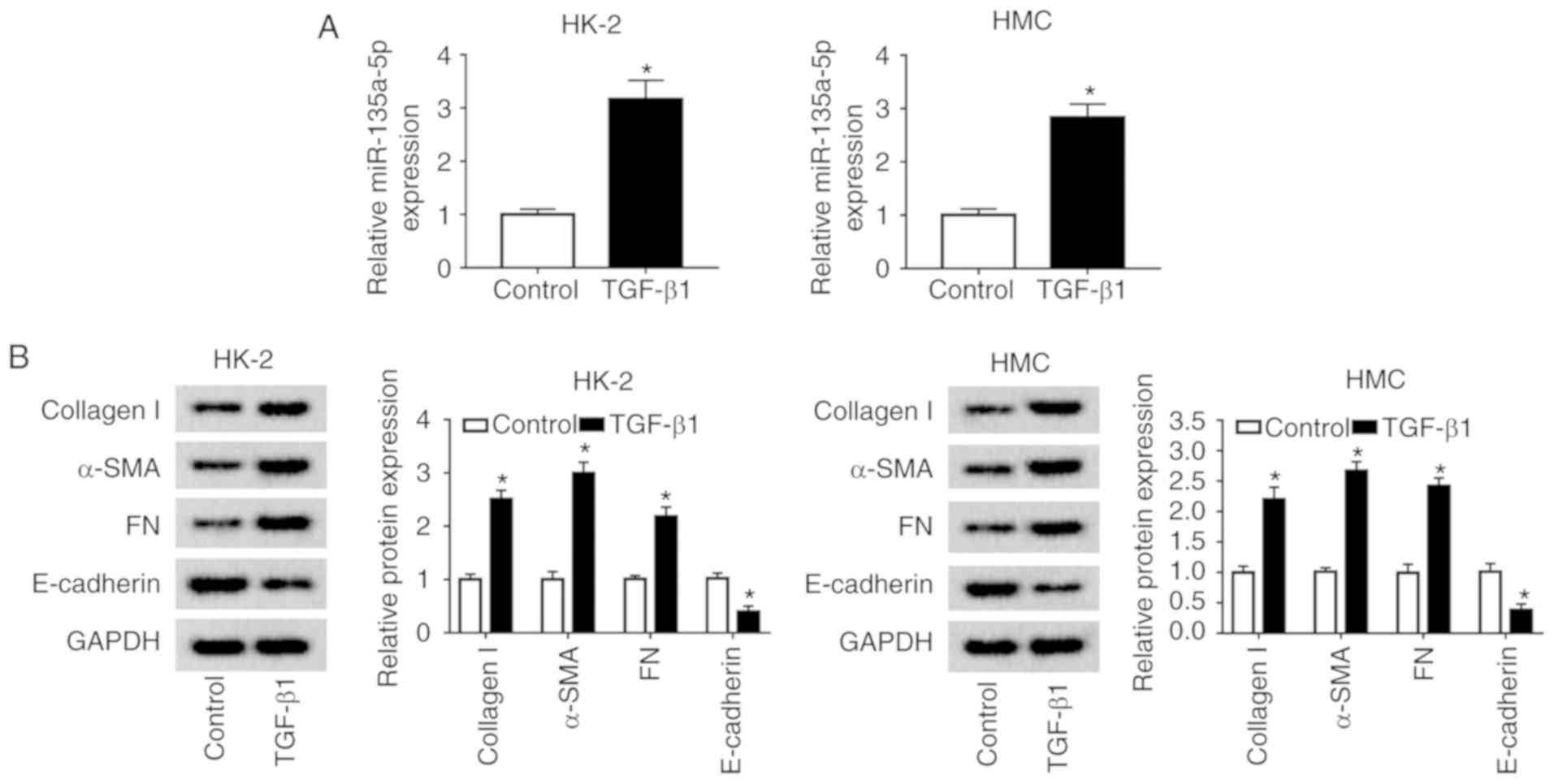
miR-135a-5p expression is increased
during TGFβ1-induced renal fibrosis in vitro
To examine the relevance of miR-135a-5p in renal
fibrosis in DN, a cell model of renal fibrosis in HMC and HK-2
cells was constructed. First, miR-135a-5p expression was monitored
in various glucose concentration stimulation in HMC and HK-2 cells.
As a result, 15-30 mmol/l of D-glucose induced an increase in
miR-135a-5p expression at 48 h (Fig.
S2). Subsequently, HMC and HK-2 cells were exposed to 10 ng/ml
TGFβ1 for 24 h for renal fibrosis analysis. As presented in
Fig. 2A, miR-135a-5p was highly
expressed in TGFβ1-induced HMC and HK-2 cells. The levels of
collagen 1A1, α-SMA and FN were significantly promoted, whereas
E-cadherin was inhibited under TGFβ1 stimulation (Fig. 2B). These data suggested that TGFβ1
treatment induced renal fibrosis in HMC and HK-2 cells, accompanied
with upregulation of miR-135a-5p.
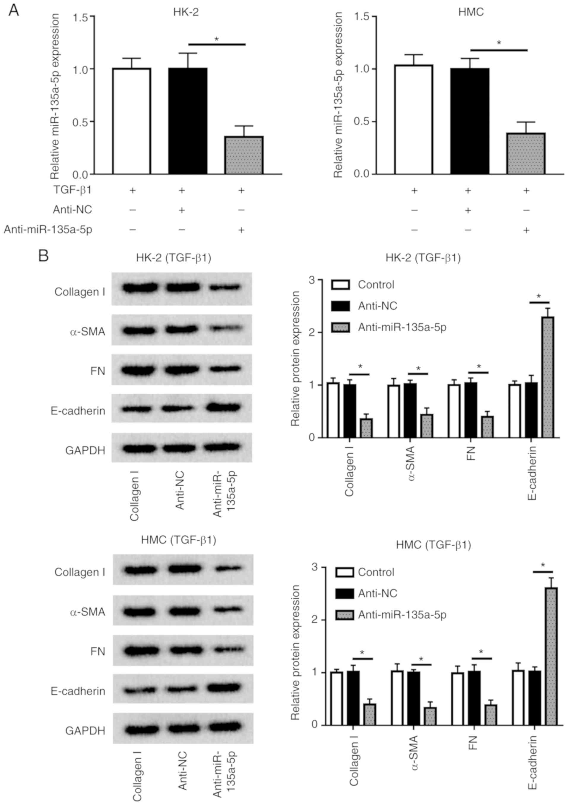
Knockdown of miR-135a-5p inhibits
TGFβ1-induced renal fibrosis in HMC and HK-2 cells
Considering the upregulation of miR-135a-5p during
renal fibrosis, miR-135a-5p was knocked down in HMC and HK-2 cells
by transient transfection of anti-miR-135a-5p. During TGFβ1
exposure, anti-miR-135a-5p-transfected cells exhibited lower
expression levels of miR-135a-5p compared with that of
anti-NC-transfected cells (Fig.
3A). In addition, the levels of collagen 1A1, α-SMA and FN were
decreased under TGFβ1 stimulation when miR-135a-5p was knocked
down, whereas that of E-cadherin was elevated compared with
anti-NC-transfected cells (Fig.
3B). These results demonstrated that miR-135a-5p knockdown may
alleviate TGFβ1-induced renal fibrosis in HMC and HK-2 cells.
SIRT1 is downregulated, and
overexpression of SIRT1 exerts a suppressive role in TGFβ1-induced
renal fibrosis in vitro
To examine the effects of SIRT1 in renal fibrosis in
DN, the present study determined its expression in a cell model of
renal fibrosis in HMC and HK-2 cells. As presented in Fig. 4A, SIRT1 was expressed at a low
level in TGFβ1-induced HMC and HK-2 cells. Thus, SIRT1 was
overexpressed in HMC and HK-2 cells using ectopic expression of a
recombinant vector pcDNA-SIRT1 (Fig.
4B). Of note, during TGFβ1 stimulation, levels of collagen 1A1,
α-SMA and FN were significantly decreased, whereas E-cadherin was
enhanced by SIRT1 upregulation compared with the negative control
(Fig. 4C). These results
demonstrated that overexpression of SIRT1 high expression may
relieve TGFβ1-induced renal fibrosis in HMC and HK-2 cells.
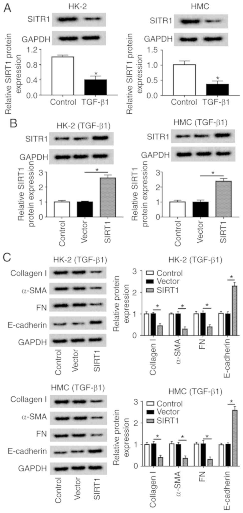 | Figure 4Role of SIRT1 in TGFβ1-induced HMC
and HK-2 cells. HMC and HK-2 cells were treated with 10 ng/ml TGFβ1
for 24 h. (A) SIRT1 protein expression was detected. (B) Levels of
SIRT1 were identified when HMC and HK-2 cells transfected
pcDNA-SIRT1 (SIRT1). (C) Expression of collagen 1A1, α-SMA, FN and
E-cadherin was measured by western blotting in SIRT1-overexpressed
HMC and HK-2 cells. *P<0.05 vs. control cells
(vector). FN, fibronectin; SMA, smooth muscle actin; TGF,
transforming growth factor; HMC, human mesangial cells; miR,
microRNA; E, epithelial; NC, negative control; SIRT1, sirtuin
1. |
miR-135a-5p regulates SIRT1 expression
via target binding
The regulatory relationship between miR-135a-5p and
SIRT1 was further investigated. Algorithms analysis by TargetScan
Human database (http://www.targetscan.org/vert_72/) identified the
targets of miR-135a-5p, and the 3′UTR of human SIRT1 exhibited a
highly conserved binding site for miR-135a-5p (Fig. 5A). To verify this, a luciferase
reporter assay was performed. The luciferase reporter vectors
integrating wt or mut SIRT1 3′UTR fragment were constructed, and
HMC and HK-2 cells were co-transfected with SIRT1-wt/mut and either
miR-135a-5p or miR-NC mimic. First, RT-qPCR analysis was used to
confirm the high miR-135a-5p expression level in the
mimic-transfected HMC and HK-2 cells (Fig. S3A). The luciferase activity was
significantly reduced in cells transfected with the miR-135a-5p
mimic and SIRT1-wt; however, no differences were observed in the
SIRT1-mut groups (Fig. 5B). RIP
assay further identified the target binding of miR-135a-5p and
SIRT1 (Fig. 5C). A western blot
assay demonstrated that SIRT1 expression was inhibited by the
miR-135a-5p mimic but promoted by anti-miR-135a-5p in HMC and HK-2
cells compared with the corresponding NCs (Fig. 5D). These data supported the
hypothesis that SIRT1 was a direct target of miR-135a-5p.
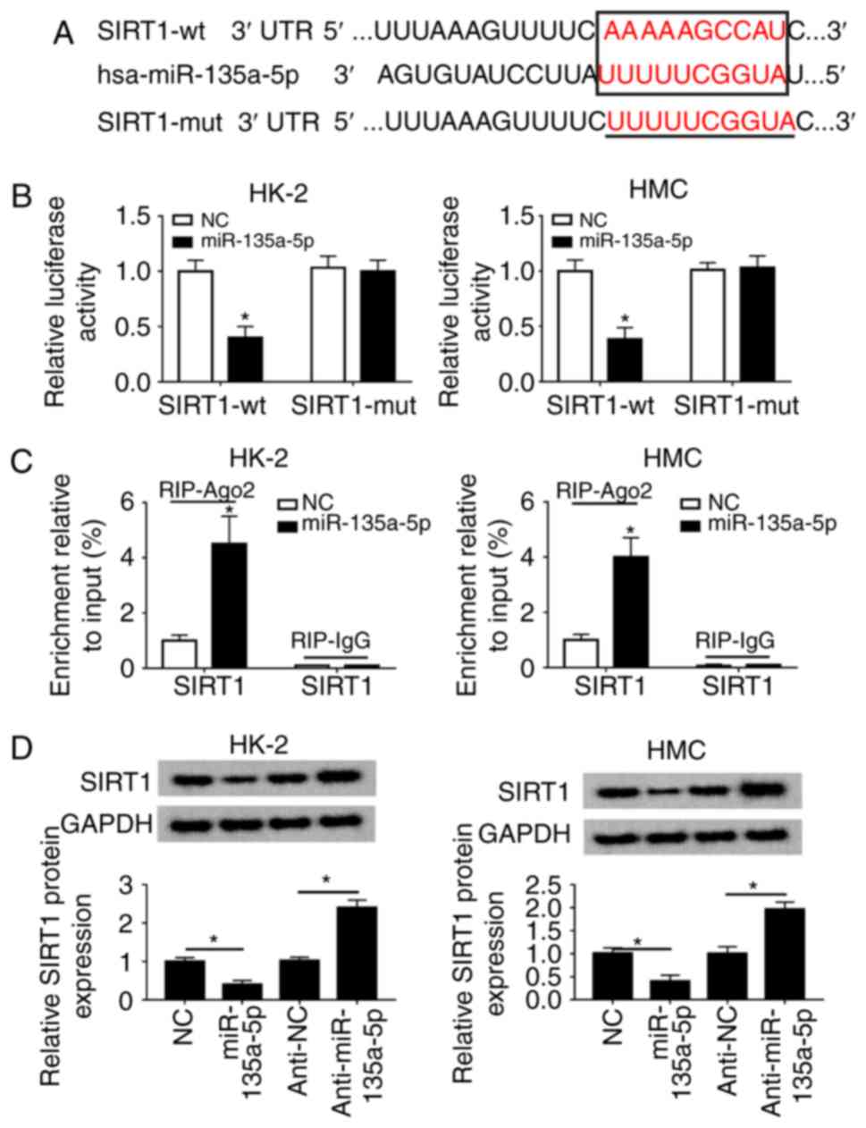 | Figure 5miR-135a-5p directly targets SIRT1
3′UTR. (A) TargetScan Human algorithm predicted the target sequence
of hsa-miR-135a-5p in the SIRT1 3′ UTR. (B) Luciferase reporter
assay was used to validate the relative luciferase activity of
vectors containing the SIRT1-wt/mut in HMC and HK-2 cells when
co-transfected with miR-135a-5p mimic or NC. (C) RNA
immunoprecipitation assay was performed to further identify the
level of SIRT1 mRNA in HMC and HK-2 cells co-transfected with
miR-135a-5p or NC. (D) Western blotting was used to analyze the
SIRT1 protein levels in HMC and HK-2 cells transfected with
anti-miR-135a-5p, miR-135a-5p and the corresponding controls. Data
were plotted as the mean ± standard error of the mean and performed
in triplicate. *P<0.05 vs. control cells (NC or
anti-NC). FN, fibronectin; SMA, smooth muscle actin; TGF,
transforming growth factor; HMC, human mesangial cells; miR,
microRNA; E, epithelial; NC, negative control; UTR, untranslated
region; mut, mutant; wt, wild-type; miR-135a-5p, miR-135a-5p mimic;
anti-miR-135a-5p, miR-135a-5p inhibitor. |
SIRT1 mediates the role of miR-135a-5p
knockdown in TGFβ1-induced renal fibrosis in vitro
Rescue experiments were performed to clarify the
effects of SIRT1 dysregulation on the role of miR-135a-5p in HK-2
and HMC cells. As presented in Fig.
6A, HMC and HK-2 cells were divided into four transfection
groups: Anti-NC, anti-miR-135a-5p, anti-miR-135a-5p + scramble and
anti-miR-135a-5p + siSIRT1. The upregulation of SIRT1 induced by
anti-miR-135a-5p was impaired by siSIRT1 (Fig. 6A), and western blotting confirmed
that siSIRT1 transfection caused a significant decrease of the
SIRT1 level in HMC and HK-2 cells compared with the scramble
siRNA-transfected cells (Fig.
S3B). When miR-135a-5p was inhibited, collagen 1A1, α-SMA and
FN synthesis was reduced compared with the NC group, which was
blocked by silencing of SIRT1 (Fig.
6B). Knockdown of SIRT1 abolished the effects of
anti-miR-135a-5p on E-cadherin expression as indicated in Fig. 6B. These results demonstrated that
miR-135a-5p knockdown inhibited TGFβ1-induced renal fibrosis by
upregulating SIRT1.
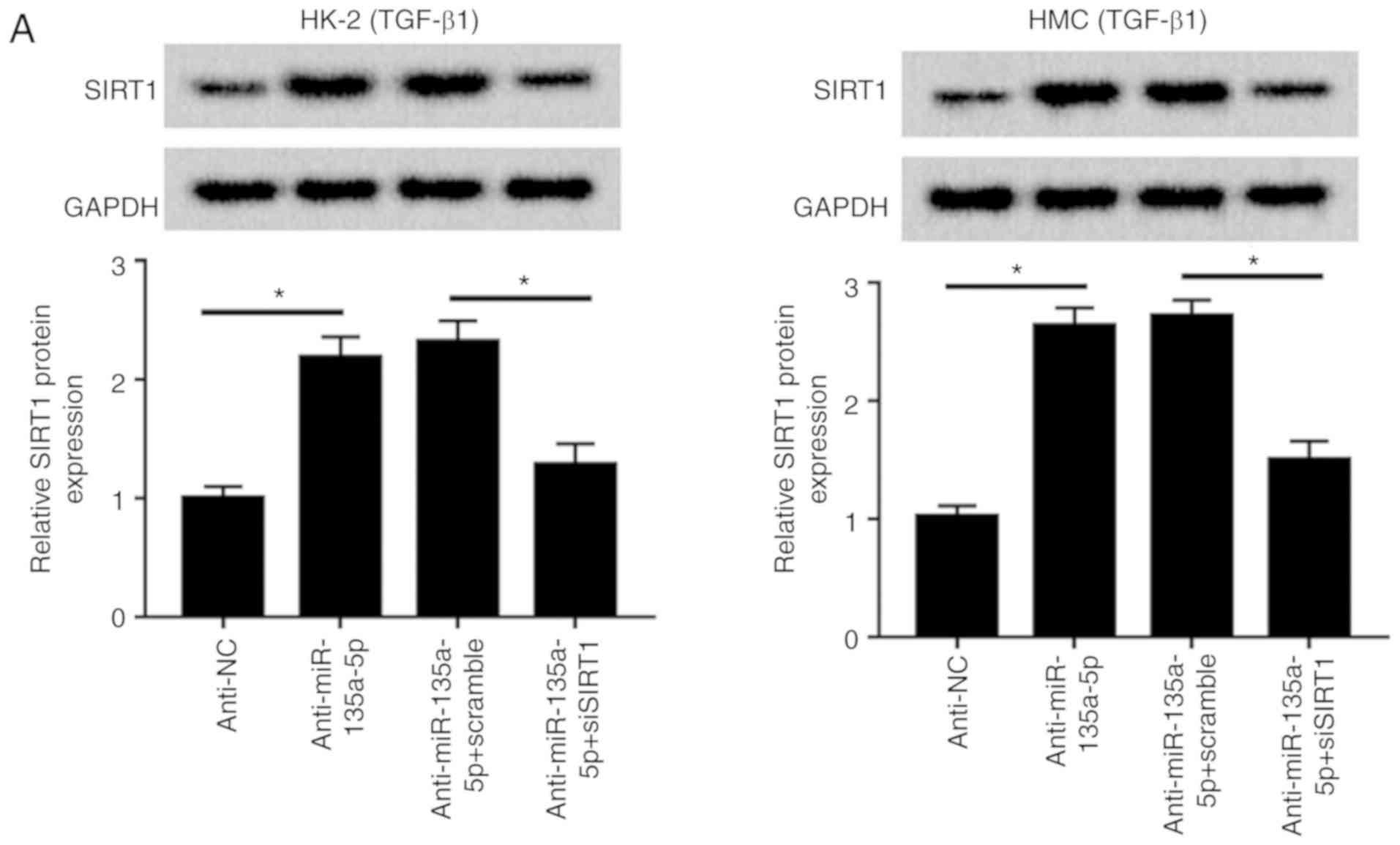 | Figure 6Influence of SIRT1 silencing on
TGFβ1-induced HMC and HK-2 cells. HMC and HK-2 cells were
transfected with siRNA against human SIRT1 (siSIRT1), and effect of
SIRT1 downregulation in miR-135a-5p-knocked down cell was evaluated
using western blotting. (A) Levels of SIRT1 were detected. (B)
Expression of collagen1A1, α-SMA, FN and E-cadherin was measured.
The quantification was performed on Image J. Data were plotted as
mean ± standard error of mean and performed in triplicate.
*P<0.05 vs. control cells (anti-NC or
anti-miR-135a-5p+scramble). FN, fibronectin; SMA, smooth muscle
actin; TGF, transforming growth factor; HMC, human mesangial cells;
miR, microRNA; E, epithelial; NC, negative control; si, small
interfering. |
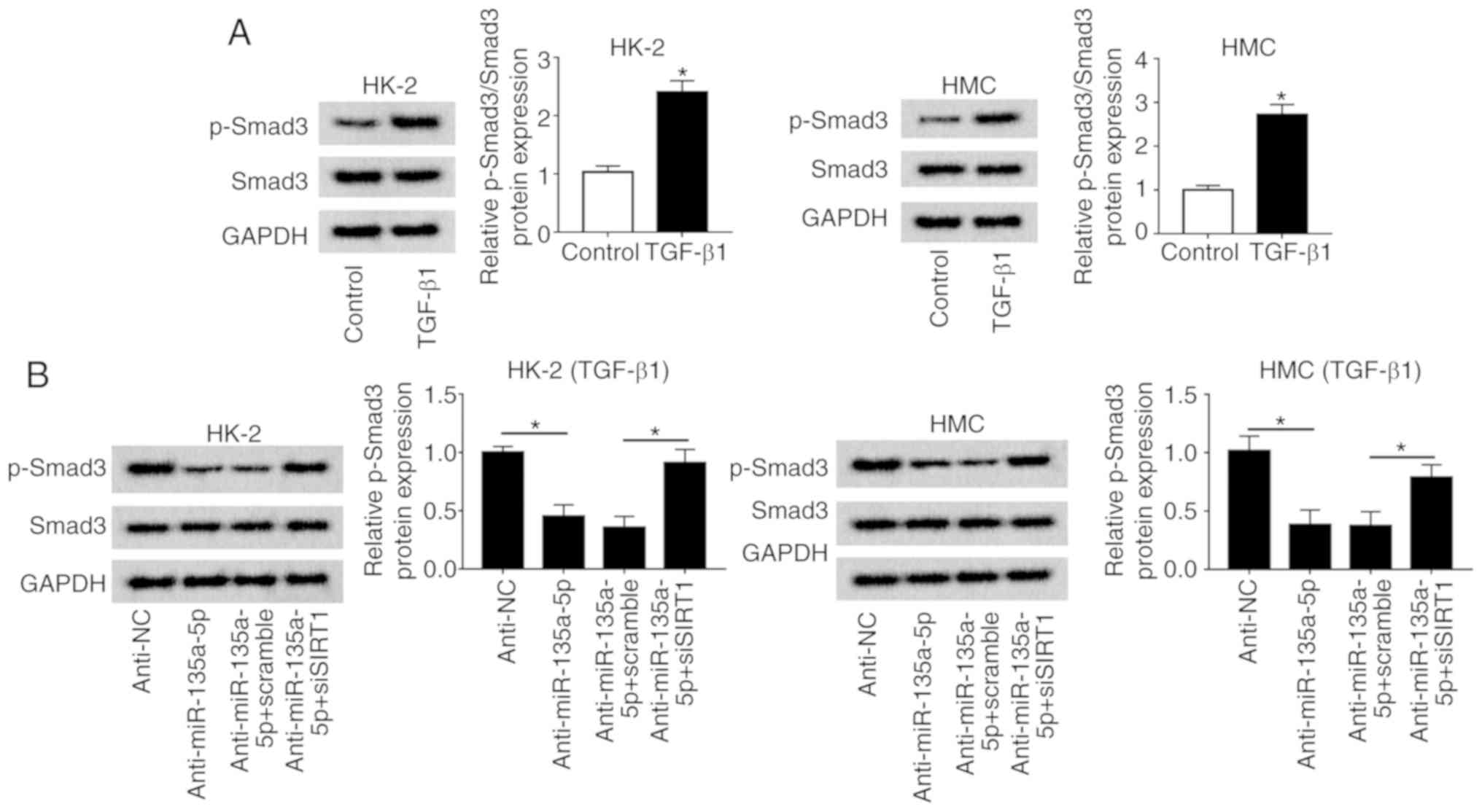
miR-135a-5p knockdown inactivates the
TGFβ1/Smad3 signaling pathway through upregulating SIRT1
To explore the signaling pathway underlying the
activity of miR-135a-5p during TGFβ1-induced renal fibrosis, Smad3
activation was measured. The upregulation of p-Smad normalized to
total Smad3 was observed in HMC and HK-2 cells under TGFβ1
stimulation compared with that in untreated cells (Fig. 7A). The relative level of p-Smad3
was significantly reduced in the anti-miR-135a-5p group compared
with that in the anti-NC group; in addition, the inactivation of
Smad3 induced by miR-135a-5p knockdown was reversed by silencing
SIRT1 in HMC and HK-2 cells (Fig.
7B). These results indicated that the inhibition of the
TGFβ1/Smad3 signaling pathway was involved in the role of
miR-135a-5p/SIRT1 in renal fibrosis in vitro.
Discussion
In China, diabetes has become a major public health
problem (24), and ~10% of
patients with diabetes suffer from DN (25). Progressive renal fibrosis is one
of the hallmark pathological characteristics of DN (8). For example, the fibrosis-related
genes collagen I, FN, E-cadherin and α-SMA were upregulated in a
mouse model of DN (26). Bai
et al (27) observed that
the levels of Snail, Vimentin, collagen IV and α-SMA were
upregulated, and E-cadherin was downregulated in 86 renal biopsies
of DN. Putta et al (28)
reported that silencing of miR-192 caused downregulation of key
profibrotic genes such as collagen 1A2, collagen 4A1 and FN in the
glomeruli and cortex of diabetic mice. In addition, it was also
suggested that the epithelial-mesenchymal transition (EMT) served
as one potential mechanism underlying renal fibrosis in DN
(29,30). In the current study, TGFβ1
treatment induced renal fibrosis in HMC and HK-2 cells as
demonstrated by the increased synthesis of collagen 1A1, α-SMA and
FN, as well as by decreased expression of E-cadherin.
Numerous miRNAs have been reported to be involved in
renal fibrosis in DN. For example, Zhao et al (31) demonstrated that miR-23b was
expressed at a lower level in the serum of patients with diabetes
mellitus and concluded that miR-23b had a protective effect against
renal fibrosis in DN. Expression of miR-192 was upregulated by
TGF-β1 in cultured glomerular mesangial cells and diabetic
glomeruli of mice (28). The
specific reduction of renal miR-192 decreased renal fibrosis and
improve proteinuria. These results supported the possibility of an
anti-miRNA-based translational approach to the treatment of DN. The
results of the present study demonstrated that inhibition of
miR-135a-5p the upregulation of E-cadherin levels, but reduced
collagen 1A1, α-SMA, and FN expression in TGFβ1-induced HMC and
HK-2 cells. The present results also revealed that SIRT1 was a
target gene of miR-135a-5p, and silencing of SIRT1 abolished the
effects of miR-135a-5p on renal fibrosis. In addition, Smad3
activation was altered by miR-135a-5p/SIRT1 in HMC and HK-2
cells.
As an oncogene, miR-135a-5p promotes cell
proliferation and metastasis in hepatocellular carcinoma (32,33) and breast cancer (34). However, miR-135a-5p served as an
anti-oncogene and targeted HOXA10 to suppress the proliferation of
head and neck squamous cell carcinoma (35). Thus, miR-135a-5p may serve a dual
role in cancers. In diabetes, expression of this miRNA is
considered to be upregulated. For instance, Agarwal et al
(36) observed elevated
miR-135a-5p levels in human diabetic skeletal muscle. Upregulation
of miR-135a-5p was also identified in the serum miRNA expression
profile and renal tissues from patients with DN (21). In addition, the biological role
and mechanism of miR-135a-5p was preliminarily explored, and the
results demonstrated that miR-135a-5p upregulation promoted
mesangial cell proliferation by decreasing G1/S arrest and
increasing synthesis of ECM proteins such as FN, Vimentin, and
collagen I by directly regulating short transient receptor
potential channel 1 in a HMC line (21); by contrast, silencing of
miR-135a-5p alleviated hyperglycemia and improved glucose tolerance
in vivo (36). Thus,
miR-135a-5p may be associated with fibrosis and diabetes. The
present study focused on the expression of miR-135a-5p in patients
with DN, as well as its biological role in TGFβ1-induced renal
fibrosis cell models in HMC and HK-2 cells. The results
demonstrated that the expression levels of miR-135a-5p were
increased in the sera and renal tissues of patients with DN as well
as in HMC and HK-2 cells under various glucose concentrations or
TGFβ1 stimulation. Functionally, anti-miR-135a-5p attenuated the
expression of collagen 1A1, α-SMA and FN, and elevated the levels
of E-cadherin under TGFβ1 stimulation in vitro, which was in
agreement with the previous findings by He et al (21). In addition, SIRT1 has been
demonstrated to mediate the inhibitory activity of miR-135a-5p in
the synthesis of fibrosis-related genes. Of note, Wu et al
(37) recently investigated the
role and possible regulatory mechanism of miR-135a-5p in cardiac
fibrosis and reported that cardiac fibroblasts from neonatal rats
induced by isoproterenol was inhibited by miR-135a-5p targeting the
transient receptor potential melastatin 7. Simultaneously,
miR-135a-5p was decreased in ISO-induced cardiac fibrosis in
vitro and in vivo (37). These results suggested the complex
and vital role of miR-135a-5p in the biological functions of
diseases, including cancer and diabetes complications.
SIRT1 has been demonstrated to serve a crucial role
in miscellaneous physiological processes through the deacetylation
of a number of nuclear proteins such as p53 and NF-κB (22,38,39). Previous studies have demonstrated
that SIRT1 reduces apoptosis in TGFβ-treated mesangial cells via
acceleration of Smad7 degradation and the TGFβ signaling pathway
(40). In addition, SIRT1
regulates fibroblast activation and tissue fibrosis by canonical
TGFβ signaling (10). Inhibition
of SIRT1 promotes TGFβ1-induced EMT and renal fibrosis in HK-2
cells (26). Accumulating
evidence has indicated SIRT1 is affected by miRNAs during
fibrogenesis, including that in the kidney. For example, inhibition
of miR-133b and miR-199b attenuated TGF-β1-induced EMT and renal
fibrosis by targeting SIRT1 (26). miR-34a targeting SIRT1 aggravated
high glucose-stimulated tubulointerstitial fibrosis in HK cells
(41,42). In the present study, the results
demonstrated that SIRT1, which is a vital regulator in the
evolution of renal fibrosis in DN (22), was directly suppressed by
miR-135a-5p. Furthermore, the current study proposed that the
miR-135a-5p/SIRT1 axis may provide a new approach for DN
treatment.
Previously, SIRT1 activation had been suggested as a
therapeutic strategy in progressive, fibrotic diseases in the
kidney, liver, lung and heart (14,43-45). Mechanically, inhibition of the
TGFβ1/Smad3 pathway has been attributed to the protective role of
SIRT1 activation in organ fibrosis including renal fibrosis
(14). For example,
co-immunoprecipitation assays have provided direct evidence of an
interaction between acetylated Smad3 and SIRT1 (14,46). In the kidney, knockdown of SIRT1
increases the levels of acetylated Smad3, thus substantially
enhancing the transcriptional activity of Smad3 following TGF-β1
treatment (14). Additionally,
the allosteric modifier of SIRT1 deacetylase ameliorates the
TGFβ1-stimulated collagen production, which is accompanied by a
reduction of Smad3 reporter activity (47). The present study indicated that
the miR-135a-5p/SIRT1/Smad3 pathway was involved in TGFβ1-induced
renal fibrosis.
One limitation of the current study was that it did
not verify the suppressive activity of miR-135a-5p in diabetic
db/db mice by injection of recombinant lentivirus
containing miR-135a-5p inhibitor (21,26,28). Furthermore, immunohistochemistry
examination of the kidney was not performed (14). TGFβ regulates biological processes
by interacting with Smads, MAPK and Jagged/Notch signaling pathways
(10). The results of the present
study suggested that the miR-135a-5p/SIRT1 axis regulated Smad3
activation; it would be interesting to verify whether the other two
signaling pathways may be altered by the functions of the
miR-135a-5p/SIRT1 axis. Therefore, the identification of molecular
pathways underlying DN would be imperative for development of new
therapeutic strategies.
In conclusion, the results of the present study
demonstrated that miR-135a-5p knockdown attenuated renal fibrosis
in DN by targeting SIRT1 and inactivating the TGFβ1/Smad3 pathway.
These results supported the hypothesis that miR-135a-5p may be a
novel therapeutic target in suppressing renal fibrosis in DN.
Supplementary Data
Abbreviations:
|
DN
|
diabetic nephropathy
|
|
SIRT1
|
sirtuin 1
|
|
HMC
|
human mesangial cells
|
|
α-SMA
|
α-smooth muscle actin
|
|
FN
|
fibronectin
|
|
ECM
|
extracellular matrix
|
|
TGFβ
|
transforming growth factor-β
|
|
Smad
|
mothers against decapentaplegic
homolog
|
|
MAPK
|
mitogen activated protein kinase
|
Acknowledgments
Not applicable.
Funding
This study was supported by the Health Commission of
Hubei Province scientific research project 'The role and mechanism
of interferon regulatory factor 8 in renal tubular injury in
diabetic nephropathy' (project no. WJ2019M210) and The Wuhan
Science and Technology Bureau (grant no. 2017060201010179).
Availability of data and materials
All data generated or analyzed during this study are
included in this published article.
Authors' contributions
JZ and LZ conceived and designed the experiments. DZ
performed the experiments and acquired funding. XW and JZ
contributed the reagents/materials/analysis tools and performed
data analysis and interpretation. LZ wrote the manuscript. All
authors read and approved the final manuscript.
Ethics approval and consent to
participate
All protocols involving human subjects were approved
by the Ethics Committee of the Zhongnan Hospital of Wuhan
University. Informed consent was obtained from all patients.
Patient consent for publication
Not applicable.
Competing interests
The authors declare that they have no competing
interests.
References
|
1
|
Liu ZH: Nephrology in China. Nat Rev
Nephrol. 9:523–528. 2013. View Article : Google Scholar : PubMed/NCBI
|
|
2
|
Coimbra TM, Janssen U, Gröne HJ, Ostendorf
T, Kunter U, Schmidt H, Brabant G and Floege J: Early events
leading to renal injury in obese Zucker (fatty) rats with type II
diabetes. Kidney Int. 57:167–182. 2000. View Article : Google Scholar : PubMed/NCBI
|
|
3
|
Kanwar YS, Sun L, Xie P, Liu FY and Chen
S: A glimpse of various pathogenetic mechanisms of diabetic
nephropathy. Annu Rev Pathol. 6:395–423. 2011. View Article : Google Scholar : PubMed/NCBI
|
|
4
|
Gilbert RE and Cooper ME: The
tubulointerstitium in progressive diabetic kidney disease: More
than an aftermath of glomerular injury? Kidney Int. 56:1627–1637.
1999. View Article : Google Scholar : PubMed/NCBI
|
|
5
|
Steffes MW, Osterby R, Chavers B and Mauer
SM: Mesangial expansion as a central mechanism for loss of kidney
function in diabetic patients. Diabetes. 38:1077–1081. 1989.
View Article : Google Scholar : PubMed/NCBI
|
|
6
|
Wang S, Yang Z, Xiong F, Chen C, Chao X,
Huang J and Huang H: Betulinic acid ameliorates experimental
diabetic-induced renal inflammation and fibrosis via inhibiting the
activation of NF-κB signaling pathway. Mol Cell Endocrinol.
434:135–143. 2016. View Article : Google Scholar : PubMed/NCBI
|
|
7
|
Mason RM and Wahab NA: Extracellular
matrix metabolism in diabetic nephropathy. J Am Soc Nephrol.
14:1358–1373. 2003. View Article : Google Scholar : PubMed/NCBI
|
|
8
|
Song KH, Park J, Park JH, Natarajan R and
Ha H: Fractalkine and its receptor mediate extracellular matrix
accumulation in diabetic nephropathy in mice. Diabetologia.
56:1661–1669. 2013. View Article : Google Scholar : PubMed/NCBI
|
|
9
|
Wynn TA: Common and unique mechanisms
regulate fibrosis in various fibroproliferative diseases. J Clin
Invest. 117:524–529. 2007. View
Article : Google Scholar : PubMed/NCBI
|
|
10
|
Hills CE and Squires PE: The role of TGF-β
and epithelial-to mesenchymal transition in diabetic nephropathy.
Cytokine Growth Factor Rev. 22:131–139. 2011.PubMed/NCBI
|
|
11
|
Sutariya B, Jhonsa D and Saraf MN: TGF-β:
The connecting link between nephropathy and fibrosis.
Immunopharmacol Immunotoxicol. 38:39–49. 2016. View Article : Google Scholar
|
|
12
|
Chen S, Hong SW, Iglesias-de la Cruz MC,
Isono M, Casaretto A and Ziyadeh FN: The key role of the
transforming growth factor-beta system in the pathogenesis of
diabetic nephropathy. Ren Fail. 23:471–481. 2001. View Article : Google Scholar : PubMed/NCBI
|
|
13
|
Sharma K, Jin Y, Guo J and Ziyadeh FN:
Neutralization of TGF-beta by anti-TGF-beta antibody attenuates
kidney hypertrophy and the enhanced extracellular matrix gene
expression in STZ-induced diabetic mice. Diabetes. 45:522–530.
1996. View Article : Google Scholar : PubMed/NCBI
|
|
14
|
Huang XZ, Wen D, Zhang M, Xie Q, Ma L,
Guan Y, Ren Y, Chen J and Hao CM: Sirt1 activation ameliorates
renal fibrosis by inhibiting the TGF-β/Smad3 pathway. J Cell
Biochem. 115:996–1005. 2014. View Article : Google Scholar
|
|
15
|
Böttinger EP and Bitzer M: TGF-beta
signaling in renal disease. J Am Soc Nephrol. 13:2600–2610. 2002.
View Article : Google Scholar : PubMed/NCBI
|
|
16
|
Massagué J and Chen YG: Controlling
TGF-beta signaling. Genes Dev. 14:627–644. 2000.PubMed/NCBI
|
|
17
|
Kato M: TGF-β-induced signaling circuit
loops mediated by microRNAs as new therapeutic targets for renal
fibrosis? Kidney Int. 84:1067–1069. 2013. View Article : Google Scholar : PubMed/NCBI
|
|
18
|
Lu Z and Wang F: Epigenetic regulations in
diabetic nephropathy. J Diabetes Res. 2017:78050582017. View Article : Google Scholar : PubMed/NCBI
|
|
19
|
Cui C, Cui Y, Fu Y, Ma S and Zhang S:
Microarray analysis reveals gene and microRNA signatures in
diabetic kidney disease. Mol Med Rep. 17:2161–2168. 2018.
|
|
20
|
Rysz J, Gluba-Brzózka A, Franczyk B,
Jabłonowski Z and Ciałkowska-Rysz A: Novel biomarkers in the
diagnosis of chronic kidney disease and the prediction of its
outcome. Int J Mol Sci. 18:17022017. View Article : Google Scholar :
|
|
21
|
He F, Peng F, Xia X, Zhao C, Luo Q, Guan
W, Li Z, Yu X and Huang F: MiR-135a promotes renal fibrosis in
diabetic nephrop-athy by regulating TRPC1. Diabetologia.
57:1726–1736. 2014. View Article : Google Scholar : PubMed/NCBI
|
|
22
|
Kume S, Kitada M, Kanasaki K, Maegawa H
and Koya D: Anti-aging molecule, Sirt1: A novel therapeutic target
for diabetic nephropathy. Arch Pharm Res. 36:230–236. 2013.
View Article : Google Scholar : PubMed/NCBI
|
|
23
|
Livak KJ and Schmittgen TD: Analysis of
relative gene expression data using real-time quantitative PCR and
the 2(-Delta Delta C(T)) method. Methods. 25:402–408. 2001.
View Article : Google Scholar
|
|
24
|
Yang W, Lu J, Weng J, Jia W, Ji L, Xiao J,
Shan Z, Liu J, Tian H, Ji Q, et al: Prevalence of diabetes among
men and women in China. N Engl J Med. 362:1090–1101. 2010.
View Article : Google Scholar : PubMed/NCBI
|
|
25
|
Roglic G, Unwin N, Bennett PH, Mathers C,
Tuomilehto J, Nag S, Connolly V and King H: The burden of mortality
attributable to diabetes: Realistic estimates for the year 2000.
Diabetes Care. 28:2130–2135. 2005. View Article : Google Scholar : PubMed/NCBI
|
|
26
|
Sun Z, Ma Y, Chen F, Wang S, Chen B and
Shi J: miR-133b and miR-199b knockdown attenuate TGF-β1-induced
epithelial to mesenchymal transition and renal fibrosis by
targeting SIRT1 in diabetic nephropathy. Eur J Pharmacol.
837:96–104. 2018. View Article : Google Scholar : PubMed/NCBI
|
|
27
|
Bai X, Geng J, Zhou Z, Tian J and Li X:
MicroRNA-130b improves renal tubulointerstitial fibrosis via
repression of Snail-induced epithelial-mesenchymal transition in
diabetic nephropathy. Sci Rep. 6:204752016. View Article : Google Scholar : PubMed/NCBI
|
|
28
|
Putta S, Lanting L, Sun G, Lawson G, Kato
M and Natarajan R: Inhibiting microRNA-192 ameliorates renal
fibrosis in diabetic nephropathy. J Am Soc Nephrol. 23:458–469.
2012. View Article : Google Scholar : PubMed/NCBI
|
|
29
|
Loeffler I and Wolf G:
Epithelial-to-mesenchymal transition in diabetic nephropathy: Fact
or fiction? Cells. 4:631–652. 2015. View Article : Google Scholar : PubMed/NCBI
|
|
30
|
Simonson MS: Phenotypic transitions and
fibrosis in diabetic nephropathy. Kidney Int. 71:846–854. 2007.
View Article : Google Scholar : PubMed/NCBI
|
|
31
|
Zhao B, Li H, Liu J, Han P, Zhang C, Bai
H, Yuan X, Wang X, Li L, Ma H, et al: MicroRNA-23b targets ras
GTPase-activating protein SH3 domain-binding protein 2 to alleviate
fibrosis and albuminuria in diabetic Nephropathy. J Am Soc Nephrol.
27:2597–2608. 2016. View Article : Google Scholar : PubMed/NCBI
|
|
32
|
Zeng YB, Liang XH, Zhang GX, Jiang N,
Zhang T, Huang JY, Zhang L and Zeng XC: miRNA-135a promotes
hepatocellular carcinoma cell migration and invasion by targeting
forkhead box O1. Cancer Cell Int. 16:632016. View Article : Google Scholar : PubMed/NCBI
|
|
33
|
Yao S, Tian C, Ding Y, Ye Q, Gao Y, Yang N
and Li Q: Down-regulation of Krüppel-like factor-4 by
microRNA-135a-5p promotes proliferation and metastasis in
hepatocellular carcinoma by transforming growth factor-β1.
Oncotarget. 7:42566–42578. 2016. View Article : Google Scholar : PubMed/NCBI
|
|
34
|
Chen Y, Zhang J, Wang H, Zhao J, Xu C, Du
Y, Luo X, Zheng F, Liu R, Zhang H and Ma D: miRNA-135a promotes
breast cancer cell migration and invasion by targeting HOXA10. BMC
Cancer. 12:1112012. View Article : Google Scholar : PubMed/NCBI
|
|
35
|
Guo LM, Ding GF, Xu W, Ge H, Jiang Y, Chen
XJ and Lu Y: MiR-135a-5p represses proliferation of HNSCC by
targeting HOXA10. Cancer Biol Ther. 19:973–983. 2018. View Article : Google Scholar
|
|
36
|
Agarwal P, Srivastava R, Srivastava AK,
Ali S and Datta M: miR-135a targets IRS2 and regulates insulin
signaling and glucose uptake in the diabetic gastrocnemius skeletal
muscle. Biochim Biophys Acta. 1832:1294–1303. 2013. View Article : Google Scholar : PubMed/NCBI
|
|
37
|
Wu Y, Liu Y, Pan Y, Lu C, Xu H, Wang X,
Liu T, Feng K and Tang Y: MicroRNA-135a inhibits cardiac fibrosis
induced by isoproterenol via TRPM7 channel. Biomed Pharmacother.
104:252–260. 2018. View Article : Google Scholar : PubMed/NCBI
|
|
38
|
Yuan F, Xie Q, Wu J, Bai Y, Mao B, Dong Y,
Bi W, Ji G, Tao W, Wang Y and Yuan Z: MST1 promotes apoptosis
through regulating Sirt1-dependent p53 deacetylation. J Biol Chem.
286:6940–6945. 2011. View Article : Google Scholar : PubMed/NCBI
|
|
39
|
Salminen A and Kaarniranta K: NF-kappaB
signaling in the aging process. J Clin Immunol. 29:397–405. 2009.
View Article : Google Scholar : PubMed/NCBI
|
|
40
|
Kume S, Haneda M, Kanasaki K, Sugimoto T,
Araki S, Isshiki K, Isono M, Uzu T, Guarente L, Kashiwagi A and
Koya D: SIRT1 inhibits transforming growth factor beta-induced
apoptosis in glomerular mesangial cells via Smad7 deacetylation. J
Biol Chem. 282:151–158. 2007. View Article : Google Scholar
|
|
41
|
Xue M, Li Y, Hu F, Jia YJ, Zheng ZJ, Wang
L and Xue YM: High glucose up-regulates microRNA-34a-5p to
aggravate fibrosis by targeting SIRT1 in HK-2 cells. Biochem
Biophys Res Commun. 498:38–44. 2018. View Article : Google Scholar : PubMed/NCBI
|
|
42
|
Li A, Peng R, Sun Y, Liu H, Peng H and
Zhang Z: LincRNA 1700020I14Rik alleviates cell proliferation and
fibrosis in diabetic nephropathy via miR-34a-5p/Sirt1/HIF-1α
signaling. Cell Death Dis. 9:4612018. View Article : Google Scholar
|
|
43
|
Bugyei-Twum A, Ford C, Civitarese R,
Seegobin J, Advani SL, Desjardins JF, Kabir G, Zhang Y, Mitchell M,
Switzer J, et al: Sirtuin 1 activation attenuates cardiac fibrosis
in a rodent pressure overload model by modifying Smad2/3
transactivation. Cardiovasc Res. 114:1629–1641. 2018. View Article : Google Scholar : PubMed/NCBI
|
|
44
|
Yang T, Wang J, Pang Y, Dang X, Ren H, Liu
Y, Chen M and Shang D: Emodin suppresses silica-induced lung
fibrosis by promoting Sirt1 signaling via direct contact. Mol Med
Rep. 14:4643–4649. 2016. View Article : Google Scholar : PubMed/NCBI
|
|
45
|
Jiang R, Zhou Y, Wang S, Pang N, Huang Y,
Ye M, Wan T, Qiu Y, Pei L, Jiang X, et al: Nicotinamide riboside
protects against liver fibrosis induced by CCl4 via
regulating the acetylation of Smads signaling pathway. Life Sci.
225:20–28. 2019. View Article : Google Scholar : PubMed/NCBI
|
|
46
|
Li J, Qu X, Ricardo SD, Bertram JF and
Nikolic-Paterson DJ: Resveratrol inhibits renal fibrosis in the
obstructed kidney: Potential role in deacetylation of Smad3. Am J
Pathol. 177:1065–1071. 2010. View Article : Google Scholar : PubMed/NCBI
|
|
47
|
Zhang Y, Connelly KA, Thai K, Wu X, Kapus
A, Kepecs D and Gilbert RE: Sirtuin 1 activation reduces
transforming growth factor-β1-induced fibrogenesis and affords
organ protection in a model of progressive, experimental kidney and
associated cardiac disease. Am J Pathol. 187:80–90. 2017.
View Article : Google Scholar
|