Introduction
Type 2 diabetes mellitus (T2DM) is a metabolic
disorder characterized by insulin resistance and hyperglycemia
(1-3). The prevalence of diabetes in adults
is 6.4%, affecting 285 million adults in 2010 and expected to
increase to 7.7% and 439 million adults by 2030 (4,5).
Diabetes increases the risk of cardiovascular disease, such as
atherosclerosis (6). Endothelial
dysfunction is a hallmark of diabetes. Vascular endothelial
complications, such as endothelial cell dysfunction and vascular
inflammation, contribute to the morbidity and mortality of
diabetes, which are common causes of limb amputation (7). Inflammation is considered as a key
event in vascular dysfunction. In diabetes, the pro-inflammatory
phenotype is enhanced by a variety of factors, including
pro-inflammatory cytokines, such as interleukin (IL)-1β, IL-6,
IL-18 and tumor necrosis factor (TNF)-α, and nuclear factor (NF)-κB
(8). The main cause of diabetic
vascular complications is hyperglycemia, which is associated with
endothelial cell dysfunction. Hyperglycemia is a major risk factor
for atherosclerosis, and atherosclerosis is the most important
cause of various cardiovascular complications (9). Endothelial cells isolated from
healthy subjects exposed to high glucose (HG) or isolated from
diabetic patients exhibit limited proliferative capacity (10). Clinical studies have demonstrated
that protecting vascular endothelial cells from damage may be a
useful approach to the treatment of cardiovascular complications in
diabetes. However, there is currently a lack of effective drugs for
preventing the development of cardiovascular diseases in diabetic
patients.
Plants have been used in traditional medicine due to
their beneficial and protective properties (11). It has been reported that carvacrol
(2-methyl-5-isopropylphenol), a monoterpenic phenol, has several
pharmacological properties, such as anti-inflammatory (12), antioxidant (13), antiapoptotic (14), anticancer (14) and antimicrobial properties
(15). Accumulating evidence from
in vitro studies confirms these properties: For example,
carvacrol was found to reduce the serum levels of inflammatory
mediators and improve respiratory symptoms in veterans exposed to
sulfur mustard (16). However,
the effects of carvacrol on diabetes remain unclear.
In the present study, genetically hyperglycemic
db/db mice were used as a T2DM model (17,18) to investigate whether carvacrol can
alleviate vascular inflammation in diabetes.
Materials and methods
Animals
A total of 45 male C57BL/KsJ db/db mice (age, 8
weeks; weight, 32-36 g) and 15 age-matched C57BL/6J control
non-diabetic db/m+ mice (age, 6 weeks; weight, 16-18 g)
were purchased from Changzhou Cavans Experimental Animal Co., Ltd.,
(SCXK2001-0003). All mice were housed in a well-ventilated
environment, with a 12-h light-dark cycle, at 23±2°C and 70±10%
humidity, with free access to water and food. All animal
experiments were performed strictly in accordance with the Guide
for the Care and Use of Laboratory Animals of the National
Institutes of Health. The research protocol was approved by the
Traditional Chinese Medicine Guizhou University Animal Care and
Ethics Committee.
Experimental design for the T2DM animal
model
All mice were randomly divided into four groups as
follows: i) Age-matched healthy control (n=15); ii) model control;
iii) db/db model + low-dose carvacrol (5 mg/kg); and iv) db/db
model + high-dose carvacrol (10 mg/kg) groups. All mice were
anesthetized by intraperitoneal injection of pentobarbital sodium
(50 mg/kg). Subsequently, the db/db mice were treated with
carvacrol (282197-50G, Sigma-Aldrich; Merck KGaA) daily for 6 weeks
by gavage. At the same time, the normal control and model control
groups were administered 0.9% saline at equal volumes. After 6
weeks, all the mice were euthanized by intraperitoneal injection of
pentobarbital sodium (200 mg/kg) according to the recommendations
of the animal ethics guidelines. Blood was quickly collected,
centrifuged (at 3,000 × g for 10 min at 4°C) to obtain the blood
serum samples, and stored at -20°C. The pancreatic tissues and
skeletal muscles were removed and immediately immersed in 4%
paraformaldehyde for 12 h at 4°C.
Oral glucose tolerance test (OGTT)
After 8 h of fasting, the mice were orally
administered glucose solution (1.2 g/kg body weight). Blood was
drawn from the tail vein, and the glucose levels were measured
using a glucose monitor (Ascensia ELITE; Bayer).
Serum lipid and insulin levels
The fasting serum levels of total cholesterol,
triglyceride (TG), high-density lipoprotein (HDL) and non-HDL were
detected using enzymatic methods (Stanbio Laboratory). Furthermore,
the serum insulin concentration was analyzed by enzyme immunoassay
(Mercodia).
Histological examination and
immunohistochemical analyses
The thoracoabdominal aorta was fixed at room
temperature for 48 h in a buffer solution of 10% formalin, and then
embedded in paraffin and sectioned at 20 µm. The sections
were subjected to Masson's trichrome and hematoxylin and eosin
(H&E) staining and observed under an optical microscope at a
magnification, ×200 (BX51, Olympus Corporation). For
immunohistochemical analyses, paraffin-embedded sections were
incubated with anti-inhibitor of NF-κB kinase (anti-IKK; 1:100,
ab32041, Abcam), anti-NF-κB inhibitor-α (anti-IKB-α; 1:100,
ab32518, Abcam), anti-NALP3 (1:150, 19771-1-AP; ProteinTech Group,
Inc.), anti-NF-κB (1:150, 14220-1-AP; ProteinTech Group, Inc.),
anti-phosphorylated insulin receptor (anti-p-InsR; 1:100, ab60946,
Abcam), anti-phosphorylated insulin receptor substrate-1
(anti-p-ISR-1; 1:100, ab3690, Abcam) and anti-toll-like receptor
(TLR)4 (1:200, ab22048, Abcam) at 4°C overnight. Subsequently, the
slides were incubated with secondary antibody (PV-9001; ZSGB-BIO,
China) for 2 h at room temperature. The optical density was
measured using ImageJ software, v1.8.0 (National Institutes of
Health). The semi-quantitative results were evaluated by scoring
the percentage of positive cells and staining intensity under the
microscope. The percentage of positive cells was scored as follows:
<5%, 0 points; 5 -25%, 1 point; 26-50%, 2 points; 51-75%, 3
points; and 76-100%, 4 points. The staining intensity was scored as
follows: 0, no staining; 1, light yellow; 2, brown; and 3, tan. The
product of the percentage of positive cells and the cell staining
intensity was defined as the grade: 0, negative (−); 1-4, weakly
positive (+); 5-8, positive (++); and 9-12, strongly positive
(+++).
Reverse transcription-quantitative PCR
(RT-qPCR) analysis
Total RNA was extracted from tissues or cells using
a TRIzol reagent kit (15596-026, Invitrogen; Thermo Fisher
Scientific, Inc.). Subsequently, the extracted RNA was
reverse-transcribed into cDNA on ice using the TaqMan cDNA
Synthesis kit (Applied Biosystems; Thermo Fisher Scientific, Inc.).
RT-qPCR was carried out with the miScript SYBR Green PCR kit
(A25742, PowerUp™ SYBR™ Green Master Mix, Applied Biosystems;
Thermo Fisher Scientific, Inc.). The conditions for RT-qPCR were as
follows: One cycle of 2 min at 50°C; one cycle of 10 min at 95°C;
40 cycles of 15 sec at 95°C and 40 cycles of 1 min at 60°C. The
primer sequences of the targeted genes are shown in Table I. The relative expression levels
of the genes were normalized to GAPDH expression. The fold changes
in expression were calculated using the 2-ΔΔCq method
(PMID: 18546601).
 | Table IPrimer sequence information for
reverse transcription-quantitative PCR. |
Table I
Primer sequence information for
reverse transcription-quantitative PCR.
| Target gene | Primer sequence
(5′-3′) |
|---|
| IKK | Forward:
ACGACCTAGAGGAGCAAGCA |
| Reverse:
AGCTCTGAATTGCCTGAAGC |
| IKB-α | Forward:
GGTGTTTGAATGTATTGCTGG |
| Reverse:
AGGCTGTTTGGCTGAGGT |
| NALP3 | Forward:
TGGATCTAGCCACGCTAATG |
| Reverse:
AAACCCATCCACTCCTCTTC |
| NF-κB | Forward:
ACCTGCCAGATACAGACGAT |
| Reverse:
GAAGCTGAGCTGCGGGAA |
| TLR4 | Forward:
TCCCTGAACCCTATGAAC |
| Reverse:
CTAAACCAGCCAGACCTT |
| GAPDH | Forward:
TGAGTCCTTCCACGATACCA |
| Reverse:
ATCCCATCACCATCTTCCAG |
Western blot analysis
Total protein was extracted from tissues or cells
using RIPA lysis buffer (R0010; Solarbio) supplemented with 1% PMSF
(P0100; Solarbio), and was determined with BCA Protein Assay kit
(Pierce; Thermo Fisher Scientific, Inc.). A total of 120 µg
protein was loaded per lane. Subsequently, the protein samples were
separated by 10% SDS-PAGE and transferred to a PVDF membrane
(IPVH00010; EMD Millipore). The membrane was then blocked with 5%
skimmed milk at room temperature for 1 h, followed by incubation
with the primary antibodies at 4°C overnight, and then incubation
with the secondary antibody for 45 min. The primary antibodies
included: Anti-IKK (1:1,000, ab32041; Abcam), anti-IKB-α (1:1,000,
ab32518, Abcam), anti-NALP3 (1:2,000, 19771-1-AP, ProteinTech
Group, Inc.), anti-NF-κB (1:2,000, 14220-1-AP, ProteinTech Group,
Inc.), anti-TLR4 (1:2,000, ab22048, Abcam) and anti-β-actin
(1:1,000; bs-0061R; BIOSS). β-actin was used as an internal
control. Finally, the blots were visualized using an ECL kit
(KGP1121; KeyGen Biotech Co., Ltd.).
Cell culture
Human umbilical vein endothelial cells (HUVECs) were
cultured for 48 h in Endothelial Cell Growth Medium-2 BulletKit™
(Gibco; Thermo Fisher Scientific, Inc.) at 37°C with 5%
CO2, and divided into three groups as follows: i)
Control; ii) endothelial cells + HG; and iii) endothelial cells +
HG + carvacrol.
ELISA
Inflammatory cytokines (IL-1β, IL-6, IL-18 and
TNF-α), insulin and TG levels in the serum were measured using a
96-well microplate and commercially available ELISA kits (cat. nos.
H007, H203 and H266, respectively; all from NanJing JianCheng Bio).
The optical density was read at 450 nm using a microplate reader
(saf-680t; Thermo Fisher Scientific Inc.). The concentrations of
IL-1β, IL-6, IL-18, TNF-α, insulin and TG were quantified based on
the standard curves that were constructed by Curve Expert 1.4
software (Beijing Boleide Development of Science and Technology
Co., Ltd.). Three wells from each sample were assayed in this
experiment.
Flow cytometry assay of cell
apoptosis
Vascular endothelial cell apoptosis was evaluated
using the Annexin V-FITC Apoptosis Detection kit (BestBio). The
cells were incubated with Annexin V-FITC and propidium iodide (PI)
in the dark for 15 min at room temperature. The number of apoptotic
cells was analyzed using flow cytometry (BD Biosciences).
Cell Counting Kit-8 (CCK-8) assay
The cells were seeded in a 96-well plate (3,000
cells/well) and were cultured under different concentrations of
carvacrol (20, 50, 100, 200, 500 and 1,000 µM/ml) at room
temperature for 24 h. Subsequently, cell viability was evaluated
using a CCK-8 Kit (Dojindo Molecular Technologies, Inc.) according
to the manufacturer's instructions.
Immunocytochemical analyses
HUVECs were fixed in 4% paraformaldehyde at 4°C for
15 min and incubated in hydrogen peroxide for 15 min. Subsequently,
the cells were blocked with goat serum (Solarbio) at 37°C for 30
min, followed by incubation with anti-IKK (1:100, ab32041; Abcam),
anti-IKB-α (1:100, ab32518; Abcam), anti-NALP3 (1:150, 19771-1-AP;
ProteinTech Group, Inc.), anti-NFκB (1:150, 14220-1-AP; ProteinTech
Group, Inc.), anti-p-INSR (1:100, ab60946; Abcam), anti-p-ISR-1
(1:100, ab3690; Abcam) and anti-TLR4 (1:200, ab22048; Abcam) at 4°C
overnight.
Statistical analysis
Statistical analyses were performed using GraphPad
Prism v7.0 (GraphPad Software, Inc.). Data are expressed as means ±
standard error of the mean. The differences between two groups were
calculated using Student's t-test. Comparisons among multiple
groups were performed using one-way analysis of variance followed
by Tukey's post hoc test. P<0.05 was considered to indicate
statistically significant differences.
Results
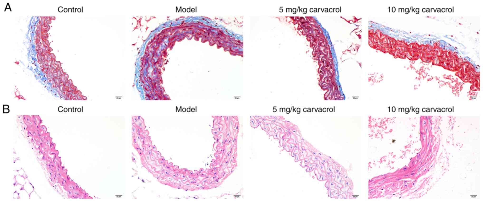
Effects of carvacrol on the pathomorphism
of the thoracoabdominal aorta in db/db mice
Fibrosis was assessed using Masson's trichrome
staining on thoracoabdominal aortic sections (Fig. 1A). Compared with the normal
control group, severe fibrosis was observed in the thoracoabdominal
aorta of the db/db mice in the model group. As described in a
previous study, 5 and 10 mg/kg carvacrol did not affect the normal
activity and movement of the mice (19). Thus, these concentrations of
carvacrol were selected for the present study. Carvacrol treatment
(5 and 10 mg/kg) significantly improved vessel fibrosis in db/db
mice. The model control group db/db mice exhibited severe
pathological changes of the thoracoab-dominal aorta on H&E
staining, such as disorderly and loosely arranged HUVECs,
hypertrophied, distorted an disordered vascular smooth muscle cells
with an increased number of layers, different nucleus sizes,
unclear cell membrane and nuclear membrane, uneven cytoplasmic
staining, and broken intracellular muscle fibers (Fig. 1B). These histological
abnormalities were significantly alleviated in the carvacrol
treatment groups (5 and 10 mg/kg) compared with the model
group.
Carvacrol reduces the levels of insulin
signaling molecules in the thoracoabdominal aorta of db/db
mice
It was first observed that carvacrol decreased the
levels of fasting blood glucose and serum insulin in the db/db
model compared with those in the db/db model control group.
Moreover, carvacrol significantly increased the Homeostatic Model
Assessment of Insulin Resistance of db/db mice compared with the
db/db model control group (Table
II). Next, the protein expression of the insulin signaling
molecules IRS-1 and InsR was found to be significantly higher in
the thoracoabdominal aorta of the db/db model group compared with
the control group (Fig. 2A-C).
However, carvacrol treatment (5 and 10 µM/ml) significantly
reduced the expression levels of these proteins (Fig. 2A-C). Similar results were observed
on immunohistochemical examination (Fig. 2D-G). Thus, carvacrol reduces the
expression of insulin signaling molecules in the thoracoabdominal
aorta of db/db mice.
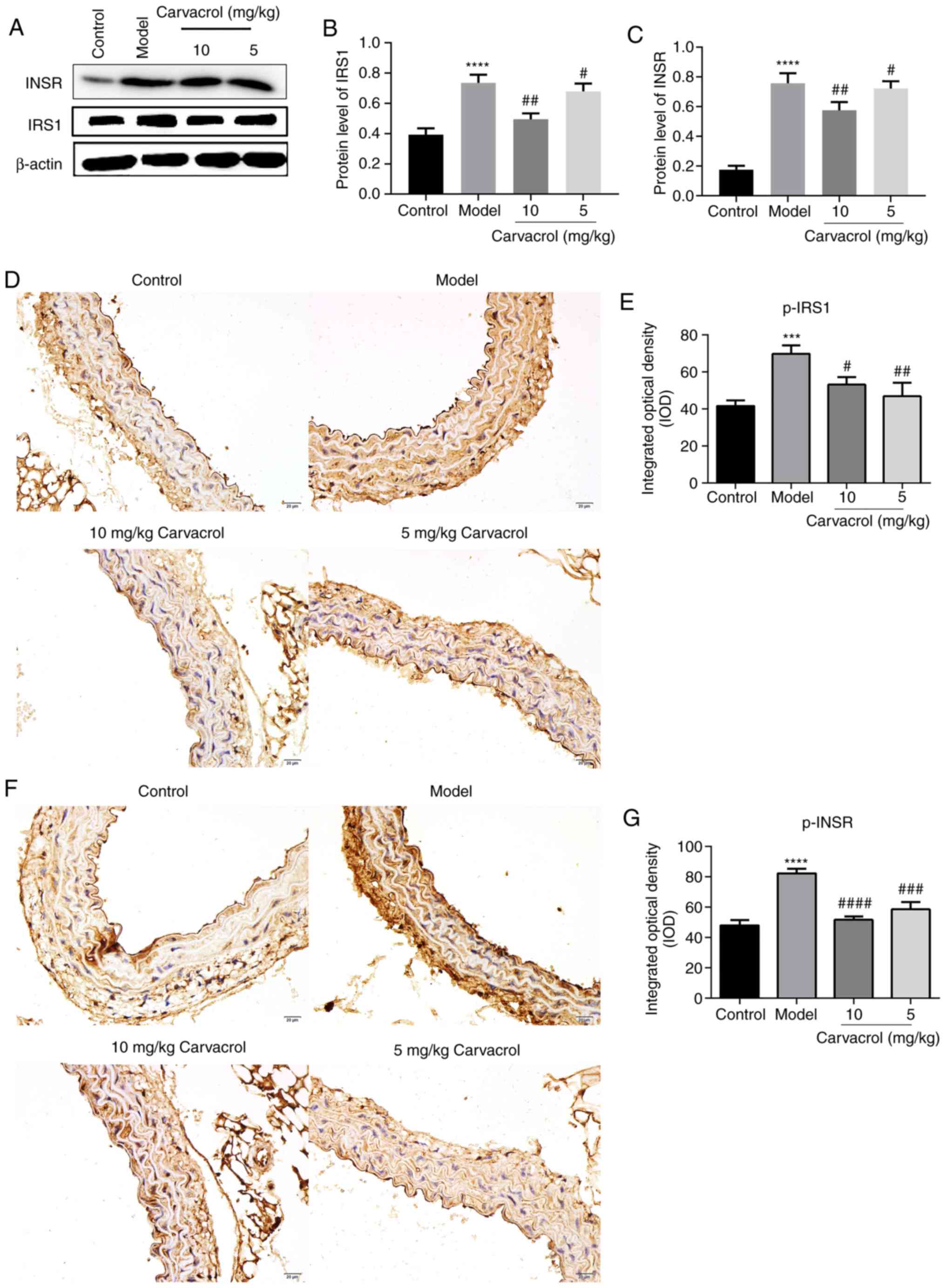 | Figure 2Carvacrol reduced the levels of
insulin signaling molecules, including IRS-1, InsR, p-IRS-1 and
p-InsR in the abdominal aorta of db/db mice. (A) Western blot
analysis of protein expression in the abdominal aorta of db/db
mice. (B and C) Western blotting demonstrated the expression levels
of IRS-1 and InsR in the abdominal aorta of db/db mice with and
without carvacrol treatment. (D and E) Immunohistochemical staining
for p-IRS-1 in the abdominal aorta of db/db mice; magnification,
×200. (F and G) Immunohistochemical staining for p-InsR in the
abdominal aorta of db/db mice; magnification, ×200. Compared with
control: ***P<0.001 and ****P<0.0001.
Compared with model: #P<0.05, ##P<0.01,
###P<0.001 and ####P<0.0001. InsR,
insulin receptor; IRS-1, insulin receptor substrate-1; p-,
phosphorylated; IKK, inhibitor of NF-κB kinase; IKB-α, NF-κB
inhibitor-α; NF-κB, nuclear factor-κB; TLR, toll-like receptor.
Scale bar, 20 µm. |
| Table IIEffect of carvacrol intervention on
insulin resistance markers in db/db mice. |
Table II
Effect of carvacrol intervention on
insulin resistance markers in db/db mice.
| Groups | Fasting blood
glucose (mmol/l) | Insulin (mU/l) | HOMA-IR |
|---|
|
Db/m+ | 6.62±3.04b | 1.20±0.15b | 0.35±0.09b |
| db/db model | 22.15±5.98 | 2.33±0.12 | 2.29±0.18 |
| db/db model +
carvacrol (5 mg/kg) | 18.15±4.73 | 1.73±0.19b | 1.39±0.12b |
| db/db model +
carvacrol (10 mg/kg) | 15.27±5.06a | 1.53±0.17b | 1.04±0.08b |
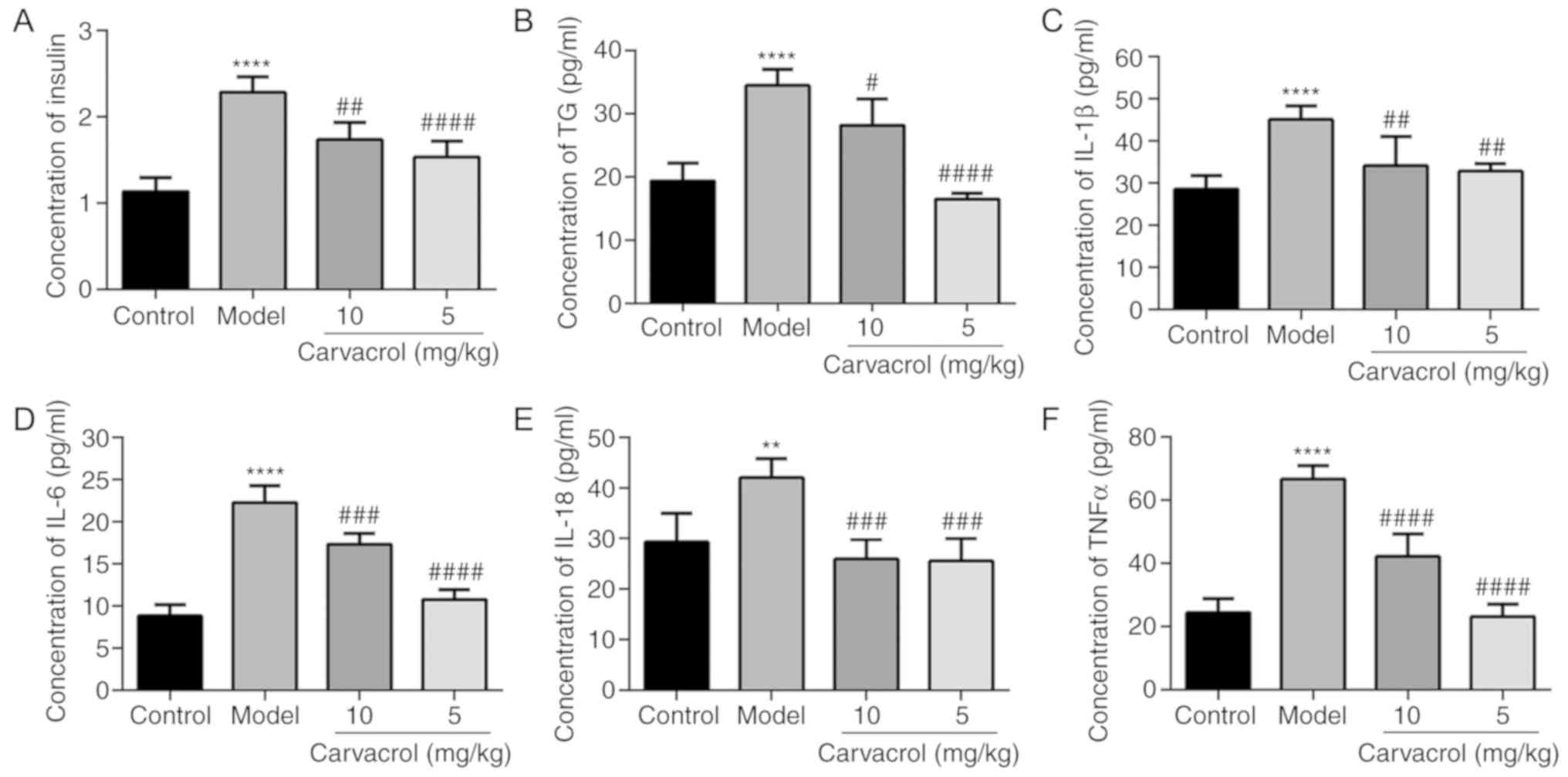
Carvacrol reduces the expression levels
of insulin, TG and markers of insulin resistance in the serum of
db/db mice
As shown in Fig.
3A, the serum insulin level in the db/db mice group was
significantly higher compared with that in the normal control
group, and carvacrol treatment (5 and 10 mg/kg) markedly decreased
the serum insulin level compared with the db/db mice of the model
control group. It was observed that the serum TG level was
significantly higher in db/db mice compared with that in the normal
control group (Fig. 3B). However,
carvacrol treatment (5 and 10 mg/kg) markedly reduced the TG level
compared with that in the db/db mouse control group.
Inflammatory markers, such as IL-1β, IL-6, IL-18 and
TNF-α, have been confirmed as the main cause of insulin resistance
in diabetic patients. Therefore, the levels of IL-1β, IL-6, IL-18
and TNF-α were measured in the serum of db/db mice. It was observed
that the serum levels of IL-1β, IL-6, IL-18 and TNF-α in the db/db
mice of the model group were significantly higher compared with
those in the normal control group (Fig. 3C-F). However, carvacrol treatment
(5 and 10 mg/kg) obviously decreased the serum levels of IL-1β,
IL-6, IL-18 and TNF-α compared with those in the db/db mice of the
model control group.
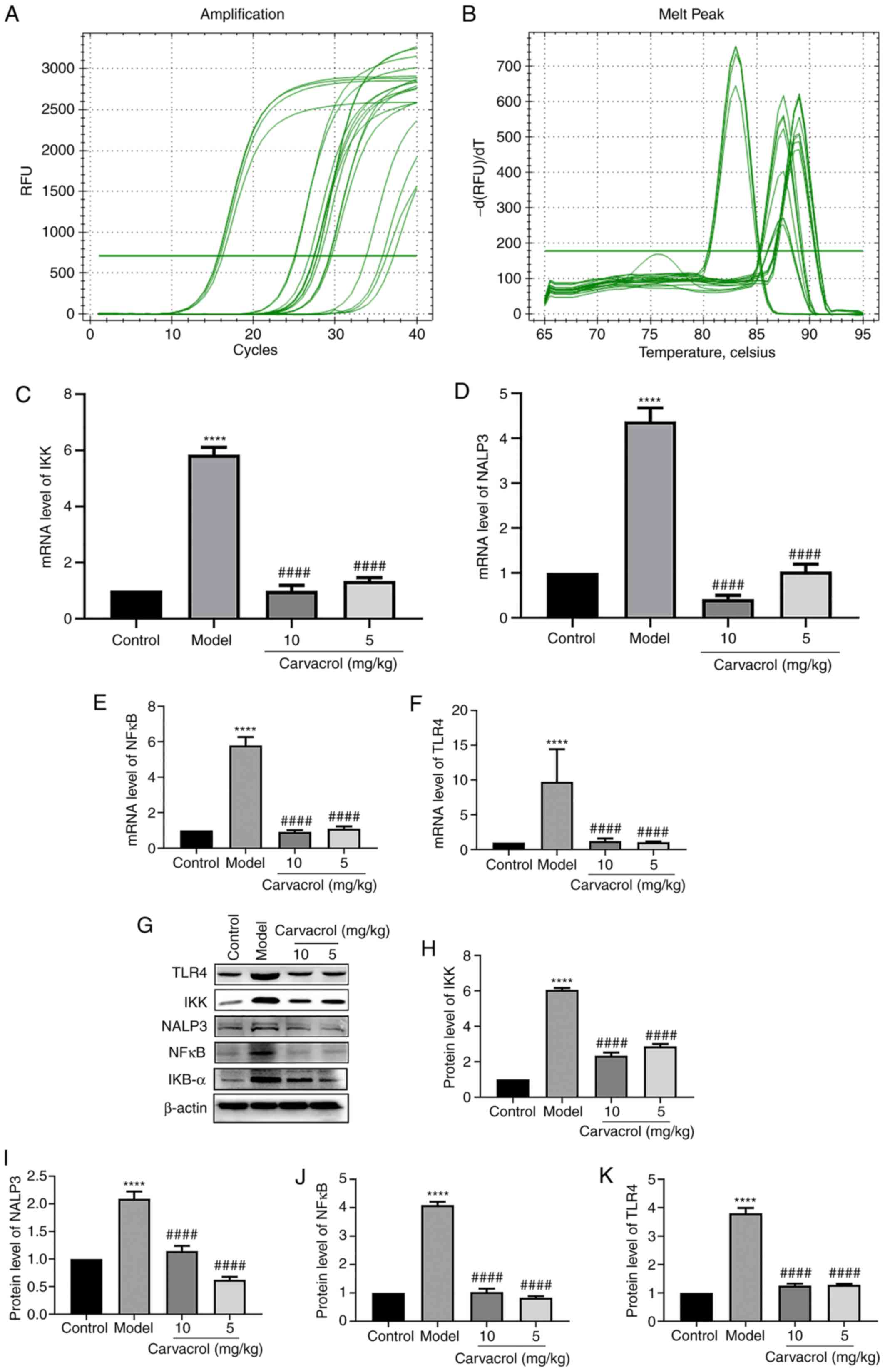
Effects of carvacrol on the TLR4/NF-κB
signaling pathway in the thoracoabdominal aorta of db/db mice
It has been demonstrated that the TLR4/NF-κB
signaling pathway is involved in the regulation of vascular
inflammatory responses (20,21). To explore the potential underlying
mechanism, the expression levels of the TLR4/NF-κB pathway
molecules, including IKK, IKB-α, NALP3, NF-κB and TLR4, were
measured in the thoracoabdominal aorta of db/db mice. The mRNA and
protein levels of IKK, NALP3, NF-κB and TLR4 were found to be
significantly higher in the db/db mice of the model group compared
with those in the normal control group (Fig. 4A-K). However, carvacrol treatment
(5 and 10 mg/kg) markedly decreased the levels of IKK, NALP3, NFκB
and TLR4 compared with the db/db mice of the model control group
(Fig. 4A-K). The results of
immunohistochemical analysis were consistent with the results
mentioned above (Fig. 5A-L).
Moreover, it was observed that the IKB-α and p-IKB-α levels were
higher in the thoracoabdominal aorta of db/db mice compared with
those in the normal control group, whereas carvacrol treatment (5
and 10 kg/kg) decreased the levels of IKB-α and p-IKB-α.
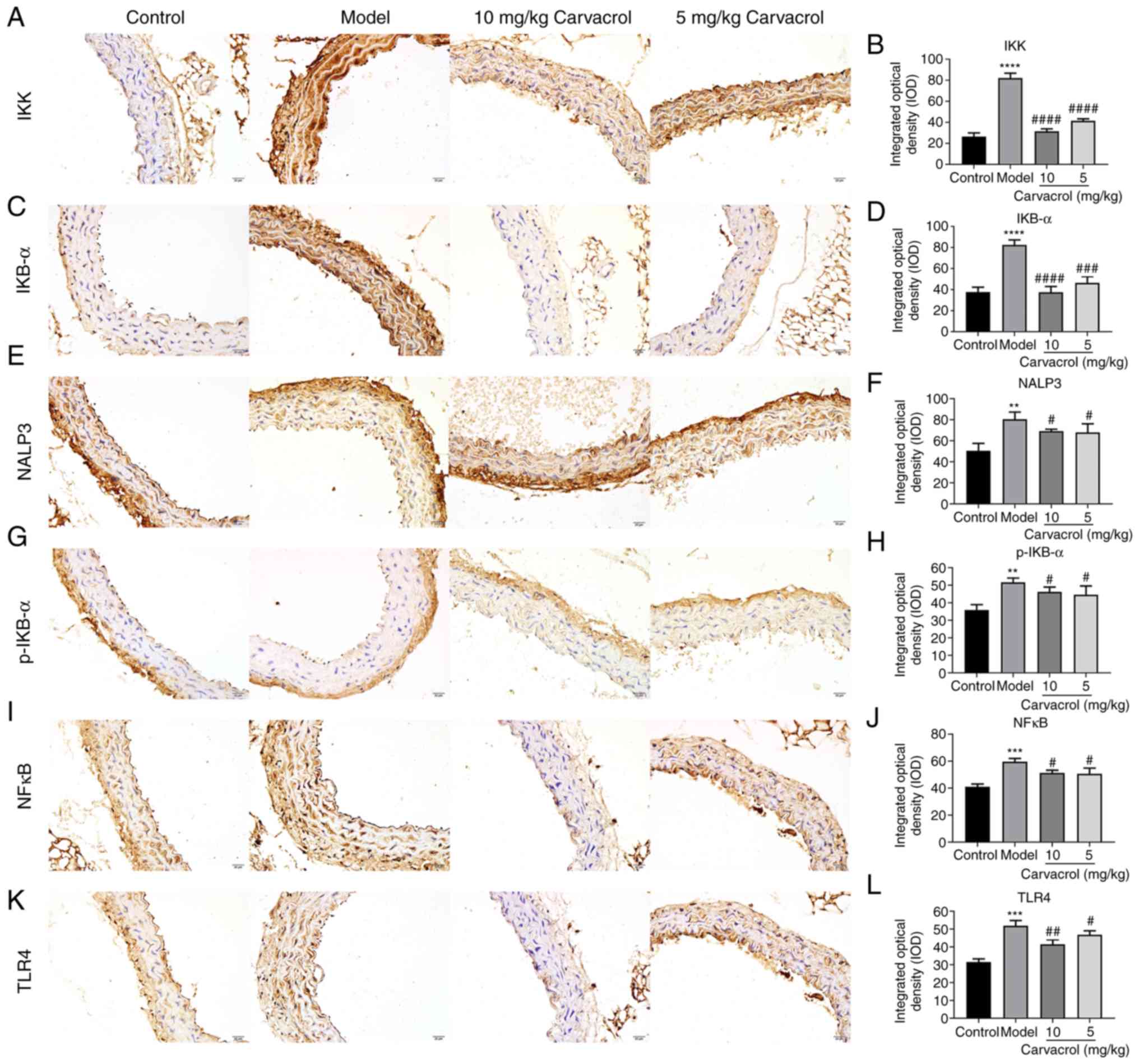 | Figure 5Immunohistochemical analyses results
showing the expression levels of (A and B) IKK, (C and D) IKB-α, (E
and F) NALP3, (G and H) IKB-α, (I and J) NF-κB and (K and L) TLR4
in the thoracoabdominal aortic tissues of db/db mice
(magnification, ×200). Compared with control:
**P<0.01; ***P<0.001 and
****P<0.0001. Compared with model:
#P<0.05, ##P<0.01,
###P<0.001 and ####P<0.0001. IKK,
inhibitor of NF-κB kinase; IKB-α, NF-κB inhibitor-α; NF-κB, nuclear
factor-κB; TLR, toll-like receptor. Scale bar, 20 µm. |
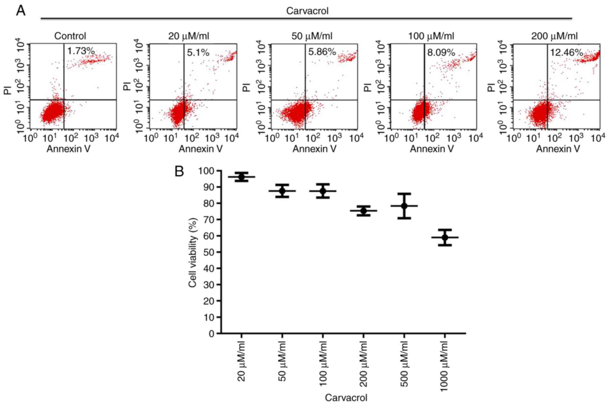
Carvacrol promotes apoptosis and inhibits
the viability of HUVECs
Endothelial cell apoptosis and viability were then
investigated. Flow cytometry was employed to evaluate HUVEC
apoptosis. It was observed that carvacrol promoted vascular
endothelial cell apoptosis in a dose-dependent manner (Fig. 6A). Cell viability was assessed
with the CCK-8 assay. The results demonstrated that carvacrol
decreased endothelial cell viability in a dose-dependent manner
(Fig. 6B).
Carvacrol reduces the levels of insulin
signaling molecules in HG-induced HUVECs
The protein levels of IRS-1 and InsR were found to
be significantly higher in HG-induced HUVECs compared with those in
the HUVECs control group (Fig.
7A-C), and carvacrol treatment (5 and 10 mg/kg) significantly
reduced the levels of p-IRS-1 and p-InsR in HG-induced HUVECs
(Fig. 7D-G). The results
mentioned above indicate that carvacrol reduces the levels of
insulin signaling molecules in HG-induced HUVECs.
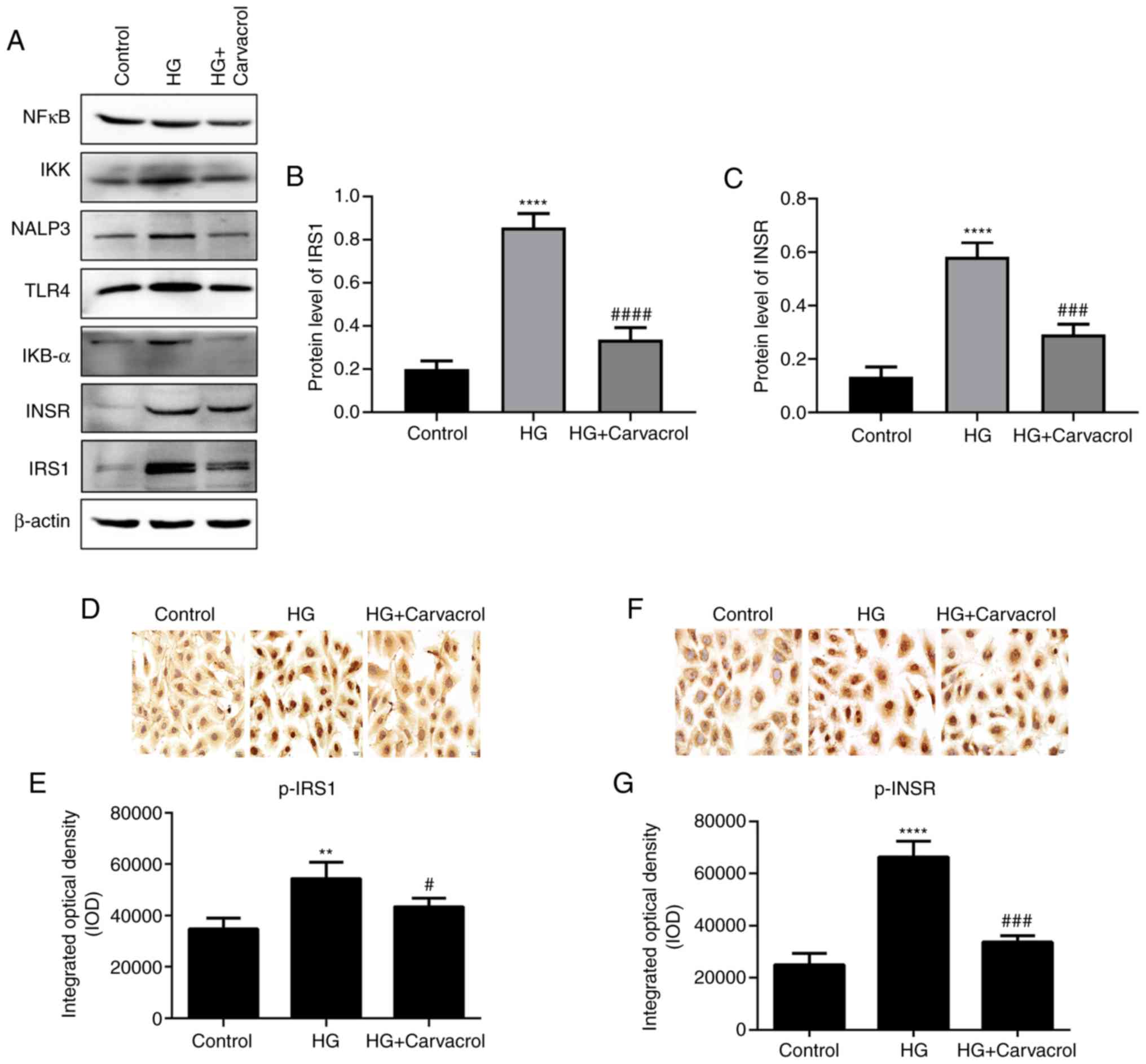 | Figure 7Carvacrol reduced the levels of
insulin signaling molecules in HG-induced HUVECs. (A) Western
blotting of different proteins in HUVECs. (B) Expression of IRS-1
in the HUVECs according to western blotting analysis. (C)
Expression of InsR in HUVECs according to western blotting
analysis. (D and E) Immunohistochemical staining for p-IRS-1 in
HG-induced HUVECs; magnification, ×200. (F and G)
Immunohistochemical staining for p-InsR in HG-induced HUVECs;
magnification, ×200. Compared with control: **P<0.01
and ****P<0.0001. Compared with model:
#P<0.05, ###P<0.001 and
####P<0.0001. HUVECs, human umbilical vein
endothelial cells; HG, high glucose; p-InsR, phosphorylated insulin
receptor; p-IRS-1, phosphorylated insulin receptor substrate-1;
IKK, inhibitor of NF-κB kinase; IKB-α, NF-κB inhibitor-α; NF-κB,
nuclear factor-κB; TLR, toll-like receptor. |
Effects of carvacrol on the TLR4/NF-κB
signaling pathway in HG-induced HUVECs
It was inferred that carvacrol reduces the
activation of the TLR4/NF-κB signaling pathway in HG-induced
HUVECs. As expected, the mRNA and protein expression of IKK, IKB-α,
NALP3, NF-κB and TLR4 were found to be increased in HG-induced
HUVECs compared with those in the control group (Fig. 8A-J). However, carvacrol
significantly decreased the expression of IKK, IKB-α, NALP3, NF-κB
and TLR4 compared with the control group (Fig. 8A-J). These data indicated that
carvacrol may be involved in the activation of the TLR4/NF-κB
signaling pathway in HG-induced HUVECs. In addition,
immunohistochemical analysis was performed. As expected, the
results of immunohistochemistry were consistent with the results
mentioned above (Fig. 8K-O). The
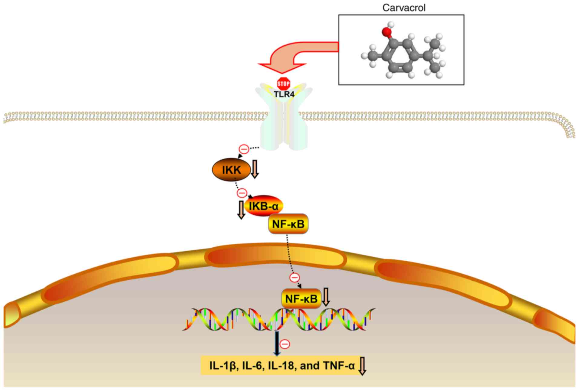
hypothesis diagram of the present study is shown in Fig. 9.
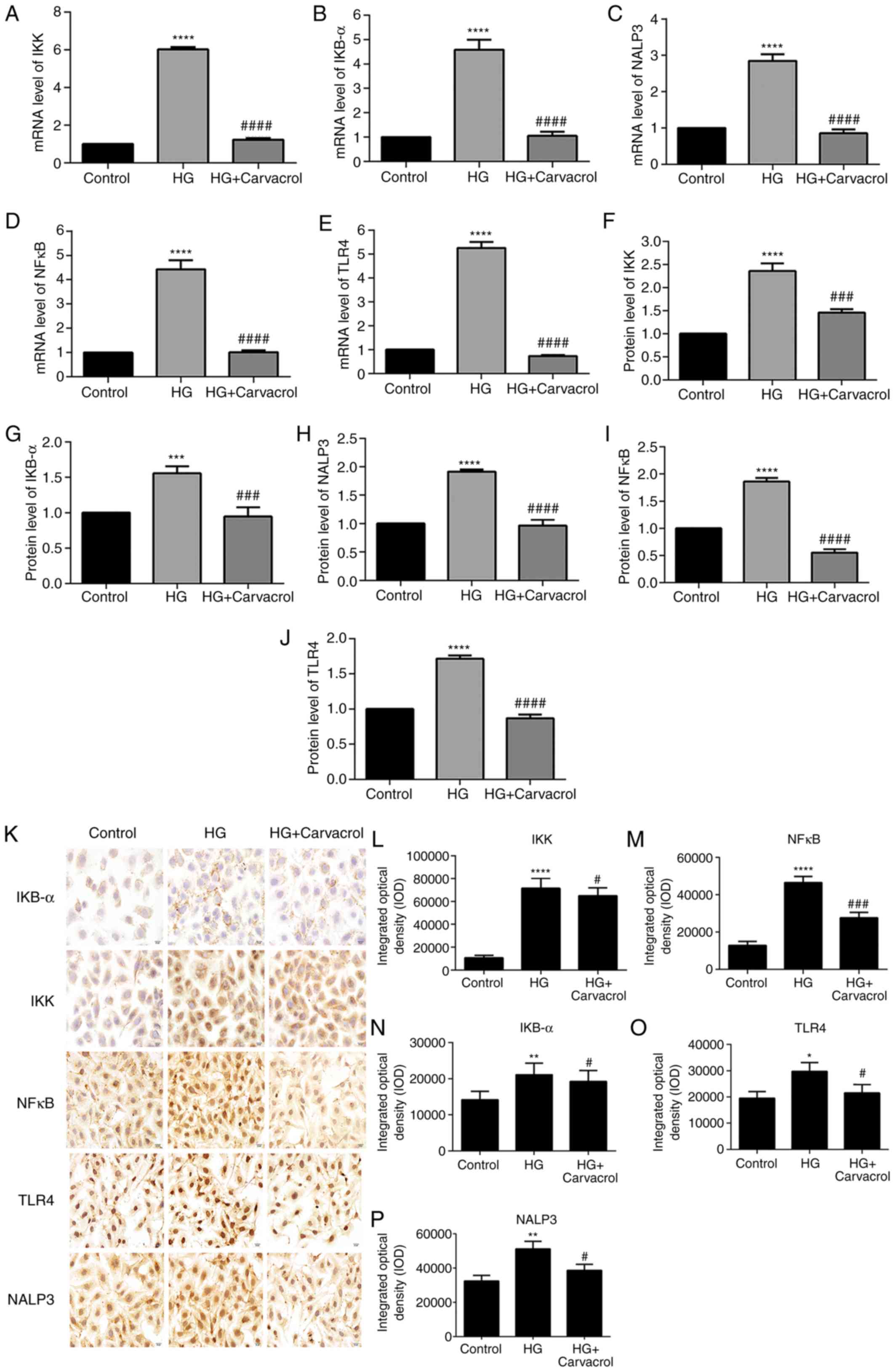 | Figure 8Effects of carvacrol on the
TLR4/NF-κB signaling pathway in HG-induced HUVECs. Reverse
transcription-quantitative PCR analysis results showing the mRNA
expression levels of (A) IKK, (B) IKB-α, (C) NALP3, (D) NF-κB and
(E) TLR4. Western blotting results showing the protein expression
levels of (F) IKK, (G) IKB-α, (H) NALP3, (I) NF-κB and (J) TLR4.
(K) Representative images of immunohistochemical analyses results
magnification, ×200. Expression levels of (L) IKK, (M) NF-κB, (N)
IKB-α, (O) TLR4 and (P) NALP3 in HG-induced HUVECs according to the
immunohistochemical analysis results. Compared with control:
*P<0.05, **P<0.01,
***P<0.001 and ****P<0.0001. Compared
with model: #P<0.05, ###P<0.001 and
####P<0.0001. IKK, inhibitor of NF-κB kinase; IKB-α,
NF-κB inhibitor-α; NF-κB, nuclear factor-κB; TLR, toll-like
receptor; HUVECs, human umbilical vein endothelial cells; HG, high
glucose. |
Discussion
The optimal antidiabetic drug would be expected to
improve insulin resistance in diabetic patients without causing any
side effects (22). However, the
currently available antidiabetic drugs are associated with
long-term side effects. Therefore, in an attempt to find safe and
effective antidiabetic drugs, the effects of carvacrol on a db/db
mouse model were investigated. The results of the present study
suggested that carvacrol may alleviate endothelial cell dysfunction
and vascular inflammation in T2DM.
In the present study, db/db mice were used to
construct a T2DM model. It was observed that the db/db model mice
exhibited major pathological changes. The db/db mice were treated
with low-dose carvacrol (5 mg/kg) or high-dose carvacrol (10
mg/kg), and it was demonstrated that both doses significantly
alleviated the histological abnormalities of the abdominal aorta in
the db/db mouse model. It is well known that T2DM-related disorders
are often associated with insulin resistance (23-25). Peripheral target tissues exhibit
reduced sensitivity to insulin, leading to abnormal insulin
secretion and even hyperglycemia. Insulin resistance is considered
to be a major therapeutic target in T2DM (26). It was herein demonstrated that
both low- and high-dose carvacrol improved insulin resistance via
suppressing the phosphorylation of the insulin signaling molecules
p-IRS-1 and p-InsR in vitro as well as in vivo,
indicating that carvacrol may be of value in the treatment of
T2DM.
T2DM is considered to be a chronic inflammatory
disease. Inflammation and immune cell dysfunction have been shown
to be associated with insulin resistance and secretion dysfunction
(27). In diabetes, vascular
inflammation and endothelial cell dysfunction play a major role in
the development of vascular disease (28). Pro-inflammatory cytokines promote
vascular dysfunction in diabetes by promoting endothelial cell
inflammation (21). For example,
the pro-inflammatory cytokine IL-1β has been reported to be a
driving factor for β-cell dysfunction (29,30). IL-1β secretion is increased in the
islets in response to high levels of glucose, which promotes the
recruitment and activation of macrophages, thereby maintaining the
islet inflammatory response (30). IL-6 has been confirmed to be
highly expressed in the serum of patients with T2DM, which induces
insulin resistance (31). It was
previously demonstrated that serum IL-18 levels are positively
correlated with the pathogenesis and development of T2DM (32). Furthermore, TNF-α is mainly
produced in adipocytes or peripheral tissues. Elevated TNF-α levels
impair insulin signaling through serine phosphorylation, which
induces insulin resistance in adipocytes and peripheral tissues
(33). It has been demonstrated
that insulin resistance in diabetic patients can increase the serum
levels of inflammatory mediators (including IL-1β, IL-6, IL-18 and
TNF-α) and plays an important role in regulating glucose
homeostasis (22). The results of
the present study indicated that carvacrol effectively reduced the
serum levels of inflammatory mediators, including IL-1β, IL-6,
IL-18 and TNF-α, in db/db mice. These findings indicated that
carvacrol may alleviate insulin resistance of db/db mice by
inhibiting the expression of these pro-inflammatory cytokines.
IKK activates the nuclear translocation of NF-κB by
the degradation inhibitor IκBα (34). In the present study, db/db mice
exhibited increased expression of IKK and IκBα. Increased IKK
expression indicated the activation of NF-κB signaling.
Furthermore, increased IκBα expression may be an adaptive response
to this activation. However, carvacrol significantly reduced IKK
and IκBα expression in db/db mice. Therefore, carvacrol may reduce
vascular inflammation by suppressing the NF-κB signaling pathway.
The TLR4/NF-κB signaling pathway is involved in the inflammatory
response of diabetes. To explore whether carvacrol also exerts
anti-inflammatory effects through the TLR4/NF-κB signaling pathway,
the expression levels of relevant markers in the TLR4/NF-κB
signaling pathway in the abdominal aorta were examined. TLR4 and
NF-κB levels were significantly upregulated in db/db mice, but they
were reduced by carvacrol at both the transcriptional and
translational levels. Therefore, the results mentioned above
indicate that carvacrol may exert its anti-inflammatory effects
through inactivation of the TLR4/NF-κB signaling pathway.
Endothelial cells are sensitive to changes in blood
glucose levels (35). Endothelial
cell injury has been identified as an early event in the
development of atherosclerosis. Diabetes, as one of the risk
factors for atherosclerosis, can damage the vascular endothelium.
There is growing evidence that diabetic atherosclerosis is
associated with hyperglycemia. Both in vivo and in
vitro studies have demonstrated that hyperglycemia can
contribute to HUVEC damage and dysfunction, ultimately leading to
atherosclerosis (36,37). In the present study, the results
revealed that carvacrol promoted apoptosis of HG-induced HUVECs in
a dose-dependent manner. As expected, the in vitro
experiment results demonstrated that the protein levels of the
TLR4/NF-κB signaling pathway molecules were elevated in HG-induced
vascular endothelial cells. Moreover, carvacrol significantly
suppressed the levels of relevant markers in the TLR4/NF-κB
signaling pathway. These results indicated that carvacrol may
protect HG-induced HUVECs through inactivation of the TLR4/NF-κB
signaling pathway.
In conclusion, the present study demonstrated that
carvacrol alleviated the histological abnormalities of the
abdominal aorta in a db/db mouse model. Furthermore, the
anti-inflammatory effect of carvacrol was confirmed in the db/db
mouse model. Further research into the underlying mechanism
demonstrated that carvacrol reduced the activation of the
TLR4/NF-κB inflammatory response signaling pathway in vitro
and in vivo. Thus, these findings indicated that carvacrol
may reduce vascular inflammation and endothelial cell dysfunction,
and it may be of value in the treatment of T2DM.
Abbreviations:
|
T2DM
|
type 2 diabetes mellitus
|
|
H&E
|
hematoxylin and eosin
|
|
p-InsR
|
phosphorylated insulin receptor
|
|
p-IRS-1
|
phosphorylated insulin receptor
substrate-1
|
|
TG
|
triglyceride
|
|
IL-1β
|
interleukin-1β
|
|
IL-6
|
interleukin-6
|
|
IL-18
|
interleukin-18
|
|
TNF-α
|
tumor necrosis factor-α
|
|
NF-κB
|
nuclear factor-κB
|
|
CCK-8
|
Cell Counting Kit-8
|
|
RT-qPCR
|
reverse transcription-quantitative
PCR
|
|
ELISA
|
enzyme-linked immunosorbent assay
|
Acknowledgments
Not applicable.
Funding
The present study was funded by grants from the
National Natural Science Foundation of China (81760813) and the
Science and Technology Cooperation Plan of Guizhou [Qiankehe LH
(2016) No.7127].
Availability of data and materials
The datasets generated and/or analyzed during the
present study are available from the corresponding author on
reasonable request.
Authors' contributions
WZ conceived and designed the study. CD and QH
conducted most of the experiments and data analysis, and wrote the
manuscript. HX and YC participated in collecting data and helped
with the drafting the manuscript. All authors have reviewed and
approved the final version of the manuscript.
Ethics approval and consent to
participate
All animal experiments were performed strictly in
accordance with the Guide for the Care and Use of Laboratory
Animals of the National Institutes of Health. The research protocol
was approved by the Traditional Chinese Medicine Guizhou University
Animal Care and Ethics Committee.
Patient consent for publication
Not applicable.
Competing interests
The authors declare that they have no competing
interests.
References
|
1
|
van der Schaft N, Schoufour JD, Nano J,
Kiefte-de Jong JC, Muka T, Sijbrands EJG, Ikram MA, Franco OH and
Voortman T: Dietary antioxidant capacity and risk of type 2
diabetes mellitus, prediabetes and insulin resistance: The
rotterdam study. Eur J Epidemiol. 34:853–861. 2019. View Article : Google Scholar : PubMed/NCBI
|
|
2
|
Zhou Z, Jardine M, Perkovic V, Matthews
DR, Mahaffey KW, de Zeeuw D, Fulcher G, Desai M, Oh R, Simpson R,
et al: Canagliflozin and fracture risk in individuals with type 2
diabetes: Results from the CANVAS program. Diabetologia.
62:1854–1867. 2019. View Article : Google Scholar : PubMed/NCBI
|
|
3
|
Zheng Y, Yang Y, Dong B, Zheng H, Lin X,
Du Y, Li X, Zhao L and Gao H: Metabonomic profiles delineate
potential role of glutamate-glutamine cycle in db/db mice with
diabetes-associated cognitive decline. Mol Brain. 9:402016.
View Article : Google Scholar : PubMed/NCBI
|
|
4
|
GBD 2015 Disease and Injury Incidence and
Prevalence Collaborators: Global, regional, and national incidence,
prevalence, and years lived with disability for 310 diseases and
injuries, 1990-2015: A systematic analysis for the global burden of
disease study 2015. Lancet. 388:1545–1602. 2016. View Article : Google Scholar : PubMed/NCBI
|
|
5
|
Shaw JE, Sicree RA and Zimmet PZ: Global
estimates of the prevalence of diabetes for 2010 and 2030. Diabetes
Res Clin Pract. 87:4–14. 2010. View Article : Google Scholar
|
|
6
|
Petersen C, Bharat D, Cutler BR, Gholami
S, Denetso C, Mueller JE, Cho JM, Kim JS, Symons JD and Anandh Babu
PV: Circulating metabolites of strawberry mediate reductions in
vascular inflammation and endothelial dysfunction in db/db mice.
Int J Cardiol. 263:111–117. 2018. View Article : Google Scholar : PubMed/NCBI
|
|
7
|
Sawada N, Jiang A, Takizawa F, Safdar A,
Manika A, Tesmenitsky Y, Kang KT, Bischoff J, Kalwa H, Sartoretto
JL, et al: Endothelial PGC-1α mediates vascular dysfunction in
diabetes. Cell Metab. 19:246–258. 2014. View Article : Google Scholar : PubMed/NCBI
|
|
8
|
Brennan E, Wang B, McClelland A, Mohan M,
Marai M, Beuscart O, Derouiche S, Gray S, Pickering R, Tikellis C,
et al: Protective effect of let-7 mirna family in regulating
inflammation in diabetes-associated atherosclerosis. Diabetes.
66:2266–2277. 2017. View Article : Google Scholar : PubMed/NCBI
|
|
9
|
Ren Y, Tao S, Zheng S, Zhao M, Zhu Y, Yang
J and Wu Y: Salvianolic acid B improves vascular endothelial
function in diabetic rats with blood glucose fluctuations via
suppression of endothelial cell apoptosis. Eur J Pharmacol.
791:308–315. 2016. View Article : Google Scholar : PubMed/NCBI
|
|
10
|
Kang H, Ma X, Liu J, Fan Y and Deng X:
High glucose-induced endothelial progenitor cell dysfunction. Diab
Vasc Dis Res. 14:381–394. 2017. View Article : Google Scholar : PubMed/NCBI
|
|
11
|
Kara M, Uslu S, Demirci F, Temel HE and
Baydemir C: Supplemental carvacrol can reduce the severity of
inflammation by influencing the production of mediators of
inflammation. Inflammation. 38:1020–1027. 2015. View Article : Google Scholar
|
|
12
|
Somensi N, Rabelo TK, Guimarães AG,
Quintans-Junior LJ, de Souza Araújo AA, Moreira JCF and Gelain DP:
Carvacrol suppresses LPS-induced pro-inflammatory activation in RAW
264.7 macrophages through ERK1/2 and NF-kB pathway. Int
Immunopharmacol. 75:1057432019. View Article : Google Scholar : PubMed/NCBI
|
|
13
|
Manouchehrabadi M, Farhadi M, Azizi Z and
Torkaman-Boutorabi A: Carvacrol protects against
6-hydroxydopamine-induced neurotoxicity in in vivo and in vitro
models of parkinson's disease. Neurotox Res. 37:156–170. 2020.
View Article : Google Scholar
|
|
14
|
Khan F, Singh VK, Saeed M, Kausar MA and
Ansari IA: Carvacrol induced program cell death and cell cycle
arrest in androgen-independent human prostate cancer cells via
inhibition of notch signaling. Anticancer Agents Med Chem.
19:1588–1608. 2019. View Article : Google Scholar
|
|
15
|
Shoorei H, Khaki A, Khaki AA, Hemmati AA,
Moghimian M and Shokoohi M: The ameliorative effect of carvacrol on
oxidative stress and germ cell apoptosis in testicular tissue of
adult diabetic rats. Biomed Pharmacother. 111:568–578. 2019.
View Article : Google Scholar : PubMed/NCBI
|
|
16
|
Khazdair MR and Boskabady MH: The effect
of carvacrol on inflammatory mediators and respiratory symptoms in
veterans exposed to sulfur mustard, a randomized,
placebo-controlled trial. Respir Med. 150:21–29. 2019. View Article : Google Scholar : PubMed/NCBI
|
|
17
|
Kobayashi K, Forte TM, Taniguchi S, Ishida
BY, Oka K and Chan L: The db/db mouse, a model for diabetic
dyslipidemia: Molecular characterization and effects of Western
diet feeding. Metabolism. 49:22–31. 2000. View Article : Google Scholar : PubMed/NCBI
|
|
18
|
Peng BY, Wang Q, Luo YH, He JF, Tan T and
Zhu H: A novel and quick PCR-based method to genotype mice with a
leptin receptor mutation (db/db mice). Acta Pharmacol Sin.
39:117–123. 2018. View Article : Google Scholar :
|
|
19
|
Mahmoodi M, Amiri H, Ayoobi F, Rahmani M,
Taghipour Z, Ghavamabadi RT, Jafarzadeh A and Sankian M: Carvacrol
ameliorates experimental autoimmune encephalomyelitis through
modulating pro- and anti-inflammatory cytokines. Life Sci.
219:257–263. 2019. View Article : Google Scholar
|
|
20
|
Chen X, Wu S, Chen C, Xie B, Fang Z, Hu W,
Chen J, Fu H and He H: Omega-3 polyunsaturated fatty acid
supplementation attenuates microglial-induced inflammation by
inhibiting the HMGB1/TLR4/NF-κB pathway following experimental
traumatic brain injury. J Neuroinflammation. 14:1432017. View Article : Google Scholar
|
|
21
|
Tang ZH, Peng J, Ren Z, Yang J, Li TT, Li
TH, Wang Z, Wei DH, Liu LS, Zheng XL and Jiang ZS: New role of
PCSK9 in athero-sclerotic inflammation promotion involving the
TLR4/NF-κB pathway. Atherosclerosis. 262:113–122. 2017. View Article : Google Scholar : PubMed/NCBI
|
|
22
|
Sharma BR, Kim HJ and Rhyu DY: Caulerpa
lentillifera extract ameliorates insulin resistance and regulates
glucose metabolism in C57BL/KsJ-db/db mice via PI3K/AKT signaling
pathway in myocytes. J Transl Med. 13:622015. View Article : Google Scholar : PubMed/NCBI
|
|
23
|
Ogata S, Ito S, Masuda T and Ohtsuki S:
Changes of blood-brain barrier and brain parenchymal protein
expression levels of mice under different insulin-resistance
conditions induced by high-fat diet. Pharm Res. 36:1412019.
View Article : Google Scholar : PubMed/NCBI
|
|
24
|
Pivari F, Mingione A, Brasacchio C and
Soldati L: Curcumin and type 2 diabetes mellitus: Prevention and
treatment. Nutrients. 11:18372019. View Article : Google Scholar :
|
|
25
|
Wang Y, Zhou H, Palyha O and Mu J:
Restoration of insulin receptor improves diabetic phenotype in T2DM
mice. JCI Insight. 4:e1249452019. View Article : Google Scholar :
|
|
26
|
Eckel RH, Grundy SM and Zimmet PZ: The
metabolic syndrome. Lancet. 365:1415–1428. 2005. View Article : Google Scholar : PubMed/NCBI
|
|
27
|
Kammoun HL, Allen TL, Henstridge DC, Barre
S, Coll RC, Lancaster GI, Cron L, Reibe S, Chan JY, Bensellam M, et
al: Evidence against a role for NLRP3-driven islet inflammation in
db/db mice. Mol Metab. 10:66–73. 2018. View Article : Google Scholar : PubMed/NCBI
|
|
28
|
Pollack RM, Donath MY, LeRoith D and
Leibowitz G: Anti-inflammatory agents in the treatment of diabetes
and its vascular complications. Diabetes Care. 39(Suppl 2):
S244–S52. 2016. View Article : Google Scholar : PubMed/NCBI
|
|
29
|
Maedler K, Sergeev P, Ris F, Oberholzer J,
Joller-Jemelka HI, Spinas GA, Kaiser N, Halban PA and Donath MY:
Glucose-induced beta cell production of IL-1beta contributes to
glucotoxicity in human pancreatic islets. J Clin Invest.
110:851–860. 2002. View Article : Google Scholar : PubMed/NCBI
|
|
30
|
Herder C, Dalmas E, Böni-Schnetzler M and
Donath MY: The IL-1 pathway in type 2 diabetes and cardiovascular
complications. Trends Endocrinol Metab. 26:551–563. 2015.
View Article : Google Scholar : PubMed/NCBI
|
|
31
|
Rehman K, Akash MSH, Liaqat A, Kamal S,
Qadir MI and Rasul A: Role of interleukin-6 in development of
insulin resistance and type 2 diabetes mellitus. Crit Rev Eukaryot
Gene Expr. 27:229–236. 2017. View Article : Google Scholar : PubMed/NCBI
|
|
32
|
Zhuang H, Han J, Cheng L and Liu SL: A
positive causal influence of IL-18 levels on the risk of T2DM: A
mendelian randomization study. Front Genet. 10:2952019. View Article : Google Scholar : PubMed/NCBI
|
|
33
|
Akash MSH, Rehman K and Liaqat A: Tumor
necrosis factor-alpha: Role in development of insulin resistance
and pathogenesis of type 2 diabetes mellitus. J Cell Biochem.
119:105–110. 2018. View Article : Google Scholar
|
|
34
|
Babu PV, Si H and Liu D: Epigallocatechin
gallate reduces vascular inflammation in db/db mice possibly
through an NF-κB-mediated mechanism. Mol Nutr Food Res.
56:1424–1432. 2012. View Article : Google Scholar : PubMed/NCBI
|
|
35
|
Silambarasan M, Tan JR, Karolina DS,
Armugam A, Kaur C and Jeyaseelan K: MicroRNAs in hyperglycemia
induced endothelial cell dysfunction. Int J Mol Sci. 17:5182016.
View Article : Google Scholar : PubMed/NCBI
|
|
36
|
Xiao X, Dong Y, Zhong J, Cao R, Zhao X,
Wen G and Liu J: Adiponectin protects endothelial cells from the
damages induced by the intermittent high level of glucose.
Endocrine. 40:386–393. 2011. View Article : Google Scholar : PubMed/NCBI
|
|
37
|
Torimoto K, Okada Y, Mori H and Tanaka Y:
Relationship between fluctuations in glucose levels measured by
continuous glucose monitoring and vascular endothelial dysfunction
in type 2 diabetes mellitus. Cardiovasc Diabetol. 12:12013.
View Article : Google Scholar : PubMed/NCBI
|