Introduction
Peripheral arterial disease results from progressive
narrowing of arteries with a great impact on lower limb, which
leads to critical limb ischemia (CLI) (1). Previous studies demonstrated that
patients with peripheral arterial disease have a 40% increased risk
of stroke and a 20-60% increased risk of myocardial infarction, and
that patients with CLI in particular would have an additional
substantial risk of limb loss (1,2).
CLI is a gradual process in which arteries become blocked, narrowed
or weakened (3). It is therefore
crucial to develop novel therapeutic options for patients who
develop CLI. In clinical, although some therapeutic strategies
exist, including surgical or interventional revascularization,
substantial number of patients are not eligible for those
treatments and amputation can be the only option (4). Previous studies have reported that
therapeutic neovascularization is a promising option that could
overcome ischemia by providing an improved vascular network
(4,5). However, the process of
neovascularization is complex and involves the induction of
capillary sprouting (angiogenesis), the maturation of newly formed
vessels and the growth of large-conductance vessels
(arteriogenesis) to improve blood supply (6-8).
Thymosin-β 4 (Tβ4) is a naturally-occurring peptide
that is encoded in humans by the TMSB4X gene on the X-chromosome
(9). Tβ4 is the most abundant and
biologically active member of the β-thymosin family, which is
presents in all body fluids and all cells, except from red blood
cells (10). Tβ4 is the major
G-actin-sequestering protein in mammalian cells and it prevents
actin polymerization into filaments, confirming its crucial role in
maintaining cytoskeletal dynamics (11). In the last decade, Tβ4 has been
reported to possess the ability to regulate multiple biological
functions. For example, Kobayash et al (12) have demonstrated that Tβ4 regulates
the motility and metastasis abilities of mouse fibrosarcoma cells.
Renga et al (13) have
reported that Tβ4 can limit inflammation by regulating autophagy,
and Kleinman and Sosne (14) have
demonstrated that Tβ4 can promote dermal healing. In addition,
previous studies reported that Tβ4 has the ability to regulate
endothelial cell angiogenesis (10,15). Tβ4 has also been reported to
promote the angiogenesis of endothelial progenitor cells, and Quan
et al (16) showed that
Tβ4 promotes the survival and angiogenesis of transplanted
endothelial progenitor cells in infarcted myocardium. Furthermore,
Zhao et al (17)
demonstrated that Tβ4 can stimulate endothelial progenitor cell
angiogenesis via a vascular endothelial growth factor-dependent
mechanism. However, whether Tβ4 has a pro-angogenic effect in CLI
needs to be further investigated and the underlying mechanisms must
be determined. The present study aimed therefore to investigate the
pro-angogenic effect and underlying mechanisms of Tβ4 in CLI
mice.
Materials and methods
Ethics statement
All animal experiments were performed in accordance
with the guidelines of the China Council on Animal Care and Use.
This study was approved by the Committee of Experimental Animals of
The First Affiliated Hospital of Zhejiang Chinese Medical
University (approval no. Z20190312G). Every effort was made to
minimize pain and discomfort to the animals. Animal experiments
were performed in The First Affiliated Hospital of Zhejiang Chinese
Medical University.
Cell culture
Human umbilical vein endothelial cells (HUVEC; cat.
no. CRL-1730) and 293T/17 cells (cat. no. CRL-11268) were obtained
from the American Type Culture Collection. HUVEC and 293T/17 cells
were both cultured in DMEM (cat. no. C11995500BT; Gibco; Thermo
Fisher Scientific, Inc.) containing 10% FBS (cat. no. 10437010;
Gibco; Thermo Fisher Scientific, Inc.) and placed at 37°C in a
humidified incubator containing 5% CO2. 293T/17 cells
were only used for the construction of Tβ4 overexpression
lentiviral. HUVEC were used for transfection and the MTT, tube
formation, western blotting and immunofluorescence assays.
Construction of Tβ4 overexpression
lentiviral vector
The sequence of the Tβ4 overexpression vector was as
follows: Forward, 5′-TGG ATT TGT ACC ATT CTT CTG-3′ and reverse,
5′-GAA GAA TGG TAC AAA TCC AAG-3′ (Shanghai GenePharma Co., Ltd.).
Once the overexpression sequence was ligated into pLJM1 plasmid
vector (cat. no. 60908-4538; Tiandz, Inc.) by 2xEasyTaq Supermix
(cat. no. AS111-11; TransGen Biotech Co., Ltd.), 10 µl
ligate product was mixed with 50 µl competent cell
(E.coli DH5α; cat. no. MCC0010; Frdbio) and uniformly coated
on the LB medium (cat. no. ST156; Beyotime Institute of
Biotechnology). After the competent cell and LB medium were
incubated for 16 h at 37°C, the clone colonies were selected.
Subsequently, plasmids were extracted using TIANprep Mini Plasmid
Kit (cat. no. DP103-03; Tiangen Biotech Co. Ltd.). The pLJM1
plasmid vector without any target sequence was used as a negative
control.
After collection of the Tβ4 overexpression plasmids,
293T/17 cells were placed into a 15 cm dish (1.2×10⁷
cells in 20 ml complete medium) and were incubated at 37°C
overnight until confluence reached 20-30%. Subsequently, 100 ng
overexpression plasmids and viral packaging plasmids psPAX2 (cat.
no. VT1444; Youbio Technology Co., Ltd.) and pmD2.G (cat. no.
VT1443; Youbio Technology Co., Ltd.) were co-transfected into
293T/17 cells using Lipofectamine 2000 (cat. no. 11668-019;
Invitrogen; Thermo Fisher Scientific, Inc.). After 8 h, medium
containing overexpression plasmids and viral packaging plasmids was
replaced by fresh complete medium and cultured for another 48 h.
The culture supernatant was then collected and centrifuged for 10
min at 4°C (14,000 × g). The supernatant was filtered with 0.45
µm filter (cat. no. 342414; Beckman Coulter, Inc.), the
lentiviral solution was centrifuged for 15 min at 4°C (4,000 × g),
the lentiviral vector was then concentrated, and the sample was
finally collected and stored at -80°C for subsequent
experiments.
Lentiviral infection
Before infection, HUVEC were seeded into 6-well
plates at the density of 1×10⁶ cells in 2 ml complete
medium and left in incubator overnight until confluence reached
20-30%. Subsequently, complete medium was replaced with serum-free
DMEM and cells were cultured at 37°C for 4 h. Subsequently, cells
were infected with the Tβ4 overexpression lentiviral vector for 6
h. Medium containing lentiviral was replaced with complete medium
and the cells were cultured for another 48 h at 37°C.
DAPT and BMS treatment
The inhibitors of Notch pathway and of NF-κB pathway
DAPT (cat. no. A8200) and BMS-345541 hydrochloride (BMS; cat. no.
B4655), respectively, were obtained from APeXBIO Technology LLC.
Following HUVEC infection with lentiviral vector, HUVEC were seeded
into 6-well plates at the density of 1×10⁶ cells in 2 ml
complete medium and cultured until attachment. Then, DAPT and BMS
were diluted in DMSO and cells were treated with 10 µm DAPT
or 1 µm BMS for 48 h. The concentrations of DAPT and BMS
were determined as previously described (18-20). Following treatment, cells were
collected for further experiments.
MTT assay
MTT (cat. no. B7777; APeXBIO Technology LLC) assay
was used to determine cell viability. Following HUVEC infection,
cells were seeded into 96-well plates at the density of
1×10⁴ cells in 100 µl complete medium. After 24
h, the cells were incubated with MTT reagent (0.5 mg/ml) for 4 h.
MTT solution was discarded and 100 µl DMSO was added to each
well. Finally, absorbance was detected at 570 nm on a microplate
reader (Infinite m200 PRo; Tecan Group, Ltd.).
Tube formation assay
Following cell infection, HUVEC were diluted at the
density of 2×10⁵ cells in 200 µl medium and
seeded into a 48 well plate which was precoated with 200 µl
ECMatix gel (cat. no. ECM625; EMD Millipore). Cells were then
incubated for 4 h. Subsequently, by using a phase-contrast optical
microscope (Axio Lab.A1 pol; Leica Microsystems GmbH), series of
tube-like structures were examined and photographed (magnification,
×100).
Wound healing assay
Following cell infection, HUVEC were seeded into
6-well plates at a density of 3.5×10⁵ cells in 2 ml of
complete medium and cultured until confluence reached 95%. Then, a
vertical wound in each well was created by using a 20 µl
pipette tip, and medium was replaced by serum-free medium. Images
in each well were collected at 0 and 48 h using a phase-contrast
optical microscope (Axio Lab.A1 pol; magnification, ×100). Image J
software (version 1.8.0; National Institutes of Health) was used
for data analysis.
Immunofluorescence
Following cell infection, HUVEC were fixed with 4%
paraformaldehyde (cat. no. P804536, macklin) for 15 min at room
temperature and washed three times with PBS. Cells were
permeabilized using 0.5% Triton X-100 in PBS (cat. no.
R-10789704001; Roche Diagnostics) for 10 min at room temperature,
washed three times with PBS and incubated overnight at 4°C with
NF-κB/p65 antibody (1:400; cat. no. 8242; Cell Signaling
Technology, Inc.). The next day, cells were washed with PBS and
incubated with Alexa Fluor 488 goat anti-rabbit IgG (1:1,000; cat.
no. ab150077; Abcam) for 1 h at room temperature. Finally, cells
were counterstained with 10 µg/ml DAPI (cat. no. D3571;
Invitrogen; Thermo Fisher Scientific, Inc.) and visualized using a
fluorescence microscope (CKX53; Olympus Corporation).
CLI model establishment
A total of 80 adult male C57BL/6J mice (8-weeks old)
were obtained from Shanghai Laboratory Animal Technology. All
animals were fed using the same animal feeding unit and given 12 h
dark/12 h light cycle. Animals were maintained under specifc
pathogen-free conditions at 20-25°C and 50-65% humidity. Animals
were randomly divided into eight groups (n=10/group) as follows:
Sham, Model, negative control (NC), Tβ4, DAPT + NC, Tβ4 + DAPT, BMS
+ NC and Tβ4 + BMS groups. Before surgery, mice were
intraperitoneally injected with ketamine (80 mg/kg; cat. no. 3131;
R&D Systems, Inc.) and xylazine (10 mg/kg; cat. no. B27154;
Yuanye). Once mice were anesthesized, 1 cm incision was made
perpendicular to the right posterior inguinal ligament. Then, the
proximal part of the right femoral artery and vein (including the
superficial and deep branches, as well as the distal part of the
saphenous artery and vein) was ligated and resected. The incision
was sutured with propylene suture line (cat. no. LAT-18-5901; Lab
Animal Technology Develop) (5).
Finally, buprenorphine hydrochloride (0.1 mg/kg; cat. no. 2808;
R&D Systems, Inc.) was injected subcutaneously to relieve
postoperative pain. The mice were observed twice daily to monitor
their health and behavior, and they did not appear to be in
distress or to exhibit obvious behavioral abnormalities. on the 7th
day following operation, mice were sacrificed with an overdose of
pentobarbital sodium (100-150 mg/kg; intraperitoneally injected;
cat. no. B005; Nanjing Jiancheng Bioengineering Institute), and the
gastrocnemius muscle of the right hind limb was collected and
stored at -80°C for later use. The humane endpoints used in the
study included the following: Animal death was verifed by the
absence of pulse, breathing, corneal reflex and inaudibility of
respiratory sounds and heartbeat sounds upon examination with a
stethoscope.
For the Sham group, mice were only cut and the skin
of a limb without ligation or resection was sutured. For the NC
group, 14 days before the model establishment, mice were injected
with 3×10¹² NC lentiviral (10 times, 5 µl each
time) into the right hind limb muscle during. For Tβ4 group, 14
days before the model establishment, mice were injected with
3×10¹² Tβ4 overexpression lentiviral (10 times, 5
µl each time) vector into the right hind limb muscle. For
the DAPT + NC and BMS + NC groups, based on the NC group and after
the establishment of the model, mice were treated orally with 10
mg/kg BMS daily or intraperitoneally injected with 10 mg/kg DAPT
daily for 7 days. For the Tβ4 + DAPT and Tβ4 + BMS groups, based on
the Tβ4 group and after the establishment of the model, mice were
intraperitoneally injected with 10 mg/kg DAPT or treated orally
with 10 mg/kg BMS for 7 days. The concentrations of DAPT and BMS
were determined according to previous studies (19,21,22).
Western blotting
HUVEC and animal samples were lysed using RIPA lysis
buffer (cat. no. P0013B; Beyotime Institute of Biotechnology).
Protein concentration was determined using a BCA assay kit (cat.
no. 23250; Pierce; Thermo Fisher Scientific, Inc.). Proteins (30
µg) were separated by 10% SDS-PAGE (cat. no. P0052A;
Beyotime Institute of Biotechnology) and transferred onto
nitrocellulose membranes (cat. no. HTS112M: EMD Millipore).
Membranes were blocked with 5% skimmed milk for 2 h at room
temperature and incubated with primary antibodies against Ang2
(1:1,000; 57 kDa; cat. no. ab155106; Abcam), tie2 (1:1,000; 126
kDa; ab24859; Abcam), VEGF-A (1:1,000; 23 kDa; cat. no. ab46154;
Abcam), N1ICD (1:500; 80 kDa; ab8925; Abcam), p-p65 (1:2,000; 70
kDa; cat. no. ab86299; Abcam), p65 (1:1,000; 64 kDa; cat. no.
ab16502; Abcam), Notch3 (1:1,000; 270 kDa; cat. no. 2889; Cell
Signaling Technology, Inc.) and GAPDH (1:1,000; 37 kDa; cat. no.
5174; Cell Signaling Technology, Inc.) at 4°C over-night. The next
day, membranes were incubated with HRP-conjugated goat anti-rabbit
IgG secondary antibody (1:5,000; cat. no. ab205718; Abcam) for 1 h
at room temperature. Bands were detected using SuperSignal West
Pico Chemiluminescent Substrate (cat. no. 34078; Thermo Fisher
Scientific, Inc.). Relative expression of various proteins was
normalized to endogenous control GAPDH using Image Lab™ Software
(version 3.0; Bio-Rad Laboratories, Inc.).
RNA extraction and reverse transcription
quantitative (RT-q) PCR
mRNA was extracted from cells and gastrocnemius
muscle samples using TRIzol (cat. no. 15596; Invitrogen; Thermo
Fisher Scientific, Inc.) and collected into a 1.5 ml centrifuge
tube (cat. no. 615001; Nest). Chloroform (160 µl; cat. no.
C805334; Shanghai Macklin Biochemical Co., Ltd.) was added into the
tube that was centrifuged at 4°C for 20 min (14,000 × g). The
supernatant was collected and mixed with an equal volume of
isopropanol (cat. no. H822173; Shanghai Macklin Biochemical Co.,
Ltd.) and the samples were centrifuged at 4°C for 5 min (14,000 ×
g). RNA sediment was diluted using RNase-free H2O. Then,
PrimeScript RT kit (cat. no. RR037A; Takara Bio, Inc.) was used to
reverse-transcribe RNA into cDNA according to the manufacturers'
instructions. Gene expression was tested by q-PCR assays using
Verso 1-step RT-qPCR Kit (cat. no. A15300; Thermo Fisher
Scientific, Inc.) in ABI 7500 Fast Real-Time PCR System (Applied
Biosystems). RT-qPCR reactions were performed as follows: 95°C for
30 sec, 60°C for 30 sec, 45 cycles at 60°C for 30 sec. The relative
expression levels were normalized to endogenous control using
2−ΔΔCq method (23).
The sequences of the primers are presented in Table I (Sangon Biotech Co., Ltd.).
 | Table ISequences of the primers used for
reverse transcription-quantitative PCR. |
Table I
Sequences of the primers used for
reverse transcription-quantitative PCR.
| Target gene | Forward primers,
5'-3' | Reverse primers,
5'-3' |
|---|
| Ang2-human |
AACTTTCGGAAGAGCATGGAC |
CGAGTCATCGTATTCGAGCGG |
| Ang2-rat |
AGAATAAGCAAGTCTCGCTTCC |
TGAACCCTTTAGAGGCTCGGT |
| Tie2-human |
TTAGCCAGCTTAGTTCTCTGTGG |
AGCATCAGATACAAGAGGTAGGG |
| Tie2-rat |
CAGCTTGCTCCTTTATGGAGTAG |
ATCAGACACAAGAGGTAGGGAAT |
| VEGFA-human |
AGGGCAGAATCATCACGAAGT |
AGGGTCTCGATTGGATGGCA |
| VEGFA-rat |
CTGCCGTCCGATTGAGACC |
CCCCTCCTTGTACCACTGTC |
| GAPDH-human |
GGAGCGAGATCCCTCCAAAAT |
GGCTGTTGTCATACTTCTCATGG |
| GAPDH-rat |
AGGTCGGTGTGAACGGATTTG |
GGGGTCGTTGATGGCAACA |
Immunohistochemistry
The density of capillaries (CD31+ cells)
and arterioles (α-SMA+ cells) was observed by
immunohistochemistry. After mice gastrocnemius muscle tissues were
paraffin-embedded, the muscle tissues were placed on a microtome
(cat. no. Rm2235; Leica microsystems GmbH) and cut into 4 µm
thick slices. Then the slices were fixed 4% paraformaldehyde for 10
min at room temperature and placed on a glass slide (cat. no.
80302-3101-16-P4; ShiTai) and followed by deparaffinization (in two
successive xylene baths) for 10 min. Following slides incubation
with antigen repair solution (cat. no. p0081; Beyotime Institute of
Biotechnology) for 10 min at room temperature, slides were
incubated with endogenous peroxidase blocker (cat. no. BF06060;
Biodragon Immunotech) for 10 min at room temperature. Tissue slides
were blocked with 5% FBS (cat. no. 10437010; Gibco; Thermo Fisher
Scientific, Inc.) for 1 h at room temperature. Slides were
incubated with primary antibodies against CD31 (cat. no. ab134168;
1:500; Abcam) and α-SMA (cat. no. ab32575; 1:500; Abcam) overnight
at 4°C. Then, all sections were incubated with a secondary antibody
(cat. no. G-21234; 1:500; Thermo Fisher Scientific, Inc.) at 37°C
for 30 min and treated with the DBA reagent (cat. no. SFQ004;
Beijing 4A Biotech Co., Ltd.) for 30 min. Sections were treated
with hematoxylin (cat. no. B25380; Yuanye) for 10 min and sealed
with resin (cat. no. G8590; Beijing Solarbio Science &
Technology Co., Ltd.). Finally, the capillaries density
(CD31+ cells) and arterioles density (α-SMA+
cells) were observed and imaged using a phase-contrast optical
microscope (Axio Lab.A1 pol; magnification, ×400). Furthermore,
immunohistochemistry quantification was evaluated by calculating
the ratio of positive cell number to the total cell number in five
fields, which were selected in each slide randomly.
Statistical analysis
Student's t-test and one-way ANoVA followed by
Tukey's post-hoc test were used for statistical analysis. Data were
analyszed using SPSS software (version 18.0, SPSS, Inc.). Data were
presented as the means ± standard deviation. All experiments were
conducted three times. P<0.05 was considered to indicate a
statistically significant difference.
Results
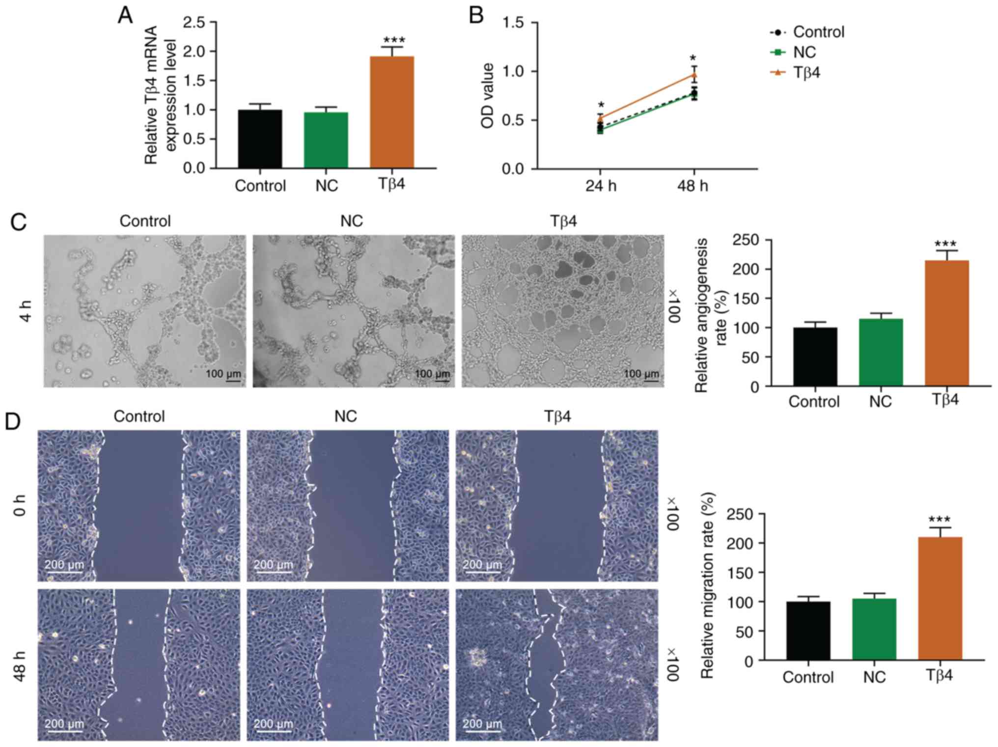
Tβ4 promotes cell viability, angiogenesis
and migration of HUVEC
Following Tβ4 overexpression in HUVEC, the
transfection efficiency was detected by RTq-PCR, and MTT, tube
formation and wound healing assays were conducted. As presented in
Fig. 1A, Tβ4 expression level was
increased in the Tβ4 group compared with NC group (P<0.001).
Furthermore, Tβ4 overexpression significantly increased HUVEC
viability compared with the NC group (Fig. 1B; P<0.05). In addition, Tβ4
enhanced the HUVEC angiogenesis (Fig.
1C) and migratory ability (Fig.
1D), compared with the NC group (both P<0.001).
Tβ4 promotes the expression of
angiogenesis-related and Notch/NF-κB pathway-related factors in
HUVEC
The expression of some angiogenesis-related factors
were detected by western blottong and RTq-PCR. As presented in
Fig. 2A and B, Tβ4 significantly
upregulated the expression of Ang2, tie2 and VEGF-A at both
translation and transcription levels, compared with the NC group
(all P<0.001). Furthermore, whether the effect of Tβ4 on HUVEC
angiogenesis was associated with Notch/NF-κB signaling pathway was
evaluated. Western blot-ting and immunofluorescence were used to
detect the expression of key proteins of Notch/NF-κB pathways. As
presented in Fig. 2C, the protein
expression of N1ICD and Notch3 was increased by Tβ4 group compared
with NC group (P<0.001 and P<0.01, respectively). In
addition, Tβ4 increased the expression of NF-κB/p65 in HUVEC
nucleus (Fig. 2D). These results
indicated that the effect of Tβ4 on HUVEC angiogenesis may be
associated with Notch/NF-κB signaling pathway.
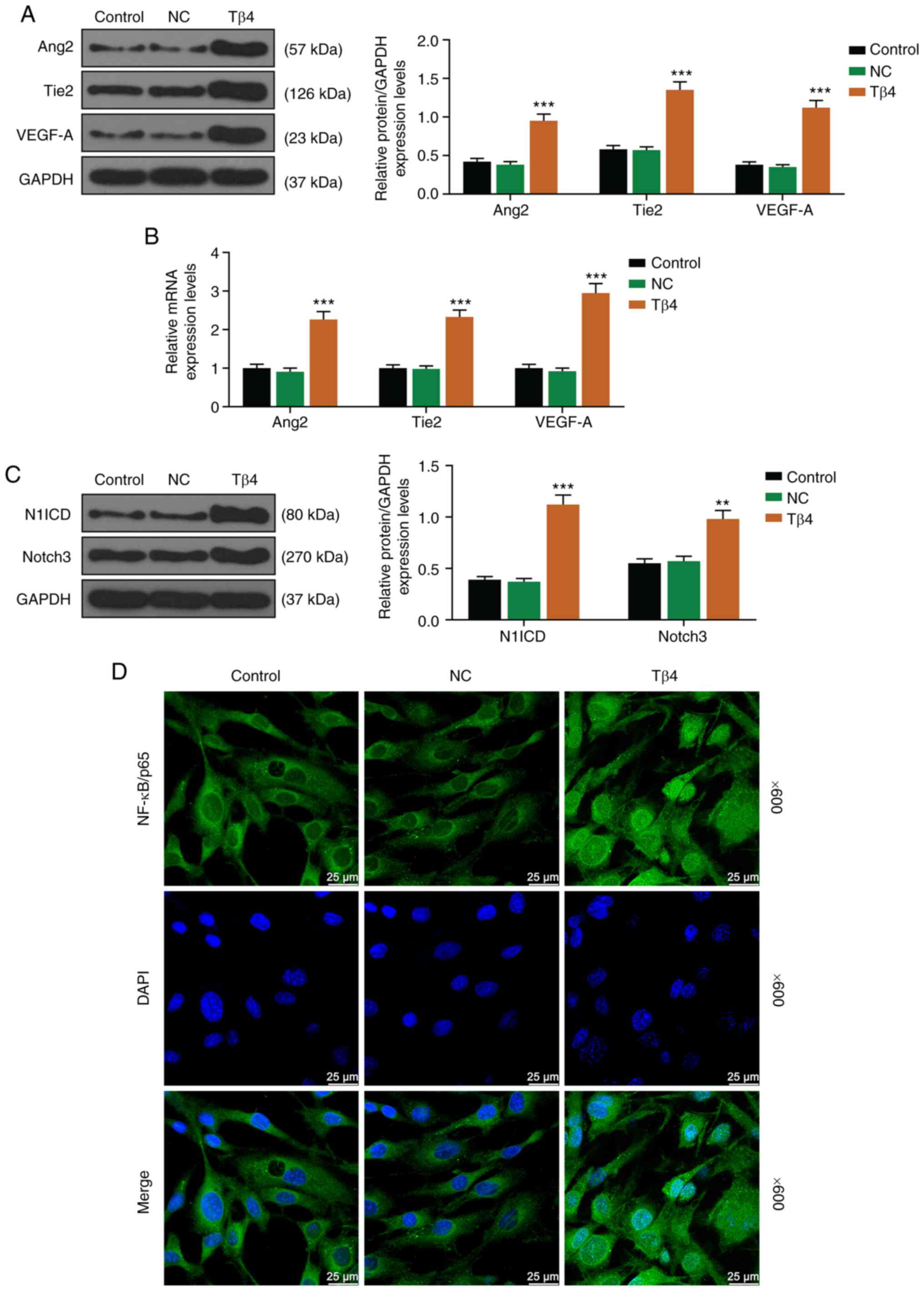 | Figure 2Tβ4 promoted the expression of
angiogenesis-related and Notch/NF-κB pathway-related proteins in
HUVEC. (A) Protein expression of Ang2, tie and VEGF-A were detected
by western blotting after infection with Tβ4 overexpression
lentiviral. GAPDH was used as an internal control. (B) mRNA
expression of Ang2, tie2 and VEGF-A were detected by reverse
transcription quantitative PCR after infection with Tβ4
overexpression lentiviral vector. GAPDH was used as an internal
control. (C) Protein expression of N1ICD and Notch3 were detected
by western blotting after infection with Tβ4 overexpression
lentiviral vector. GAPDH was used as an internal control. (D)
Expression of NF-κB/p65 in HUVEC nucleus was detected by
immunofluorescence. Magnification, ×600. All experiments were
conducted three times. **P<0.01 and
***P<0.001 vs. NC. VEGF-A, vascular endothelial
growth factor A; Ang2, angiopoietin-2; tie2, tyrosine kinase 2; NC,
negative control; N1ICD, NOTCH1 intracellular domain; Notch3, Notch
receptor 3; Tβ4, thymosin-β 4. |
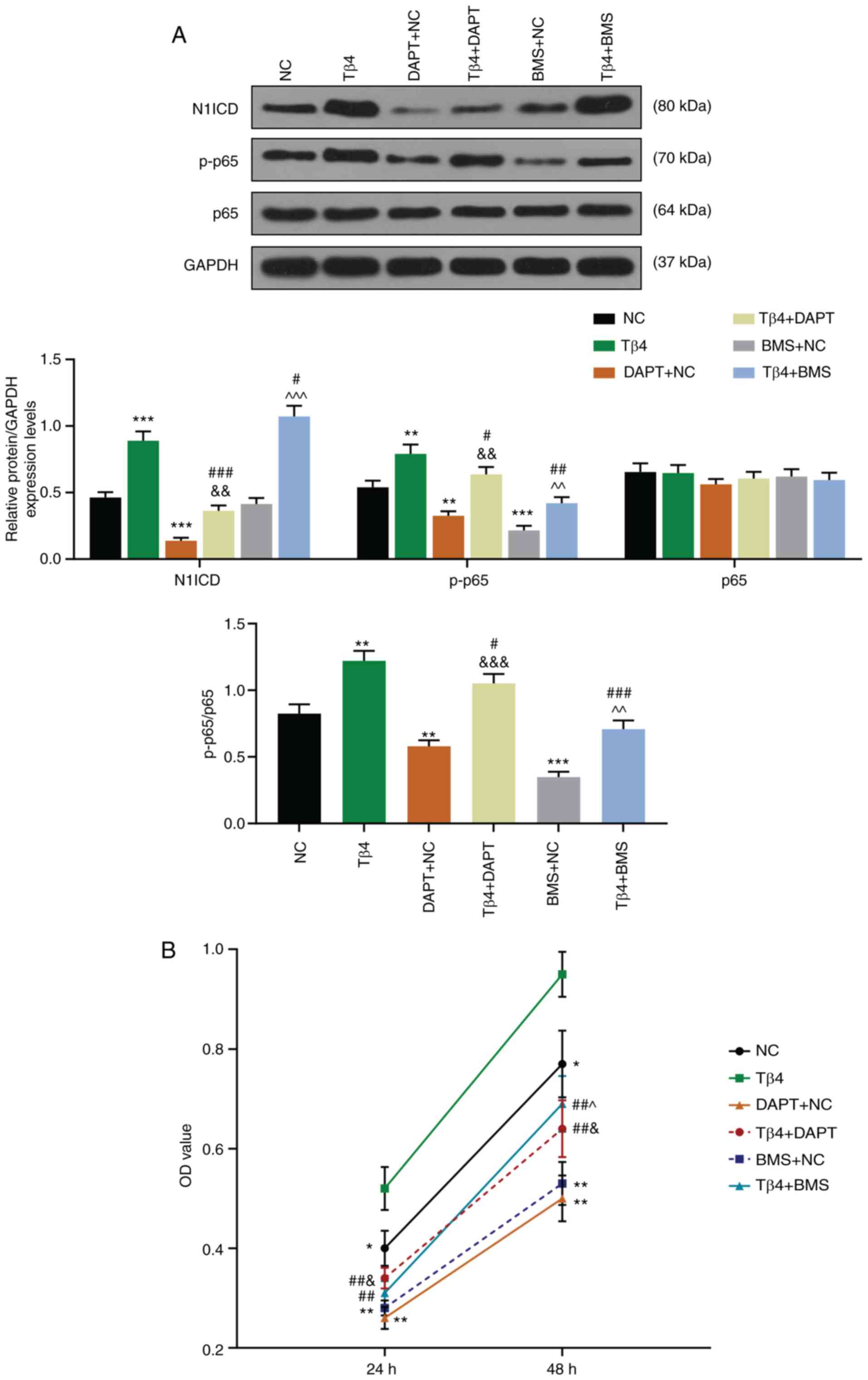
The promotion effects of Tβ4 on HUVEC
viability and on N1ICD and p-p65 expression are mediated by
Notch/NF-κB pathway
The inhibitors of Notch and NF-κB pathways (DAPT and
BMS, respectively) were used in the present study. As presented in
Fig. 3A, the expression of N1ICD
and p-p65, as well as the ratio p-p65/p65 were significantly
increased by Tβ4, but were decreased following treatment with DAPT
and BMS compared with NC group (P<0.001, P<0.01 and
P<0.05, respectively). Furthermore, in Tβ4 + DAPT and Tβ4 + BMS
groups, the promotion effect of Tβ4 on the expression of theses
proteins was reversed by treatment with DAPT and BMS compared with
Tβ4 and DAPT + NC or BMS + NC groups (P<0.05 and P<0.001,
respectively). Similarly, as presented in Fig. 3B, the promotion effect of Tβ4 on
HUVEC viability was reversed by DAPT and BMS compared with Tβ4 and
DAPT + NC or BMS + NC groups (P<0.05 and P<0.01,
respectively). These results demonstrated that the promotion
effects of Tβ4 on HUVEC viability and N1ICD and p-p65 expression
may be mediated by Notch/NF-κB signaling pathway.
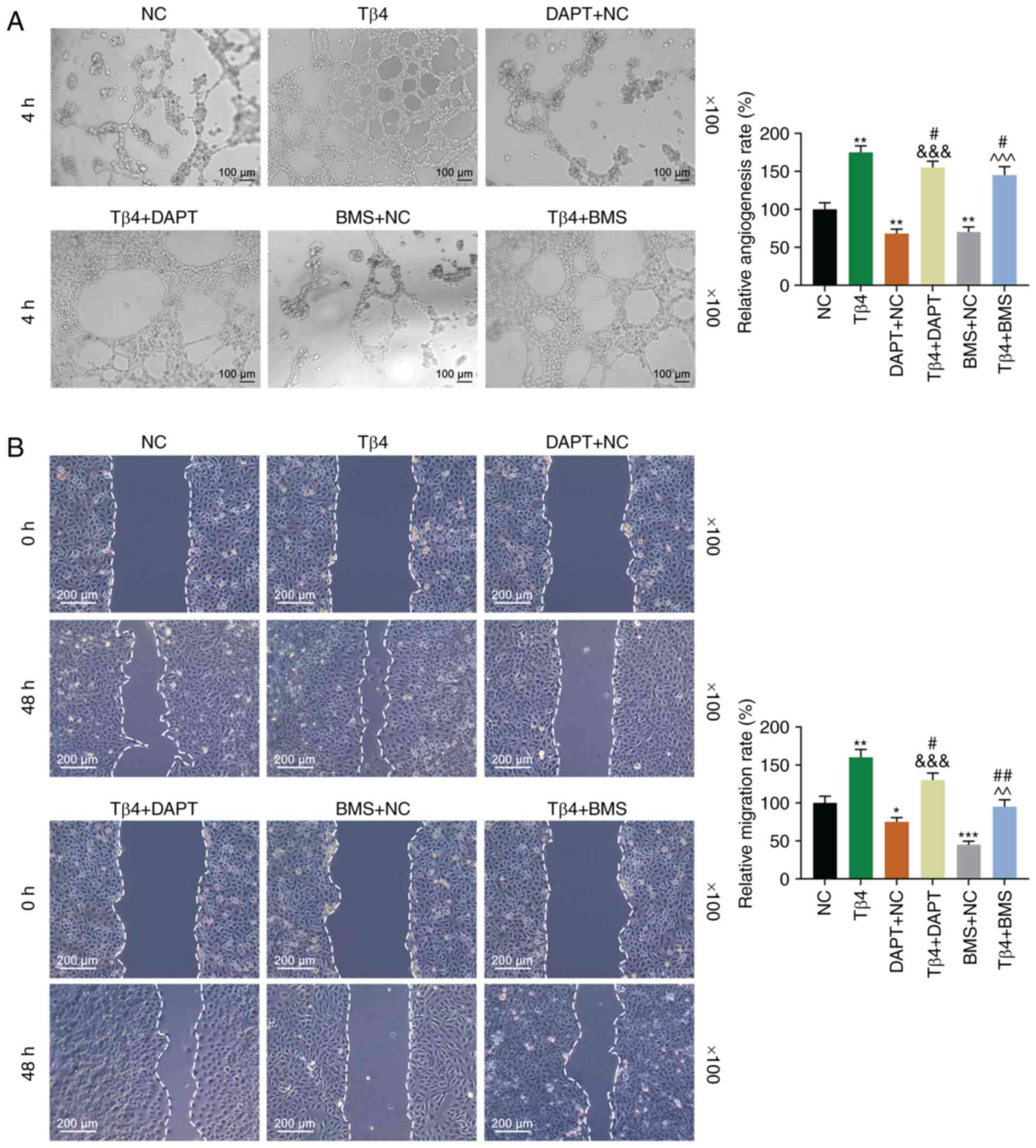
The promotion effects of Tβ4 on HUVEC
angiogenesis and migratory ability are mediated by Notch/NF-κB
pathway
Regarding the effect of Tβ4 on HUVEC angiogenesis
and migratory ability (Fig. 4A and
B), the results demonstrated that the relative angiogenesis and
migration rates of HUVEC were significantly increased by Tβ4 but
were decreased following treatment with DAPT and BMS compared with
the NC group (P<0.001, P<0.01 and P<0.05, respectively).
Furthermore, in Tβ4 + DAPT and Tβ4 + BMS groups, the promotion
effects of Tβ4 on HUVEC angiogenesis and migratory ability was
reversed by DAPT and BMS treatments compared with Tβ4 and DAPT + NC
or BMS + NC groups (P<0.05 and P<0.001, respectively). These
results suggested that the promotion effects of Tβ4 on HUVEC
angiogenesis and migratory ability may be mediated by Notch/NF-κB
signaling pathway.
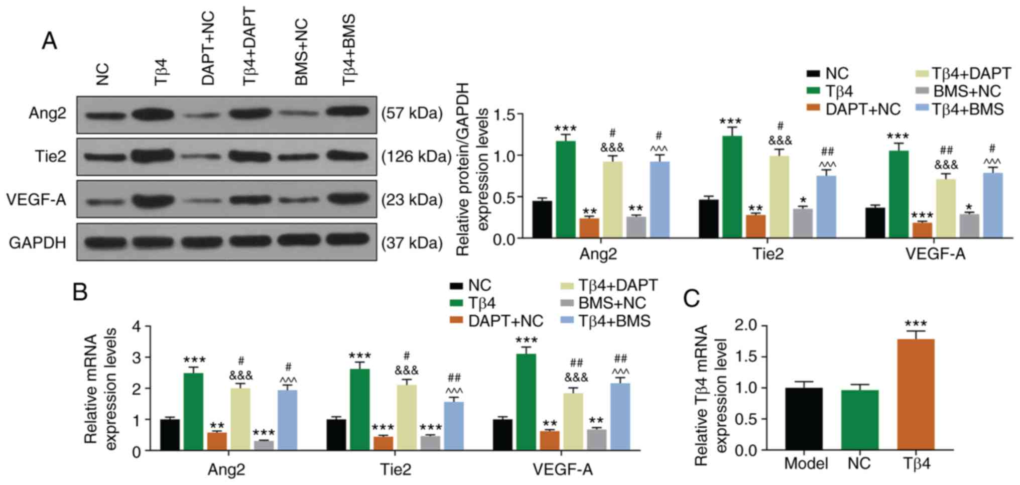
The promotion effects of Tβ4 on the
expression of angiogenesis-related factors are mediated by
Notch/NF-κB pathway
The changes in angiogenesis-related protein
expression were detected following HUVEC treatment with DAPT and
BMS. As presented in Fig. 5A and
B, the protein and gene expression of Ang2, tie2 and VEGF-A
were significantly increased by Tβ4 but were decreased following
cell treatment with DAPT and BMS compared with the NC group
(P<0.001 and P<0.01, respectively). Furthermore, in Tβ4 +
DAPT and Tβ4 + BMS groups, the promotion effects of Tβ4 on the
expression of these proteins were reversed by DAPT and BMS
treatments compared with Tβ4 and DAPT + NC or BMS + NC groups
(P<0.05, P<.01, and P<0.001, respectively). These findings
further suggested that the promotion effects of Tβ4 on HUVEC
angiogenesis and migratory ability were mediated by Notch/NF-κB
signaling pathway.
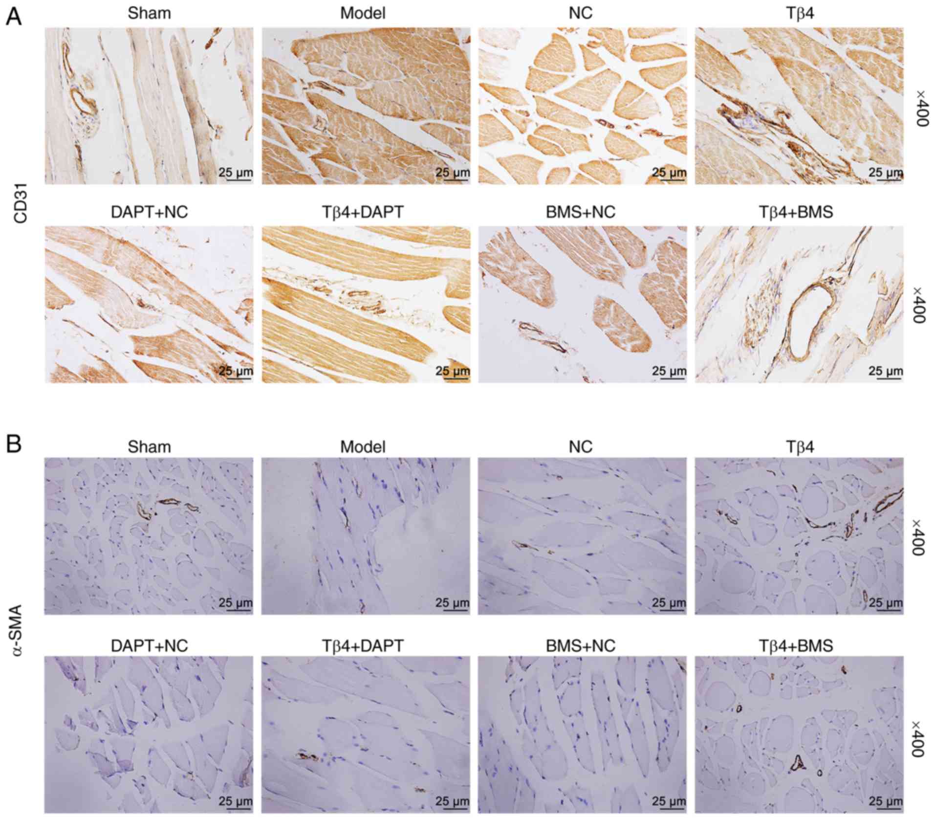
Tβ4 enhances the capillary and arteriolar
densities through regulating Notch/NF-κB pathway in CLI mice
In order to confirm the pro-angiogenesis effect of
Tβ4, in vivo experiments were preformed. The expression of
Tβ4 was increased in the Tβ4 group compared with NC group in
gastrocnemius of right hind limb tissues (Fig. 5C; P<0.001). Once CLI mice model
was established, the capillary and arteriolar densities were
observed by immunohistochemical staining. As presented in Fig. 6A and B, capillary density
(CD31+ cells) and arteriolar density (α-SMA+
cells) were remarkably decreased in Model, NC, DAPT + NC, and BMC +
NC groups, but were increased in Tβ4 group. Furthermore, in Tβ4 +
DAPT and Tβ4 + BMS groups, the promotion effects of Tβ4 on the
densities of capillary and arteriolar were reversed by DAPT and BMS
treatment. These results demonstrated that Tβ4 may have the ability
to increase capillary and arteriolar densities in CLI mice, and
that these effects might be mediated by Notch/NF-κB signaling
pathway.
Tβ4 enhances the expression of
angiogenesis-related proteins by regulating Notch/NF-κB pathway in
CLI mice
To further verify the current findings, the
expression of angiogenesis-related proteins was detected in CLI
mice. As presented in Fig. 7A and
B, the protein and gene expression of Ang2, tie2 and VEGF-A
were significantly increased by Tβ4, but were decreased following
treatment with DAPT and BMS compared with the NC group (P<0.001
and P<0.05, respectively). Furthermore, in Tβ4 + DAPT and Tβ4 +
BMS groups, the promotion effects of Tβ4 on the expression of these
proteins were reversed by DAPT and BMS treatment, compared with Tβ4
and DAPT + NC or BMS + NC groups (P<0.05, P<0.01 and
P<0.001, respectively). Regarding the expressions of key
proteins of Notch/NF-κB signaling pathway (Fig. 7C), the results demonstrated that
expression of N1ICD, p-p65 and the ratio of p-p65/p65 were
significantly increased by Tβ4, but were decreased following
treatment with DAPT and BMS compared with the NC group
(P<0.001). Furthermore, in Tβ4 + DAPT and Tβ4 + BMS groups, the
promotion effects of Tβ4 on the expression of these proteins were
reversed by DAPT and BMS treatment, compared with Tβ4 and DAPT + NC
or BMS + NC groups (P<0.001). These findings suggested that Tβ4
had the ability to enhance angiogenesis by regulating Notch/NF-κB
signaling pathway in CLI mice.
 | Figure 7Tβ4 enhanced the expression of
angiogenesis-related proteins by regulating Notch/NF-κB signaling
pathway in CLI mice. (A) Protein expression of Ang2, tie2 and
VEGF-A was detected by western blotting in CLI mice muscle tissues.
GAPDH was used as an internal control. (B) mRNA expression of Ang2,
tie2 and VEGF-A was detected by reverse transcription quantitative
PCR in CLI mice muscle tissues. GAPDH was used as an internal
control. (C) Protein expression of N1ICD, p-p65 and p65 was
detected by western blotting in CLI mice muscle tissues. GAPDH was
used as an internal control. All experiments were conducted three
times. ‡P<0.05, ‡‡P<0.01 and
‡‡‡P<0.001 vs. Sham; *P<0.05,
**P<0.01 and ***P<0.001 vs. NC;
#P<0.05 and ###P<0.001 vs. Tβ4;
&&&P<0.001 vs. DAPT + NC;
^^P<0.01 and ^^^P<0.001 vs. BMS + NC.
VEGF-A, vascular endothelial growth factor A; Ang2, angiopoietin-2;
tie2, tyrosine kinase 2; NC, negative control; N1ICD, NOTCH1
intracellular domain; Tβ4, thymosin-β 4; CLI, critical limb
ischemia. |
Discussion
Therapeutic angiogenesis and arteriogenesis remain
important therapeutic goals in patients with CLI without
revascularization possibilities. Numerous therapies have been
attempted, with some encouraging effects (24-26). The present study aimed to explore
the underlying mechanisms and pro-angiogenic effects of Tβ4 in CLI
mice. The effects of Tβ4 on HUVEC were first investigated, and the
results demonstrated Tβ4 could promote the migratory ability and
angiogenesis of HUVEC, which were mediated by Notch/NF-κB signaling
pathway. Furthermore, a CLI mice model was established followed by
treatment with Tβ4 overexpression vector, the inhibitor of Notch
pathway DAPT and the inhibitor of NF-κB pathway BMS. The results
further demonstrated that Tβ4 could increase the capillary and
arteriolar densities in CLI mice muscle tissues by regulating
Notch/NF-κB signaling pathway. These findings suggested that Tβ4
may promote angiogenesis in CLI mice via regulation of Notch/NF-κB
pathway.
Tβ4 was first isolated from the thymus and belongs
to the β-thymosin family, which consists of several structurally
related amino acid polypeptides (27). Tβ4 lacks a secretion signal,
therefore it is speculated that its presence in body fluids might
be due to damaged cell (10,27,28). Tβ4 was reported to take part in
tissue damage-related diseases, such as dermal injuries, cerebral
ischemia-reperfusion injury, heart injury and CLI (14,15,28-30). Furthermore, during tissue injury,
Tβ4 can promote endothelial cell migration and tube formation
(10,12). Similarly, the present study
demonstrated that Tβ4 may stimulate the migratory ability and tube
formation of HUVEC. In addition, this study demonstrated that Tβ4
could increase the capillary and arteriolar densities of CLI mice
muscle tissues. These findings suggested that Tβ4 may have the
ability to induce angiogenesis in CLI mice.
Previous studies reported that Ang-tie signaling is
an important regulator signal in vascular development, angiogenesis
and remodeling (31,32). In the Ang family, Ang2 is usually
secreted in diseased or remodeling vessels and has been identified
as the main ligand for tie2 (31,33). Tie2 plays a central role in
promoting vascular stability, and therapeutic drugs that target the
Angtie signaling axis to enhance tie2 activation have been
extensively explored in ischemic vascular diseases, various types
of cancer and inflammation (31,32). The expression of Ang2 and tie2 in
HUVEC and CLI mice muscle tissues following Tβ4 overexpression was
therefore determined in the present study. The results demonstrated
that Tβ4 upregulated the expression of Ang2 and Tie2 in both HUVEC
and CLI mice muscle tissues, which further confirmed previous
results. In addition, VEGFA is one vascular endothelial growth
factor, which has been strongly associated with angiogenesis in
endothelial cells (34,35). In the present study, the
expression of VEGFA was therefore evaluated and the results
demonstrated that Tβ4 also increased VEGFA expression, which
confirmed that Tβ4 could induce angiogenesis in CLI mice.
Previous studies have reported that Notch/NF-κB
signaling pathway is highly related to the process of angiogenesis
(36-40). Notch1 is part of the Notch family
that regulates cell fate and VEGF expression in various types of
cell, including HUVEC (10,39,41). Once ischemia occurrs, Notch1 would
bind to its ligands (Jagged1 or Jagged2, or Delta-like 1, 3, or 4)
to form a dimer and cleaved into N1CID, leading to VEGFA
overexpression (42,43). Previous studies reported that
activation of Notch signaling pathway could also promote NF-κB
pathway following ischemia (40).
After ischemia onset, Notch induces free NF-κB to translocate into
the nucleus and to activate the expression of pro-angogenic
factors, such as VEGFA, leading to angiogenesis of endothelial cell
(37,40,44). The present study hypothesized
therefore that Notch/NF-κB signaling pathway could be involved in
the process of Tβ4-induced angiogenesis. After inhibiting Notch and
NF-κB signaling pathway, the results demonstrated that Notch/NF-κB
pathway might be related to Tβ4-induced angiogenesis.
In conclusion, the presents tudy demonstrated that
Tβ4 may induce angiogenesis in CLI mice by regulating Notch/NF-κB
signaling pathway, and may be considered as a therapeutic target
for CLI treatment. However, whether Tβ4 could induce angiogenesis
in a clinical setup requires further investigation focusing on
application, duration, dosage and safety of Tβ4 treatment.
Funding
No funding was received.
Availiability of data and materials
The datasets used and/or analyzed during the current
study are available from the corresponding author on reasonable
request.
Authors' contributions
SL and HC made substantial contributions to the
conception and design of the study. YX, JD, XR and LZ were involved
in data acquisition, analysis and interpretation. SL and HC were
responsible for drafting the article and critically revising it for
important intellectual content. All authors agreed to be
accountable for all aspects of the work, in ensuring that questions
related to the accuracy or integrity of the work were appropriately
investigated and resolved. All authors read and approved the final
manuscript.
Ethics approval and consent to
participate
This study was approved by the Committee of
Experimental Animals of The First Affiliated Hospital of Zhejiang
Chinese Medical University (approval no. Z20190312G).
Patient consent for publication
Not applicable.
Competing interests
The authors declare that they have no competing
interests.
Acknowledgments
Not applicable.
Abbreviations:
|
Tβ4
|
thymosin-β 4
|
|
CLI
|
critical limb ischemia
|
References
|
1
|
Conte SM and Vale PR: Peripheral arterial
disease. Heart Lung Circ. 27:427–432. 2018. View Article : Google Scholar
|
|
2
|
Nehler MR, Duval S, Diao L, Annex BH,
Hiatt WR, Rogers K, Zakharyan A and Hirsch AT: Epidemiology of
peripheral arterial disease and critical limb ischemia in an
insured national population. J Vasc Surg. 60:686–695 e682. 2014.
View Article : Google Scholar : PubMed/NCBI
|
|
3
|
Dua A and Lee CJ: Epidemiology of
peripheral arterial disease and critical limb ischemia. Tech Vasc
Interv Radiol. 19:91–95. 2016. View Article : Google Scholar : PubMed/NCBI
|
|
4
|
Albrecht-Schgoer K, Barthelmes J, Schgoer
W, Theurl M, Nardin I, Lener D, Gutmann C, Dünnhaupt S,
Bernkop-Schnürch A and Kirchmair R: Nanoparticular delivery system
for a secretoneurin derivative induces angiogenesis in a hind limb
ischemia model. J Control Release. 250:1–8. 2017. View Article : Google Scholar : PubMed/NCBI
|
|
5
|
Constantinescu IM, Bolfa P, Constantinescu
D, Mironiuc AI and Gherman CD: Treatment with sildenafil and
donepezil improves angiogenesis in experimentally induced critical
limb ischemia. Biomed Res Int. 2017:95323812017. View Article : Google Scholar : PubMed/NCBI
|
|
6
|
Ishibashi T and Ryan SJ: Maturation of
newly-formed subretinal vessels. EXS. 61:59–63. 1992.PubMed/NCBI
|
|
7
|
Zhao C, Wang X, Zhao Y, Li Z, Lin S, Wei Y
and Yang H: A novel xenograft model in zebrafish for
high-resolution investigating dynamics of neovascularization in
tumors. PLoS One. 6:e217682011. View Article : Google Scholar : PubMed/NCBI
|
|
8
|
Sajib S, Zahra FT, Lionakis MS, German NA
and Mikelis CM: mechanisms of angiogenesis in microbe-regulated
inflammatory and neoplastic conditions. Angiogenesis. 21:1–14.
2018. View Article : Google Scholar
|
|
9
|
Vasilopoulou E, Riley PR and Long DA:
Thymosin-β4: A key modifier of renal disease. Expert Opin Biol
Ther. 18:185–192. 2018. View Article : Google Scholar : PubMed/NCBI
|
|
10
|
Lv S, Cheng G, Zhou Y and Xu G: Thymosin
β4 induces angiogenesis through Notch signaling in endothelial
cells. Mol Cell Biochem. 381:283–290. 2013. View Article : Google Scholar : PubMed/NCBI
|
|
11
|
Sanders MC, Goldstein AL and Wang YL:
Thymosin beta 4 (Fx peptide) is a potent regulator of actin
polymerization in living cells. Proc Natl Acad Sci USA.
89:4678–4682. 1992. View Article : Google Scholar : PubMed/NCBI
|
|
12
|
Kobayashi T, Okada F, Fujii N, Tomita N,
Ito S, Tazawa H, Aoyama T, Choi SK, Shibata T, Fujita H and
Hosokawa M: Thymosin-beta4 regulates motility and metastasis of
malignant mouse fibrosarcoma cells. Am J Pathol. 160:869–882. 2002.
View Article : Google Scholar : PubMed/NCBI
|
|
13
|
Renga G, Oikonomou V, Stincardini C,
Pariano M, Borghi M, Costantini C, Bartoli A, Garaci E, Goldstein
AL and Romani L: Thymosin β4 limits inflammation through autophagy.
Expert Opin Biol Ther. 18(Suppl 1): S171–S175. 2018. View Article : Google Scholar
|
|
14
|
Kleinman HK and Sosne G: Thymosin β4
promotes dermal healing. Vitam Horm. 102:251–275. 2016. View Article : Google Scholar
|
|
15
|
Trenkwalder T, Deindl E, Bongiovanni D,
Lee S, Schunkert H, Kupatt C and Hinkel R: Thymosin-β4-mediated
therapeutic neovascularization: Role of the PI3K/AKT pathway.
Expert Opin Biol Ther. 15(Suppl 1): S175–S185. 2015. View Article : Google Scholar
|
|
16
|
Quan Z, Wang QL, Zhou P, Wang GD, Tan YZ
and Wang HJ: Thymosin β4 promotes the survival and angiogenesis of
trans-planted endothelial progenitor cells in the infarcted
myocardium. Int J Mol Med. 39:1347–1356. 2017. View Article : Google Scholar : PubMed/NCBI
|
|
17
|
Zhao Y, Song J, Bi X, Gao J, Shen Z, Zhu J
and Fu G: Thymosin β4 promotes endothelial progenitor cell
angiogenesis via a vascular endothelial growth factor-dependent
mechanism. Mol Med Rep. 18:2314–2320. 2018.PubMed/NCBI
|
|
18
|
Zhao J, Liang Y, Song F, Xu S, Nian L,
Zhou X and Wang S: TSG attenuates LPC-induced endothelial cells
inflammatory damage through notch signaling inhibition. IUBMB Life.
68:37–50. 2016. View
Article : Google Scholar
|
|
19
|
MacMaster JF, Dambach DM, Lee DB, Berry
KK, Qiu Y, Zusi FC and Burke JR: An inhibitor of IkappaB kinase,
BMS-345541, blocks endothelial cell adhesion molecule expression
and reduces the severity of dextran sulfate sodium-induced colitis
in mice. Inflamm Res. 52:508–511. 2003. View Article : Google Scholar
|
|
20
|
Grimaldo S, Tian F and Li LY:
Sensitization of endothelial cells to VEGI-induced apoptosis by
inhibiting the NF-kappaB pathway. Apoptosis. 14:788–795. 2009.
View Article : Google Scholar : PubMed/NCBI
|
|
21
|
Burke JR, Pattoli MA, Gregor KR, Brassil
PJ, Macmaster JF, McIntyre KW, Yang X, Iotzova VS, Clarke W, Strnad
J, et al: BMS-345541 is a highly selective inhibitor of I kappa B
kinase that binds at an allosteric site of the enzyme and blocks
NF-kappa B-dependent transcription in mice. J Biol Chem.
278:1450–1456. 2003. View Article : Google Scholar
|
|
22
|
Peng X, Zhou J, Li B, Zhang T, Zuo Y and
Gu X: Notch1 and PI3K/Akt signaling blockers DAPT and LY294002
coordinately inhibit metastasis of gastric cancer through mutual
enhancement. Cancer Chemother Pharmacol. 85:309–320. 2020.
View Article : Google Scholar
|
|
23
|
Schmittgen TD and Livak KJ: Analyzing
real-time PCR data by the comparative C(T) method. Nat Protoc.
3:1101–1108. 2008. View Article : Google Scholar : PubMed/NCBI
|
|
24
|
Farber A and Eberhardt RT: The current
state of critical limb ischemia: A systematic review. JAMA Surg.
151:1070–1077. 2016. View Article : Google Scholar : PubMed/NCBI
|
|
25
|
Falluji N and Mukherjee D: Critical and
acute limb ischemia: An overview. Angiology. 65:137–146. 2014.
View Article : Google Scholar
|
|
26
|
Liew A, Bhattacharya V, Shaw J and Stansby
G: Cell therapy for critical limb ischemia: A meta-analysis of
randomized controlled trials. Angiology. 67:444–455. 2016.
View Article : Google Scholar
|
|
27
|
Sosne G, Qiu P, Goldstein AL and Wheater
M: Biological activities of thymosin beta4 defined by active sites
in short peptide sequences. FASEB J. 24:2144–2151. 2010. View Article : Google Scholar : PubMed/NCBI
|
|
28
|
Yang WS, Kang S, Sung J and Kleinman HK:
Thymosin β4: Potential to treat epidermolysis bullosa and other
severe dermal injuries. Eur J Dermatol. 29:459–467. 2019.
View Article : Google Scholar : PubMed/NCBI
|
|
29
|
Zhang Z, Liu S and Huang S: Effects of
thymosin β4 on neuronal apoptosis in a rat model of cerebral
ischemiareperfusion injury. Mol Med Rep. 20:4186–4192.
2019.PubMed/NCBI
|
|
30
|
Bjørklund G, Dadar M, Aaseth J and
Chirumbolo S: Thymosin β4: A multi-faceted tissue repair
stimulating protein in heart injury. Curr Med Chem. July
31–2019.Epub ahead of print. View Article : Google Scholar
|
|
31
|
Zhang Y, Kontos CD, Annex BH and Popel AS:
Angiopoietin-tie signaling pathway in endothelial cells: A
computational model. iScience. 20:497–511. 2019. View Article : Google Scholar : PubMed/NCBI
|
|
32
|
Saharinen P, Eklund L and Alitalo K:
Therapeutic targeting of the angiopoietin-TIE pathway. Nat Rev Drug
Discov. 16:635–661. 2017. View Article : Google Scholar : PubMed/NCBI
|
|
33
|
Thurston G and Daly C: The complex role of
angiopoietin-2 in the angiopoietin-tie signaling pathway. Cold
Spring Harb Perspect Med. 2:a0065502012. View Article : Google Scholar : PubMed/NCBI
|
|
34
|
Perez-Moral N, Needs PW, moyle CWA and
Kroon PA: Hydrophobic interactions drive binding between vascular
endothelial growth factor-A (VEGFA) and polyphenolic inhibitors.
Molecules. 24:27852019. View Article : Google Scholar :
|
|
35
|
Bhisitkul RB: Vascular endothelial growth
factor biology: Clinical implications for ocular treatments. Br J
Ophthalmol. 90:1542–1547. 2006. View Article : Google Scholar : PubMed/NCBI
|
|
36
|
Shukla K, Sonowal H, Saxena A and Ramana
KV: Didymin by suppressing NF-κB activation prevents VEGF-induced
angiogenesis in vitro and in vivo. Vascul Pharmacol. 115:18–25.
2019. View Article : Google Scholar : PubMed/NCBI
|
|
37
|
Huang DY, Dai ZR, Li WM, Wang RG and Yang
SM: Inhibition of EGF expression and NF-κB activity by treatment
with quercetin leads to suppression of angiogenesis in
nasopharyngeal carcinoma. Saudi J Biol Sci. 25:826–831. 2018.
View Article : Google Scholar : PubMed/NCBI
|
|
38
|
Si W, Xie W, Deng W, Xiao Y, Karnik SS, Xu
C, Chen Q and Wang QK: Angiotensin II increases angiogenesis by
NF-κB-mediated transcriptional activation of angiogenic factor
AGGF1. FASEB J. 32:5051–5062. 2018. View Article : Google Scholar : PubMed/NCBI
|
|
39
|
Li L, Tang P, Zhou Z, Wang Q, Xu T, Zhao
S, Huang Y, Kong F, Liu W, Cheng L, et al: GIT1 regulates
angiogenic factor secretion in bone marrow mesenchymal stem cells
via NF-κB/notch signalling to promote angiogenesis. Cell Prolif.
52:e126892019. View Article : Google Scholar
|
|
40
|
Xu D, Xia N, Hou K, Li F, Chen S, Hu Y,
Fang W and Li Y: Clematichinenoside facilitates recovery of
neurological and motor function in rats after cerebral ischemic
injury through inhibiting notch/NF-κB pathway. J Stroke Cerebrovasc
Dis. 28:1042882019. View Article : Google Scholar
|
|
41
|
Saito T and Tanaka S: Molecular mechanisms
underlying osteoarthritis development: Notch and NF-κB. Arthritis
Res Ther. 19:942017. View Article : Google Scholar
|
|
42
|
Chen Y, Zhao B, Zhu Y, Zhao H and Ma C:
HIF-1-VEGF-notch mediates angiogenesis in temporomandibular joint
osteoarthritis. Am J Transl Res. 11:2969–2982. 2019.PubMed/NCBI
|
|
43
|
Luo Z, Shang X, Zhang H, Wang G, Massey
PA, Barton SR, Kevil CG and Dong Y: Notch signaling in
osteogenesis, osteoclastogenesis, and angiogenesis. Am J Pathol.
189:1495–1500. 2019. View Article : Google Scholar : PubMed/NCBI
|
|
44
|
Huang F, Yao Y, Wu J, Liu Q, zhang J, Pu
X, zhang Q and Xia L: Curcumin inhibits gastric cancer-derived
mesenchymal stem cells mediated angiogenesis by regulating
NF-κB/VEGF signaling. Am J Transl Res. 9:5538–5547. 2017.
|