Diabetes mellitus, a disease affecting individuals
worldwide with a complex course and a number of complications, has
garnered increasing attention. The International Diabetes
Federation predicts that the number of adults aged 20-79 years with
diabetes mellitus globally will increase to 700 million by 2045,
and 3 out of 4 individuals will be of working age (1). The incidence of this disease is
influenced by a number of factors, such as body mass index, body
fat distribution, metabolic syndrome and sex differences (2-4).
Even sleep deprivation and work-related stress can influence its
incidence (5,6). The complications of diabetes
mellitus can be classified into macrovascular complications [for
example, cardiovascular disease (CVD)] and microvascular
complications (for example, complications affecting the retina,
kidneys and the nervous systems). Diabetic retinopathy (DR) is
closely related to hyperglycemia caused by diabetes mellitus.
Traditionally, DR was considered to be a microvascular disease;
however, new research has found that retinal neurodegeneration is
also closely related to its occurrence (7). Thus, the pathogenic mechanisms
underlying DR are very complex.
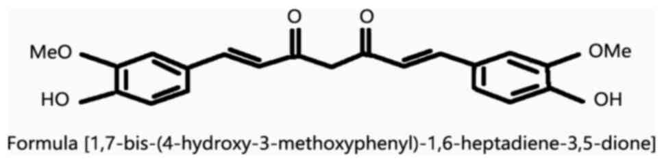
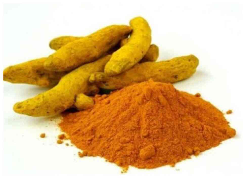
Curcumin represents the major constituent of
turmeric. In addition to being widely recognized as a spice,
curcumin also has a variety of therapeutic effects. For example, as
an anticancer agent, it can prevent ovarian, prostate, breast and
gastric cancers (8-11). Moreover, curcumin exerts
anti-inflammatory effects, and it can be used in the treatment of
chronic obstructive pulmonary disease (COPD), hepatic inflammation
and even acute vascular inflammation (12-14). As a natural antioxidant, the
reno-protective effects of curcumin have been demonstrated
(15). In diabetic rats and
Müller cells, the increase in histone acetylation plays an
important role in the regulation of the inflammatory response.
Curcumin can exert protective effects on diabetes by inhibiting
histone acetylation (16-18).
The preventive and curative effects of curcumin on DR and its
mechanisms of action have, however, not been thoroughly defined.
The present review article aimed to investigate the therapeutic
effects of curcumin on diabetes mellitus and DR, and to demonstrate
the possible mechanisms through which it exerts these effects.
The low bioavailability of curcumin may be related
to the low solubility of curcumin, the poor permeability of the
gastrointestinal mucosa and the rapid clearance of curcumin in
vivo. Although curcumin exerts therapeutic effects against
diabetes and related diseases, data from clinical trials have
demonstrated that even when a large dose (12,000 mg/day) of
curcumin is used in the treatment of the human body, the curcumin
content in the serum remains low (22). Further research has also
indicated that the use of dendritic nanoparticles can enhance the
solubility of curcumin in vivo (23) and improve the bioavailability of
curcumin. In an in vitro study, scholars have found that
solid lipid nanoparticles loaded with curcumin can prolong the
release time of curcumin in vitro to 12 h with good
stability (24). Furthermore, in
an in vivo experiment in rats with asthma, the use of
curcumin lipid nanocarriers has also been found to increase the
concentration of curcumin in plasma suspension and tissue (25). In addition, it has been found
that the administration of piperine within 1 h following the
curcumin administration can significantly increase the serum
concentration of curcumin in rats and humans, enhance its
bioavailability, and exerts no side-effects (26). As a therapeutic drug, curcumin
has other structural analogs that can enhance its therapeutic
effect. Compared with curcumin, its structural analog EF24 has
significant activity in the treatment of leukemia (27). The above-mentioned methods can
enhance the bioavailability of curcumin to enhance its therapeutic
effect.
Diabetes represents a group of metabolic diseases.
Over the past decades, its incidence has increased exponentially
(28), and as a chronic disease
that can cause damage to a variety of organs, the prevention and
treatment of diabetes have garnered increasing attention. Studies
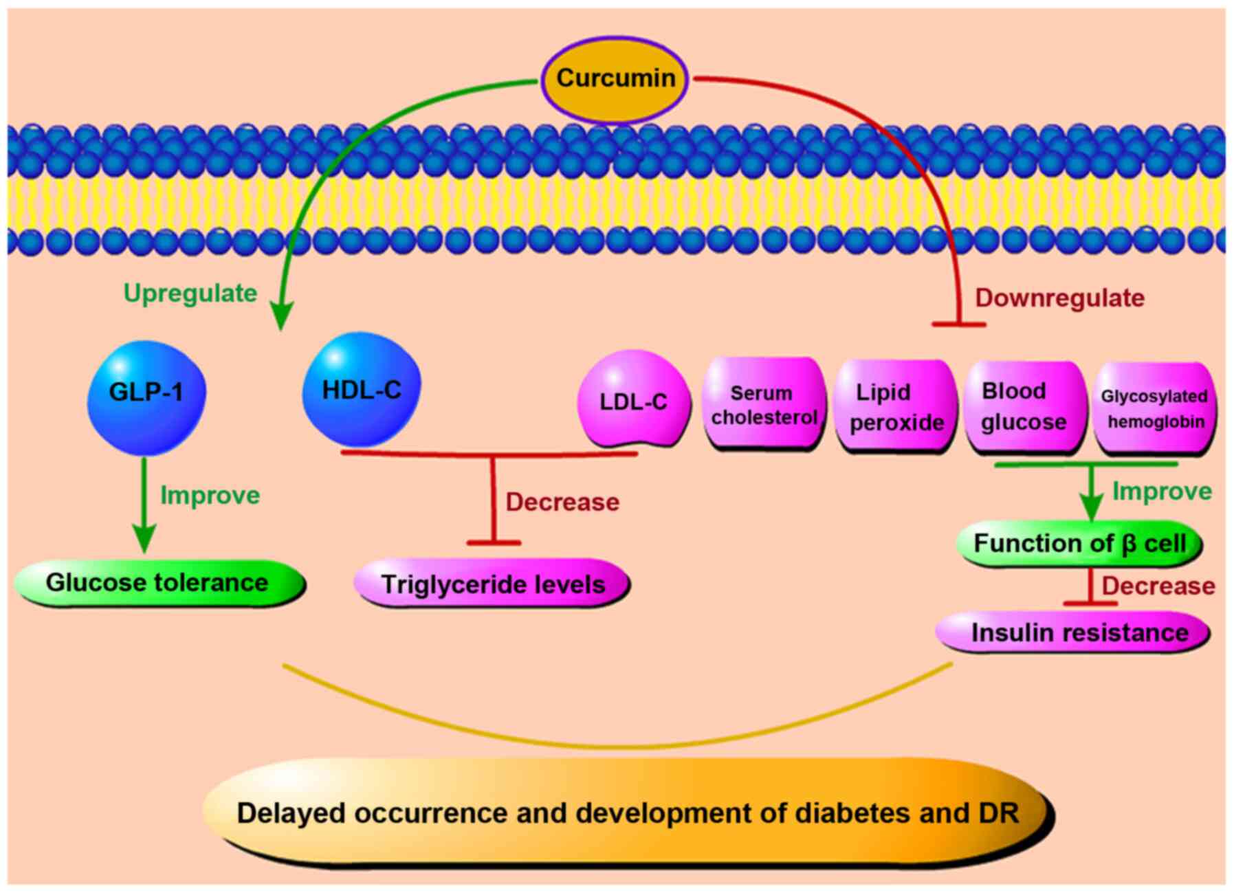
have indicated that curcumin can protect diabetic patients or
cultured cells in vitro (29). In terms of prevention, it has
been found that curcumin can significantly reduce the number of
patients who eventually progress to type 2 diabetes after 9 months
of curcumin intervention at the pre-diabetic stage. In addition,
curcumin treatment has been shown to improve the overall function
of β cells (30). Al-Saud found
that curcumin treatment exerted hypoglycemic effects in rats with
diabetes induced by a high-fat diet combined with a streptozotocin
(STZ) injection; furthermore, curcumin reduced insulin resistance
in tissues and improved the diabetic status (31).
Similarly, some scholars have found that curcumin
can significantly reduce fasting blood glucose in diabetic rats. In
rats treated with curcumin, pancreatic tissue damage has been shown
to be mild, and the expression levels of interleukin (IL)-1, IL-6,
monocyte chemoattractant protein-1 (MCP-1), tumor necrosis factor
(TNF)-α, Bax and caspase-3 have been found to be significantly
reduced. These results indicate that curcumin can curb pancreatic
islet cell apoptosis and inflammation, all the while promoting the
recovery of pancreatic function (32,33).
Curcumin also plays an important protective role in
cells under high-glucose conditions in in vitro experiments
(34,35). In a study that exposed insulin
(INS)-1 cells to high glucose with or without curcumin, the
proliferation of INS-1 cells, the morphological changes of islet
cells, the production of reactive oxygen species (ROS) as well as
the activities of superoxide dismutase and catalase, insulin
levels, the expression of nicotinamide adenine dinucleotide
phosphate (NADPH) oxidase subunits, and the expression of apoptotic
factors were observed (34). The
results of that study revealed that curcumin effectively inhibited
the proliferation-induced damage and oxidative stress caused by
high glucose in INS-1 cells, increased the insulin level, and
reduced the expression of NADPH oxidase subunits and apoptotic
factors. These results also suggested that curcumin alleviated
high-glucose/palmitate-induced oxidative stress in islet cells by
regulating the NADPH pathway and protected islet cells from
apoptosis (34). Xia et
al found that following curcumin administration, the activity
of rat insulinoma RIN-m5F β-cells increased, the apoptotic rate of
the cells decreased and the cells were protected (35). These results indicate that
curcumin protects islet cells from the damaging effects of high
glucose through anti-inflammatory and anti-apoptotic pathways, and
it can play a certain therapeutic role in diabetes by directly
reducing blood glucose levels.
Oxidative stress plays a key role in diabetic
microvascular complications. Under abnormal metabolic conditions
created by diabetes, superoxide production in microvascular
endothelial cells and the mitochondria is increased. Since
glutathione (GSH) is one of the important scavengers of ROS, a
reduced expression of NADPH, which is required to generate GSH, is
consumed excessively under a high glucose state, eventually leading
to an increased ROS generation. Increased intracellular ROS levels,
aggravated by oxidative stress and accompanied by an increased
local inflammatory response, ultimately lead to the apoptosis of
endothelial cells and pericytes (36,37). Under this premise, the barrier
function of the tight junctions of retinal capillary epithelial
cells is disrupted, leading to the leakage of intraretinal fluid.
Kowluru and Kanwar demonstrated that in the diabetic rat retina,
the level of oxidation-modified DNA in the cells continued to
increase (38). Moreover,
compared to that in the age-matched normal control group, the
antioxidant capacity of the retina of diabetic rats decreased by
approximately 30-35%. Curcumin may not only reduce the level of
oxidative stress, but may also inhibit the increase in the retinal
nitrotyrosine level, effectively reducing retinal damage caused by
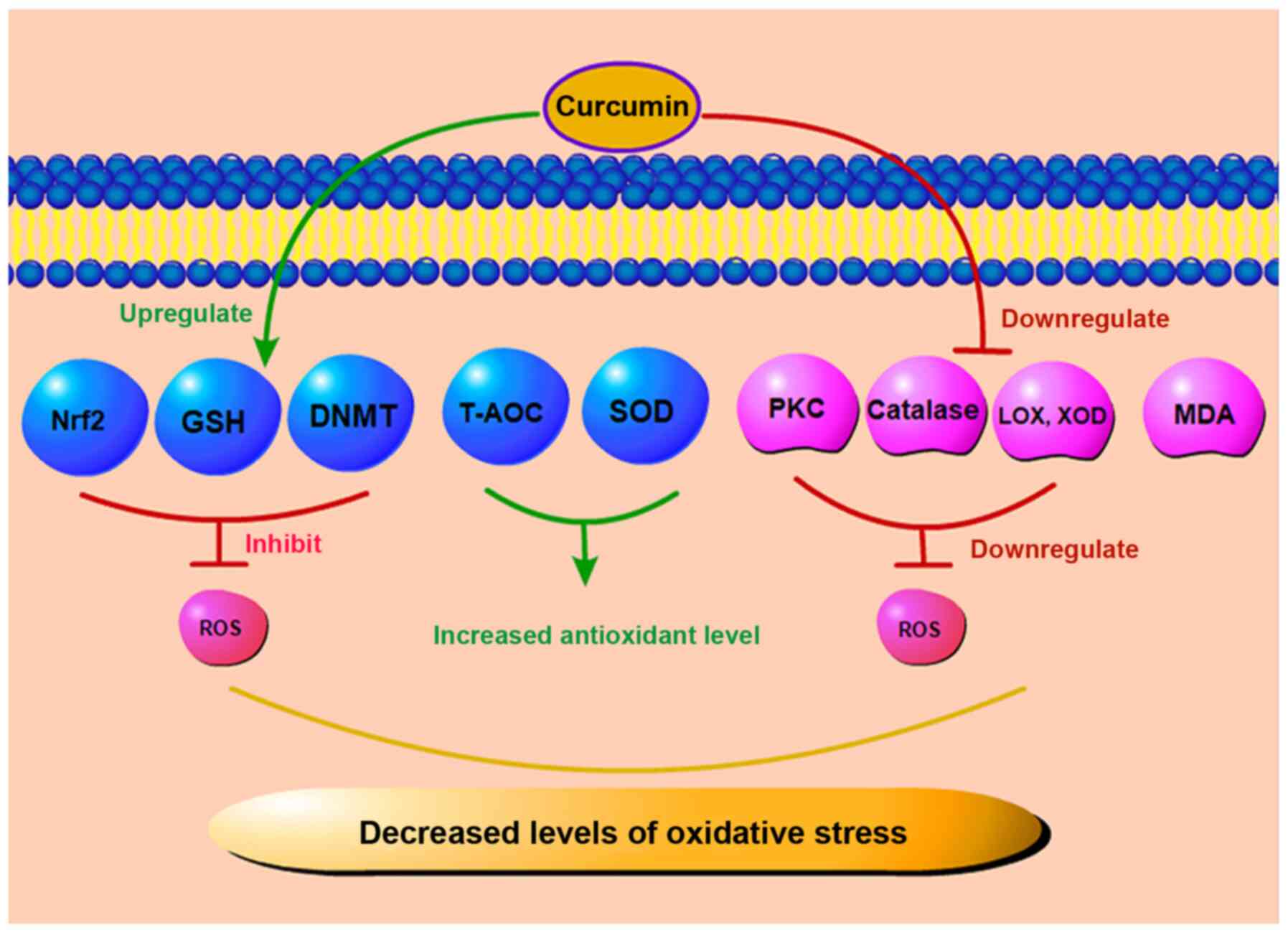
the accumulation of nitrotyrosine in the retina (38). A schematic diagram of the effects
of curcumin on oxidative stress effects is depicted in Fig. 3. Some scholars have indicated
that the combination of curcumin and oral anti-diabetic drugs or
other natural antioxidants exerts a more beneficial effect on the
treatment of diabetes and its complications compared with the use
of a single drug (39,40). As a regulator of antioxidant
reduction, nuclear factor E2-related factor 2 (Nrf2) plays an
important anti-DR role in the diabetic state. Curcumin can increase
the Nrf2 level, thus promoting its anti-fibrotic effect (41). In Müller cells of rats with
STZ-induced DR, curcumin has been shown to reduce the level of
malondialdehyde (MDA) in the retina and increase the level of
glutathione (GSH), thereby inhibiting oxidative stress in diabetic
retina and protecting Müller cells (42). ROS are normal products of cell
metabolism which can be leveraged as regulators of cell signal
transduction. However, high levels of ROS may exert cytotoxic and
mutagenic effects on cells, resulting in the destruction of lipids,
proteins, DNA and carbohydrates, and finally leading to cell
apoptosis (43). In DR,
oxidative stress increases with the progression of the disease.
Curcumin can mitigate oxidative stress by scavenging various forms
of free radicals, including ROS and reactive nitrogen species. In a
previous study, curcumin restored the expression and function of
DNA methyltransferase (DNMT) and inhibited oxidative stress and ROS
in cultured ARPE-19 cells (44).
In addition, it can inhibit protein kinase C (PKC) and other
enzymes, such as lipoxygenase (LOX) and xanthine oxidase (XOD),
which can produce ROS (45,46). As a key molecule in oxidative
stress, PKC plays a role in the pathogenesis of DR. Some scholars
have found that curcumin can inhibit PKC to a certain extent,
reduce the degree of oxidative stress, and thus exert a protective
effect with regard to retinal tissue (47). The total antioxidant capacity
(T-AOC) can reflect the antioxidant capacity of an organism. Yang
et al found that in a rat model of STZ-induced DR, curcumin
lowered the level of MDA and significantly increased the level of
superoxide dismutase (SOD) in rat retinal tissue after 16 weeks of
treatment (48). In parallel,
the activity of T-AOC also significantly increased (48). In addition, Bulboacă et al
found that curcumin significantly reduced the levels of total
oxidative stress and catalase in diabetic rats (49). In short, curcumin can
downregulate the level of ROS and reduce the apoptosis of
endothelial cells by increasing the levels of antioxidant stress
substances and reducing the levels of ROS related enzymes. Thus,
curcumin plays an important antioxidant role in DR.
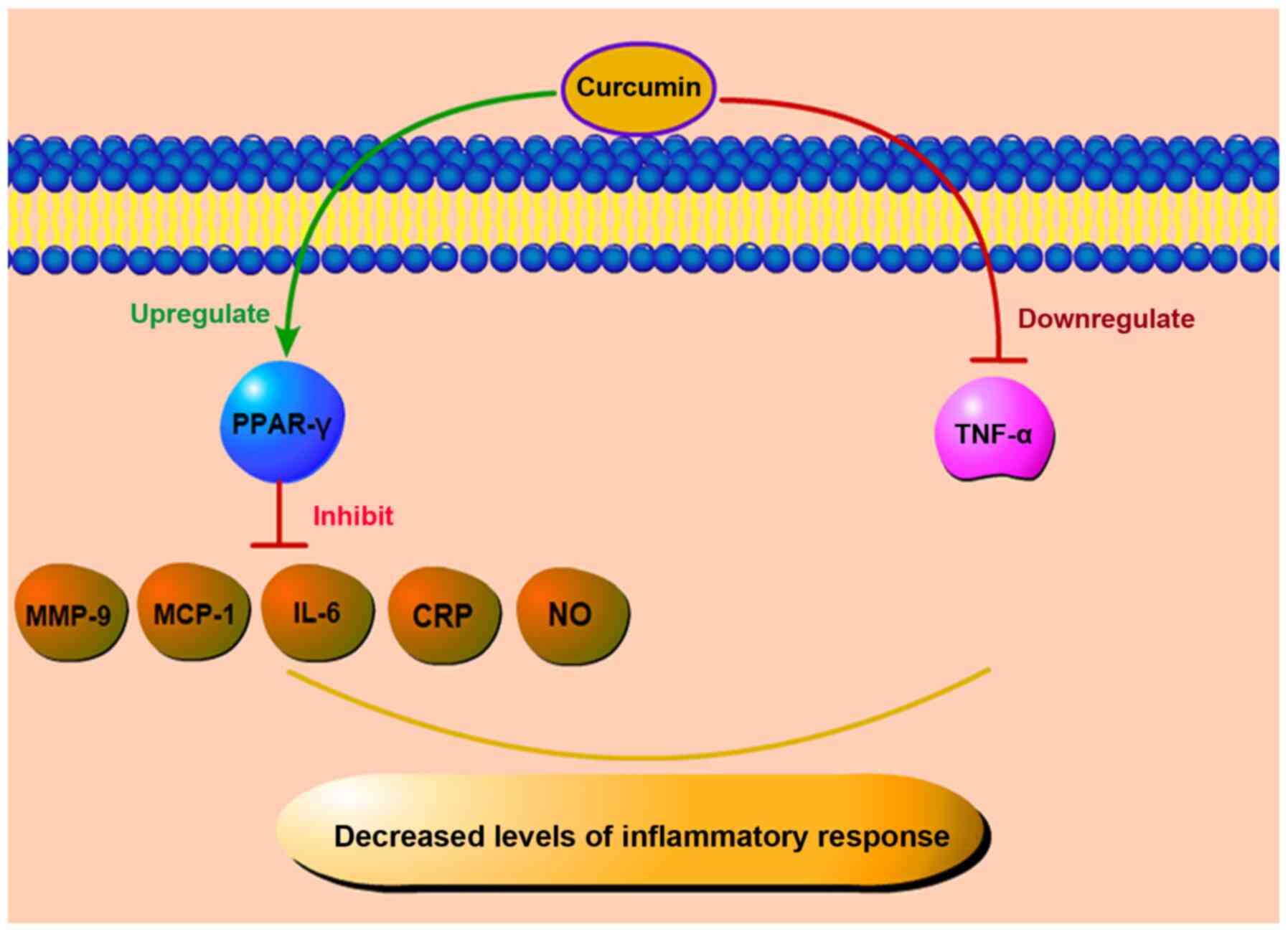
Peroxisome proliferator-activated receptor-γ
(PPAR-γ), a receptor activated by peroxisome proliferators, is a
transcription factor induced by ligands. In peripheral monocytes,
PPAR-γ is induced in the process of infiltration of blood vessels
into tissues and the activation of pro-inflammatory stimulation.
Some scholars also believe that PPAR-γ and its agonist not only
improve glucose homeostasis, but also regulate inflammation
(61,62). In currently existing animal
research models, PPAR-γ agonists can inhibit the formation of
retinal leukocytes and the leakage of retinal barrier in rats with
STZ-induced DR, alongside reducing the levels of various
inflammatory markers, such as MCP-1, IL-6, matrix
metalloproteinase-9 (MMP-9), C-reactive protein (CRP), etc.
(63,64). Curcumin activates PPAR-γ, reduces
the level of inflammatory factors in the peripheral blood of
patients with type 2 diabetes and elevates the total antioxidant
capacity (65). In addition,
curcumin can also play a protective role in improving the activity
of PPAR-γ to reduce the production of nitric oxide (NO) and inhibit
the proliferation of vascular smooth muscle cells and inflammatory
reaction (66). Curcumin
inhibits the expression of related cytokines by enhancing PPAR-γ
activity to reduce the stimulation of ROS and NF-κB and
subsequently, inhibits inflammation and oxidative stress. It can
also play an anti-inflammatory role by directly reducing the levels
of the inflammatory factor, TNF-α, and the damage to retinal cells
in a high glucose state.
In DR, the levels of STAT3 can be upregulated by the
pro-inflammatory factor, IL-6. Furthermore, its own activation can
induce the production of VEGF and increase the permeability of
endothelial cells, as well as pathological angiogenesis in retinal
endothelial cells (67). On the
one hand, it can be stated that while pro-inflammatory cytokines in
diabetes may lead to retinal inflammatory injury, resulting in an
altered retinal permeability, on the other hand, they may also
indirectly result in pathological angiogenesis, causing greater
damage to the eye.
VEGF represents another key factor involved in DR.
Under physiological conditions, VEGF is a key factor involved in
angiogenesis (68). In the DR
state, VEGF is associated with the breakdown of the blood-retinal
barrier. With the release of VEGF, the level of ICAM-1 increases
(53), which further promotes
the adhesion of leukocytes to retinal blood vessels and ultimately
mediates retinal barrier breakdown and leakage, and retinal edema
(69). With the increase in
capillary occlusion, the state of retinal ischemia becomes more
severe, leading to increased levels of VEGF, which in turn promote
pathological neovascularization. The new vessels thus formed are
very fragile, and their abnormal structure often leads to bleeding.
The resulting progressive hemorrhage can further increase the
expression of VEGF and aggravate chronic inflammatory response.
It is worth noting that diabetic rats treated with
curcumin exhibit a significantly reduced VEGF expression. In a
previous study, following treatment with curcumin, a significant
reduction in the thickness of the retinal capillary basement
membrane was observed in diabetic rats treated with curcumin, but
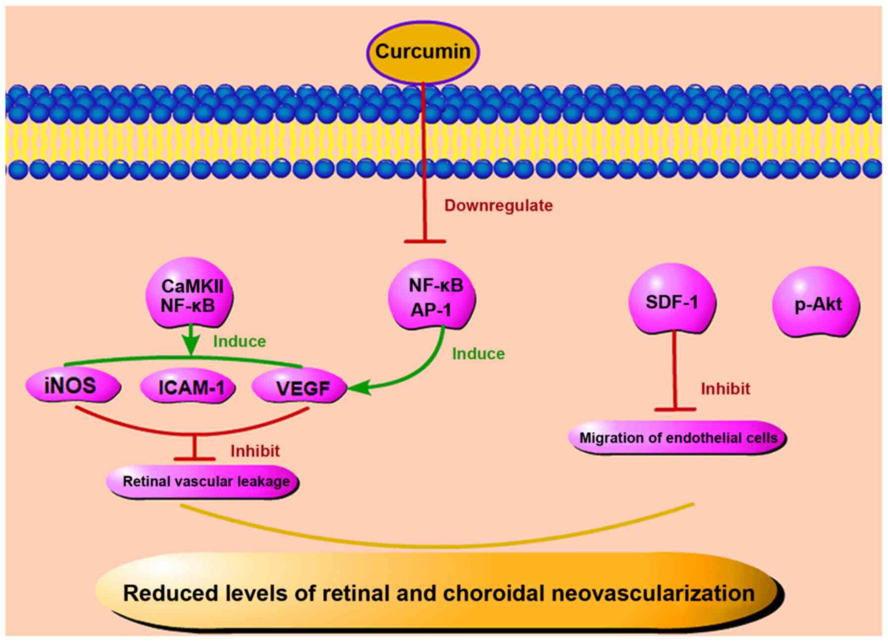
not in the untreated diabetic rats (70). Some scholars have confirmed that
curcumin can reduce the expression of VEGF, inducible nitric oxide
synthase (iNOS) and ICAM-1 by inhibiting the activation of
CaMKII/NF-κB signaling in the diabetic rat retina and in Müller
cells cultured in high glucose (71,72). Curcumin not only exerts anti-VEGF
effects on the retina, but also exerts anti-angiogenetic effects on
the choroidal vascular system of diabetic rats. In another study,
following treatment with curcumin, the size of blood vessels
significantly increased, while both the degree of tortuosity of the
diabetic microvascular system and the amount of generated
microaneurysms decreased (73).
Some scholars have found that when the expression of
phosphorylated-Akt (p-Akt) is downregulated, the angiogenesis of
human umbilical vein endothelial cells (HUVEC) in vitro is
also inhibited (74). It has
been suggested that Akt phosphorylation may represent a key factor
in angiogenesis. However, in RPECs that were treated with high
glucose, curcumin was shown to reduce Akt phosphorylation and thus
play a therapeutic role (75,76). Okamoto et al demonstrated
that the activation of NF-κB and activator protein-1 (AP-1) can
contribute to age-induced angiogenesis through the overproduction
of autocrine VEGF; however, curcumin, which is an inhibitor of
NF-κB and AP-1, can prevent the upregulation of VEGF mRNA and the
subsequent increase of DNA synthesis in endothelial cells (77). Stromal cell-derived factor-1
(SDF-1) is another important factor in angiogenesis. The level of
SDF-1 is increased whether in patients with PDR or endothelial
cells cultured in high glucose in vitro (78,79). Curcumin can significantly inhibit
the SDF-1-induced migration of human retinal endothelial cells
(80,81). In summary, it is suggested that
curcumin may play a therapeutic role in DR by reducing the level of
SDF-1 and angiogenesis (Fig. 5).
Studies have found that inflammation is involved in the process of
angiogenesis. In mice, the subcutaneous injection of IL-1α can
induce a strong local angiogenic response, and VEGF receptor 2
(VEGFR-2)-neutralizing antibody can block the angiogenic effects of
IL-1α. Therefore, pro-inflammatory factors can induce angiogenesis
by activating the VEGF signaling pathway between inflammatory cells
and vascular endothelial cells in a concentration-dependent manner
(82). Curcumin not only
directly inhibits abnormal angiogenesis, but may also rely on the
regulation of inflammatory factors to reduce the level of
inflammation, to further reduce the number of new blood vessels. On
the whole, curcumin not only reduces the severity of DR through
anti-inflammatory effects, but also regulates the number of
abnormal retinal neovascularization events, reduces bleeding of
pathological vessels, and further protects retinal tissue through
anti-angiogenic mechanisms.
Apoptosis or autophagy is a self-protective
mechanism through which eukaryotic cells degrade toxic substances
or damaged organelles through lysosomes. In the high glucose state,
the expression of MMP-2 increases, while the expression of heat
shock protein 60 and connexin 43 decreases, leading to
mitochondrial membrane degradation, increased apoptosis, and more
severe DR (83,84). On the basis of in vitro
and in vivo experiments, researchers have demonstrated that
in retinal ganglion cells of mice with STZ-induced diabetes, the
expression of caspase-3 gradually increases (85). In addition, as previously
demonstrated, the apoptosis of adult retinal pigment epithelial
cells (ARPE-19 cells) that were cultured under high glucose
conditions also significantly increased (86). These results indicate that
increased apoptosis is also one of the key pathological mechanisms
in diabetes.
Dysfunctional autophagy is associated with cancer,
neurodegeneration, microbial infection and aging (87-90). Some scholars have shown that
autophagy-related markers are increased in Müller cells cultured
in vitro when exposed to a high glucose environment
(91). In a high glucose
environment, the inhibition of autophagy can lead to a higher
apoptotic rate of Müller cells, while rapamycin, an autophagy
inducer, can prevent the release of VEGF and prevent apoptosis. It
is suggested that the promotion of autophagy may protect cells
cultured in high glucose (92).
Another explanation that has been proposed for the association
between apoptosis, autophagy and DR is endoplasmic reticulum (ER)
stress. ER stress is involved in cellular metabolism in
physiological or pathological states. When ER stress remains mild,
it helps cells adapt to an abnormal state; however, long-term,
severe ER stress, which occurs in the diabetic state, can lead to
increased autophagy and apoptosis (93).
Curcumin can also reduce insulin resistance in the
diabetic state, and it has been proven to induce autophagy in
gastric cancer cells. This effect is ROS concentration-dependent,
suggesting that the control of ER stress by curcumin may be
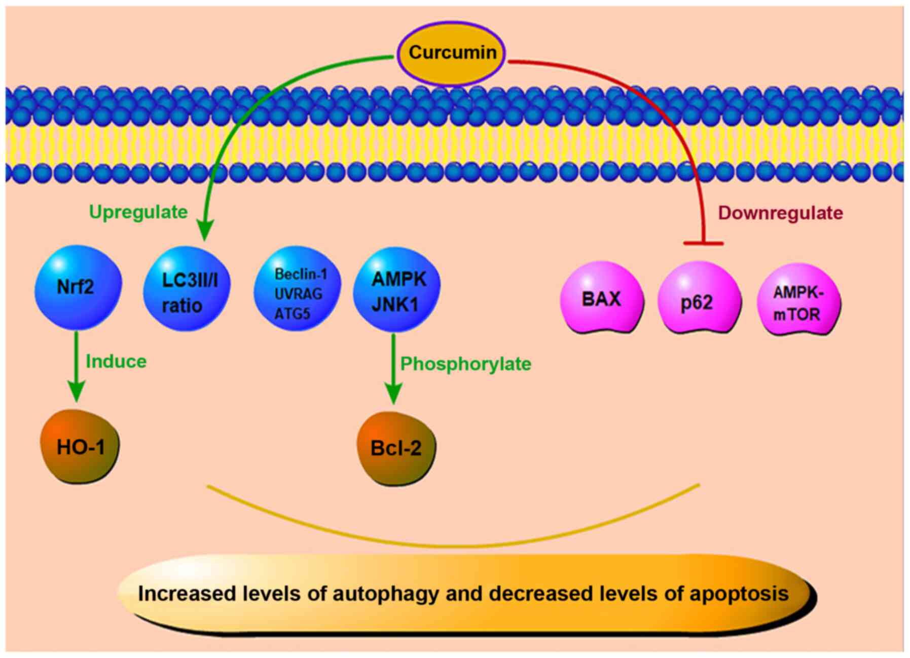
achieved by regulating the ROS level (96). Some scholars have found that
curcumin can inhibit ER stress in HUVECs and enhance autophagy,
which may maintain ER homeostasis by degrading denatured and
damaged proteins (97). In a
previous study, in mice with STZ-induced diabetic nephropathy (DN),
following 8 weeks of treatment with curcumin, compared with the
control group, the curcumin group had a significantly increased
light chain 3 (LC3) II/I ratio, and the levels of autophagy-related
proteins Beclin-1, ultraviolet radiation resistance-associated gene
protein (UVRAG) and autophagy-related protein 5 (ATG5) were also
increased. In the same vein, curcumin reduced the expression of p62
and enhanced the autophagy of podocytes in a high-glucose culture,
thus exerting a protective effect on cells (98). Yao et al found that in
mice with diabetic cardiomyopathy induced by a high-fat diet
combined with a low-dose STZ injection, the apoptosis of cardiac
cells increased, and autophagy was inhibited (99). Curcumin phosphorylated Bcl-2 by
activating the AMP-activated protein kinase (AMPK) and c-Jun
N-terminal kinase-1 (JNK1) pathways, and then destroyed their
interaction with Beclin-1; the AMPK-mammalian target of rapamycin
(mTOR) pathway was also inhibited; both these effects ultimately
promoted autophagy and reduced apoptosis, thus the protecting cells
(99). Therefore, it can be
hypothesized that the protective effects of curcumin on DR involve
the reduction of abnormal apoptosis of the retinal tissue in a
high-glucose state by regulating the level of apoptosis-related
proteins. On the contrary, by properly enhancing autophagy and
removing denatured and damaged proteins, curcumin can reduce the
damage to the retinal barrier and the state of vascular leakage. In
parallel, it can restore the normal level of apoptosis and reduce
the damage to the retina caused by high glucose.
Hyperglycemia has been inevitably implicated as the
main cause of DR. In addition to increasing the production of ROS
through the autoxidation of glucose in a hyperglycemic environment,
oxidative stress is also associated with hyperglycemia-induced
metabolic pathways in DR. With an increase in the glucose
concentration, NF-κB, one of the key mediators of pro-inflammation,
is also constantly activated. Moreover, high glucose levels can
induce the oxidative stress pathway, promote the production of
multiple free radicals and participate in the pathogenic mechanisms
of DR (100).
Although hyperglycemia is the main cause of DR,
hyperlipidemia is also considered to play a decisive role in its
pathogenesis. Triglyceride levels are directly related to the
incidence and severity of DR, and long-term lipid-lowering therapy
can reduce the need for laser therapy for patients with DR
(101). In recent years, an
increasing number of studies have investigated lipids in
association with diabetes. For DR, an abnormal vascular endothelial
function leads to increased plasma exudation, and the lipids
themselves aggravate endothelial cell damage (102). In diabetic rat models, the
development of DR has been found to be enhanced in a hyperlipidemic
environment, and the combination of hyperlipidemia and
hyperglycemia enhances the activation of the apoptotic pathway in
retinal capillary cells by accelerating mitochondrial damage
(103).
DR represents one of the severe microvascular
complications of diabetes mellitus. Its underlying pathogenic
mechanisms represent a complex process characterized by
multi-factorial participation, multigenic regulation, and
multi-step evolutionary processes. A variety of mechanisms jointly
contribute to the occurrence and progression of DR. Due to the
large number of patients with DR worldwide, particularly
individuals of working-age, increasing attention is being paid to
DR as a severe eye disease that leads to blindness. It has been
found that curcumin can improve glucose and lipid metabolism,
improve insulin sensitivity, reduce insulin resistance, and improve
oxidative stress and inflammatory pathway conditions in diabetes
and DR. In the future, further clinical trials may be designed to
assess whether the bioavailability of curcumin could be improved by
combining it with other available therapies. The regulatory effects
of curcumin on autophagy and apoptosis may provide insight for the
development of novel therapeutics for DR. Further studies are
required to explore the therapeutic potential of curcumin and
evaluate whether it can be used as a novel drug for DR in clinical
practice.
Not applicable.
JY, XM and FJY were involved in the conception of
the study, and in the writing and preparation of the manuscript, as
well as in the supervision of figure preparation. JY, JFC and XL
were involved in the writing of the manuscript and preparation of
the figures. JY, JLF and GFS were involved in the writing and
supervision of the manuscript.
Not applicable.
Not applicable.
The authors declare that they have no competing
interests.
Not applicable.
This study was supported by the National Natural Science
Foundation (81970836), the International Cooperation Project of
Science and Technology Department of Jilin Province
(20190701044GH), Training Program for Outstanding Young Teachers of
Jilin University (419080520313), and the Natural Science Foundation
Project of Science and Technology Department of Jilin Province
(20200201360JC).
|
1
|
International Diabetes Federation (IDF):
IDF Diabetes Atlas. 8th Edition. IDF; 2017, https://www.diabetesatlas.org/upload/resources/previous/files/8/IDF_DA_8e-EN-final.pdf.
|
|
2
|
Logue J, Walker JJ, Colhoun HM, Leese GP,
Lindsay RS, McKnight JA, Morris AD, Pearson DW, Petrie JR, Philip
S, et al: Do men develop type 2 diabetes at lower body mass indices
than women? Diabetologia. 54:3003–3006. 2011. View Article : Google Scholar : PubMed/NCBI
|
|
3
|
Hartwig S, Greiser KH, Medenwald D, Tiller
D, Herzog B, Schipf S, Ittermann T, Völzke H, Müller G, Haerting J
and Kluttig A: Association of change of anthropometric measurements
with incident type 2 diabetes mellitus: A pooled analysis of the
prospective population-based CARLA and SHIP cohort studies.
Medicine(Baltimore). 94:e13942015.
|
|
4
|
Wannamethee SG, Papacosta O, Lawlor DA,
Whincup PH, Lowe GD, Ebrahim S and Sattar N: Do women exhibit
greater differences in established and novel risk factors between
diabetes and non-diabetes than men? the British regional heart
study and British women's heart health study. Diabetologia.
55:80–87. 2012. View Article : Google Scholar
|
|
5
|
Shigiyama F, Kumashiro N, Tsuneoka Y,
Igarashi H, Yoshikawa F, Kakehi S, Funato H and Hirose T:
Mechanisms of sleep deprivation-induced hepatic steatosis and
insulin resistance in mice. Am J Physiol Endocrinol Metab.
315:E848–E858. 2018. View Article : Google Scholar : PubMed/NCBI
|
|
6
|
Pan KY, Xu W, Mangialasche F, Fratiglioni
L and Wang HX: Work-related psychosocial stress and the risk of
type 2 diabetes in later life. J Intern Med. 281:601–610. 2017.
View Article : Google Scholar : PubMed/NCBI
|
|
7
|
Sohn EH, van Dijk HW, Jiao C, Kok PHB,
Jeong W, Demirkaya N, Garmager A, Wit F, Kucukevcilioglu M, van
Velthoven ME, et al: Retinal neurodegeneration may precede
microvascular changes characteristic of diabetic retinopathy in
diabetes mellitus. Proc Natl Acad Sci USA. 113:E2655–E2664. 2016.
View Article : Google Scholar : PubMed/NCBI
|
|
8
|
Sahin K, Orhan C, Tuzcu M, Sahin N, Tastan
H, Özercan IH, Güler O, Kahraman N, Kucuk O and Ozpolat B:
Chemopreventive and antitumor efficacy of curcumin in a
spontaneously developing hen ovarian cancer model. Cancer Prev Res
(Phila). 11:59–67. 2018. View Article : Google Scholar
|
|
9
|
Fu H, Wang C, Yang D, Wei Z, Xu J, Hu Z,
Zhang Y, Wang W, Yan R and Cai Q: Curcumin regulates proliferation,
autophagy, and apoptosis in gastric cancer cells by affecting PI3K
and P53 signaling. J Cell Physiol. 233:4634–4642. 2018. View Article : Google Scholar
|
|
10
|
Kronski E, Fiori ME, Barbieri O, Astigiano
S, Mirisola V, Killian PH, Bruno A, Pagani A, Rovera F, Pfeffer U,
et al: miR181b is induced by the chemopreventive polyphenol
curcumin and inhibits breast cancer metastasis via down-regulation
of the inflammatory cytokines CXCL1 and -2. Mol Oncol. 8:581–595.
2014. View Article : Google Scholar : PubMed/NCBI
|
|
11
|
Ide H, Lu Y, Noguchi T, Muto S, Okada H,
Kawato S and Horie S: Modulation of AKR1C2 by curcumin decreases
testosterone production in prostate cancer. Cancer Sci.
109:1230–1238. 2018. View Article : Google Scholar : PubMed/NCBI
|
|
12
|
Yuan J, Liu R, Ma Y, Zhang Z and Xie Z:
Curcumin attenuates airway inflammation and airway remolding by
inhibiting NF-κB signaling and COX-2 in cigarette smoke-induced
COPD mice. Inflammation. 41:1804–1814. 2018. View Article : Google Scholar : PubMed/NCBI
|
|
13
|
Ding XQ, Wu WY, Jiao RQ, Gu TT, Xu Q, Pan
Y and Kong LD: Curcumin and allopurinol ameliorate fructose-induced
hepatic inflammation in rats via miR-200a-mediated TXNIP/NLRP3
inflammasome inhibition. Pharmacol Res. 137:64–75. 2018. View Article : Google Scholar : PubMed/NCBI
|
|
14
|
Xiao Y, Xia J, Wu S, Lv Z, Huang S, Huang
H, Su X, Cheng J and Ke Y: Curcumin inhibits acute vascular
inflammation through the activation of heme oxygenase-1. Oxid Med
Cell Longev. 2018:32958072018. View Article : Google Scholar : PubMed/NCBI
|
|
15
|
Sharma S, Kulkarni SK and Chopra K:
Curcumin, the active principle of turmeric (Curcuma longa),
ameliorates diabetic nephropathy in rats. Clin Exp Pharmacol
Physiol. 33:940–945. 2006. View Article : Google Scholar : PubMed/NCBI
|
|
16
|
Kadiyala CSR, Zheng L, Du Y, Yohannes E,
Kao HY, Miyagi M and Kern TS: Acetylation of retinal histones in
diabetes increases inflammatory proteins: Effects of minocycline
and manipulation of histone acetyltransferase (HAT) and histone
deacetylase (HDAC). J Biol Chem. 287:25869–25880. 2012. View Article : Google Scholar : PubMed/NCBI
|
|
17
|
Wang LL, Chen H, Huang K and Zheng L:
Elevated histone acetylations in Müller cells contribute to
inflammation: A novel inhibitory effect of minocycline. Glia.
60:1896–1905. 2012. View Article : Google Scholar : PubMed/NCBI
|
|
18
|
Yun JM, Jialal I and Devaraj S: Epigenetic
regulation of high glucose-induced proinflammatory cytokine
production in monocytes by curcumin. J Nutr Biochem. 22:450–458.
2011. View Article : Google Scholar
|
|
19
|
Gui L, Jiang S, Xie D, Yu L, Huang Y,
Zhang Z and Liu Y: Analysis of complete chloroplast genomes of
Curcuma and the contribution to phylogeny and adaptive evolution.
Gene. 732:1443552020. View Article : Google Scholar : PubMed/NCBI
|
|
20
|
Nishino H, Tokuda H, Satomi Y, Masuda M,
Osaka Y, Yogosawa S, Wada S, Mou XY, Takayasu J, Murakoshi M, et
al: Cancer prevention by antioxidants. Biofactors. 22:57–61. 2004.
View Article : Google Scholar
|
|
21
|
Boyanapalli SSS, Huang Y, Su Z, Cheng D,
Zhang C, Guo Y, Rao R, Androulakis IP and Kong AN: Pharmacokinetics
and pharmacodynamics of curcumin in regulating anti-inflammatory
and epigenetic gene expression. Biopharm Drug Dispos. 39:289–297.
2018. View Article : Google Scholar : PubMed/NCBI
|
|
22
|
Lao CD, Ruffin MT, Normolle D, Heath DD,
Murray SI, Bailey JM, Boggs ME, Crowell J, Rock CL and Brenner DE:
Dose escalation of a curcuminoid formulation. BMC Complement Altern
Med. 6:102006. View Article : Google Scholar : PubMed/NCBI
|
|
23
|
Falconieri MC, Adamo M, Monasterolo C,
Bergonzi MC, Coronnello M and Bilia AR: New dendrimer-based
nanoparticles enhance curcumin solubility. Planta Med. 83:420–425.
2017.
|
|
24
|
Tiyaboonchai W, Tungpradit W and
Plianbangchang P: Formulation and characterization of curcuminoids
loaded solid lipid nanoparticles. Int J Pharm. 337:299–306. 2007.
View Article : Google Scholar : PubMed/NCBI
|
|
25
|
Wang W, Zhu R, Xie Q, Li A, Xiao Y, Li K,
Liu H, Cui D, Chen Y and Wang S: Enhanced bioavailability and
efficiency of curcumin for the treatment of asthma by its
formulation in solid lipid nanoparticles. Int J Nanomedicine.
7:3667–3677. 2012. View Article : Google Scholar : PubMed/NCBI
|
|
26
|
Shoba G, Joy D, Joseph T, Majeed M,
Rajendran R and Srinivas PS: Influence of piperine on the
pharmacokinetics of curcumin in animals and human volunteers.
Planta Med. 64:353–356. 1998. View Article : Google Scholar : PubMed/NCBI
|
|
27
|
Skoupa N, Dolezel P, Ruzickova E and
Mlejnek P: Apoptosis induced by the curcumin analogue EF-24 is
neither mediated by oxidative stress-related mechanisms nor
affected by expression of main drug transporters ABCB1 and ABCG2 in
human leukemia cells. Int J Mol Sci. 18:22892017. View Article : Google Scholar :
|
|
28
|
Ingelfinger JR and Jarcho JA: Increase in
the incidence of diabetes and its implications. N Engl J Med.
376:1473–1474. 2017. View Article : Google Scholar : PubMed/NCBI
|
|
29
|
Javidi MA, Kaeidi A, Mortazavi Farsani SS,
Babashah S and Sadeghizadeh M: Investigating curcumin potential for
diabetes cell therapy, in vitro and in vivo study. Life Sci.
239:1169082019. View Article : Google Scholar : PubMed/NCBI
|
|
30
|
Chuengsamarn S, Rattanamongkolgul S,
Luechapudiporn R, Phisalaphong C and Jirawatnotai S: Curcumin
extract for prevention of type 2 diabetes. Diabetes Care.
35:2121–2127. 2012. View Article : Google Scholar : PubMed/NCBI
|
|
31
|
Al-Saud NBS: Impact of curcumin treatment
on diabetic albino rats. Saudi J Biol Sci. 27:689–694. 2020.
View Article : Google Scholar : PubMed/NCBI
|
|
32
|
Qihui L, Shuntian D, Xin Z, Xiaoxia Y and
Zhongpei C: Protection of curcumin against streptozocin-induced
pancreatic cell destruction in T2D rats. Planta Med. 86:113–120.
2020. View Article : Google Scholar
|
|
33
|
Bulboacă AE, Boarescu PM, Bolboacă SD,
Blidaru M, Feștilă D, Dogaru G and Nicula CA: Comparative effect of
curcumin versus liposomal curcumin on systemic pro-inflammatory
cytokines profile, MCP-1 and RANTES in experimental diabetes
mellitus. Int J Nanomedicine. 14:8961–8972. 2019. View Article : Google Scholar
|
|
34
|
Li J, Wu N, Chen X, Chen H, Yang X and Liu
C: Curcumin protects islet cells from glucolipotoxicity by
inhibiting oxidative stress and NADPH oxidase activity both in
vitro and in vivo. Islets. 11:152–164. 2019. View Article : Google Scholar
|
|
35
|
Xia ZH, Jiang X, Li K, Li LX, Chen WB,
Wang YX and Liu YQ: Curcumin inhibits alloxan-induced pancreatic
islet cell damage via antioxidation and antiapoptosis. J Biochem
Mol Toxicol. 34:e224992020. View Article : Google Scholar : PubMed/NCBI
|
|
36
|
Gupta A, Tripathi AK, Tripathi RL, Madhu
SV and Banerjee BD: Advanced glycosylated end products-mediated
activation of polymorphonuclear neutrophils in diabetes mellitus
and associated oxidative stress. Indian J Biochem Biophys.
44:373–378. 2007.
|
|
37
|
Aplin AC, Gelati M, Fogel E, Carnevale E
and Nicosia RF: Angiopoietin-1 and vascular endothelial growth
factor induce expression of inflammatory cytokines before
angiogenesis. Physiol Genomics. 27:20–28. 2006. View Article : Google Scholar : PubMed/NCBI
|
|
38
|
Kowluru RA and Kanwar M: Effects of
curcumin on retinal oxidative stress and inflammation in diabetes.
Nutr Metab (Lond). 4:82007. View Article : Google Scholar
|
|
39
|
Assis RP, Arcaro CA, Gutierres VO,
Oliveira JO, Costa PI, Baviera AM and Brunetti IL: Combined effects
of curcumin and lycopene or bixin in yoghurt on inhibition of LDL
oxidation and increases in HDL and paraoxonase levels in
streptozotocin-diabetic rats. Int J Mol Sci. 18:3322017. View Article : Google Scholar :
|
|
40
|
Gutierres VO, Pinheiro CM, Assis RP,
Vendramini RC, Pepato MT and Brunetti IL: Curcumin-supplemented
yoghurt improves physiological and biochemical markers of
experimental diabetes. Br J Nutr. 108:440–448. 2012. View Article : Google Scholar
|
|
41
|
Jiménez-Osorio AS, González-Reyes S and
Pedraza-Chaverri J: Natural Nrf2 activators in diabetes. Clin Chim
Acta. 448:182–192. 2015. View Article : Google Scholar : PubMed/NCBI
|
|
42
|
Zuo ZF, Zhang Q and Liu XZ: Protective
effects of curcumin on retinal Müller cell in early diabetic rats.
Int J Ophthalmol. 6:422–424. 2013.
|
|
43
|
Liu Z, Lin H, Ye S, Liu QY, Meng Z, Zhang
CM, Xia Y, Margoliash E, Rao Z and Liu XJ: Remarkably high
activities of testicular cytochrome c in destroying reactive oxygen
species and in triggering apoptosis. Proc Natl Acad Sci USA.
103:8965–8970. 2006. View Article : Google Scholar : PubMed/NCBI
|
|
44
|
Maugeri A, Mazzone MG, Giuliano F,
Vinciguerra M, Basile G, Barchitta M and Agodi A: Curcumin
modulates DNA methyltransferase functions in a cellular model of
diabetic retinopathy. Oxid Med Cell Longev. 2018:54074822018.
View Article : Google Scholar : PubMed/NCBI
|
|
45
|
Shishodia S, Sethi G and Aggarwal BB:
Curcumin: Getting back to the roots. Ann N Y Acad Sci.
1056:206–217. 2005. View Article : Google Scholar
|
|
46
|
Chen Y, Li C, Duan S, Yuan X, Liang J and
Hou S: Curcumin attenuates potassium oxonate-induced hyperuricemia
and kidney inflammation in mice. Biomed Pharmacother.
118:1091952019. View Article : Google Scholar : PubMed/NCBI
|
|
47
|
Balasubramanyam M, Koteswari AA, Kumar RS,
Monickaraj SF, Maheswari JU and Mohan V: Curcumin-induced
inhibition of cellular reactive oxygen species generation: Novel
therapeutic implications. J Biosci. 28:715–721. 2003. View Article : Google Scholar : PubMed/NCBI
|
|
48
|
Yang F, Yu J, Ke F, Lan M and Li D, Tan K,
Ling J, Wang Y, Wu K and Li D: Curcumin alleviates diabetic
retinopathy in experimental diabetic rats. Ophthalmic Res.
60:43–54. 2018. View Article : Google Scholar : PubMed/NCBI
|
|
49
|
Bulboacă AE, Porfire AS, Tefas LR,
Boarescu PM, Bolboacă SD, Stănescu IC, Bulboacă AC and Dogaru G:
Liposomal curcumin is better than curcumin to alleviate
complications in experimental diabetic mellitus. Molecules.
24:8462019. View Article : Google Scholar
|
|
50
|
Zhou P, Xie W, Meng X, Zhai Y, Dong X,
Zhang X, Sun G and Sun X: Notoginsenoside R1 ameliorates diabetic
retinopathy through PINK1-dependent activation of mitophagy. Cells.
8:2132019. View Article : Google Scholar :
|
|
51
|
Brucklacher RM, Patel KM, VanGuilder HD,
Bixler GV, Barber AJ, Antonetti DA, Lin CM, LaNoue KF, Gardner TW,
Bronson SK and Freeman WM: Whole genome assessment of the retinal
response to diabetes reveals a progressive neurovascular
inflammatory response. BMC Med Genomics. 1:262008. View Article : Google Scholar : PubMed/NCBI
|
|
52
|
Yuuki T, Kanda T, Kimura Y, Kotajima N,
Tamura J, Kobayashi I and Kishi S: Inflammatory cytokines in
vitreous fluid and serum of patients with diabetic
vitreoretinopathy. J Diabetes Complications. 15:257–259. 2001.
View Article : Google Scholar : PubMed/NCBI
|
|
53
|
Joussen AM, Murata T, Tsujikawa A,
Kirchhof B, Bursell SE and Adamis AP: Leukocyte-mediated
endothelial cell injury and death in the diabetic retina. Am J
Pathol. 158:147–152. 2001. View Article : Google Scholar : PubMed/NCBI
|
|
54
|
Khalfaoui T, Lizard G and Ouertani-Meddeb
A: Adhesion molecules (ICAM-1 and VCAM-1) and diabetic retinopathy
in type 2 diabetes. J Mol Histol. 39:243–249. 2008. View Article : Google Scholar : PubMed/NCBI
|
|
55
|
Boss JD, Singh PK, Pandya HK, Tosi J, Kim
C, Tewari A, Juzych MS, Abrams GW and Kumar A: Assessment of
neurotrophins and inflammatory mediators in vitreous of patients
with diabetic retinopathy. Invest Ophthalmol Vis Sci. 58:5594–5603.
2017. View Article : Google Scholar : PubMed/NCBI
|
|
56
|
Luo DW, Zheng Z, Wang H, Fan Y, Chen F,
Sun Y, Wang WJ, Sun T and Xu X: UPP mediated diabetic retinopathy
via ROS/PARP and NF-κB inflammatory factor pathways. Curr Mol Med.
15:790–799. 2015. View Article : Google Scholar
|
|
57
|
Hollanders K, Van Hove I, Sergeys J, Van
Bergen T, Lefevere E, Kindt N, Castermans K, Vandewalle E, van Pelt
J, Moons L and Stalmans I: AMA0428, a potent rock inhibitor,
attenuates early and late experimental diabetic retinopathy. Curr
Eye Res. 42:260–272. 2017. View Article : Google Scholar
|
|
58
|
Ran Z, Zhang Y, Wen X and Ma J: Curcumin
inhibits high glucose induced inflammatory injury in human retinal
pigment epithelial cells through the ROS PI3K/AKT/mTOR signaling
pathway. Mol Med Rep. 19:1024–1031. 2019.
|
|
59
|
Costagliola C, Romano V, De Tollis M,
Aceto F, dell'Omo R, Romano MR, Pedicino C and Semeraro F:
TNF-alpha levels in tears: A novel biomarker to assess the degree
of diabetic retinopathy. Mediators Inflamm. 2013:6295292013.
View Article : Google Scholar : PubMed/NCBI
|
|
60
|
Gupta SK, Kumar B, Nag TC, Agrawal SS,
Agrawal R, Agrawal P, Saxena R and Srivastava S: Curcumin prevents
experimental diabetic retinopathy in rats through its hypoglycemic,
anti-oxidant, and anti-inflammatory mechanisms. J Ocul Pharmacol
Ther. 27:123–130. 2011. View Article : Google Scholar : PubMed/NCBI
|
|
61
|
Jiang C, Ting AT and Seed B: PPAR-gamma
agonists inhibit production of monocyte inflammatory cytokines.
Nature. 391:82–86. 1998. View
Article : Google Scholar : PubMed/NCBI
|
|
62
|
Charrier A, Wang L, Stephenson EJ, Ghanta
SV, Ko CW, Croniger CM, Bridges D and Buchner DA: Zinc finger
protein 407 overexpression upregulates PPAR target gene expression
and improves glucose homeostasis in mice. Am J Physiol Endocrinol
Metab. 311:E869–E880. 2016. View Article : Google Scholar : PubMed/NCBI
|
|
63
|
Aljada A, Garg R, Ghanim H, Mohanty P,
Hamouda W, Assian E and Dandona P: Nuclear factor-kappaB
suppressive and inhibitor-kappaB stimulatory effects of
troglitazone in obese patients with type 2 diabetes: Evidence of an
antiinflammatory action? J Clin Endocrinol Metab. 86:3250–3256.
2001.PubMed/NCBI
|
|
64
|
Haffner SM, Greenberg AS, Weston WM, Chen
H, Williams K and Freed MI: Effect of rosiglitazone treatment on
nontraditional markers of cardiovascular disease in patients with
type 2 diabetes mellitus. Circulation. 106:679–684. 2002.
View Article : Google Scholar : PubMed/NCBI
|
|
65
|
Shafabakhsh R, Mobini M, Raygan F,
Aghadavod E, Ostadmohammadi V, Amirani E, Mansournia MA and Asemi
Z: Curcumin administration and the effects on psychological status
and markers of inflammation and oxidative damage in patients with
type 2 diabetes and coronary heart disease. Clin Nutr ESPEN.
40:77–82. 2020. View Article : Google Scholar : PubMed/NCBI
|
|
66
|
Li HY, Yang M, Li Z and Meng Z: Curcumin
inhibits angiotensin II-induced inflammation and proliferation of
rat vascular smooth muscle cells by elevating PPAR-γ activity and
reducing oxidative stress. Int J Mol Med. 39:1307–1316. 2017.
View Article : Google Scholar : PubMed/NCBI
|
|
67
|
Yun JH, Park SW, Kim KJ, Bae JS, Lee EH,
Paek SH, Kim SU, Ye S, Kim JH and Cho CH: Endothelial STAT3
activation increases vascular leakage through downregulating tight
junction proteins: Implications for diabetic retinopathy. J Cell
Physiol. 232:1123–1134. 2017. View Article : Google Scholar
|
|
68
|
Shibuya M: Vascular endothelial growth
factor and its receptor system: Physiological functions in
angiogenesis and pathological roles in various diseases. J Biochem.
153:13–19. 2013. View Article : Google Scholar
|
|
69
|
Miyamoto K, Khosrof S, Bursell SE, Rohan
R, Murata T, Clermont AC, Aiello LP, Ogura Y and Adamis AP:
Prevention of leukostasis and vascular leakage in
streptozotocin-induced diabetic retinopathy via intercellular
adhesion molecule-1 inhibition. Proc Natl Acad Sci USA.
96:10836–10841. 1999. View Article : Google Scholar : PubMed/NCBI
|
|
70
|
Mrudula T, Suryanarayana P, Srinivas PNBS
and Reddy GB: Effect of curcumin on hyperglycemia-induced vascular
endothelial growth factor expression in streptozotocin-induced
diabetic rat retina. Biochem Biophys Res Commun. 361:528–532. 2007.
View Article : Google Scholar : PubMed/NCBI
|
|
71
|
Li J, Wang P, Ying J, Chen Z and Yu S:
Curcumin attenuates retinal vascular leakage by inhibiting
calcium/calmodulin-dependent protein kinase II activity in
streptozotocin-induced diabetes. Cell Physiol Biochem.
39:1196–1208. 2016. View Article : Google Scholar : PubMed/NCBI
|
|
72
|
Pradhan D, Dasmohapatra T and Tripathy G:
Pharmacognostic evaluation of curcumin on diabetic retinopathy in
alloxan-induced diabetes through NF-KB and Brn3a related mechanism.
Pharmacogn J. 10:324–332. 2018. View Article : Google Scholar
|
|
73
|
Khimmaktong W, Petpiboolthai H, Sriya P
and Anupunpisit V: Effects of curcumin on restoration and
improvement of microvasculature characteristic in diabetic rat's
choroid of eye. J Med Assoc Thai. 97(Suppl 2): S39–S46.
2014.PubMed/NCBI
|
|
74
|
Lee TK, Park JY, Yu JS, Jang TS, Oh ST,
Pang C, Ko YJ, Kang KS and Kim KH: 7α,15-Dihydroxydehydroabietic
acid from Pinus koraiensis inhibits the promotion of angiogenesis
through down-regulation of VEGF, p-Akt and p-ERK in HUVECs. Bioorg
Med Chem Lett. 28:1084–1089. 2018. View Article : Google Scholar : PubMed/NCBI
|
|
75
|
Farajipour H, Rahimian S and Taghizadeh M:
Curcumin: A new candidate for retinal disease therapy? J Cell
Biochem. 2018.Epub ahead of print. PubMed/NCBI
|
|
76
|
Ran Z, Zhang Y, Wen X and Ma J: Curcumin
inhibits high glucose induced inflammatory injury in human retinal
pigment epithelial cells through the ROS PI3K/AKT/mTOR signaling
pathway. Mol Med Rep. 19:1024–1031. 2019.
|
|
77
|
Okamoto T, Yamagishi SI, Inagaki Y, Amano
S, Koga K, Abe R, Takeuchi M, Ohno S, Yoshimura A and Makita Z:
Angiogenesis induced by advanced glycation end products and its
prevention by cerivastatin. FASEB J. 16:1928–1930. 2002. View Article : Google Scholar : PubMed/NCBI
|
|
78
|
Salvucci O, Basik M, Yao L, Bianchi R and
Tosato G: Evidence for the involvement of SDF-1 and CXCR4 in the
disruption of endothelial cell-branching morphogenesis and
angiogenesis by TNF-alpha and IFN-gamma. J Leukoc Biol. 76:217–226.
2004. View Article : Google Scholar : PubMed/NCBI
|
|
79
|
Butler JM, Guthrie SM, Koc M, Afzal A,
Caballero S, Brooks HL, Mames RN, Segal MS, Grant MB and Scott EW:
SDF-1 is both necessary and sufficient to promote proliferative
retinopathy. J Clin Invest. 115:86–93. 2005. View Article : Google Scholar : PubMed/NCBI
|
|
80
|
Arbiser JL, Klauber N, Rohan R, van
Leeuwen R, Huang MT, Fisher C, Flynn E and Byers HR: Curcumin is an
in vivo inhibitor of angiogenesis. Mol Med. 4:376–383. 1998.
View Article : Google Scholar
|
|
81
|
Sameermahmood Z, Balasubramanyam M,
Saravanan T and Rema M: Curcumin modulates SDF-1alpha/CXCR4-induced
migration of human retinal endothelial cells (HRECs). Invest
Ophthalmol Vis Sci. 49:3305–3311. 2008. View Article : Google Scholar : PubMed/NCBI
|
|
82
|
Salven P, Hattori K, Heissig B and Rafii
S: Interleukin-1alpha promotes angiogenesis in vivo via VEGFR-2
pathway by inducing inflammatory cell VEGF synthesis and secretion.
FASEB J. 16:1471–1473. 2002. View Article : Google Scholar : PubMed/NCBI
|
|
83
|
Mohammad G and Kowluru RA: Novel role of
mitochondrial matrix metalloproteinase-2 in the development of
diabetic retinopathy. Invest Ophthalmol Vis Sci. 52:3832–3841.
2011. View Article : Google Scholar : PubMed/NCBI
|
|
84
|
Mohammad G and Kowluru RA: Matrix
metalloproteinase-2 in the development of diabetic retinopathy and
mitochondrial dysfunction. Lab Invest. 90:1365–1372. 2010.
View Article : Google Scholar : PubMed/NCBI
|
|
85
|
Fu Y, Wang Y, Gao X, Li H and Yuan Y:
Dynamic expression of HDAC3 in db/db mouse RGCs and its
relationship with apoptosis and autophagy. J Diabetes Res.
2020:60867802020. View Article : Google Scholar : PubMed/NCBI
|
|
86
|
Zhang X, He N, Xing Y and Lu Y: Knockdown
of GCN2 inhibits high glucose-induced oxidative stress and
apoptosis in retinal pigment epithelial cells. Clin Exp Pharmacol
Physiol. 47:591–598. 2020. View Article : Google Scholar
|
|
87
|
Mathew R and White E: Why sick cells
produce tumors: The protective role of autophagy. Autophagy.
3:502–505. 2007. View Article : Google Scholar : PubMed/NCBI
|
|
88
|
Hara T, Nakamura K, Matsui M, Yamamoto A,
Nakahara Y, Suzuki-Migishima R, Yokoyama M, Mishima K, Saito I,
Okano H and Mizushima N: Suppression of basal autophagy in neural
cells causes neurodegenerative disease in mice. Nature.
441:885–889. 2006. View Article : Google Scholar : PubMed/NCBI
|
|
89
|
Pahari S, Negi S, Aqdas M, Arnett E,
Schlesinger LS and Agrewala JN: Induction of autophagy through
CLEC4E in combination with TLR4: An innovative strategy to restrict
the survival of mycobacterium tuberculosis. Autophagy.
16:1021–1043. 2020. View Article : Google Scholar :
|
|
90
|
Fernández ÁF, Sebti S, Wei Y, Zou Z, Shi
M, McMillan KL, He C, Ting T, Liu Y, Chiang WC, et al: Disruption
of the beclin 1-BCL2 autophagy regulatory complex promotes
longevity in mice. Nature. 558:136–140. 2018. View Article : Google Scholar : PubMed/NCBI
|
|
91
|
Luo Y, Dong X, Lu S, Gao Y, Sun G and Sun
X: Gypenoside XVII alleviates early diabetic retinopathy by
regulating Müller cell apoptosis and autophagy in db/db mice. Eur J
Pharmacol. 895:1738932021. View Article : Google Scholar
|
|
92
|
de Faria JML, Duarte DA, Montemurro C,
Papadimitriou A, Consonni SR and de Faria JBL: Defective autophagy
in diabetic retinopathy. Invest Ophthalmol Vis Sci. 57:4356–4366.
2016. View Article : Google Scholar
|
|
93
|
Pereira C: Crosstalk between endoplasmic
reticulum stress and protein misfolding in neurodegenerative
diseases. ISRN Cell Biol. 2013:2013. View Article : Google Scholar
|
|
94
|
Pittalà V, Fidilio A, Lazzara F, Platania
CBM, Salerno L, Foresti R, Drago F and Bucolo C: Effects of novel
nitric oxide-releasing molecules against oxidative stress on
retinal pigmented epithelial cells. Oxid Med Cell Longev.
2017:14208922017. View Article : Google Scholar : PubMed/NCBI
|
|
95
|
Bucolo C, Drago F, Maisto R, Romano GL,
D'Agata V, Maugeri G and Giunta S: Curcumin prevents high glucose
damage in retinal pigment epithelial cells through ERK1/2-mediated
activation of the Nrf2/HO-1 pathway. J Cell Physiol.
234:17295–17304. 2019. View Article : Google Scholar : PubMed/NCBI
|
|
96
|
Chen W, Zou P, Zhao Z, Weng Q, Chen X,
Ying S, Ye Q, Wang Z, Ji J and Liang G: Selective killing of
gastric cancer cells by a small molecule via targeting TrxR1 and
ROS-mediated ER stress activation. Oncotarget. 7:16593–16609. 2016.
View Article : Google Scholar : PubMed/NCBI
|
|
97
|
Ye M, Qiu H, Cao Y, Zhang M, Mi Y, Yu J
and Wang C: Curcumin improves palmitate-induced insulin resistance
in human umbilical vein endothelial cells by maintaining
proteostasis in endoplasmic reticulum. Front Pharmacol. 8:1482017.
View Article : Google Scholar : PubMed/NCBI
|
|
98
|
Zhang P, Fang J, Zhang J, Ding S and Gan
D: Curcumin inhibited podocyte cell apoptosis and accelerated cell
autophagy in diabetic nephropathy via regulating
beclin1/UVRAG/Bcl2. Diabetes Metab Syndr Obes. 13:641–652. 2020.
View Article : Google Scholar : PubMed/NCBI
|
|
99
|
Yao Q, Ke ZQ, Guo S, Yang XS, Zhang FX,
Liu XF, Chen X, Chen HG, Ke HY and Liu C: Curcumin protects against
diabetic cardiomyopathy by promoting autophagy and alleviating
apoptosis. J Mol Cell Cardiol. 124:26–34. 2018. View Article : Google Scholar : PubMed/NCBI
|
|
100
|
Gürler B, Vural H, Yilmaz N, Oguz H,
Satici A and Aksoy N: The role of oxidative stress in diabetic
retinopathy. Eye (Lond). 5:730–735. 2000. View Article : Google Scholar
|
|
101
|
Chew EY, Klein ML, Ferris FL III, Remaley
NA, Murphy RP, Chantry K, Hoogwerf BJ and Miller D: Association of
elevated serum lipid levels with retinal hard exudate in diabetic
retinopathy. Early treatment diabetic retinopathy study (ETDRS)
report 22. Arch Ophthalmol. 114:1079–1084. 1996. View Article : Google Scholar : PubMed/NCBI
|
|
102
|
Kumar B, Kowluru A and Kowluru RA:
Lipotoxicity augments glucotoxicity-induced mitochondrial damage in
the development of diabetic retinopathy. Invest Ophthalmol Vis Sci.
56:2985–2995. 2015. View Article : Google Scholar : PubMed/NCBI
|
|
103
|
Kowluru RA, Mishra M, Kowluru A and Kumar
B: Hyperlipidemia and the development of diabetic retinopathy:
Comparison between type 1 and type 2 animal models. Metabolism.
65:1570–1581. 2016. View Article : Google Scholar : PubMed/NCBI
|
|
104
|
de Melo ISV, Dos Santos AF and Bueno NB:
Curcumin or combined curcuminoids are effective in lowering the
fasting blood glucose concentrations of individuals with
dysglycemia: Systematic review and meta-analysis of randomized
controlled trials. Pharmacol Res. 128:137–144. 2018. View Article : Google Scholar
|
|
105
|
Seo KI, Choi MS, Jung UJ, Kim HJ, Yeo J,
Jeon SM and Lee MK: Effect of curcumin supplementation on blood
glucose, plasma insulin, and glucose homeostasis related enzyme
activities in diabetic db/db mice. Mol Nutr Food Res. 52:995–1004.
2008. View Article : Google Scholar : PubMed/NCBI
|
|
106
|
Das KK, Razzaghi-Asl N, Tikare SN, Di
Santo R, Costi R, Messore A, Pescatori L, Crucitti GC, Jargar JG,
Dhundasi SA and Saso L: Hypoglycemic activity of curcumin synthetic
analogues in alloxan-induced diabetic rats. J Enzyme Inhib Med
Chem. 31:99–105. 2016. View Article : Google Scholar
|
|
107
|
Kaur G, Invally M and Chintamaneni M:
Influence of piperine and quercetin on antidiabetic potential of
curcumin. J Complement Integr Med. 13:247–255. 2016.PubMed/NCBI
|
|
108
|
Song Z, Wang H, Zhu L, Han M, Gao Y, Du Y
and Wen Y: Curcumin improves high glucose-induced INS-1 cell
insulin resistance via activation of insulin signaling. Food Funct.
6:461–469. 2015. View Article : Google Scholar
|
|
109
|
Pivari F, Mingione A, Brasacchio C and
Soldati L: Curcumin and type 2 diabetes mellitus: Prevention and
treatment. Nutrients. 11:18372019. View Article : Google Scholar :
|
|
110
|
Kato M, Nishikawa S, Ikehata A, Dochi K,
Tani T, Takahashi T, Imaizumi A and Tsuda T: Curcumin improves
glucose tolerance via stimulation of glucagon-like peptide-1
secretion. Mol Nutr Food Res. 61:2017. View Article : Google Scholar
|
|
111
|
Yang YS, Su YF, Yang HW, Lee YH, Chou JI
and Ueng KC: Lipid-lowering effects of curcumin in patients with
metabolic syndrome: A randomized, double-blind, placebo-controlled
trial. Phytother Res. 28:1770–1777. 2014. View Article : Google Scholar : PubMed/NCBI
|
|
112
|
Peschel D, Koerting R and Nass N: Curcumin
induces changes in expression of genes involved in cholesterol
homeostasis. J Nutr Biochem. 18:113–119. 2007. View Article : Google Scholar
|
|
113
|
Dou X, Fan C, Wo L, Yan J, Qian Y and Wo
X: Curcumin up-regulates LDL receptor expression via the sterol
regulatory element pathway in HepG2 cells. Planta Med.
74:1374–1379. 2008. View Article : Google Scholar : PubMed/NCBI
|
|
114
|
Fan C, Qian Y, Wo X, Yan J and Gao L:
Effect of curcumin on the gene expression of low density
lipoprotein receptors. Chin J Integr Med. 11:201–204. 2005.In
Chinese. View Article : Google Scholar : PubMed/NCBI
|
|
115
|
Soni KB and Kuttan R: Effect of oral
curcumin administration on serum peroxides and cholesterol levels
in human volunteers. Indian J Physiol Pharmacol. 36:273–275.
1992.PubMed/NCBI
|
|
116
|
Um MY, Hwang KH, Choi WH, Ahn J, Jung CH
and Ha TY: Curcumin attenuates adhesion molecules and matrix
metalloproteinase expression in hypercholesterolemic rabbits. Nutr
Res. 34:886–893. 2014. View Article : Google Scholar : PubMed/NCBI
|