Introduction
Breast cancer is the most common malignancy
affecting women worldwide. Although the development of novel
therapies has improved the prognosis of patients with breast
cancer, with the 5-year survival rate being >80%, some patients
with breast cancer develop metastatic lesions in the lungs, bones
and brain, and only a few effective therapies for metastatic breast
cancer are currently available (1-4).
Triple-negative breast cancer (TNBC) is a subtype of breast cancer
that does not express estrogen receptor (ER), progesterone receptor
(PR) and human epidermal growth factor receptor (HER)2 (5). Compared with other breast cancer
subtypes, TNBC indicates an early risk of distant recurrence,
higher rates of metastases and a poor overall survival (6). In addition, the 5-year survival rate
is ~90% for patients with TNBC with localized tumors and only 10%
for those with distant metastasis (1). Therefore, the effective treatment of
TNBC metastasis poses a challenge.
A molecular aberration in cancer dysregulates
receptor tyrosine kinases (RTKs), including the HER family members,
c-mesenchymal-epithelial transition (c-Met), insulin-like growth
factor 1 receptor (IGF1R) and insulin receptor (IR) (7-10).
The HER family comprises four homologous RTKs, including epidermal
growth factor receptor (EGFR), HER2, HER3 and HER4, and plays a
critical role in the survival, proliferation, angiogenesis and
migration of cancer cells (11).
Moreover, the abnormalities in HER family members resulting in
receptor hyperactivation (due to mutation or overexpression) have
been shown to be associated with tumor progression and adverse
clinical outcomes in patients with breast cancer (12,13). In addition, c-Met is a
high-affinity receptor for hepatocyte growth factor and is
frequently dysregulated in breast cancer, and the extent of its
overexpression is associated with the poor prognosis of patients
with breast cancer (14).
Furthermore, IGF1R and IR are highly expressed in breast cancer and
play roles in various phases of cancer development and progression
by promoting mitogenic, pro-invasive and proangiogenic programs
(10,15,16). However, whether RTKs, such as HER
family members, contribute to TNBC metastasis and serve as useful
therapeutic targets is unclear.
The present study investigated whether RTK levels
are associated with the metastatic phenotype of TNBC cells, thereby
determining the potential of RTKs to function as therapeutic
targets. The findings presented herein demonstrate that HER3
activation is associated with a poor distant metastasis-free
survival (DMFS). Furthermore, the present study examined the
ability of HER3 inhibition to suppress the migration, invasion and
metastasis of metastatic TNBC cells. Finally, the findings
confirmed that everolimus, a small molecule that inhibits mammalian
target of rapamycin (mTOR), is a potential therapeutic agent for
the suppression of the metastasis of TNBC cells. The findings of
the present study indicate potential therapeutic targets for TNBC
metastasis, thereby facilitating the development of multiple
treatment options.
Materials and methods
Cells and cell culture conditions
The mouse TNBC cell line, luciferase-expressing 4T1
(cat. no. JCRB1447), was obtained from the Japanese Collection of
Research Bioresources (JCRB). Subsequently, 4T1 and a highly
metastatic 4T1 mouse TNBC cell line termed 4T1-L8 (established as
described below) were cultured in RPMI-1640 (MilliporeSigma) with
10% fetal bovine serum (FBS) (Biosera, Inc.), 2 mM L-glutamine
(Wako Pure Chemical Industries, Ltd.), 25 mM
4-(2-hydroxyethyl)-1-piperazineethanesulfonic acid (Wako Pure
Chemical Industries, Ltd.) and 1% penicillin/streptomycin (Gibco;
Thermo Fisher Scientific, Inc.) and maintained at 37°C in a
CO2 humid incubator (Sanyo Co., Ltd.) with 95% air and
5% CO2.
Establishment of a highly metastatic TNBC
4T1-L8 cells from 4T1 cells
All animal experiments were approved by the Animal
Care and Use Committee of the Kindai University (Higashi-Osaka,
Japan). Female BALB/c mice (age, 6 weeks; weight, 20±3 g; n=34)
were purchased from Shimizu Laboratory Supplies Co., Ltd. The mice
were housed in a room at a controlled temperature of 22-25°C and a
humidity of 40-60%, under a 12-h light/dark cycle. The mice were
provided wiht free access to water and commercially available chow.
The 4T1-L8 cell line that can metastasize to the lungs in a highly
efficient manner was established from the 4T1 cells. Parental 4T1
cells [1×10⁶ cells in 0.1 ml phosphate-buffered saline
(PBS)] were injected via the tail vein into the mice, and pulmonary
metastasis of 4T1 cells was subsequently monitored using an in
vivo imaging system (IVIS). The mice were then sacrificed by
asphyxiation with CO2. The mortality of the mice was
confirmed by examining their breathing. The lungs were then
harvested from the mice and cultured. Tumor cells that grew were
then injected into the tail vein of the mice, and the lungs were
harvested. In total, eight rounds of intravenous inoculation were
performed, resulting in the selection of the independent metastatic
subline, 4T1-L8. The animals were monitored daily, and sacrificed
via CO2 inhalation when they met the following humane
endpoint criteria: Posture, gait or mobility that interfere with
feeding behavior, such as hind limb paralysis, consistent foot
dragging or spinal curvature. During euthanasia, the mice were kept
in a 5 liter cage, and 100% CO2 was introduced at a flow
rate of 50%/min. The mortality of the mice was confirmed by
respiratory arrest, no heartbeat, no response to any external
stimuli and rigor mortis.
Luminex magnetic bead assays
Luminex magnetic bead assays were conducted using
the 7-Plex RTK Mitogenesis Phosphoprotein Magnetic Bead kit (cat.
no. 48-671MAG; Merck Life Science UK, Ltd.), according to the
manufacturer's protocol. First, radioimmunoprecipitation assay
buffer with 1 µg/ml leupeptin (MilliporeSigma), 1
µg/ml pepstatin (MilliporeSigma), 1 mM sodium orthovanadate
(MilliporeSigma), PhosSTOP™ phosphatase inhibitor cocktail tablets
(Roche, Ltd.), and 2 mM phenylmethylsulfonyl fluoride
(MilliporeSigma) were added to the 4T1 and 4T1-L8 cells. Second,
the protein concentration was determined using a bicinchoninic acid
assay (Wako Pure Chemical Industries, Ltd.). Third, the samples
were mixed with 7-Plex RTK mitogenesis magnetic beads and incubated
overnight at 4°C. Fourth, the samples were washed and mixed with a
biotin-labeled detection antibody (dilution 1:20; cat. no.
48-671MAG, Merck Life Science UK, Ltd.). Finally, RTK expression
was measured using a Luminex® 200 instrument (Luminex
Corporation).
Western blotg analysis
Protein extracts were prepared as previously
described (17). The extracts
were quantified using the BCA Protein assay kit (Thermo Fischer
Scientific, Inc.). The samples (40 µg) were separated using
10% sodium dodecyl sulfate (Wako Pure Chemical Industries,
Ltd.)-polyacrylamide gel electrophoresis (SDS-PAGE) and transferred
onto an Immobilon-P membrane (Merck Life Science UK, Ltd.). The
membranes were blocked with 5% skim milk for 30 min at room
temperature and incubated with antibodies against phosphorylated
(p-)HER3 (cat. no. 2235; dilution 1:1,000), HER3 (cat. no. 4754;
dilution 1:1,000), p-Akt (cat. no. 9271; dilution 1:3,000), Akt
(cat. no. 9272; dilution 1:3,000), p-mTOR (cat. no. 2971; dilution
1:3,000), mTOR (cat. no. 2972; dilution 1:3,000), p-extracellular
signal-regulated kinase (p-ERK) (cat. no. 4370; dilution 1:3,000),
ERK (cat. no. 9102; dilution 1:3,000), chemokine receptor (CCR)2
(cat. no. 12199; dilution 1:3,000) (all from Cell Signaling
Technology, Inc.), CCR7 (cat. no. sc-9701; dilution 1:3,000), C-X-C
chemokine receptor type 4 (CXCR4) (cat. no. sc-9046; dilution
1:3,000) (all from Santa Cruz Biotechnology, Inc.) and β-actin
(cat. no. A2228; dilution 1:3,000; MilliporeSigma) overnight at
4°C. Subsequently, the membranes were incubated with anti-rabbit
secondary antibody (cat. no. 7074; dilution 1:5,000) or anti-mouse
secondary antibody (cat. no. 7076; dilution 1:5,000) conjugated
with horseradish peroxidase (HRP) (both from Cell Signaling
Technology, Inc.) for 2 h at room temperature. HRP activity was
visualized with Immobilon Forte Western HRP Substrate (Merck Life
Science UK, Ltd.). β-actin was used as the loading control. The
bands were analyzed using Densitograph software CS Analyzer version
3.0 (Atto Corporation).
Survival analysis using Kaplan-Meier
Plotter
The Kaplan-Meier Plotter database (http://kmplot.com/analysis/) is an online database
containing gene expression profiles and survival data from GEO, EGA
and TCGA cancer microarray datasets. In the present study, the
clinical relevance of HER3 expression and DMFS was evaluated in 424
TNBC cases. Briefly, the parameters of ER-negative, PR-negative and
HER2-negative in breast cancer were selected. The median was then
selected and Affymetrix probe ID 215638 was selected based on the
default suggestion. Log-rank P-values and hazard ratios (HRs) with
95% confidence intervals were determined on the website.
siRNA transfection
HER3 siRNA (HSS140802; 5′-GGC CAT GAA TGA ATT CTC
TAC TCT A-3′) and control siRNA (Stealth RNAi Negative Control)
were purchased from Invitrogen (Thermo Fisher Scientific, Inc.).
The 4T1-L8 cells were transfected with HER3 siRNA (50 nM) or
control siRNA using Lipofectamine 3000® (Invitrogen;
Thermo Fisher Scientific, Inc.), according to the manufacturer's
instructions. Lipofectamine 3000® and siRNA were diluted
in RPMI-1640 medium, respectively, and were incubated for 5 min at
room temperature. The diluted Lipofectamine 3000 and siRNA were
then mixed at a ratio of 1:1, and were subsequently incubated for
15 min at room temperature. The complexes were then added to cells
and incubated for 24 h at 37°C in a 5% CO2. Following
transfection, 4T1-L8 cells were treated according to experimental
requirements.
Cell viability analysis
The 4T1-L8 cells (2×10³ cells per plate)
were seeded into 96-well plates and transfected with HER3 siRNA or
treated with everolimus (Chemscene LLC; 1, 5 and 10 µM) at
1, 3 and 5 days. All cells were stained with 0.4% trypan blue (Wako
Pure Chemical Industries, Ltd.) for 3 min at room temperature, and
counted at a magnification of ×100 under a light microscope
(Olympus CK2; Olympus Corporation).
Transwell migration and invasion
assay
Cell culture inserts (8.0 µm pore size;
Corning, Inc.) were used to detect cell migration and invasion. For
the cell invasion assay, the cell culture insert was pre-coated
with 20 µl Matrigel (Corning, Inc.) for 30 min at 37°C. The
4T1 and 4T1-L8 cells (2×10⁴ cells) were added to the
upper chamber with 0.5% FBS medium, whereas 10% FBS medium or
stromal derived factor-1 (SDF-1; R&D Systems, Inc.) was seeded
into the lower well. Following incubation for 24 h at 37°C, the
cells that had not moved to the lower wells were removed using a
cotton swab, and the cells that had moved to the lower layer were
stained with the Diff-Quik kit (Sysmex Corporation), according to
the manufacturer's instructions. The cells were fixed with methanol
for 30 sec at room temperature, and dyed with Diff-Quik I for 15
sec at room temperature, and then dyeed with Diff-Quik Ⅱ for 15 sec
at room temperature. The cells passing through the cell culture
insert were counted at a magnification of ×200 under a light
microscope (Olympus BX50; Olympus Corporation). For the cell
migration assay, the upper inserts were not pre-coated with
Matrigel.
Experimental mouse model of
metastasis
Female BALB/c (age, 6 weeks; weight, 20±3 g; n=50)
mice were purchased from Shimizu Laboratory Supplies Co., Ltd. For
the anti-metastatic activity of HER3 siRNA, 4T1-L8 cells
(1×10⁶ cells in 0.1 ml PBS) transfected with HER3 siRNA
(50 nM) or control siRNA were injected into the tail vein of the
mice to evaluate lung colonization. The mice were divided into the
HER3 siRNA groups and control siRNA groups (10 mice per group). For
the anti-metastatic activity of everolimus, the 4T1-L8 cells
(1×10⁶ cells in 0.1 ml of PBS) were injected into the
tail vein of the mice to evaluate lung colonization. The mice were
divided, and orally treated with 5 mg/kg everolimus, 10 mg/kg
everolimus and PBS (as the control) once a day (10 mice per group).
For in vivo imaging, the mice were intraperitoneally
injected with D-luciferin (Cosmo Bio Co., Ltd.) at a dose of 150
mg/kg in PBS. Lung metastasis was quantified by bioluminescence
imaging using the IVIS Lumina XRMS (SPI Engineering Co., Ltd.). The
mice were sacrificed 8 days after the injection by asphyxiation
with CO2, and metastatic nodules in the lungs were
counted. The mortality of the mice was confirmed by examining their
breathing. The animals were monitored daily, and sacrificed via
CO2 inhalation when they met the following humane
endpoint criteria: Posture, gait or mobility that interfere with
feeding behavior, such as hind limb paralysis, consistent foot
dragging or spinal curvature. During euthanasia, the mice were kept
in a 5 liter cage, and 100% CO2 was then introduced at a
flow rate of 50%/min. The mortality of the mice was confirmed by
respiratory arrest, no heartbeat, no response to any external
stimuli and rigor mortis.
Statistical analysis
GraphPad Prism 9.0 (GraphPad Prism software, Inc.)
was used for statistical analysis. Data are presented as the mean ±
standard deviation (SD). Data comparisons between two groups were
performed using an unpaired Student's t-test. Comparisons among
multiple groups were performed using one-way analysis of variance
(ANOVA), and Tukey's post hoc test. P-values <0.05 were
considered to indicate statistically significant differences.
Results
Activation of HER3/Akt/mTOR pathway is
associated with a high levels of migration, invasion and metastasis
of TNBC cells
To clarify whether RTK contributes to TNBC
metastasis and serves as a useful therapeutic target, the present
study established a new highly metastatic TNBC cell line (Fig. S1). Subsequently, RTK expression
was examined using the Luminex assay. It was found that the
activation levels of HER3 were higher in the 4T1-L8 cells than in
the 4T1 cells (Fig. 1A). However,
no changes in the activation levels of other RTKs, including c-Met,
EGFR, IGF1R and IR, were observed between the 4T1 and 4T1-L8 cells.
The activation levels of HER3, downstream Akt, mTOR, and ERK n 4T1
and 4T1-L8 cells were then confirmed using western blot analysis.
The results indicated that the activation of HER3/Akt/mTOR was
elevated in 4T1-L8 cells compared with te 4T1 cells (Fig. 1B). However, no marked changes in
ERK activation were observed between the two cell lines. In
addition, the clinical relevance of HER3 in TNBC was examined using
Kaplan-Meier Plotter. It was observed that high expression levels
of HER3 contributed to DMFS in TNBC (Fig. 1C). These results suggest that the
activation of the HER3/Akt/mTOR pathway is associated with high
levels of migration, invasion, and metastasis of TNBC cells.
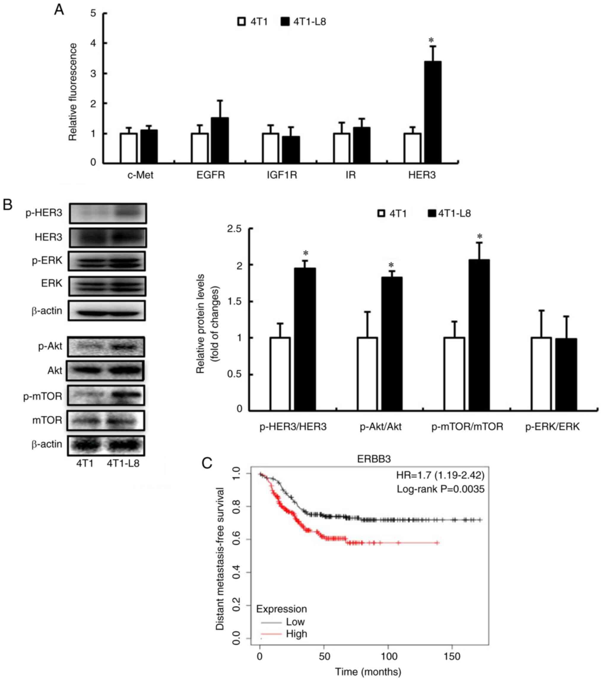 | Figure 1The HER3 pathway is associated with
the DMFS of patients with TNBC. (A) c-Met, EGFR, IGF1R, IR and HER3
protein phosphorylation levels were measured using
Luminex® 200. The experiments were performed in
duplicate and repeated three times. Data are presented as the mean
± SD. *P<0.05, compared with 4T1 cells. (B) The
expression levels of p-HER3, HER3, p-Akt, Akt, p-mTOR, mTOR, p-ERK
and ERK were detected using western blot analysis. The expression
levels of β-actin were used as internal controls. Quantification of
signals is presented as fold of changes relative to phosphorylated
proteins vs. total proteins. The experiments were repeated three
times. Data are presented as the mean ± SD. *P<0.05,
compared with 4T1 cells. (C) Kaplan-Meier Plotter analysis
indicated that DMFS was associated with HER3 expression (low
expression group, n=212; high expression group, n=212) among 424
TNBC cases (log-rank, P=0.0035). DMFS, distant metastasis-free
survival; TNBC, triple-negative breast cancer; HER3, human
epidermal growth factor receptor 3; EGFR, epidermal growth factor
receptor; IGF1R, insulin-like growth factor 1 receptor; IR, insulin
receptor; mTOR, mammalian target of rapamycin;
p-phosphorylated. |
Knockdown of HER3 decreases the
migration, invasion and metastasis of metastatic TNBC cells via the
inhibition of Akt and mTOR
To determine whether HER3 serves as a useful
therapeutic target for TNBC metastasis, the migration, invasion and
metastasis of TNBC cells were examined when HER3 was silenced using
siRNA. The efficacy of HER3 siRNA was first confirmed on the
expression of HER3/Akt/mTOR and the viability of 4T1-L8 cells. It
was confirmed that HER3 siRNA decreased the expression of HER3,
downstream p-Akt, and that of p-mTOR in 4T1-L8 cells (Fig. 2A). In addition, HER3 siRNA
inhibited the migration and invasion of 4T1-L8 cells, although it
did not affect their viability (Fig.
2B and C). To validate the anti-metastatic activity of HER3
siRNA, the number of metastatic nodules were counted in the lungs
of the mice. Treatment with HER3 siRNA reduced the number of lung
metastatic nodules compared with those in the group without HER3
siRNA treatment (Fig. 2D). These
results suggest that the HER3/Akt/mTOR pathway serves as a
therapeutic target for TNBC metastasis.
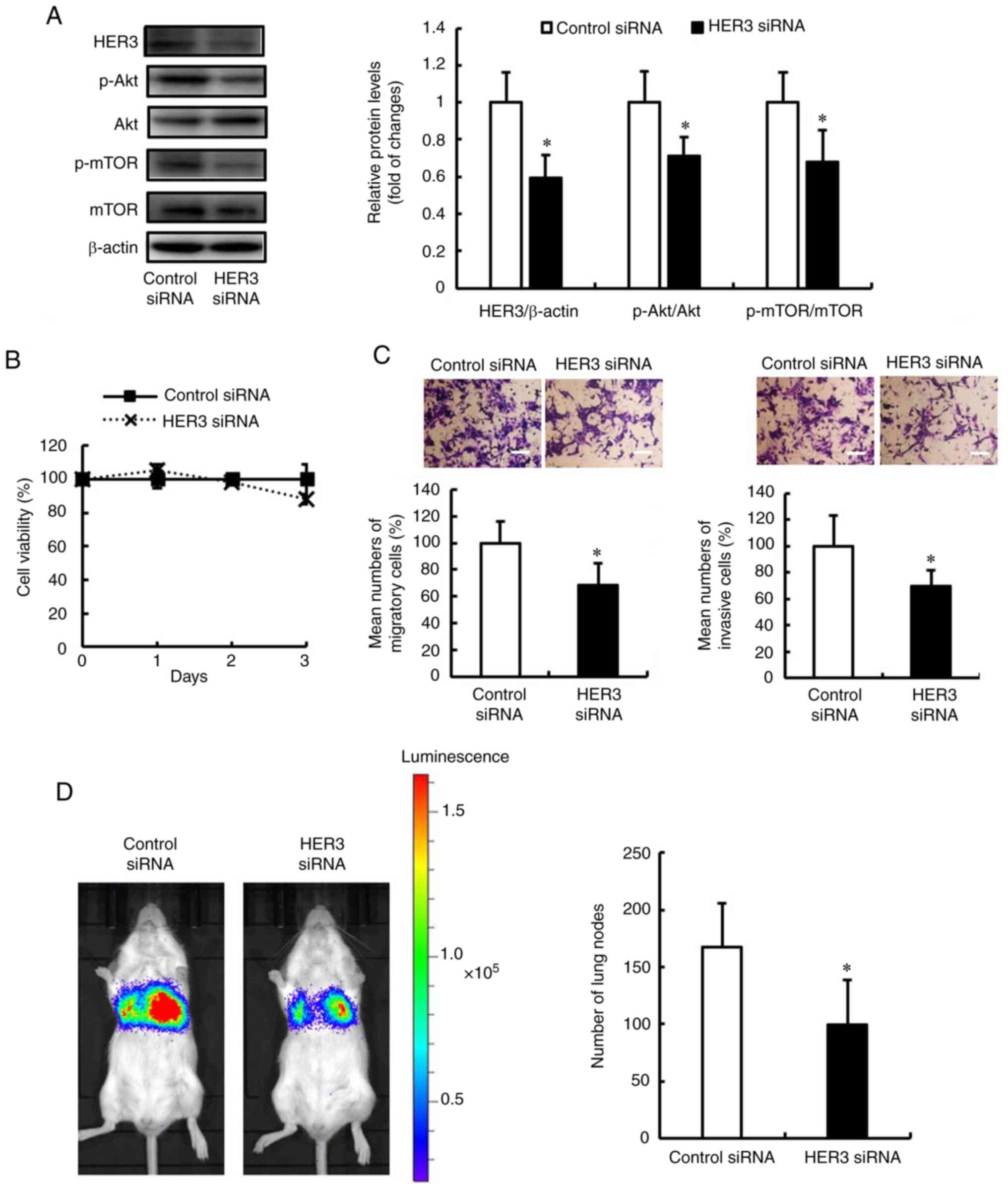 | Figure 2Knockdown of HER3 decreases
the migration, invasion and metastasis of metastatic TNBC cells by
inhibiting Akt and mTOR. (A-C) The 4T1-L8 cells were transfected
with HER siRNA (50 nM) or control siRNA. (A) The expression levels
of HER3, p-Akt, Akt, p-mTOR and mTOR were detected using western
blot analysis. The expression levels of β-actin were used as
internal controls. Quantification of signals is presented as fold
of changes relative to phosphorylated protein vs. total proteins or
total proteins vs. β-actin. The experiments were repeated four
times. Data are presented as the mean ± SD. *P<0.05,
compared with control siRNA. (B) The number of cells stained with
trypan blue counted on days 1, 2 and 3. The experiments were
performed in triplicate and repeated three times. Data are
presented as the mean ± SD. *P<0.05, compared with
control siRNA. (C) Cell migration and invasion analysis was
performed using the cell culture insert. The cells passing through
the cell culture insert were counted. The experiments were repeated
three times. Data are presented as the mean ± SD.
*P<0.05, compared with control siRNA. Representative
images of cells transfected with control siRNA and HER3 siRNA are
shown. Scale bars, 100 µm. (D) The 4T1-L8 cells transfected
with HER siRNA (50 nM) or control siRNA were injected into BALB/c
mice via the tail vain. Metastasis in the lungs was monitored using
IVIS. After 8 days, the mice were sacrificed and the number of
metastatic nodules in the lungs were counted. The results are
expressed as the mean ± SD. *P<0.05, compared with
control siRNA. TNBC, triple-negative breast cancer; HER3, human
epidermal growth factor receptor 3; mTOR, mammalian target of
rapamycin; p-phosphorylated. |
HER3/Akt/mTOR pathway promotes the
migration and invasion of TNBC cells by increasing CXCR4 expression
in metastatic TNBC
Subsequently, the present study investigated the
molecular mechanisms through which the HER3/Akt/mTOR pathway
promotes the metastatic phenotype. There is evidence to indicate
that the interaction between chemokines (members of the
chemoattractant cytokines) and chemokine receptors may play
critical roles in several key steps of tumorigenesis and/or
metastasis (18,19). Therefore, the present study
examined the expression of chemokine receptors, such as CCR2, CCR7
and CXCR4 in 4T1 and 4T1-L8 cells. Notably, CXCR4 expression was
higher in the 4T1-L8 cells than that in the 4T1 cells (Fig. 3A). However, no changes in the
expression of CCR2 and CCR7 proteins were observed between the two
cell lines. To validate the role of CXCR4 in the migration and
invasion of 4T1-L8 cells, the cells were treated with the CXCR4
ligand, SDF-1, and the migration and invasion were detected. It was
found that the migration and invasion of the 4T1-L8 cells treated
with SDF-1 were higher compared with those of the 4T1 cells treated
with SDF-1 (Fig. 3B). To
determine whether the HER3/Akt/mTOR pathway regulates CXCR4
expression, and promotes the migration and invasion of TNBC cells,
CXCR4 expression was examined in 4T1-L8 cells transfected with HER3
siRNA. Notably, HER3 siRNA suppressed CXCR4 expression in the
4T1-L8 cells (Fig. 3C). In
addition, HER3 siRNA decreased the migration and invasion of 4T1-L8
cells treated with SDF-1 (Fig.
3D). These results suggest that the HER3/Akt/mTOR pathway
promotes migration and invasion by increasing CXCR4 expression in
metastatic TNBC.
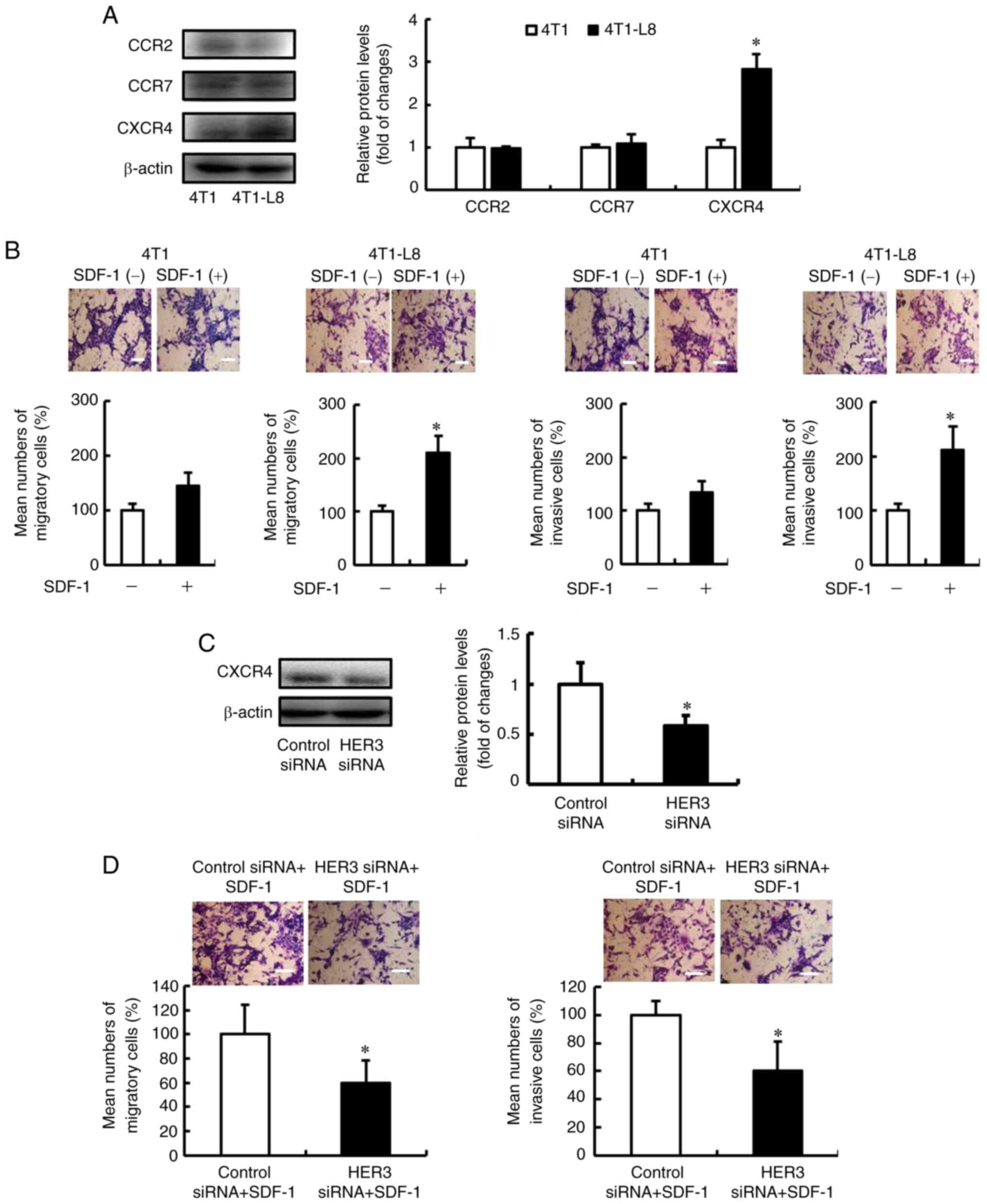 | Figure 3The HER3/Akt/mTOR pathway promotes
the migration and invasion of metastatic triple-negative breast
cancer cells by increasing CXCR4 expression. (A) The expression
levels of CCR2, CCR7 and CXCR4 were detected using western blot
analysis. The expression levels of β-actin were used as internal
controls. Quantification of signals is presented as fold of changes
relative to total proteins vs. β-actin. The experiments were
repeated three times. Data are presented as the mean ± SD.
*P<0.05, compared with 4T1 cells. (B) Cell migration
and invasion analysis was performed using the cell culture insert.
The cells passing through the cell culture insert were counted. The
experiments were repeated three times. Data are presented as the
mean ± SD. *P<0.05, compared with SDF-1(-).
Representative images of SDF-1-untreated 4T1 or 4T1-L8 and
SDF-1-treated 4T1 or 4T1-L8 cells are shown. Scale bars, 100
µm. (C and D) 4T1-L8 cells were transfected with HER siRNA
(50 nM) or control siRNA (Stealth™ RNAi Negative Control). (C) The
expression levels of CXCR4 were detected using western blotting.
The expression levels of β-actin were used as internal controls.
Quantification of signals is presented as fold change or relative
levels of CXCR4 vs. β-actin. The experiments were repeated three
times. Data are presented as the mean ± SD. *P<0.05,
compared with control siRNA. (D) Cell migration and invasion
analysis was performed using the cell culture insert. The cells
passing through the cell culture insert were counted. The
experiments were repeated three times. Data are presented as the
mean ± SD. *P<0.05, compared with control siRNA.
Representative images of control siRNA and HER3 siRNA are shown.
Scale bars, 100 µm. HER3, human epidermal growth factor
receptor 3; mTOR, mammalian target of rapamycin; CCR, chemokine
receptor; CXCR4, C-X-C chemokine receptor type 4; SDF-1, stromal
derived factor-1. |
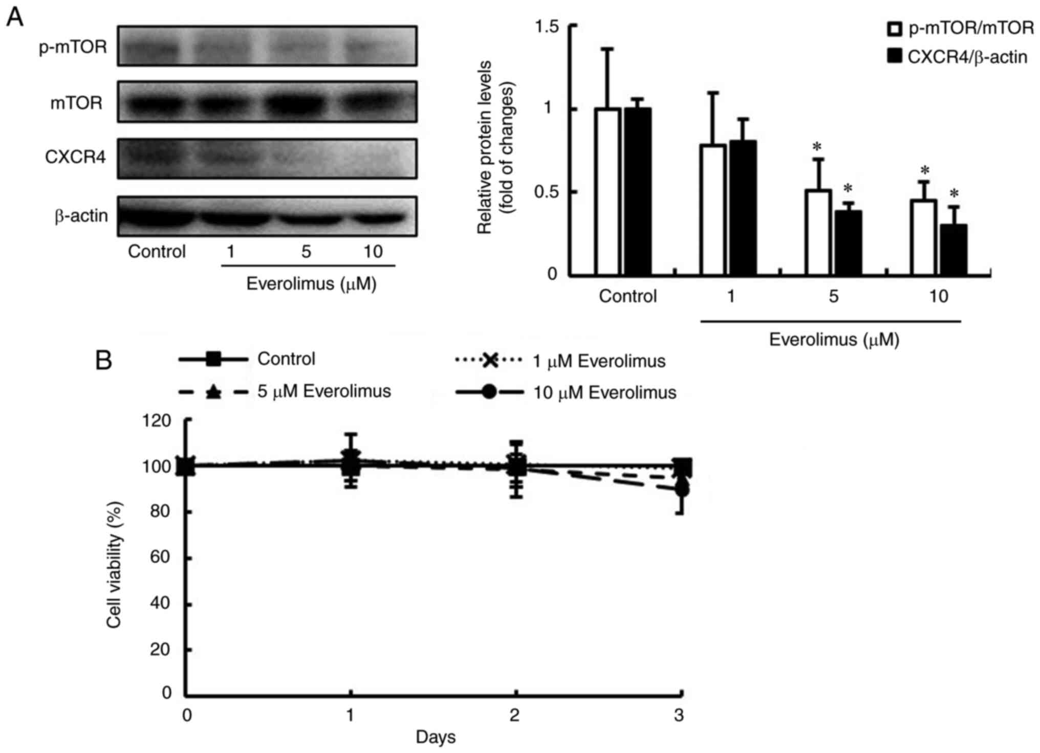
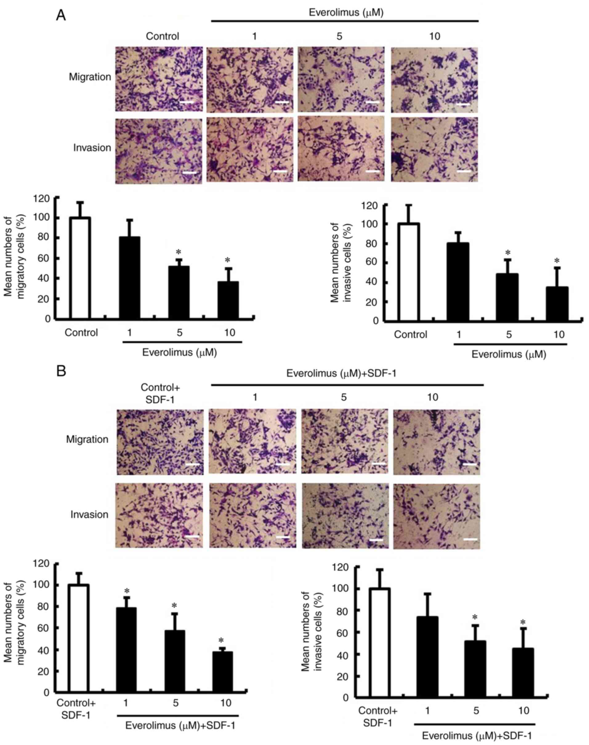
Everolimus suppresses migration, invasion
and metastasis by inhibiting CXCR4
Everolimus, an orally administered drug, is an
inhibitor of the mTOR serine/threonine kinase signal transduction
pathway that is currently used to treat advanced renal cell
carcinoma, neuroendocrine tumors and hormone receptor-positive
advanced breast cancer (20-22). Therefore, it was hypothesized that
everolimus may be effective in treating metastatic TNBC and thus
its effects on the 4T1-L8 cells were examined herein. Everolimus
decreased CXCR4 expression by inhibiting mTOR activation (Fig. 4A). In addition, everolimus
suppressed the migration and invasion of both 4T1-L8 and
SDF-1-treated 4T1-L8 cells, but did not affect cell viability
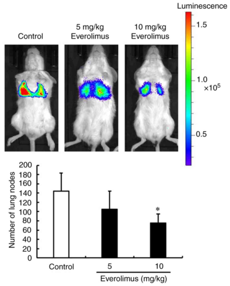
(Figs. 4B and 5). To validate the anti-metastatic
activity of everolimus, the number of metastatic nodules were
counted in the lungs of mice. The doses of everolimus used were 5
and 10 mg/kg once a day, based on previous studies (23-25). Treatment of the mice with
everolimus reduced the number of lung metastatic nodules compared
with those in the untreated mice (Fig. 6). Thus, these results indicated
that everolimus decreased the migration, invasion and metastasis of
TNBC cells by inhibiting CXCR4.
Discussion
TNBC is a subtype of breast cancer that is
associated with the worst prognosis among all subtypes, owing to
the high frequency of metastases (1,6).
However, no effective systematic therapy for TNBC metastasis is
currently available, at least to the best of our knowledge. The
present study demonstrated that the HER3/Akt/mTOR pathway plays a
critical role in the migration, invasion and metastasis of TNBC
cells. The HER3 pathway transduces extracellular signals into the
cell, resulting in several changes in regulatory processes,
including proliferation, survival, apoptosis and migration
(11,26). HER3 overexpression participates in
the regulation of various physiological and pathological pathways
in several types of cancer (26,27). Additionally, HER3 knockdown has
reported to suppress cell migration and metastasis via the
inhibition of Akt activation in gastric cancer (28). These findings suggest that the
HER3/Akt/mTOR pathway is a prospective biomarker and therapeutic
target for TNBC therapy.
Furthermore, HER3 is mediated through downstream
signals, including PI3K/Akt/mTOR and mitogen-activated protein
kinase (MAPK)/ERK1/2, and leads to cancer progression and
metastasis (29). In the present
study, it was demonstrated that HER3/Akt/mTOR activation was higher
in the 4T1-L8 metastatic cells compared with the 4T1 cells.
However, no changes in ERK activation were observed between the 4T1
and 4T1-L8 cells. Notably, breast cancer is more dependent on the
dysfunctional PI3K/Akt/mTOR pathway than the MAPK/ERK pathway
(30). In addition, HER3 allows
the recruitment of the p85 regulatory subunit of PI3K to activate
PI3K/Akt/mTOR signaling (31,32). These results suggest that a subset
of TNBC cells preferentially employ the HER3/Akt/mTOR pathway and
not the HER3/ERK pathway.
CXCR4 is a seven-transmembrane G-protein-coupled
chemokine receptor known for its ability to mediate directed cell
migration when activated by its cognate ligand SDF-1 (33). In addition, CXCR4 is responsible
for metastasis to predilection sites in the body, including the
lymph nodes, lungs, liver, bone marrow and brain, that are enriched
by SDF-1 (34). Notably, the
results of the present study indicated that HER3 siRNA suppressed
the migration and invasion of 4T1-L8 cells by decreasing CXCR4
expression via the inhibition of the Akt/mTOR pathway. A previous
study reported that Akt inhibition decreased CXCR4 expression via
mTOR inhibition in hepatocellular carcinoma (35). In addition, chromatin
immunoprecipitation with p-mTOR as a target revealed a physical
interaction between p-mTOR and the CXCR4 gene promoter (35). These results support the
hypothesis that the HER3/Akt/mTOR pathway regulates CXCR4
expression in TNBC cells.
The present study demonstrated that the
HER3/Akt/mTOR pathway may be a potential therapeutic target for
TNBC metastasis. One potential agent to target the HER3/Akt/mTOR
pathway is everolimus that has been FDA-approved for the treatment
of advanced renal cell carcinoma, neuroendocrine tumors and hormone
receptor-positive advanced breast cancer (22). Everolimus is an oral mTOR pathway
inhibitor that binds to its intracellular target, FK-506 binding
protein-12, to form a complex that inhibits mTOR in an allosteric
manner (36). In the present
study, it was demonstrated that everolimus suppressed the
migration, invasion and metastasis of TNBC cells by inhibiting
CXCR4 expression. As previously demonstrated, DHM25, a covalent
mTOR inhibitor, suppressed the tumor growth and metastasis of TNBC
by inhibiting mTOR in vivo (37). In addition, everolimus has been
shown to prolong the progression-free survival of patients
diagnosed with ER+ breast cancer, who have developed
resistance to hormonal therapy, in an advanced/metastatic setting
(38-40). Notably, everolimus is already
being used in the treatment of cancers, such as breast cancer.
Therefore, repurposing everolimus to prevent metastasis in patients
with TNBC may be more effective and cost-effective than traditional
drug development. These results suggest that everolimus has the
potential to treat TNBC metastasis.
In conclusion, the present study indicated that the
activation of the HER3/Akt/mTOR pathway was associsated with high
levels of migration, invasion and metastasis of TNBC cells.
Furthermore, the inhibition of the HER3/Akt/mTOR pathway decreased
the migration, invasion and metastasis of TNBC cells by decreasing
CXCR4 expression. In addition, treatment of metastatic TNBC cells
with everolimus inhibited cell migration, invasion and metastasis
by decreasing CXCR4 expression. Thus, targeting the HER3/Akt/mTOR
pathway may serve as a novel avenue for the development of
therapeutics against TNBC metastasis, and everolimus may be an
effective therapeutic agent which can be used to suppress TNBC
metastasis.
Supplementary Data
Availability of data and materials
The datasets used and/or analyzed during the current
study are available from the corresponding author on reasonable
request.
Authors' contributions
SN conceived the study. TT and MT designed the
experiments. TT, SG, KT and RT performed the experiments. TT and MT
analyzed the data. TT drafted the manuscript. MT and SN revised the
manuscript. SN and TT confirm the authenticity of all the raw data.
All authors have read and approved the final manuscript.
Ethics approval and consent to
participate
The animal experiments were approved by the Animal
Care and Use Committee of the Kindai University (project
identification code KAPS-27-021).
Patient consent for publication
Not applicable.
Competing interests
The authors declare that they have no competing
interests.
Acknowledgments
Not applicable.
Funding
The present study was partly supported by a Grant-in-Aid for
Young Scientists from the Japan Society for the Promotion of
Science (JSPS) (grant no. 20K16343).
References
|
1
|
Seto-Tetsuo F, Arioka M, Miura K, Inoue T,
Igawa K, Tomooka K, Takahashi-Yanaga F and Sasaguri T: DIF-1
inhibits growth and metastasis of triple-negative breast cancer
through AMPK-mediated inhibition of the mTORC1-S6K signaling
pathway. Oncogene. 40:5579–5589. 2021. View Article : Google Scholar : PubMed/NCBI
|
|
2
|
DeSantis CE, Fedewa SA, Goding Sauer A,
Kramer JL, Smith RA and Jemal A: Breast cancer statistics, 2015:
Convergence of incidence rates between black and white women. CA
Cancer J Clin. 66:31–42. 2016. View Article : Google Scholar
|
|
3
|
Chen L, Linden HM, Anderson BO and Li CI:
Trends in 5-year survival rates among breast cancer patients by
hormone receptor status and stage. Breast Cancer Res Treat.
147:609–616. 2014. View Article : Google Scholar : PubMed/NCBI
|
|
4
|
Weigelt B, Peterse JL and van 't Veer LJ:
Breast cancer metastasis: Markers and models. Nat Rev Cancer.
5:591–602. 2005. View
Article : Google Scholar : PubMed/NCBI
|
|
5
|
Saraiva DP, Guadalupe Cabral M, Jacinto A
and Braga S: How many diseases is triple negative breast cancer:
The protagonism of the immune microenvironment. ESMO Open.
2:e0002082017. View Article : Google Scholar : PubMed/NCBI
|
|
6
|
Dent R, Trudeau M, Pritchard KI, Hanna WM,
Kahn HK, Sawka CA, Lickley LA, Rawlinson E, Sun P and Narod SA:
Triple-negative breast cancer: Clinical features and patterns of
recurrence. Clin Cancer Res. 13:4429–4434. 2007. View Article : Google Scholar : PubMed/NCBI
|
|
7
|
Roskoski R Jr: The ErbB/HER family of
protein-tyrosine kinases and cancer. Pharmacol Res. 79:34–74. 2014.
View Article : Google Scholar
|
|
8
|
Peruzzi B and Bottaro DP: Targeting the
c-Met signaling pathway in cancer. Clin Cancer Res. 12:3657–3560.
2006. View Article : Google Scholar : PubMed/NCBI
|
|
9
|
Farabaugh SM, Boone DN and Lee AV: Role of
IGF1R in Breast Cancer Subtypes, Stemness, and Lineage
Differentiation. Front Endocrinol (Lausanne). 6:592015. View Article : Google Scholar : PubMed/NCBI
|
|
10
|
Belfiore A and Frasca F: IGF and insulin
receptor signaling in breast cancer. J Mammary Gland Biol
Neoplasia. 13:381–406. 2008. View Article : Google Scholar : PubMed/NCBI
|
|
11
|
Eccles SA: The epidermal growth factor
receptor/Erb-B/HER family in normal and malignant breast biology.
Int J Dev Biol. 55:685–696. 2011. View Article : Google Scholar : PubMed/NCBI
|
|
12
|
Koutras AK and Evans TR: The epidermal
growth factor receptor family in breast cancer. Onco Targets Ther.
1:5–19. 2008. View Article : Google Scholar : PubMed/NCBI
|
|
13
|
Witton CJ, Reeves JR, Going JJ, Cooke TG
and Bartlett JM: Expression of the HER1-4 family of receptor
tyrosine kinases in breast cancer. J Pathol. 200:290–297. 2003.
View Article : Google Scholar : PubMed/NCBI
|
|
14
|
Ho-Yen CM, Jones JL and Kermorgant S: The
clinical and functional significance of c-Met in breast cancer: A
review. Breast Cancer Res. 17:522015. View Article : Google Scholar : PubMed/NCBI
|
|
15
|
Tamimi RM, Colditz GA, Wang Y, Collins LC,
Hu R, Rosner B, Irie HY, Connolly JL and Schnitt SJ: Expression of
IGF1R in normal breast tissue and subsequent risk of breast cancer.
Breast Cancer Res Treat. 128:243–250. 2011. View Article : Google Scholar : PubMed/NCBI
|
|
16
|
Buck E and Mulvihill M: Small molecule
inhibitors of the IGF-1R/IR axis for the treatment of cancer.
Expert Opin Investig Drugs. 20:605–621. 2011. View Article : Google Scholar : PubMed/NCBI
|
|
17
|
Takeda T, Tsubaki M, Matsuda T, Kimura A,
Jinushi M, Obana T, Takegami M and Nishida S: EGFR inhibition
reverses epithelial-mesenchymal transition, and decreases tamoxifen
resistance via Snail and Twist downregulation in breast cancer
cells. Oncol Rep. 47:1092022. View Article : Google Scholar
|
|
18
|
Kakinuma T and Hwang ST: Chemokines,
chemokine receptors, and cancer metastasis. J Leukoc Biol.
79:639–651. 2006. View Article : Google Scholar : PubMed/NCBI
|
|
19
|
Tanaka T, Bai Z, Srinoulprasert Y, Yang
BG, Hayasaka H and Miyasaka M: Chemokines in tumor progression and
metastasis. Cancer Sci. 96:317–322. 2005. View Article : Google Scholar : PubMed/NCBI
|
|
20
|
Motzer RJ, Escudier B, Oudard S, Hutson
TE, Porta C, Bracarda S, Grünwald V, Thompson JA, Figlin RA,
Hollaender N, et al: Efficacy of everolimus in advanced renal cell
carcinoma: A double-blind, randomised, placebo-controlled phase III
trial. Lancet. 372:449–456. 2008. View Article : Google Scholar : PubMed/NCBI
|
|
21
|
Yao JC, Shah MH, Ito T, Bohas CL, Wolin
EM, Van Cutsem E, Hobday TJ, Okusaka T, Capdevila J, de Vries EG,
et al: Everolimus for advanced pancreatic neuroendocrine tumors. N
Engl J Med. 364:514–523. 2011. View Article : Google Scholar : PubMed/NCBI
|
|
22
|
Lebwohl D, Anak O, Sahmoud T, Klimovsky J,
Elmroth I, Haas T, Posluszny J, Saletan S and Berg W: Development
of everolimus, a novel oral mTOR inhibitor, across a spectrum of
diseases. Ann N Y Acad Sci. 1291:14–32. 2013. View Article : Google Scholar : PubMed/NCBI
|
|
23
|
Fuereder T, Jaeger-Lansky A, Hoeflmayer D,
Preusser M, Strommer S, Cejka D, Koehrer S, Crevenna R and Wacheck
V: mTOR inhibition by everolimus counteracts VEGF induction by
sunitinib and improves anti-tumor activity against gastric cancer
in vivo. Cancer Lett. 296:249–256. 2010. View Article : Google Scholar : PubMed/NCBI
|
|
24
|
Bradshaw-Pierce EL, Pitts TM, Kulikowski
G, Selby H, Merz AL, Gustafson DL, Serkova NJ, Eckhardt SG and
Weekes CD: Utilization of quantitative in vivo pharmacology
approaches to assess combination effects of everolimus and
irinotecan in mouse xenograft models of colorectal cancer. PLoS
One. 8:e580892013. View Article : Google Scholar : PubMed/NCBI
|
|
25
|
Pantaleo MA, Nicoletti G, Nanni C, Gnocchi
C, Landuzzi L, Quarta C, Boschi S, Nannini M, Di Battista M,
Castellucci P, et al: Preclinical evaluation of KIT/PDGFRA and mTOR
inhibitors in gastrointestinal stromal tumors using small animal
FDG PET. J Exp Clin Cancer Res. 29:1732010. View Article : Google Scholar
|
|
26
|
Lyu H, Han A, Polsdofer E, Liu S and Liu
B: Understanding the biology of HER3 receptor as a therapeutic
target in human cancer. Acta Pharm Sin B. 8:503–510. 2018.
View Article : Google Scholar : PubMed/NCBI
|
|
27
|
Beji A, Horst D, Engel J, Kirchner T and
Ullrich A: Toward the prognostic significance and therapeutic
potential of HER3 receptor tyrosine kinase in human colon cancer.
Clin Cancer Res. 18:956–968. 2012. View Article : Google Scholar
|
|
28
|
Wu X, Chen Y, Li G, Xia L, Gu R, Wen X,
Ming X and Chen H: Her3 is associated with poor survival of gastric
adenocarcinoma: Her3 promotes proliferation, survival and migration
of human gastric cancer mediated by PI3K/AKT signaling pathway. Med
Oncol. 31:9032014. View Article : Google Scholar : PubMed/NCBI
|
|
29
|
Mishra R, Patel H, Alanazi S, Yuan L and
Garrett JT: HER3 signaling and targeted therapy in cancer. Oncol
Rev. 12:3552018.PubMed/NCBI
|
|
30
|
Saini KS, Loi S, de Azambuja E,
Metzger-Filho O, Saini ML, Ignatiadis M, Dancey JE and
Piccart-Gebhart MJ: Targeting the PI3K/AKT/mTOR and Raf/MEK/ERK
pathways in the treatment of breast cancer. Cancer Treat Rev.
39:935–946. 2013. View Article : Google Scholar : PubMed/NCBI
|
|
31
|
Desbois-Mouthon C, Baron A, Blivet-Van
Eggelpoël MJ, Fartoux L, Venot C, Bladt F, Housset C and Rosmorduc
O: Insulin-like growth factor-1 receptor inhibition induces a
resistance mechanism via the epidermal growth factor
receptor/HER3/AKT signaling pathway: Rational basis for cotargeting
insulin-like growth factor-1 receptor and epidermal growth factor
receptor in hepatocellular carcinoma. Clin Cancer Res. 15:5445–456.
2009. View Article : Google Scholar : PubMed/NCBI
|
|
32
|
Sithanandam G and Anderson LM: The ERBB3
receptor in cancer and cancer gene therapy. Cancer Gene Ther.
15:413–448. 2008. View Article : Google Scholar : PubMed/NCBI
|
|
33
|
Zlotnik A: New insights on the role of
CXCR4 in cancer metastasis. J Pathol. 215:211–213. 2008. View Article : Google Scholar : PubMed/NCBI
|
|
34
|
Müller A, Homey B, Soto H, Ge N, Catron D,
Buchanan ME, McClanahan T, Murphy E, Yuan W, Wagner SN, et al:
Involvement of chemokine receptors in breast cancer metastasis.
Nature. 410:50–56. 2001. View
Article : Google Scholar : PubMed/NCBI
|
|
35
|
Zhu M, Guo J, Xia H, Li W, Lu Y, Dong X,
Chen Y, Xie X, Fu S and Li M: Alpha-fetoprotein activates AKT/mTOR
signaling to promote CXCR4 expression and migration of hepatoma
cells. Oncoscience. 2:59–70. 2015. View Article : Google Scholar : PubMed/NCBI
|
|
36
|
Anandappa G, Hollingdale A and Eisen T:
Everolimus-a new approach in the treatment of renal cell carcinoma.
Cancer Manag Res. 2:61–70. 2010.PubMed/NCBI
|
|
37
|
Fouqué A, Delalande O, Jean M, Castellano
R, Josselin E, Malleter M, Shoji KF, Hung MD, Rampanarivo H,
Collette Y, et al: A Novel Covalent mTOR Inhibitor, DHM25, Shows in
Vivo Antitumor Activity against Triple-Negative Breast Cancer
Cells. J Med Chem. 58:6559–6573. 2015. View Article : Google Scholar : PubMed/NCBI
|
|
38
|
O'Shaughnessy J, Thaddeus Beck J and Royce
M: Everolimusbased combination therapies for HR+, HER2-metastatic
breast cancer. Cancer Treat Rev. 69:204–214. 2018. View Article : Google Scholar : PubMed/NCBI
|
|
39
|
Hortobagyi GN: Everolimus plus exemestane
for the treatment of advanced breast cancer: A review of
subanalyses from BOLERO-2. Neoplasia. 17:279–288. 2015. View Article : Google Scholar : PubMed/NCBI
|
|
40
|
Fan Y, Sun T, Shao Z, Zhang Q, Ouyang Q,
Tong Z, Wang S, Luo Y, Teng Y, Wang X, et al: Effectiveness of
Adding Everolimus to the First-line Treatment of Advanced Breast
Cancer in Premenopausal Women Who Experienced Disease Progression
While Receiving Selective Estrogen Receptor Modulators: A Phase 2
Randomized Clinical Trial. JAMA Oncol. 7:e2134282021. View Article : Google Scholar : PubMed/NCBI
|