Spandidos Publications Logo
  • About
    • About Spandidos
    • Aims and Scopes
    • Abstracting and Indexing
    • Editorial Policies
    • Reprints and Permissions
    • Job Opportunities
    • Terms and Conditions
    • Contact
  • Journals
    • All Journals
    • Oncology Letters
      • Oncology Letters
      • Information for Authors
      • Editorial Policies
      • Editorial Board
      • Aims and Scope
      • Abstracting and Indexing
      • Bibliographic Information
      • Archive
    • International Journal of Oncology
      • International Journal of Oncology
      • Information for Authors
      • Editorial Policies
      • Editorial Board
      • Aims and Scope
      • Abstracting and Indexing
      • Bibliographic Information
      • Archive
    • Molecular and Clinical Oncology
      • Molecular and Clinical Oncology
      • Information for Authors
      • Editorial Policies
      • Editorial Board
      • Aims and Scope
      • Abstracting and Indexing
      • Bibliographic Information
      • Archive
    • Experimental and Therapeutic Medicine
      • Experimental and Therapeutic Medicine
      • Information for Authors
      • Editorial Policies
      • Editorial Board
      • Aims and Scope
      • Abstracting and Indexing
      • Bibliographic Information
      • Archive
    • International Journal of Molecular Medicine
      • International Journal of Molecular Medicine
      • Information for Authors
      • Editorial Policies
      • Editorial Board
      • Aims and Scope
      • Abstracting and Indexing
      • Bibliographic Information
      • Archive
    • Biomedical Reports
      • Biomedical Reports
      • Information for Authors
      • Editorial Policies
      • Editorial Board
      • Aims and Scope
      • Abstracting and Indexing
      • Bibliographic Information
      • Archive
    • Oncology Reports
      • Oncology Reports
      • Information for Authors
      • Editorial Policies
      • Editorial Board
      • Aims and Scope
      • Abstracting and Indexing
      • Bibliographic Information
      • Archive
    • Molecular Medicine Reports
      • Molecular Medicine Reports
      • Information for Authors
      • Editorial Policies
      • Editorial Board
      • Aims and Scope
      • Abstracting and Indexing
      • Bibliographic Information
      • Archive
    • World Academy of Sciences Journal
      • World Academy of Sciences Journal
      • Information for Authors
      • Editorial Policies
      • Editorial Board
      • Aims and Scope
      • Abstracting and Indexing
      • Bibliographic Information
      • Archive
    • International Journal of Functional Nutrition
      • International Journal of Functional Nutrition
      • Information for Authors
      • Editorial Policies
      • Editorial Board
      • Aims and Scope
      • Abstracting and Indexing
      • Bibliographic Information
      • Archive
    • International Journal of Epigenetics
      • International Journal of Epigenetics
      • Information for Authors
      • Editorial Policies
      • Editorial Board
      • Aims and Scope
      • Abstracting and Indexing
      • Bibliographic Information
      • Archive
    • Medicine International
      • Medicine International
      • Information for Authors
      • Editorial Policies
      • Editorial Board
      • Aims and Scope
      • Abstracting and Indexing
      • Bibliographic Information
      • Archive
  • Articles
  • Information
    • Information for Authors
    • Information for Reviewers
    • Information for Librarians
    • Information for Advertisers
    • Conferences
  • Language Editing
Spandidos Publications Logo
  • About
    • About Spandidos
    • Aims and Scopes
    • Abstracting and Indexing
    • Editorial Policies
    • Reprints and Permissions
    • Job Opportunities
    • Terms and Conditions
    • Contact
  • Journals
    • All Journals
    • Biomedical Reports
      • Information for Authors
      • Editorial Policies
      • Editorial Board
      • Aims and Scope
      • Abstracting and Indexing
      • Bibliographic Information
      • Archive
    • Experimental and Therapeutic Medicine
      • Information for Authors
      • Editorial Policies
      • Editorial Board
      • Aims and Scope
      • Abstracting and Indexing
      • Bibliographic Information
      • Archive
    • International Journal of Epigenetics
      • Information for Authors
      • Editorial Policies
      • Editorial Board
      • Aims and Scope
      • Abstracting and Indexing
      • Bibliographic Information
      • Archive
    • International Journal of Functional Nutrition
      • Information for Authors
      • Editorial Policies
      • Editorial Board
      • Aims and Scope
      • Abstracting and Indexing
      • Bibliographic Information
      • Archive
    • International Journal of Molecular Medicine
      • Information for Authors
      • Editorial Policies
      • Editorial Board
      • Aims and Scope
      • Abstracting and Indexing
      • Bibliographic Information
      • Archive
    • International Journal of Oncology
      • Information for Authors
      • Editorial Policies
      • Editorial Board
      • Aims and Scope
      • Abstracting and Indexing
      • Bibliographic Information
      • Archive
    • Medicine International
      • Information for Authors
      • Editorial Policies
      • Editorial Board
      • Aims and Scope
      • Abstracting and Indexing
      • Bibliographic Information
      • Archive
    • Molecular and Clinical Oncology
      • Information for Authors
      • Editorial Policies
      • Editorial Board
      • Aims and Scope
      • Abstracting and Indexing
      • Bibliographic Information
      • Archive
    • Molecular Medicine Reports
      • Information for Authors
      • Editorial Policies
      • Editorial Board
      • Aims and Scope
      • Abstracting and Indexing
      • Bibliographic Information
      • Archive
    • Oncology Letters
      • Information for Authors
      • Editorial Policies
      • Editorial Board
      • Aims and Scope
      • Abstracting and Indexing
      • Bibliographic Information
      • Archive
    • Oncology Reports
      • Information for Authors
      • Editorial Policies
      • Editorial Board
      • Aims and Scope
      • Abstracting and Indexing
      • Bibliographic Information
      • Archive
    • World Academy of Sciences Journal
      • Information for Authors
      • Editorial Policies
      • Editorial Board
      • Aims and Scope
      • Abstracting and Indexing
      • Bibliographic Information
      • Archive
  • Articles
  • Information
    • For Authors
    • For Reviewers
    • For Librarians
    • For Advertisers
    • Conferences
  • Language Editing
Login Register Submit
  • This site uses cookies
  • You can change your cookie settings at any time by following the instructions in our Cookie Policy. To find out more, you may read our Privacy Policy.

    I agree
Search articles by DOI, keyword, author or affiliation
Search
Advanced Search
presentation
International Journal of Molecular Medicine
Join Editorial Board Propose a Special Issue
Print ISSN: 1107-3756 Online ISSN: 1791-244X
Journal Cover
December-2025 Volume 56 Issue 6

Full Size Image

Sign up for eToc alerts
Recommend to Library

Journals

International Journal of Molecular Medicine

International Journal of Molecular Medicine

International Journal of Molecular Medicine is an international journal devoted to molecular mechanisms of human disease.

International Journal of Oncology

International Journal of Oncology

International Journal of Oncology is an international journal devoted to oncology research and cancer treatment.

Molecular Medicine Reports

Molecular Medicine Reports

Covers molecular medicine topics such as pharmacology, pathology, genetics, neuroscience, infectious diseases, molecular cardiology, and molecular surgery.

Oncology Reports

Oncology Reports

Oncology Reports is an international journal devoted to fundamental and applied research in Oncology.

Experimental and Therapeutic Medicine

Experimental and Therapeutic Medicine

Experimental and Therapeutic Medicine is an international journal devoted to laboratory and clinical medicine.

Oncology Letters

Oncology Letters

Oncology Letters is an international journal devoted to Experimental and Clinical Oncology.

Biomedical Reports

Biomedical Reports

Explores a wide range of biological and medical fields, including pharmacology, genetics, microbiology, neuroscience, and molecular cardiology.

Molecular and Clinical Oncology

Molecular and Clinical Oncology

International journal addressing all aspects of oncology research, from tumorigenesis and oncogenes to chemotherapy and metastasis.

World Academy of Sciences Journal

World Academy of Sciences Journal

Multidisciplinary open-access journal spanning biochemistry, genetics, neuroscience, environmental health, and synthetic biology.

International Journal of Functional Nutrition

International Journal of Functional Nutrition

Open-access journal combining biochemistry, pharmacology, immunology, and genetics to advance health through functional nutrition.

International Journal of Epigenetics

International Journal of Epigenetics

Publishes open-access research on using epigenetics to advance understanding and treatment of human disease.

Medicine International

Medicine International

An International Open Access Journal Devoted to General Medicine.

Journal Cover
December-2025 Volume 56 Issue 6

Full Size Image

Sign up for eToc alerts
Recommend to Library

  • Article
  • Citations
    • Cite This Article
    • Download Citation
    • Create Citation Alert
    • Remove Citation Alert
    • Cited By
  • Similar Articles
    • Related Articles (in Spandidos Publications)
    • Similar Articles (Google Scholar)
    • Similar Articles (PubMed)
  • Download PDF
  • Download XML
  • View XML

  • Supplementary Files
    • Supplementary_Data1.pdf
    • Supplementary_Data2.pdf
Article Open Access

C3a/C3aR axis is involved in diabetic kidney injury by regulating podocyte mitophagy in diabetic nephropathy

  • Authors:
    • Mengjie Weng
    • Xiaoting Wu
    • Siyi Rao
    • Kun Nie
    • Danyu You
    • Tingting Zheng
    • Enqin Lin
    • Jing Zheng
    • Jiong Cui
    • Jianxin Wan
  • View Affiliations / Copyright

    Affiliations: Department of Nephrology, Blood Purification Research Center, The First Affiliated Hospital, Fujian Medical University, Fuzhou, Fujian 350005, P.R. China
    Copyright: © Weng et al. This is an open access article distributed under the terms of Creative Commons Attribution License.
  • Article Number: 223
    |
    Published online on: October 14, 2025
       https://doi.org/10.3892/ijmm.2025.5664
  • Expand metrics +
Metrics: Total Views: 0 (Spandidos Publications: | PMC Statistics: )
Metrics: Total PDF Downloads: 0 (Spandidos Publications: | PMC Statistics: )
Cited By (CrossRef): 0 citations Loading Articles...

This article is mentioned in:


Abstract

The C3a/C3aR axis has been confirmed to be associated with the pathogenesis of diabetic nephropathy (DN) and mitochondrial dysfunction; however, the exact mechanisms underlying its role in diabetic podocytopathy remain unclear. The present study investigated the involvement of C3a/C3aR signaling in regulating mitophagy during the progression of DN. Diabetic db/db mice exhibited elevated renal C3 and C3aR levels, concurrent with podocyte injury, proteinuria and glomerular damage. Administration of the C3aR antagonist (C3aRA) SB290157 attenuated podocyte loss, reduced albuminuria and mitigated glomerular pathology. Ultrastructural and functional analyses revealed that C3aRA restored mitochondrial integrity in podocytes, resolving diabetes‑associated fragmentation and bioenergetic deficits. In vitro, high glucose‑exposed human podocytes displayed suppressed mitophagy and mitochondrial dysfunction, which were exacerbated by exogenous C3a. Conversely, C3aRA treatment enhanced mitophagy and preserved mitochondrial membrane potential, while small interfering RNA‑mediated C3aR or PINK1 knockdown abolished these protective effects. Notably, C3aRA activated the PI3K/AKT/FoxO1 pathway, driving both mitochondrial biogenesis and mitophagy. Disruption of this axis via FoxO1 inhibition reversed the therapeutic benefits of C3aRA, confirming its mechanistic centrality. In conclusion, the C3a/C3aR/PI3K/AKT/FoxO1 axis represents a previously unrecognized molecular bridge between complement activation and mitophagy failure in DN. Pharmacological disruption of this pathway could preserve podocyte homeostasis, offering a precision strategy against diabetic kidney injury.
View Figures

Figure 1

Heatmap of the C3 and
C3aR transcriptome data in different kidney-related
diseases. A set of kidney disease-related transcriptome data was
collected from the Gene Expression Omnibus database using
Affymetrix microarray sequencing technology. This dataset
(GSE47183) contained 122 human renal podocyte samples (including 14
samples of diabetic nephropathy, 23 samples of focal and segmental
glomerulosclerosis, 6 samples of focal and segmental
glomerulosclerosis and MCD, 21 samples of membranous
glomerulonephritis, 15 samples of MCD, 23 samples of rapidly
progressive glomerulonephritis, 3 samples of thin membrane disease
and 17 samples of tumor nephrectomy). Red indicates high
expression, while blue indicates low expression. MCD, minimal
change disease.

Figure 2

Expression of complement C3,
C3aR and renal injury in each mouse group. (A) Protein
levels of C3 and C3aR in kidney tissues (n=6).
Corresponding histograms of the representative protein bands are
shown on the bottom panel. (B) Immunohistochemical detection of the
deposition and expression of complement C3 and C3aR
in kidney tissue specimens of each mouse group. Microscopic images
are shown at x100 and x400 magnification. (C) Hematoxylin and
eosin, and Periodic Acid-Schiff staining of kidney tissues in each
group of mice. Microscopic images are shown at x100 magnification.
(D) Levels of blood creatinine, 24 h-urinary total protein and
urine albumin/creatinine ratio in each group of mice (n=6). (E)
Transmission electron microscopy observation of the ultrastructure
of glomerular podocytes. Scale bar, 2.0 μm. (F) TdT-mediated
dUTP nick-end labeling assay detection of apoptotic cells in mouse
kidney tissues for each group. Immunofluorescence images are shown,
and statistical analysis of the percentage of apoptotic cells for
each group is shown on the right side of the representative image
(n=6). Scale bar, 20 μm. db/db mice were used to
establish a diabetic nephropathy model. SB290157 is a C3aR
antagonist. Data are expressed as the mean ± SEM and analyzed by
two-way ANOVA with Bonferroni correction. *P<0.05,
**P<0.01 and ***P<0.001. C3aRA, C3aR
antagonist; ns, no statistically significant difference.

Figure 3

Function of glomerular podocytes and
mitochondrial damage in each group of mice. (A) Immunofluorescence
images of the podocyte-specific functional proteins
synaptopodin and nephrin in each group of mice. Scale
bar, 20 μm. (B) Protein levels of ZO-1 and podocin in
the kidney tissues (n=6). Corresponding histograms are shown on the
bottom panel of representative protein bands. (C)
Immunohistochemical analysis of the mitochondrial fusion protein
Mnf2. Scale bar, 50 μm. (D) Representative images and
quantification of double immunofluorescence staining for
TOMM20 (red) and nephrin (green) in the glomerulus of
mice. The yellow areas indicate TOMM20 and nephrin
colocalization in podocytes (n=6). Scale bar, 20 μm. (E)
Electron microscopy image of mitochondrial morphology in podocytes.
Scale bar, 2.0 μm and 500 nm. Data are expressed as the mean
± SEM and analyzed by two-way ANOVA with Bonferroni correction.
*P<0.05, **P<0.01 and
***P<0.001. C3aRA, C3aR antagonist.

Figure 4

Expression levels of mitophagy and
related pathway proteins in mouse renal tissues. (A)
Immunohistochemical analysis of the mitophagy-specific protein
PINK1. Magnification, x100. (B) Protein levels of LC3B I/II, parkin
and PINK1 in kidney tissues (n=6). Corresponding histograms are
shown on the right panel of representative protein bands. (C)
Protein levels of PI3K [PI3-kinase p85α (54 + 85 kDa)],
phosphorylated-AKT and FoxO1 in kidney tissues (n=6). Corresponding
histograms are shown on the right panel of representative protein
bands. *P<0.05, **P<0.01 and
***P<0.001. p-, phosphorylated.

Figure 5

Effects of HG and C3a on podocyte
damage and mitophagy. Normal glucose refers to 5.5 mM glucose,
while the mannitol high osmotic control group was subjected to 24.5
mM mannitol + 5.5 mM glucose, and HG represents the intervention
group (30 mM glucose). (A) Representative images (left) and
quantification (right) of podocyte cytoskeleton, with F-actin
(green) stained using phalloidin (n=6). Scale bar, 20 μm.
(B) Protein levels of synaptopodin and podocin in podocytes (n=6).
Corresponding histograms are shown on the bottom panel of
representative protein bands. (C) ELISA detection of C3a levels in
podocytes at different time points (n=6). (D) Protein levels of C3
and C3aR in podocytes (n=6). Corresponding histograms are shown on
the right panel of representative protein bands. (E) Protein levels
of LC3B I/II, parkin and PINK1 in podocytes (n=8). Corresponding
histograms are shown on the bottom panel of representative protein
bands. (F) Protein levels of parkin and PINK1 in podocytes (n=5).
Corresponding histograms are shown on the right panel of
representative protein bands. (G) Representative images and
quantification (bottom) of podocyte cytoskeleton induced by
different times and concentrations of C3a (10−7 M for
12, 24 and 48 h; or 10−8, 10−7 and
10−6 M for 24 h) (n=6). Scale bar, 20 μm.
*P<0.05, **P<0.01 and
***P<0.001. ns, no statistically significant
difference; HG, high glucose.

Figure 6

Inhibition of C3aR improves
HG-induced podocyte and mitochondrial autophagic damage. (A)
Changes in cell cytoskeleton after blocking C3aR in a HG
environment. Scale bar, 20 μm. (B) Immunofluorescence images
of podocytes in each group using a TdT-mediated dUTP nick-end
labeling assay. Scale bar, 50 μm. The lower panel presents
the statistical analysis of apoptosis ratios in podocytes for each
group (n=6). (C) Transmission electron microscopy examination of
mitochondrial morphology. Scale bar, 2.0 μm and 500 nm. The
statistical charts on the right side of the electron microscopy
images represent the percentages of damaged mitochondria and
mitochondria enveloped by autophagosomes (n=5). (D) Transfection of
immortalized human podocytes with adenovirus GFP-LC3B (green) and
DsRed-Mito (red). Scale bar, 20 μm. The statistical charts
on the right side represent the number of GFP-LC3B-positive puncta
per cell and the proportion of LC3B spots on mitochondria (Mito) to
total LC3B. Quantification of GFP-LC3B (green)-associated
Mito-DsRed (red) staining intensity normalized by GFP-LC3B area
(n=5). (E) Protein levels of LC3B I/II, parkin and PINK1 in
podocytes (n=6). Corresponding histograms are shown on the right
panel of representative protein bands. *P<0.05,
**P<0.01 and ***P<0.001. ns, no
statistically significant difference; HG, high glucose; NG, normal
glucose; siRNA, small interfering RNA; NC, negative control.

Figure 7

C3a downregulates mitophagy levels
through the PI3K/AKT/FoxO1 signaling pathway in a HG environment,
leading to podocyte damage. (A) Immunofluorescence images of
F-actin-stained podocyte cytoskeleton. Scale bar, 20 μm.
Flow cytometric analysis of podocyte apoptosis under different
intervention conditions using annexin V-FITC PI. (B) Expression of
podocyte functional and mitophagy-related proteins after PINK1
inhibition (n=5). Corresponding histograms are shown on the right
panel of representative protein bands. (C) Expression of downstream
signaling molecules after direct stimulation of podocytes by C3a
(n=6). (D) Expression of downstream signaling molecules after
inhibiting C3aR in a HG environment (n=5). Corresponding histograms
are shown on the bottom panel of the representative protein bands.
(E) Confocal microscopy images capturing fluorescence of
immortalized human podocytes transfected with adenovirus GFP-LC3B
(green) and Mito-DsRed (red). Scale bar, 20 μm. The right
panel represents the statistical analysis of the number of
GFP-LC3B-positive spots per cell and the proportion of LC3B spots
on mitochondria (Mito) to total LC3B. Quantification of
GFP-LC3B-associated Mito-DsRed staining intensity normalized by
GFP-LC3B area (n=5). (F) Effects of PI3K inhibition on downstream
pathway proteins and mitophagy proteins under HG conditions with
C3a overexpression (n=5). Corresponding histograms are shown
on the bottom panel of representative protein bands. (G)
Immunofluorescence micrographs demonstrating the
nuclear/cytoplasmic distribution of FoxO1 in podocytes, with
dual-color staining of phosphoryalted-FoxO1 (red) and FoxO1
(green). Scale bar, 20 μm. *P<0.05,
**P<0.01 and ***P<0.001. ns, no
statistically significant difference; HG, high-glucose.

Figure 8

Role and mechanism of C3a/C3aR in a
DN model. In a high-glucose environment, the complement component
C3 is activated. The C3a/C3aR axis modulates the PI3K-AKT signaling
pathway, resulting in an enhanced phosphorylation level of FoxO1,
leading to the loss of its transcriptional activity. Consequently,
there is inhibition of PINK1/parkin-mediated mitophagy,
contributing to podocyte injury and DN progression. DN, diabetic
nephropathy; C3aRA, C3aR antagonist.
View References

1 

Ma J, Yiu WH and Tang SCW: Complement anaphylatoxins: Potential therapeutic target for diabetic kidney disease. Diabet Med. 42:e154272025. View Article : Google Scholar :

2 

Li X, Zhao S, Xie J, Li M, Tong S, Ma J, Yang R, Zhao Q, Zhang J and Xu A: Targeting the NF-κB p65-MMP28 axis: Wogonoside as a novel therapeutic agent for attenuating podocyte injury in diabetic nephropathy. Phytomedicine. 138:1564062025. View Article : Google Scholar

3 

Wang T, Chen Y, Liu Z, Zhou J, Li N, Shan Y and He Y: Long noncoding RNA Glis2 regulates podocyte mitochondrial dysfunction and apoptosis in diabetic nephropathy via sponging miR-328-5p. J Cell Mol Med. 28:e182042024. View Article : Google Scholar : PubMed/NCBI

4 

Woroniecka KI, Park ASD, Mohtat D, Thomas DB, Pullman JM and Susztak K: Transcriptome analysis of human diabetic kidney disease. Diabetes. 60:2354–2369. 2011. View Article : Google Scholar : PubMed/NCBI

5 

Wehner H, Höhn D, Faix-Schade U, Huber H and Walzer P: Glomerular changes in mice with spontaneous hereditary diabetes. Lab Invest. 27:331–340. 1972.PubMed/NCBI

6 

Li L, Chen L, Zang J, Tang X, Liu Y, Zhang J, Bai L, Yin Q, Lu Y, Cheng J, et al: C3a and C5a receptor antagonists ameliorate endothelial-myofibroblast transition via the Wnt/β-catenin signaling pathway in diabetic kidney disease. Metabolism. 64:597–610. 2015. View Article : Google Scholar : PubMed/NCBI

7 

Li L, Yin Q, Tang X, Bai L, Zhang J, Gou S, Zhu H, Cheng J, Fu P and Liu F: C3a receptor antagonist ameliorates inflammatory and fibrotic signals in type 2 diabetic nephropathy by suppressing the activation of TGF-β/smad3 and IKBα pathway. PLoS One. 9:e1136392014. View Article : Google Scholar

8 

Morigi M, Perico L, Corna D, Locatelli M, Cassis P, Carminati CE, Bolognini S, Zoja C, Remuzzi G, Benigni A and Buelli S: C3a receptor blockade protects podocytes from injury in diabetic nephropathy. JCI Insight. 5:e1318492020. View Article : Google Scholar : PubMed/NCBI

9 

Ma K, Chen G, Li W, Kepp O, Zhu Y and Chen Q: Mitophagy, mitochondrial homeostasis, and cell fate. Front Cell Dev Biol. 8:4672020. View Article : Google Scholar : PubMed/NCBI

10 

Stanigut AM, Tuta L, Pana C, Alexandrescu L, Suceveanu A, Blebea NM and Vacaroiu IA: Autophagy and mitophagy in diabetic kidney disease-a literature review. Int J Mol Sci. 26:8062025. View Article : Google Scholar : PubMed/NCBI

11 

Tagawa A, Yasuda M, Kume S, Yamahara K, Nakazawa J, Chin-Kanasaki M, Araki H, Araki S, Koya D, Asanuma K, et al: Impaired podocyte autophagy exacerbates proteinuria in diabetic nephropathy. Diabetes. 65:755–767. 2016. View Article : Google Scholar

12 

Zhou D, Zhou M, Wang Z, Fu Y, Jia M, Wang X, Liu M, Zhang Y, Sun Y, Zhou Y, et al: Progranulin alleviates podocyte injury via regulating CAMKK/AMPK-mediated autophagy under diabetic conditions. J Mol Med (Berl). 97:1507–1520. 2019. View Article : Google Scholar : PubMed/NCBI

13 

Zhou D, Zhou M, Wang Z, Fu Y, Jia M, Wang X, Liu M, Zhang Y, Sun Y, Lu Y, et al: PGRN acts as a novel regulator of mitochondrial homeostasis by facilitating mitophagy and mitochondrial biogenesis to prevent podocyte injury in diabetic nephropathy. Cell Death Dis. 10:5242019. View Article : Google Scholar : PubMed/NCBI

14 

Martini S, Nair V, Keller BJ, Eichinger F, Hawkins JJ, Randolph A, Böger CA, Gadegbeku CA, Fox CS, Cohen CD, et al: Integrative biology identifies shared transcriptional networks in CKD. J Am Soc Nephrol. 25:2559–2572. 2014. View Article : Google Scholar : PubMed/NCBI

15 

Ju W, Greene CS, Eichinger F, Nair V, Hodgin JB, Bitzer M, Lee YS, Zhu Q, Kehata M, Li M, et al: Defining cell-type specificity at the transcriptional level in human disease. Genome Res. 23:1862–1873. 2013. View Article : Google Scholar : PubMed/NCBI

16 

National Research Council Committee for the Update of the Guide for the C. and A. Use of Laboratory. The National Academies Collection: Reports funded by National Institutes of Health, in Guide for the Care and Use of Laboratory Animals. National Academies Press. Copyright© 2011. National Academy of Sciences; Washington, DC: 2011

17 

Ames RS, Lee D, Foley JJ, Jurewicz AJ, Tornetta MA, Bautsch W, Settmacher B, Klos A, Erhard KF, Cousins RD, et al: Identification of a selective nonpeptide antagonist of the anaphylatoxin C3a receptor that demonstrates antiinflammatory activity in animal models. J Immunol. 166:6341–6348. 2001. View Article : Google Scholar : PubMed/NCBI

18 

Yasuda-Yamahara M, Kume S, Tagawa A, Maegawa H and Uzu T: Emerging role of podocyte autophagy in the progression of diabetic nephropathy. Autophagy. 11:2385–2386. 2015. View Article : Google Scholar : PubMed/NCBI

19 

Flyvbjerg A: The role of the complement system in diabetic nephropathy. Nat Rev Nephrol. 13:311–318. 2017. View Article : Google Scholar : PubMed/NCBI

20 

Tesch GH: Diabetic nephropathy-is this an immune disorder? Clin Sci (Lond). 131:2183–2199. 2017. View Article : Google Scholar : PubMed/NCBI

21 

Sahu A and Lambris JD: Structure and biology of complement protein C3, a connecting link between innate and acquired immunity. Immunol Rev. 180:35–48. 2001. View Article : Google Scholar : PubMed/NCBI

22 

Kelly KJ, Liu Y, Zhang J and Dominguez JH: Renal C3 complement component: Feed forward to diabetic kidney disease. Am J Nephrol. 41:48–56. 2015. View Article : Google Scholar : PubMed/NCBI

23 

Racine KC, Iglesias-Carres L, Herring JA, Wieland KL, Ellsworth PN, Tessem JS, Ferruzzi MG, Kay CD and Neilson AP: The high-fat diet and low-dose streptozotocin type-2 diabetes model induces hyperinsulinemia and insulin resistance in male but not female C57BL/6J mice. Nutr Res. 131:135–146. 2024. View Article : Google Scholar : PubMed/NCBI

24 

Wang L, Zhou R, Li G, Zhang X, Li Y, Shen Y and Fang J: Multi-omics characterization of diabetic nephropathy in the db/db mouse model of type 2 diabetes. Comput Struct Biotechnol J. 27:3399–3409. 2025. View Article : Google Scholar : PubMed/NCBI

25 

Jin J, Shi Y, Gong J, Zhao L, Li Y, He Q and Huang H: Exosome secreted from adipose-derived stem cells attenuates diabetic nephropathy by promoting autophagy flux and inhibiting apoptosis in podocyte. Stem Cell Res Ther. 10:952019. View Article : Google Scholar : PubMed/NCBI

26 

Yuen DA, Stead BE, Zhang Y, White KE, Kabir MG, Thai K, Advani SL, Connelly KA, Takano T, Zhu L, et al: eNOS deficiency predisposes podocytes to injury in diabetes. J Am Soc Nephrol. 23:1810–1823. 2012. View Article : Google Scholar : PubMed/NCBI

27 

Angeletti A, Cantarelli C, Petrosyan A, Andrighetto S, Budge K, D'Agati VD, Hartzell S, Malvi D, Donadei C, Thurman JM, et al: Loss of decay-accelerating factor triggers podocyte injury and glomerulosclerosis. J Exp Med. 217:e201916992020. View Article : Google Scholar : PubMed/NCBI

28 

Galvan DL, Green NH and Danesh FR: The hallmarks of mitochondrial dysfunction in chronic kidney disease. Kidney Int. 92:1051–1057. 2017. View Article : Google Scholar : PubMed/NCBI

29 

Chen K, Dai H, Yuan J, Chen J, Lin L, Zhang W, Wang L, Zhang J, Li K and He Y: Optineurin-mediated mitophagy protects renal tubular epithelial cells against accelerated senescence in diabetic nephropathy. Cell Death Dis. 9:1052018. View Article : Google Scholar : PubMed/NCBI

30 

Nguyen TN, Padman BS and Lazarou M: Deciphering the molecular signals of PINK1/parkin mitophagy. Trends Cell Biol. 26:733–744. 2016. View Article : Google Scholar : PubMed/NCBI

31 

Li W, Du M, Wang Q, Ma X, Wu L, Guo F, Ji H, Huang F and Qin G: FoxO1 promotes mitophagy in the podocytes of diabetic male mice via the PINK1/parkin pathway. Endocrinology. 158:2155–2167. 2017. View Article : Google Scholar : PubMed/NCBI

32 

Zhao Y and Sun M: Metformin rescues Parkin protein expression and mitophagy in high glucose-challenged human renal epithelial cells by inhibiting NF-κB via PP2A activation. Life Sci. 246:1173822020. View Article : Google Scholar

33 

Yi X, Yan W, Guo T, Liu N, Wang Z, Shang J, Wei X, Cui X, Sun Y, Ren S and Chen L: Erythropoietin mitigates diabetic nephropathy by restoring PINK1/Parkin-mediated mitophagy. Front Pharmacol. 13:8830572022. View Article : Google Scholar : PubMed/NCBI

34 

Sun J, Zhu H, Wang X, Gao Q, Li Z and Huang H: CoQ10 ameliorates mitochondrial dysfunction in diabetic nephropathy through mitophagy. J Endocrinol. 240:445–465. 2019. View Article : Google Scholar : PubMed/NCBI

35 

Liu X, Wang W, Song G, Wei X, Zeng Y, Han P, Wang D, Shao M, Wu J, Sun H, et al: Astragaloside IV ameliorates diabetic nephropathy by modulating the mitochondrial quality control network. PLoS One. 12:e01825582017. View Article : Google Scholar : PubMed/NCBI

36 

Liu X, Lu J, Liu S, Huang D, Chen M, Xiong G and Li S: Huangqi-Danshen decoction alleviates diabetic nephropathy in db/db mice by inhibiting PINK1/Parkin-mediated mitophagy. Am J Transl Res. 12:989–998. 2020.PubMed/NCBI

37 

Yang M, Li C, Yang S, Xiao Y, Chen W, Gao P, Jiang N, Xiong S, Wei L, Zhang Q, et al: Mitophagy: A novel therapeutic target for treating DN. Curr Med Chem. 28:2717–2728. 2021. View Article : Google Scholar

38 

Zhang L, Li W, Gong M, Zhang Z, Xue X, Mao J, Zhang H, Li S, Liu X, Wu F, et al: C-reactive protein inhibits C3a/C3aR-dependent podocyte autophagy in favor of diabetic kidney disease. FASEB J. 36:e223322022. View Article : Google Scholar : PubMed/NCBI

39 

Wang C, Wang Z, Xu J, Ma H, Jin K, Xu T, Pan X, Feng X and Zhang W: C3aR antagonist alleviates C3a induced tubular profibrotic phenotype transition via restoring PPARα/CPT-1α mediated mitochondrial fatty acid oxidation in renin-dependent hypertension. Front Biosci (Landmark Ed). 28:2382023. View Article : Google Scholar

40 

Chen Y, Zheng YF, Lin XH, Zhang JP, Lin F and Shi H: Dendrobium mixture attenuates renal damage in rats with diabetic nephropathy by inhibiting the PI3K/Akt/mTOR pathway. Mol Med Rep. 24:5902021. View Article : Google Scholar : PubMed/NCBI

41 

Ma Z, Liu Y, Li C, Zhang Y and Lin N: Repurposing a clinically approved prescription Colquhounia root tablet to treat diabetic kidney disease via suppressing PI3K/AKT/NF-kB activation. Chin Med. 17:22022. View Article : Google Scholar : PubMed/NCBI

42 

Dong R, Zhang X, Liu Y, Zhao T, Sun Z, Liu P, Xiang Q, Xiong J, Du X, Yang X, et al: Rutin alleviates EndMT by restoring autophagy through inhibiting HDAC1 via PI3K/AKT/mTOR pathway in diabetic kidney disease. Phytomedicine. 112:1547002023. View Article : Google Scholar : PubMed/NCBI

43 

Zhang Y, Yang S, Cui X, Yang J, Zheng M, Jia J, Han F, Yang X, Wang J, Guo Z, et al: Hyperinsulinemia can cause kidney disease in the IGT stage of OLETF rats via the INS/IRS-1/PI3-K/Akt signaling pathway. J Diabetes Res. 2019:47097152019. View Article : Google Scholar : PubMed/NCBI

44 

Zheng D, Tao M, Liang X, Li Y, Jin J and He Q: p66Shc regulates podocyte autophagy in high glucose environment through the Notch-PTEN-PI3K/Akt/mTOR pathway. Histol Histopathol. 35:405–415. 2020.

45 

Wang X, Jiang L, Liu XQ, Huang YB, Wang AL, Zeng HX, Gao L, Zhu QJ, Xia LL and Wu YG: Paeoniflorin binds to VEGFR2 to restore autophagy and inhibit apoptosis for podocyte protection in diabetic kidney disease through PI3K-AKT signaling pathway. Phytomedicine. 106:1544002022. View Article : Google Scholar : PubMed/NCBI

46 

Chen Y, Li Z, Li H, Su W, Xie Y, Pan Y, Chen X and Liang D: Apremilast regulates the Teff/Treg balance to ameliorate uveitis via PI3K/AKT/FoxO1 signaling pathway. Front Immunol. 11:5816732020. View Article : Google Scholar : PubMed/NCBI

47 

Miao Z, Liu Y, Xu Y, Bu J and Yang Q: Oxaloacetate promotes the transition from glycolysis to gluconeogenesis through the Akt-FoxO1 and JNK/c-Jun-FoxO1 axes and inhibits the survival of liver cancer cells. Int Immunopharmacol. 161:1150512025. View Article : Google Scholar : PubMed/NCBI

48 

Xu Z, Liu K, Zhang G, Yang F, He Y, Nan W, Li Y and Lin J: Transcriptome analysis reveals that the injection of mesenchymal stem cells remodels extracellular matrix and complement components of the brain through PI3K/AKT/FOXO1 signaling pathway in a neuroinflammation mouse model. Genomics. 117:1110332025. View Article : Google Scholar : PubMed/NCBI

49 

Deng A, Wang Y, Huang K, Xie P, Mo P, Liu F, Chen J, Chen K, Wang Y and Xiao B: Artichoke (Cynara scolymus L.) water extract alleviates palmitate-induced insulin resistance in HepG2 hepatocytes via the activation of IRS1/PI3K/AKT/FoxO1 and GSK-3β signaling pathway. BMC Complement Med Ther. 23:4602023. View Article : Google Scholar

50 

Cosenso-Martin LN, Takaoka LY and Vilela-Martin JF: Randomized study comparing vildagliptin vs glibenclamide on glucose variability and endothelial function in patients with type 2 diabetes mellitus and hypertension. Diabetes Metab Syndr Obes. 13:3221–3229. 2020. View Article : Google Scholar : PubMed/NCBI
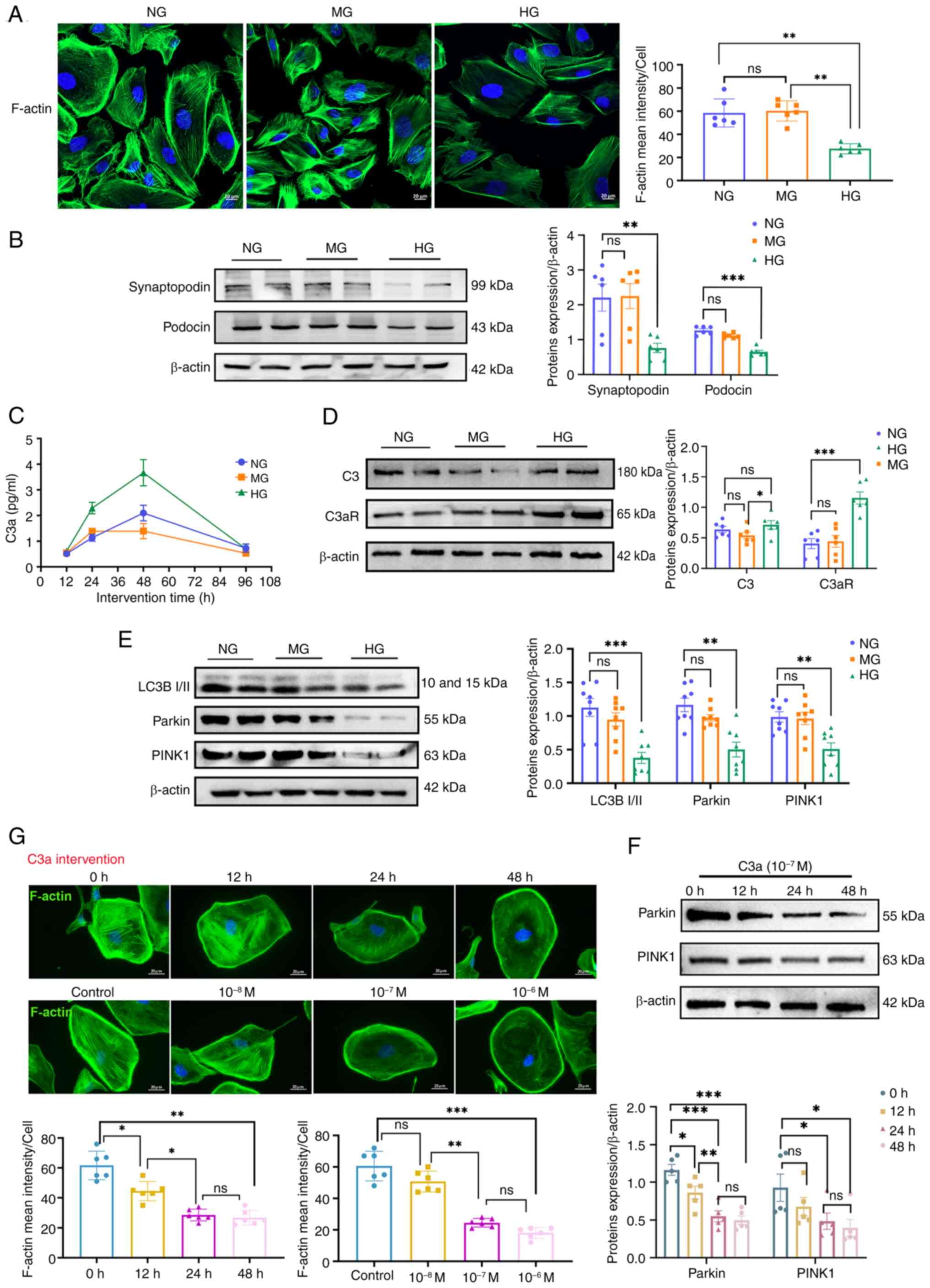
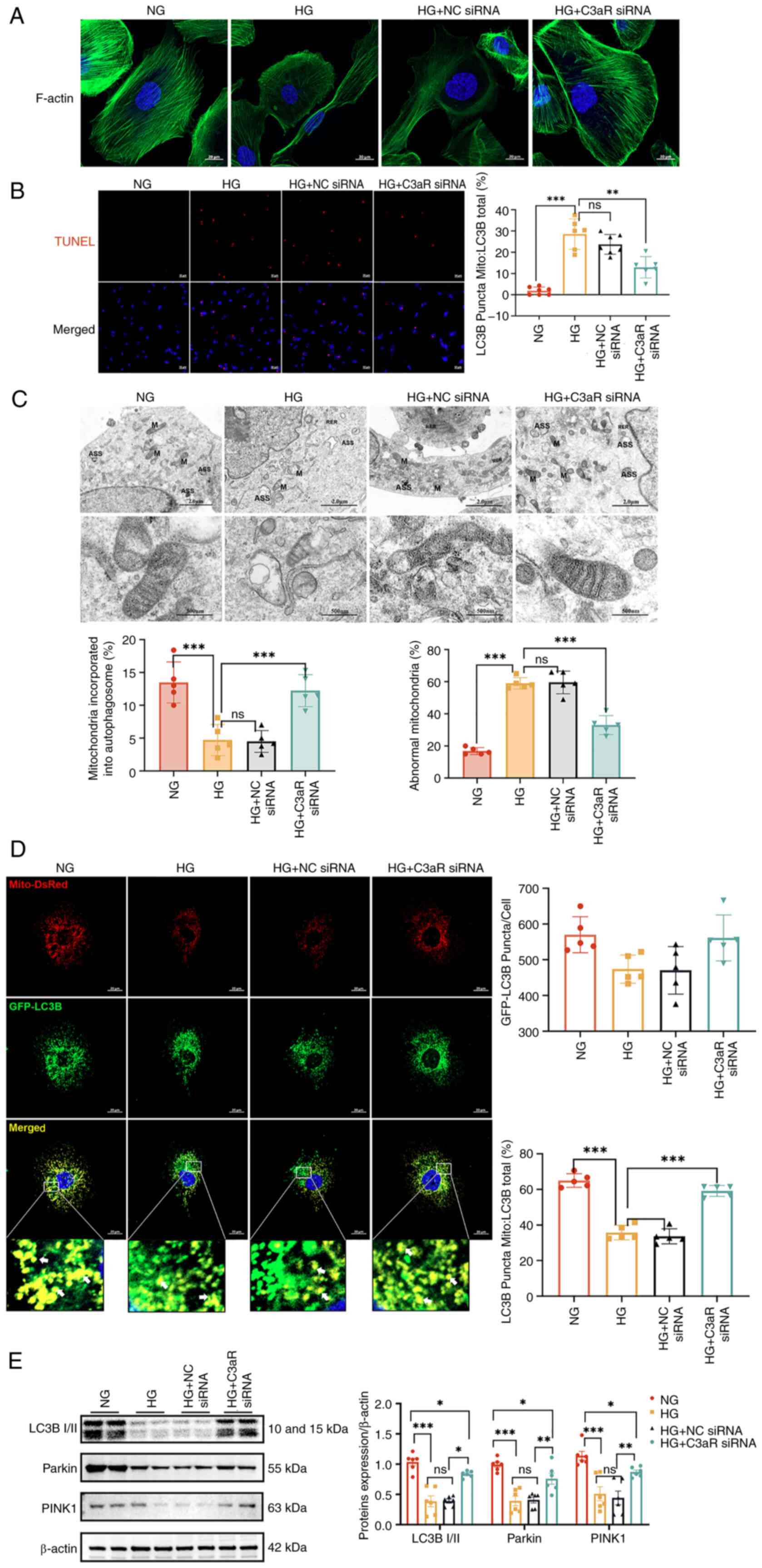
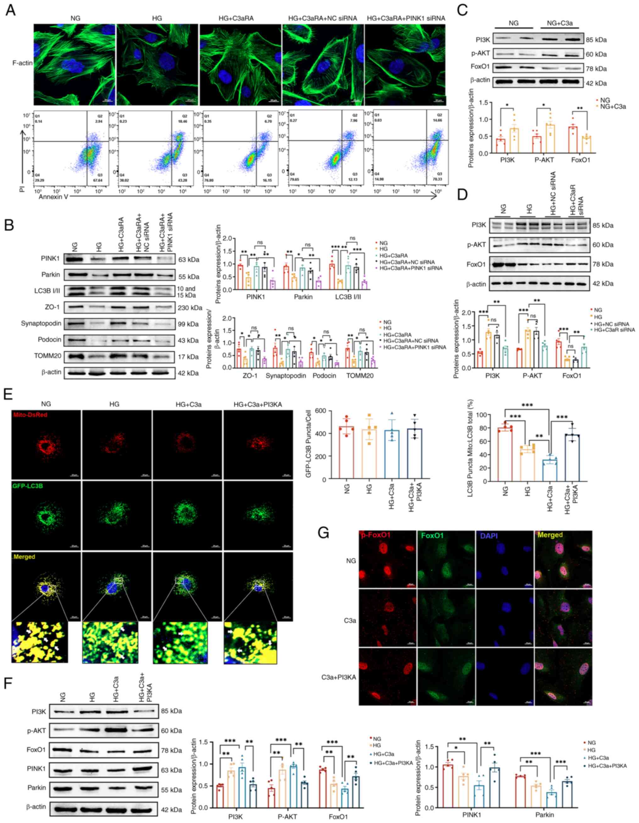
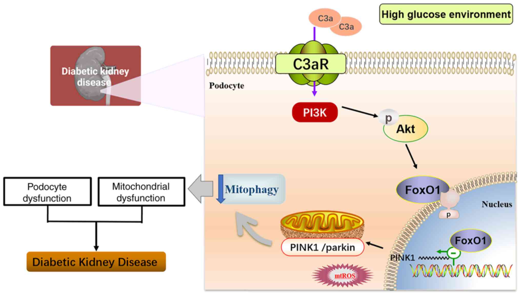
Related Articles

  • Abstract
  • View
  • Download
  • Twitter
Copy and paste a formatted citation
Spandidos Publications style
Weng M, Wu X, Rao S, Nie K, You D, Zheng T, Lin E, Zheng J, Cui J, Wan J, Wan J, et al: C3a/C3aR axis is involved in diabetic kidney injury by regulating podocyte mitophagy in diabetic nephropathy. Int J Mol Med 56: 223, 2025.
APA
Weng, M., Wu, X., Rao, S., Nie, K., You, D., Zheng, T. ... Wan, J. (2025). C3a/C3aR axis is involved in diabetic kidney injury by regulating podocyte mitophagy in diabetic nephropathy. International Journal of Molecular Medicine, 56, 223. https://doi.org/10.3892/ijmm.2025.5664
MLA
Weng, M., Wu, X., Rao, S., Nie, K., You, D., Zheng, T., Lin, E., Zheng, J., Cui, J., Wan, J."C3a/C3aR axis is involved in diabetic kidney injury by regulating podocyte mitophagy in diabetic nephropathy". International Journal of Molecular Medicine 56.6 (2025): 223.
Chicago
Weng, M., Wu, X., Rao, S., Nie, K., You, D., Zheng, T., Lin, E., Zheng, J., Cui, J., Wan, J."C3a/C3aR axis is involved in diabetic kidney injury by regulating podocyte mitophagy in diabetic nephropathy". International Journal of Molecular Medicine 56, no. 6 (2025): 223. https://doi.org/10.3892/ijmm.2025.5664
Copy and paste a formatted citation
x
Spandidos Publications style
Weng M, Wu X, Rao S, Nie K, You D, Zheng T, Lin E, Zheng J, Cui J, Wan J, Wan J, et al: C3a/C3aR axis is involved in diabetic kidney injury by regulating podocyte mitophagy in diabetic nephropathy. Int J Mol Med 56: 223, 2025.
APA
Weng, M., Wu, X., Rao, S., Nie, K., You, D., Zheng, T. ... Wan, J. (2025). C3a/C3aR axis is involved in diabetic kidney injury by regulating podocyte mitophagy in diabetic nephropathy. International Journal of Molecular Medicine, 56, 223. https://doi.org/10.3892/ijmm.2025.5664
MLA
Weng, M., Wu, X., Rao, S., Nie, K., You, D., Zheng, T., Lin, E., Zheng, J., Cui, J., Wan, J."C3a/C3aR axis is involved in diabetic kidney injury by regulating podocyte mitophagy in diabetic nephropathy". International Journal of Molecular Medicine 56.6 (2025): 223.
Chicago
Weng, M., Wu, X., Rao, S., Nie, K., You, D., Zheng, T., Lin, E., Zheng, J., Cui, J., Wan, J."C3a/C3aR axis is involved in diabetic kidney injury by regulating podocyte mitophagy in diabetic nephropathy". International Journal of Molecular Medicine 56, no. 6 (2025): 223. https://doi.org/10.3892/ijmm.2025.5664
Follow us
  • Twitter
  • LinkedIn
  • Facebook
About
  • Spandidos Publications
  • Careers
  • Cookie Policy
  • Privacy Policy
How can we help?
  • Help
  • Live Chat
  • Contact
  • Email to our Support Team