Spandidos Publications Logo
  • About
    • About Spandidos
    • Aims and Scopes
    • Abstracting and Indexing
    • Editorial Policies
    • Reprints and Permissions
    • Job Opportunities
    • Terms and Conditions
    • Contact
  • Journals
    • All Journals
    • Oncology Letters
      • Oncology Letters
      • Information for Authors
      • Editorial Policies
      • Editorial Board
      • Aims and Scope
      • Abstracting and Indexing
      • Bibliographic Information
      • Archive
    • International Journal of Oncology
      • International Journal of Oncology
      • Information for Authors
      • Editorial Policies
      • Editorial Board
      • Aims and Scope
      • Abstracting and Indexing
      • Bibliographic Information
      • Archive
    • Molecular and Clinical Oncology
      • Molecular and Clinical Oncology
      • Information for Authors
      • Editorial Policies
      • Editorial Board
      • Aims and Scope
      • Abstracting and Indexing
      • Bibliographic Information
      • Archive
    • Experimental and Therapeutic Medicine
      • Experimental and Therapeutic Medicine
      • Information for Authors
      • Editorial Policies
      • Editorial Board
      • Aims and Scope
      • Abstracting and Indexing
      • Bibliographic Information
      • Archive
    • International Journal of Molecular Medicine
      • International Journal of Molecular Medicine
      • Information for Authors
      • Editorial Policies
      • Editorial Board
      • Aims and Scope
      • Abstracting and Indexing
      • Bibliographic Information
      • Archive
    • Biomedical Reports
      • Biomedical Reports
      • Information for Authors
      • Editorial Policies
      • Editorial Board
      • Aims and Scope
      • Abstracting and Indexing
      • Bibliographic Information
      • Archive
    • Oncology Reports
      • Oncology Reports
      • Information for Authors
      • Editorial Policies
      • Editorial Board
      • Aims and Scope
      • Abstracting and Indexing
      • Bibliographic Information
      • Archive
    • Molecular Medicine Reports
      • Molecular Medicine Reports
      • Information for Authors
      • Editorial Policies
      • Editorial Board
      • Aims and Scope
      • Abstracting and Indexing
      • Bibliographic Information
      • Archive
    • World Academy of Sciences Journal
      • World Academy of Sciences Journal
      • Information for Authors
      • Editorial Policies
      • Editorial Board
      • Aims and Scope
      • Abstracting and Indexing
      • Bibliographic Information
      • Archive
    • International Journal of Functional Nutrition
      • International Journal of Functional Nutrition
      • Information for Authors
      • Editorial Policies
      • Editorial Board
      • Aims and Scope
      • Abstracting and Indexing
      • Bibliographic Information
      • Archive
    • International Journal of Epigenetics
      • International Journal of Epigenetics
      • Information for Authors
      • Editorial Policies
      • Editorial Board
      • Aims and Scope
      • Abstracting and Indexing
      • Bibliographic Information
      • Archive
    • Medicine International
      • Medicine International
      • Information for Authors
      • Editorial Policies
      • Editorial Board
      • Aims and Scope
      • Abstracting and Indexing
      • Bibliographic Information
      • Archive
  • Articles
  • Information
    • Information for Authors
    • Information for Reviewers
    • Information for Librarians
    • Information for Advertisers
    • Conferences
  • Language Editing
Spandidos Publications Logo
  • About
    • About Spandidos
    • Aims and Scopes
    • Abstracting and Indexing
    • Editorial Policies
    • Reprints and Permissions
    • Job Opportunities
    • Terms and Conditions
    • Contact
  • Journals
    • All Journals
    • Biomedical Reports
      • Information for Authors
      • Editorial Policies
      • Editorial Board
      • Aims and Scope
      • Abstracting and Indexing
      • Bibliographic Information
      • Archive
    • Experimental and Therapeutic Medicine
      • Information for Authors
      • Editorial Policies
      • Editorial Board
      • Aims and Scope
      • Abstracting and Indexing
      • Bibliographic Information
      • Archive
    • International Journal of Epigenetics
      • Information for Authors
      • Editorial Policies
      • Editorial Board
      • Aims and Scope
      • Abstracting and Indexing
      • Bibliographic Information
      • Archive
    • International Journal of Functional Nutrition
      • Information for Authors
      • Editorial Policies
      • Editorial Board
      • Aims and Scope
      • Abstracting and Indexing
      • Bibliographic Information
      • Archive
    • International Journal of Molecular Medicine
      • Information for Authors
      • Editorial Policies
      • Editorial Board
      • Aims and Scope
      • Abstracting and Indexing
      • Bibliographic Information
      • Archive
    • International Journal of Oncology
      • Information for Authors
      • Editorial Policies
      • Editorial Board
      • Aims and Scope
      • Abstracting and Indexing
      • Bibliographic Information
      • Archive
    • Medicine International
      • Information for Authors
      • Editorial Policies
      • Editorial Board
      • Aims and Scope
      • Abstracting and Indexing
      • Bibliographic Information
      • Archive
    • Molecular and Clinical Oncology
      • Information for Authors
      • Editorial Policies
      • Editorial Board
      • Aims and Scope
      • Abstracting and Indexing
      • Bibliographic Information
      • Archive
    • Molecular Medicine Reports
      • Information for Authors
      • Editorial Policies
      • Editorial Board
      • Aims and Scope
      • Abstracting and Indexing
      • Bibliographic Information
      • Archive
    • Oncology Letters
      • Information for Authors
      • Editorial Policies
      • Editorial Board
      • Aims and Scope
      • Abstracting and Indexing
      • Bibliographic Information
      • Archive
    • Oncology Reports
      • Information for Authors
      • Editorial Policies
      • Editorial Board
      • Aims and Scope
      • Abstracting and Indexing
      • Bibliographic Information
      • Archive
    • World Academy of Sciences Journal
      • Information for Authors
      • Editorial Policies
      • Editorial Board
      • Aims and Scope
      • Abstracting and Indexing
      • Bibliographic Information
      • Archive
  • Articles
  • Information
    • For Authors
    • For Reviewers
    • For Librarians
    • For Advertisers
    • Conferences
  • Language Editing
Login Register Submit
  • This site uses cookies
  • You can change your cookie settings at any time by following the instructions in our Cookie Policy. To find out more, you may read our Privacy Policy.

    I agree
Search articles by DOI, keyword, author or affiliation
Search
Advanced Search
presentation
International Journal of Molecular Medicine
Join Editorial Board Propose a Special Issue
Print ISSN: 1107-3756 Online ISSN: 1791-244X
Journal Cover
February-2026 Volume 57 Issue 2

Full Size Image

Sign up for eToc alerts
Recommend to Library

Journals

International Journal of Molecular Medicine

International Journal of Molecular Medicine

International Journal of Molecular Medicine is an international journal devoted to molecular mechanisms of human disease.

International Journal of Oncology

International Journal of Oncology

International Journal of Oncology is an international journal devoted to oncology research and cancer treatment.

Molecular Medicine Reports

Molecular Medicine Reports

Covers molecular medicine topics such as pharmacology, pathology, genetics, neuroscience, infectious diseases, molecular cardiology, and molecular surgery.

Oncology Reports

Oncology Reports

Oncology Reports is an international journal devoted to fundamental and applied research in Oncology.

Experimental and Therapeutic Medicine

Experimental and Therapeutic Medicine

Experimental and Therapeutic Medicine is an international journal devoted to laboratory and clinical medicine.

Oncology Letters

Oncology Letters

Oncology Letters is an international journal devoted to Experimental and Clinical Oncology.

Biomedical Reports

Biomedical Reports

Explores a wide range of biological and medical fields, including pharmacology, genetics, microbiology, neuroscience, and molecular cardiology.

Molecular and Clinical Oncology

Molecular and Clinical Oncology

International journal addressing all aspects of oncology research, from tumorigenesis and oncogenes to chemotherapy and metastasis.

World Academy of Sciences Journal

World Academy of Sciences Journal

Multidisciplinary open-access journal spanning biochemistry, genetics, neuroscience, environmental health, and synthetic biology.

International Journal of Functional Nutrition

International Journal of Functional Nutrition

Open-access journal combining biochemistry, pharmacology, immunology, and genetics to advance health through functional nutrition.

International Journal of Epigenetics

International Journal of Epigenetics

Publishes open-access research on using epigenetics to advance understanding and treatment of human disease.

Medicine International

Medicine International

An International Open Access Journal Devoted to General Medicine.

Journal Cover
February-2026 Volume 57 Issue 2

Full Size Image

Sign up for eToc alerts
Recommend to Library

  • Article
  • Citations
    • Cite This Article
    • Download Citation
    • Create Citation Alert
    • Remove Citation Alert
    • Cited By
  • Similar Articles
    • Related Articles (in Spandidos Publications)
    • Similar Articles (Google Scholar)
    • Similar Articles (PubMed)
  • Download PDF
  • Download XML
  • View XML

  • Supplementary Files
    • Supplementary_Data.pdf
Article Open Access

Recombinant ADAMTS‑1 promotes muscle regeneration accompanied by downregulation of Notch signaling

  • Authors:
    • Jin-Hwa Kim
    • Sang-Hyup Lee
    • Sang-Yoon Kim
    • Jeong-Won Kim
    • Ji-Soo Jeong
    • Eun-Hye Chung
    • Su-Ha Lee
    • Chang-Yeop Kim
    • Bong-Keun Choi
    • Je-Won Ko
    • Tae-Won Kim
  • View Affiliations / Copyright

    Affiliations: Department of Veterinary Medicine, College of Veterinary Medicine (BK21 FOUR Program), Chungnam National University, Daejeon 34131, Republic of Korea, Nuon Bio Co., Ltd., Seongnam, Gyeonggi 13201, Republic of Korea
    Copyright: © Kim et al. This is an open access article distributed under the terms of Creative Commons Attribution License.
  • Article Number: 47
    |
    Published online on: December 22, 2025
       https://doi.org/10.3892/ijmm.2025.5718
  • Expand metrics +
Metrics: Total Views: 0 (Spandidos Publications: | PMC Statistics: )
Metrics: Total PDF Downloads: 0 (Spandidos Publications: | PMC Statistics: )
Cited By (CrossRef): 0 citations Loading Articles...

This article is mentioned in:


Abstract

Skeletal muscle satellite cells (MuSCs) play a central role in muscle regeneration; however, their capacity declines with age, contributing to sarcopenia. A disintegrin and metalloproteinase with thrombospondin motifs‑1 (ADAMTS‑1) regulates MuSC activation and differentiation. The present study aimed to investigate the potential of recombinant ADAMTS‑1 (rADAMTS‑1) as a therapeutic strategy to enhance MuSC proliferation and improve regeneration. After barium chloride injection, mice received daily intraperitoneal injections of rADAMTS‑1 at 5 or 10 mg/kg for 1, 3, 7, or 14 days to monitor recovery. Primary skeletal muscle and C2C12 cells were also treated with rADAMTS‑1 to evaluate its effects on gene and protein expression during proliferation and differentiation in vitro. The number of MuSCs and the expression of myogenic markers increased in all injured groups by day 3 post‑injury in vivo. These levels were particularly elevated in the high‑dose rADAMTS‑1 group and remained sustained until day 14. Grip strength recovered to normal levels by day 7 in the high‑dose rADAMTS‑1 group, suggesting improved functional recovery compared with the untreated controls. In vitro, rADAMTS‑1 treatment induced a dose‑dependent increase in muscle fiber length and upregulation of regeneration‑related factors in primary skeletal muscle cells. Furthermore, C2C12 cells treated with rADAMTS‑1 exhibited enhanced expression of myocyte developmental genes during differentiation. The findings highlighted the therapeutic potential of rADAMTS‑1 for sarcopenia, potentially addressing limitations associated with conventional MuSC‑based treatments.
View Figures

Figure 1

Effects of rADAMTS-1 on recovery
following BaCl2-induced TA muscle injury. Groups
included NC, BaCl2, rADAMTS-1 treatment groups (L) and
(H). Daily intraperitoneal injections of rADAMTS-1 (L) or (H) were
initiated immediately after injury and continued until the
designated endpoint. (A) Grip strength measurements, normalized to
body weight, were recorded throughout the experimental period.
Statistical significance was determined using two-way analysis of
variance as follows: ###P<0.001 vs. BaCl2
group at day 0; ††P<0.01 and †††P<0.001
vs. rADAMTS-1 (L) at day 0; ‡‡P<0.01 vs. rADAMTS-1
(H) at day 0. (B-E) Grip strength at 1, 3, 7, and 14 dpi. Each dot
represents an individual mouse, with error bars indicating the
standard deviation. Statistical significance was determined using
one-way analysis of variance, followed by Tukey's post hoc test;
*P<0.05, **P<0.01 and
***P<0.001 vs. NC group; #P<0.05 and
##P<0.01 vs. BaCl2 group;
†P<0.05 vs. rADAMTS-1 (L). (F) Representative
histological images of TA muscle sections stained with hematoxylin
and eosin (scale bar, 60 μm) at the corresponding time
points. ADAMTS-1, a disintegrin and metalloproteinase with
thrombospondin motifs 1; rADAMTS-1, recombinant ADAMTS-1;
BaCl2, barium chloride; TA, tibialis anterior; NC,
non-injured control; rADAMTS-1 (L), rADAMTS-1 at 5 mg/kg; rADAMTS-1
(H), rADAMTS-1 at 10 mg/kg.

Figure 2

Effect of rADAMTS-1 on MuSCs
following BaCl2-induced TA muscle injury. Groups
included NC, BaCl2, and two treatment groups receiving
rADAMTS-1 (L) and (H). Daily intraperitoneal injections of
rADAMTS-1 were initiated immediately after injury and continued
until the designated time point for analysis. (A) Flow cytometry
analysis of MuSCs isolated from injured TA muscles, defined by
surface marker expression
(CD45−/CD31−/Sca1−/VCAM1+).
(B) Representative gating strategy for MuSCs across experimental
groups at different time points. (C) Relative number of MuSCs
normalized to the NC group over time. Statistical significance was
determined using one-way analysis of variance, followed by Tukey's
post hoc test: *P<0.05, **P<0.01 and
***P<0.001 vs. NC; ##P<0.01 and
###P<0.001 vs. BaCl2 group. (D)
Representative immunofluorescence images of Pax7+ cells
in injured TA muscle at different time points (scale bar, 25
μm). (E) Quantification of Pax7+ cell percentages
over time. Statistical significance was determined using one-way
analysis of variance with Tukey's post hoc test:
*P<0.05 and ***P<0.001 vs. NC;
###P<0.001 vs. BaCl2;
†††P<0.001 vs. rADAMTS-1 (L). ADAMTS-1, a disintegrin
and metalloproteinase with thrombospondin motifs 1; rADAMTS-1,
recombinant ADAMTS-1; MuSCs, skeletal muscle satellite cells;
BaCl2, barium chloride; TA, tibialis anterior; NC,
non-injured control; rADAMTS-1 (L), rADAMTS-1 at 5 mg/kg; rADAMTS-1
(H), rADAMTS-1 at 10 mg/kg; CD, cluster of differentiation; Sca1,
stem cell antigen 1; VCAM1, vascular cell adhesion molecule; Pax7,
paired box protein.

Figure 3

Effects of rADAMTS-1 on the
expression of ADAMTS-1-associated markers in
BaCl2-injured TA muscle. Groups included NC,
BaCl2, rADAMTS-1 treatment groups (L), and (H). Daily
intraperitoneal injections of rADAMTS-1 were initiated immediately
after injury and continued until the designated time point for
analysis. (A) Temporal expression patterns of ADAMTS-1, NICD, and
Hes-1 in the NC and BaCl2 groups at 1, 3, 7, and 14 days
post-injury (dpi). Comparative expression levels of (B) ADAMTS-1,
(C) NICD, and (D) Hes-1 among treatment groups [NC,
BaCl2, rADAMTS-1 (L), and rADAMTS-1 (H)] at each time
point. Mice received daily intraperitoneal injections of rADAMTS-1
until the designated dpi. Each data point represents an individual
biological replicate; error bars indicate standard deviation.
Statistical significance in panels (A) was determined using one-way
analysis of variance, followed by Tukey's honestly significant
difference post hoc test, as indicated as follows:
**P<0.01 and ***P<0.001 vs. 1 dpi;
##P<0.01 and ###P<0.001 vs. 3 dpi. For
panels (B-D), statistical comparisons were made using one-way
analysis of variance with Tukey's post hoc test:
*P<0.05, **P<0.01 and
***P<0.001 vs. NC; #P<0.05
##P<0.01 and ###P<0.001 vs.
BaCl2; †P<0.05, ††P<0.01 and
†††P<0.001 vs. rADAMTS-1 (L). ADAMTS-1, a disintegrin
and metalloproteinase with thrombospondin motifs 1; rADAMTS-1,
recombinant ADAMTS-1; BaCl2, barium chloride; TA,
tibialis anterior; NC, non-injured control; rADAMTS-1 (L),
rADAMTS-1 at 5 mg/kg; rADAMTS-1 (H), rADAMTS-1 at 10 mg/kg; NICD,
Notch intracellular domain; Hes-1, hairy and enhancer of split-1;
dpi, days post-injury.

Figure 4

Effects of rADAMTS-1 on muscle
regeneration markers in BaCl2-injured TA muscle. Groups
included NC, BaCl2, rADAMTS-1 treatment groups (L), and
(H). Daily intraperitoneal injections were given starting
immediately after injury and maintained until the designated
analysis point. (A) Representative immunohistochemical staining of
eMyHC in injured TA muscles treated with rADAMTS-1 at 1, 3, 7, and
14 days dpi (scale bar, 60 μm). (B-E) Quantification was
performed using image analysis software to assess the
eMyHC-positive area at each time point. (F) Representative
immunofluorescence images of MyoD-positive cells in injured TA
muscle (scale bar, 25 μm) treated with rADAMTS-1 at 1, 3, 7,
and 14 dpi. (G) Quantification of MyoD-positive cell percentages
over time across groups. All data are presented as mean ± standard
deviation. Statistical significance was determined using one-way
analysis of variance, followed by Tukey's post hoc test:
*P<0.05, **P<0.01 and
***P<0.001 vs. NC group; #P<0.05
##P<0.01 and ###P<0.001 vs.
BaCl2 group; †P<0.05 and
†††P<0.001 vs. rADAMTS-1 (L) group. ADAMTS-1, a
disintegrin and metalloproteinase with thrombospondin motifs 1;
rADAMTS-1, recombinant ADAMTS-1; BaCl2, barium chloride;
TA, tibialis anterior; NC, non-injured control; rADAMTS-1 (L),
rADAMTS-1 at 5 mg/kg; rADAMTS-1 (H), rADAMTS-1 at 10 mg/kg; eMyHC,
embryonic myosin heavy chain; dpi, days post-injury; MyoD, myoblast
determination protein 1; dpi, days post-injury.

Figure 5

Effects of rADAMTS-1 on early
myogenic markers in BaCl2-injured TA muscle. Groups
included NC, BaCl2, rADAMTS-1 treatment groups (L), and
(H). Daily intraperitoneal injections of rADAMTS-1 were
administered at two doses starting immediately after injury and
continuing until the designated time point of analysis. Temporal
expression of (A) MyoD and MyoG in the injured TA at 1, 3, 7, and
14 dpi. Comparative expression of (B) MyoD and (C) MyoG across
experimental groups at each time point. Each data point represents
an individual replicate; error bars indicate standard deviation.
Statistical significance in (A) was determined using one-way
analysis of variance, followed by Tukey's post hoc test and is
indicated as follows: *P<0.05 and
***P<0.001 vs. 1 dpi; ##P<0.01 and
###P<0.001 vs. 3 dpi; †††P<0.001 vs. 7
dpi. Statistical significance in (B) and (C) is indicated as
follows: *P<0.05, **P<0.01 and
***P<0.001 vs. the NC group; #P<0.05
##P<0.01 and ###P<0.001 vs. the
BaCl2 group; †P<0.05 and
††P<0.01 vs. the rADAMTS-1 (L) group. ADAMTS-1, a
disintegrin and metalloproteinase with thrombospondin motifs 1;
rADAMTS-1, recombinant ADAMTS-1; BaCl2, barium chloride;
TA, tibialis anterior; NC, non-injured control; rADAMTS-1 (L),
rADAMTS-1 at 5 mg/kg; rADAMTS-1 (H), rADAMTS-1 at 10 mg/kg; MyoD,
myoblast determination protein 1; MyoG, myogenin; dpi, days
post-injury.

Figure 6

Effects of rADAMTS-1 on proliferation
and differentiation of primary skeletal muscle cells. (A)
Dose-dependent proliferative effects of rADAMTS-1 (0.001-10 ng/ml)
on primary skeletal muscle cells, assessed using the
bromodeoxyuridine assay. (B) Dose-dependent effect of rADAMTS-1
(0.001-10 ng/ml) on the differentiation of primary skeletal muscle
cells. Quantification of myotube length following differentiation
(scale bar, 200 μm). (C) Western blot analysis showing the
expression of myogenic and ADAMTS-1 related markers (MyoD, MyoG,
ADAMTS-1, NICD, Hes-1, and β-actin) following rADAMTS-1 treatment
at a concentration of 10 ng/ml, indicating its promotion of
early-stage myogenic differentiation through inhibition of NICD
signaling. Each data point represents an individual measurement;
error bars indicate standard deviation. Statistical significance
was determined using one-way analysis of variance followed by
Tukey's honestly significant difference post hoc test and is
indicated as follows: (A) *P<0.05 vs. Con group; (B)
**P<0.01 and ***P<0.001 vs. Con group;
(C) *P<0.05 and **P<0.01 vs. Con group.
ADAMTS-1, a disintegrin and metalloproteinase with thrombospondin
motifs 1; rADAMTS-1, recombinant ADAMTS-1; MyoD, myoblast
determination protein 1; MyoG, myogenin; NICD, Notch intracellular
domain; HES-1, hairy and enhancer of split-1; Con, control.

Figure 7

Transcriptomic analysis and GO
enrichment of muscle cell differentiation following rADAMTS-1
treatment. C2C12 cells were treated with 10 ng/ml rADAMTS-1 and
differentiated for 3 days before RNA-seq analysis. (A) Volcano plot
showing DEGs. Blue and red dots indicate significantly upregulated
and downregulated DEGs, respectively. (B) Validation of selected
DEGs from RNA-sequencing by quantitative PCR. Results are presented
as mean ± SD. Statistical significance was determined using one-way
analysis of variance with Tukey's post hoc test and is indicated as
follows: *P<0.05 and **P<0.01 vs. the
control group. GO enrichment analysis of the top 20 terms
associated with (C) Biological Process, (D) Cellular Component and
(E) Molecular Function, based on the identified DEGs. GO, Gene
Ontology; ADAMTS-1, a disintegrin and metalloproteinase with
thrombospondin motifs 1; rADAMTS-1, recombinant ADAMTS-1; RNA-seq,
RNA sequencing; DEG, differentially expressed genes.
View References

1 

Tournadre A, Vial G, Capel F, Soubrier M and Boirie Y: Sarcopenia. Joint Bone Spine. 86:309–314. 2019. View Article : Google Scholar

2 

Larsson L, Degens H, Li M, Salviati L, Lee YI, Thompson W, Kirkland JL and Sandri M: Sarcopenia: Aging-related loss of muscle mass and function. Physiol Rev. 99:427–511. 2019. View Article : Google Scholar :

3 

Cruz-Jentoft AJ and Sayer AA: Sarcopenia. Lancet. 393:2636–2646. 2019. View Article : Google Scholar : PubMed/NCBI

4 

Petrocelli JJ, Mahmassani ZS, Fix DK, Montgomery JA, Reidy PT, McKenzie AI, de Hart NM, Ferrara PJ, Kelley JJ, Eshima H, et al: Metformin and leucine increase satellite cells and collagen remodeling during disuse and recovery in aged muscle. FASEB J. 35:e218622021. View Article : Google Scholar : PubMed/NCBI

5 

Kim HJ, Jung DW and Williams DR: Age is just a number: Progress and obstacles in the discovery of new candidate drugs for sarcopenia. Cells. 12:26082023. View Article : Google Scholar : PubMed/NCBI

6 

Huo F, Liu Q and Liu H: Contribution of muscle satellite cells to sarcopenia. Front Physiol. 13:8927492022. View Article : Google Scholar : PubMed/NCBI

7 

Schmidt M, Schüler SC, Hüttner SS, von Eyss B and von Maltzahn J: Adult stem cells at work: Regenerating skeletal muscle. Cell Mol Life Sci. 76:2559–2570. 2019. View Article : Google Scholar : PubMed/NCBI

8 

Haroon M, Boers HE, Bakker AD, Bloks NGC, Hoogaars WMH, Giordani L, Musters RJP, Deldicque L, Koppo K, Le Grand F, et al: Reduced growth rate of aged muscle stem cells is associated with impaired mechanosensitivity. Aging (Albany NY). 14:28–53. 2022. View Article : Google Scholar : PubMed/NCBI

9 

Qazi TH, Duda GN, Ort MJ, Perka C, Geissler S and Winkler T: Cell therapy to improve regeneration of skeletal muscle injuries. J Cachexia Sarcopenia Muscle. 10:501–516. 2019. View Article : Google Scholar : PubMed/NCBI

10 

Judson RN and Rossi FMV: Towards stem cell therapies for skeletal muscle repair. NPJ Regen Med. 5:102020. View Article : Google Scholar : PubMed/NCBI

11 

Du H, Shih CH, Wosczyna MN, Mueller AA, Cho J, Aggarwal A, Rando TA and Feldman BJ: Macrophage-released ADAMTS1 promotes muscle stem cell activation. Nat Commun. 8:6692017. View Article : Google Scholar : PubMed/NCBI

12 

Mourikis P, Sambasivan R, Castel D, Rocheteau P, Bizzarro V and Tajbakhsh S: A critical requirement for notch signaling in maintenance of the quiescent skeletal muscle stem cell state. Stem Cells. 30:243–252. 2012. View Article : Google Scholar

13 

Lee SH, Kim SY, Gwon YG, Lee C, Cho IH, Kim TW and Choi BK: Recombinant ADAMTS1 promotes muscle cell differentiation and alleviates muscle atrophy by repressing NOTCH1. BMB Rep. 57:539–545. 2024. View Article : Google Scholar : PubMed/NCBI

14 

Tarum J, Degens H, Turner MD, Stewart C, Sale C and Santos L: Modelling skeletal muscle ageing and repair in vitro. J Tissue Eng Regen Med. 2023:98022352023. View Article : Google Scholar : PubMed/NCBI

15 

Pawlikowski B, Betta ND, Antwine T and Olwin BB: Skeletal muscle stem cell self-renewal and differentiation kinetics revealed by EdU lineage tracing during regeneration. bioRxiv. https://doi.org/10.1101/627851.

16 

Al Shoyaib A, Archie SR and Karamyan VT: Intraperitoneal route of drug administration: Should it be used in experimental animal studies? Pharm Res. 37:122019. View Article : Google Scholar : PubMed/NCBI

17 

Ibraheem D, Elaissari A and Fessi H: Administration strategies for proteins and peptides. Int J Pharm. 477:578–589. 2014. View Article : Google Scholar : PubMed/NCBI

18 

Takeshita H, Yamamoto K, Nozato S, Inagaki T, Tsuchimochi H, Shirai M, Yamamoto R, Imaizumi Y, Hongyo K, Yokoyama S, et al: Modified forelimb grip strength test detects aging-associated physiological decline in skeletal muscle function in male mice. Sci Rep. 7:423232017. View Article : Google Scholar : PubMed/NCBI

19 

Ghaibour K, Rizk J, Ebel C, Ye T, Philipps M, Schreiber V, Metzger D and Duteil D: An efficient protocol for CUT&RUN analysis of FACS-isolated mouse satellite cells. J Vis Exp. 197:e652152023.

20 

Gilda JE and Gomes AV: Stain-Free total protein staining is a superior loading control to β-actin for Western blots. Anal Biochem. 440:186–188. 2013. View Article : Google Scholar : PubMed/NCBI

21 

Fortes MA, Marzuca-Nassr GN, Vitzel KF, da Justa Pinheiro CH, Newsholme P and Curi R: Housekeeping proteins: How useful are they in skeletal muscle diabetes studies and muscle hypertrophy models? Anal Biochem. 504:38–40. 2016. View Article : Google Scholar : PubMed/NCBI

22 

Paul RG, Hennebry AS, Elston MS, Conaglen JV and McMahon CD: Regulation of murine skeletal muscle growth by STAT5B is age- and sex-specific. Skelet Muscle. 9:192019. View Article : Google Scholar : PubMed/NCBI

23 

Wang R, Kumar B, Doud EH, Mosley AL, Alexander MS, Kunkel LM and Nakshatri H: Skeletal muscle-specific overexpression of miR-486 limits mammary tumor-induced skeletal muscle functional limitations. Mol Ther Nucleic Acids. 28:231–248. 2022. View Article : Google Scholar : PubMed/NCBI

24 

Livak KJ and Schmittgen TD: Analysis of relative gene expression data using real-time quantitative PCR and the 2(-Delta Delta C(T)) Method. Methods. 25:402–408. 2001. View Article : Google Scholar

25 

Forcina L, Cosentino M and Musarò A: Mechanisms regulating muscle regeneration: Insights into the interrelated and time-dependent phases of tissue healing. Cells. 9:12972020. View Article : Google Scholar : PubMed/NCBI

26 

Choo HJ, Canner JP, Vest KE, Thompson Z and Pavlath GK: A tale of two niches: Differential functions for VCAM-1 in satellite cells under basal and injured conditions. Am J Physiol Cell Physiol. 313:C392–C404. 2017. View Article : Google Scholar : PubMed/NCBI

27 

Gustafsson T and Ulfhake B: Aging skeletal muscles: What are the mechanisms of age-related loss of strength and muscle mass, and can we impede its development and progression? Int J Mol Sci. 25:109322024. View Article : Google Scholar : PubMed/NCBI

28 

Xing HY, Liu N and Zhou MW: Satellite cell proliferation and myofiber cross-section area increase after electrical stimulation following sciatic nerve crush injury in rADAMTS-1. Chin Med J (Engl). 133:1952–1960. 2020. View Article : Google Scholar : PubMed/NCBI

29 

Meng J, Lv Z, Chen X, Sun C, Jin C, Ding K and Chen C: LBP1C-2 from Lycium barbarum maintains skeletal muscle satellite cell pool by interaction with FGFR1. iScience. 26:1065732023. View Article : Google Scholar : PubMed/NCBI

30 

Agarwal M, Sharma A, Kumar P, Kumar A, Bharadwaj A, Saini M, Kardon G and Mathew SJ: Myosin heavy chain-embryonic regulates skeletal muscle differentiation during mammalian development. Development. 147:dev1845072020. View Article : Google Scholar : PubMed/NCBI

31 

Wang Y, Xiao Y, Zheng Y, Yang L and Wang D: An anti-ADAMTS1 treatment relieved muscle dysfunction and fibrosis in dystrophic mice. Life Sci. 281:1197562021. View Article : Google Scholar : PubMed/NCBI

32 

Mierzejewski B, Grabowska I, Michalska Z, Zdunczyk K, Zareba F, Irhashava A, Chrzaszcz M, Patrycy M, Streminska W, Janczyk-Ilach K, et al: SDF-1 and NOTCH signaling in myogenic cell differentiation: The role of miRNA10a, 425, and 5100. Stem Cell Res Ther. 14:2042023. View Article : Google Scholar : PubMed/NCBI

33 

Buas MF and Kadesch T: Regulation of skeletal myogenesis by Notch. Exp Cell Res. 316:3028–3033. 2010. View Article : Google Scholar : PubMed/NCBI

34 

Luo D, de Morree A, Boutet S, Quach N, Natu V, Rustagi A and Rando TA: Deltex2 represses MyoD expression and inhibits myogenic differentiation by acting as a negative regulator of Jmjd1c. Proc Natl Acad Sci USA. 114:E3071–E3080. 2017. View Article : Google Scholar : PubMed/NCBI

35 

Isesele PO and Mazurak VC: Regulation of skeletal muscle satellite cell differentiation by Omega-3 polyunsaturated fatty acids: A critical review. Front Physiol. 12:6820912021. View Article : Google Scholar : PubMed/NCBI

36 

Adhikari A, Kim W and Davie J: Myogenin is required for assembly of the transcription machinery on muscle genes during skeletal muscle differentiation. PLoS One. 16:e02456182021. View Article : Google Scholar : PubMed/NCBI

37 

Faralli H and Dilworth FJ: Turning on myogenin in muscle: A paradigm for understanding mechanisms of tissue-specific gene expression. Comp Funct Genomics. 2012:8363742012. View Article : Google Scholar :

38 

Owens J, Moreira K and Bain G: Characterization of primary human skeletal muscle cells from multiple commercial sources. In Vitro Cell Dev Biol Anim. 49:695–705. 2013. View Article : Google Scholar : PubMed/NCBI

39 

Ding R, Horie M, Nagasaka S, Ohsumi S, Shimizu K, Honda H, Nagamori E, Fujita H and Kawamoto T: Effect of cell-extracellular matrix interaction on myogenic characteristics and artificial skeletal muscle tissue. J Biosci Bioeng. 130:98–105. 2020. View Article : Google Scholar : PubMed/NCBI

40 

Sanvee GM, Bouitbir J and Krähenbühl S: C2C12 myoblasts are more sensitive to the toxic effects of simvastatin than myotubes and show impaired proliferation and myotube formation. Biochem Pharmacol. 190:1146492021. View Article : Google Scholar : PubMed/NCBI

41 

Henderson CA, Gomez CG, Novak SM, Mi-Mi L and Gregorio CC: Overview of the muscle cytoskeleton. Compr Physiol. 7:891–944. 2017. View Article : Google Scholar : PubMed/NCBI

42 

Lehka L and Rędowicz MJ: Mechanisms regulating myoblast fusion: A multilevel interplay. Semin Cell Dev Biol. 104:81–92. 2020. View Article : Google Scholar

43 

Lazure F, Blackburn DM, Corchado AH, Sahinyan K, Karam N, Sharanek A, Nguyen D, Lepper C, Najafabadi HS, Perkins TJ, et al: Myf6/MRF4 is a myogenic niche regulator required for the maintenance of the muscle stem cell pool. EMBO Rep. 21:e494992020. View Article : Google Scholar : PubMed/NCBI

44 

Krampert M, Kuenzle S, Thai SN, Lee N, Iruela-Arispe ML and Werner S: ADAMTS1 proteinase is up-regulated in wounded skin and regulates migration of fibroblasts and endothelial cells. J Biol Chem. 280:23844–23852. 2005. View Article : Google Scholar : PubMed/NCBI

45 

Dungan CM, Murach KA, Zdunek CJ, Tang ZJ, Nolt GL, Brightwell CR, Hettinger Z, Englund DA, Liu Z, Fry CS, et al: Deletion of SA β-Gal+ cells using senolytics improves muscle regeneration in old mice. Aging Cell. 21:e135282022. View Article : Google Scholar

46 

Always SE, Paez HG, Pitzer CR, Ferrandi PJ, Khan MM, Mohamed JS, Carson JA and Deschenes MR: Mitochondria transplant therapy improves regeneration and restoration of injured skeletal muscle. J Cachexia Sarcopenia Muscle. 14:493–507. 2023. View Article : Google Scholar

47 

Kim JW, Manickam R, Sinha P, Xuan W, Huang J, Awad K, Brotto M and Tipparaju SM: P7C3 ameliorates barium chloride-induced skeletal muscle injury activating transcriptomic and epigenetic modulation of myogenic regulatory factors. J Cell Physiol. 239:e313462024. View Article : Google Scholar : PubMed/NCBI

48 

Wang YX and Rudnicki MA: Sataellite cells, the engines of muscle repair. Nat Rev Mol Cell Biol. 13:127–133. 2011. View Article : Google Scholar : PubMed/NCBI

49 

Kerr HL, Krumm K, Anderson B, Christiani A, Strait L, Li T, Irwin B, Jiang S, Rybachok A, Chen A, et al: Mouse sarcopenia model reveals sex-and age-specific differences in phenotypic and molecular characteristics. J Clin Invest. 134:e1728902024. View Article : Google Scholar

50 

Owen AM and Fry CS: Decoding the decline: Unveiling drivers of sarcopenia. J Clin Invest. 134:e1833022024. View Article : Google Scholar : PubMed/NCBI

51 

Tao L, Huang W, Li Z, Wang W, Lei X, Chen J, Song X, Lu F, Fan S and Zhang L: Transcriptome analysis of differentially expressed genes and molecular pathways involved in C2C12 cells myogenic differentiation. Mol Biotechnol. 67:3640–3655. 2025. View Article : Google Scholar
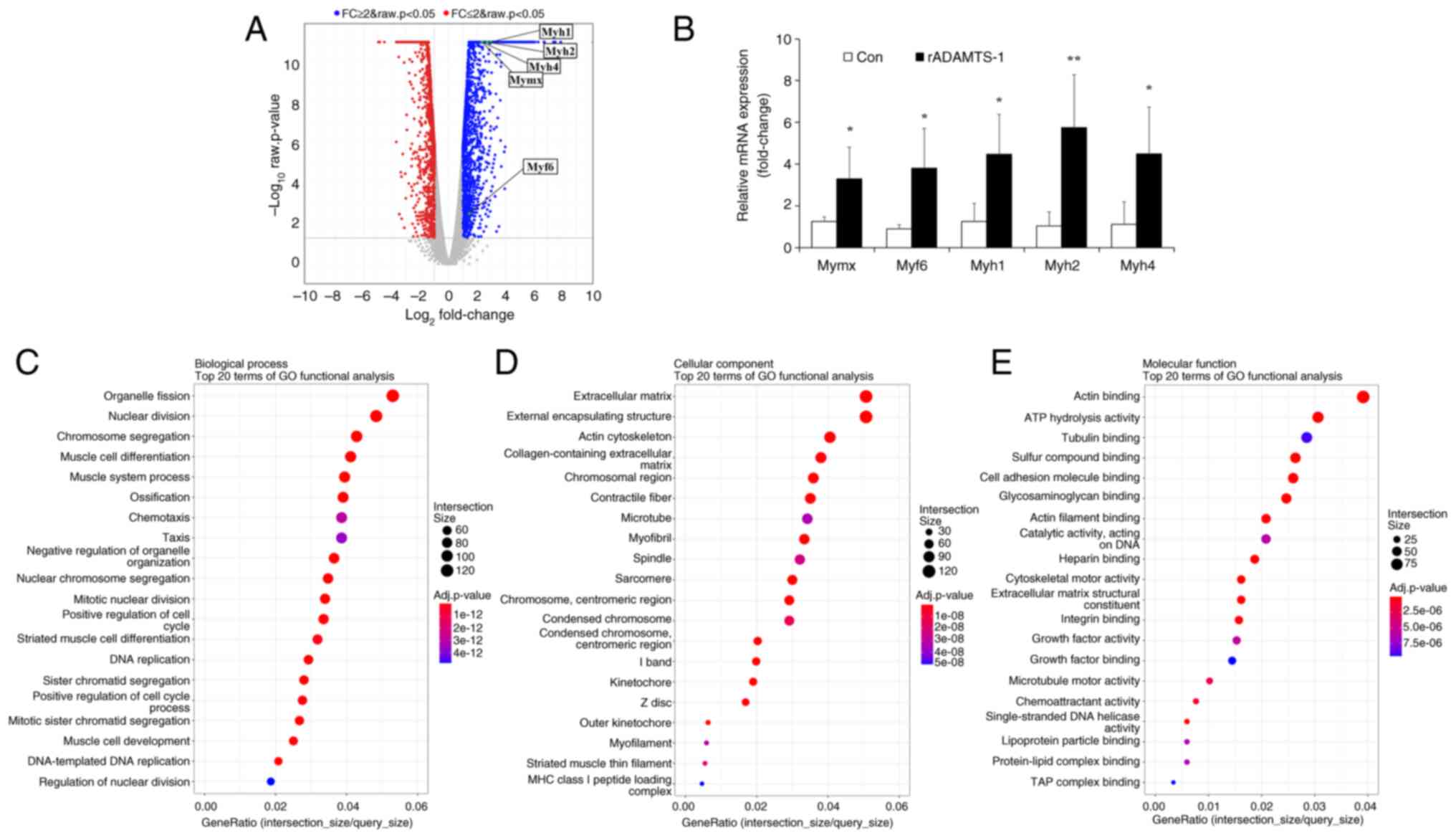
Related Articles

  • Abstract
  • View
  • Download
  • Twitter
Copy and paste a formatted citation
Spandidos Publications style
Kim J, Lee S, Kim S, Kim J, Jeong J, Chung E, Lee S, Kim C, Choi B, Ko J, Ko J, et al: Recombinant ADAMTS‑1 promotes muscle regeneration accompanied by downregulation of Notch signaling. Int J Mol Med 57: 47, 2026.
APA
Kim, J., Lee, S., Kim, S., Kim, J., Jeong, J., Chung, E. ... Kim, T. (2026). Recombinant ADAMTS‑1 promotes muscle regeneration accompanied by downregulation of Notch signaling. International Journal of Molecular Medicine, 57, 47. https://doi.org/10.3892/ijmm.2025.5718
MLA
Kim, J., Lee, S., Kim, S., Kim, J., Jeong, J., Chung, E., Lee, S., Kim, C., Choi, B., Ko, J., Kim, T."Recombinant ADAMTS‑1 promotes muscle regeneration accompanied by downregulation of Notch signaling". International Journal of Molecular Medicine 57.2 (2026): 47.
Chicago
Kim, J., Lee, S., Kim, S., Kim, J., Jeong, J., Chung, E., Lee, S., Kim, C., Choi, B., Ko, J., Kim, T."Recombinant ADAMTS‑1 promotes muscle regeneration accompanied by downregulation of Notch signaling". International Journal of Molecular Medicine 57, no. 2 (2026): 47. https://doi.org/10.3892/ijmm.2025.5718
Copy and paste a formatted citation
x
Spandidos Publications style
Kim J, Lee S, Kim S, Kim J, Jeong J, Chung E, Lee S, Kim C, Choi B, Ko J, Ko J, et al: Recombinant ADAMTS‑1 promotes muscle regeneration accompanied by downregulation of Notch signaling. Int J Mol Med 57: 47, 2026.
APA
Kim, J., Lee, S., Kim, S., Kim, J., Jeong, J., Chung, E. ... Kim, T. (2026). Recombinant ADAMTS‑1 promotes muscle regeneration accompanied by downregulation of Notch signaling. International Journal of Molecular Medicine, 57, 47. https://doi.org/10.3892/ijmm.2025.5718
MLA
Kim, J., Lee, S., Kim, S., Kim, J., Jeong, J., Chung, E., Lee, S., Kim, C., Choi, B., Ko, J., Kim, T."Recombinant ADAMTS‑1 promotes muscle regeneration accompanied by downregulation of Notch signaling". International Journal of Molecular Medicine 57.2 (2026): 47.
Chicago
Kim, J., Lee, S., Kim, S., Kim, J., Jeong, J., Chung, E., Lee, S., Kim, C., Choi, B., Ko, J., Kim, T."Recombinant ADAMTS‑1 promotes muscle regeneration accompanied by downregulation of Notch signaling". International Journal of Molecular Medicine 57, no. 2 (2026): 47. https://doi.org/10.3892/ijmm.2025.5718
Follow us
  • Twitter
  • LinkedIn
  • Facebook
About
  • Spandidos Publications
  • Careers
  • Cookie Policy
  • Privacy Policy
How can we help?
  • Help
  • Live Chat
  • Contact
  • Email to our Support Team