Introduction
Kidney disease refers to structural or functional
impairment of the kidney caused by various factors and is the
seventh leading cause of death globally. It has been indicated that
~850 million individuals worldwide suffer from kidney disease, with
projections suggesting that 5.4 million individuals will require
renal replacement therapy by 2030. Furthermore, 15-20% of patients
succumb within 12 months of initiating dialysis, posing a severe
threat to human health and imposing a substantial economic burden
(1). Currently, the precise
mechanisms underlying kidney disease remain unclear, and specific
therapeutic approaches are lacking. Therefore, exploring renal
physiological and pathological mechanisms and developing novel
treatment strategies are urgently needed. However, the complex and
highly differentiated cellular composition of the kidney poses
challenges for investigating the molecular mechanisms of kidney
disease.
In recent years, the advancement of single-cell
omics technologies has enabled the study of kidney disease
mechanisms at the cellular level. Through high-throughput
sequencing and high-precision analysis at the single-cell level,
this technology has revealed distinct cell subpopulations and
functional alterations in kidney diseases, mapping the renal cell
landscape. These findings provide novel perspectives for
investigating kidney cell development, ageing, and patterns of
change under disease conditions (2-4).
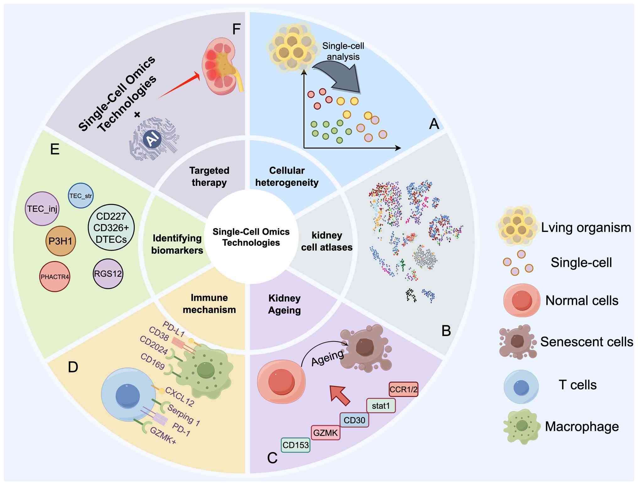
Accordingly, the present review summarizes the application of
single-cell omics technologies in acute kidney injury (AKI),
diabetic nephropathy (DN), IgA nephropathy (IgAN) and lupus
nephritis (LN). The aim of the present study was to provide novel
insights for elucidating the mechanisms of kidney diseases and
identifying therapeutic targets.
Overview of single-cell omics
technologies
Single-cell omics technologies collectively refer to
a class of techniques capable of performing multidimensional
molecular analysis on individual cells. They primarily encompass
single-cell transcriptomics, single-cell proteomics (SCP),
single-cell metabolomics (SCMET) and single-cell spatial
transcriptomics. Over the past decade, these technologies have
undergone continuous updates and iterations, enabling rapid and
precise acquisition of multidimensional molecular information from
tens of thousands of cells. They have thus become among the most
influential technologies in the field of life sciences research. In
kidney research, these technologies are not applied in isolation
but form an integrated technical framework characterized by
'interconnectedness, complementary validation and synergistic
empowerment'. By integrating transcriptional, proteomic, metabolic
and spatial localization information from individual cells, they
collectively advance the identification of kidney cell identities,
spatial organization analysis, and functional output clarification.
This provides an unprecedented high-precision perspective for
elucidating kidney physiological functions and disease mechanisms
and identifying therapeutic targets (Fig. 1).
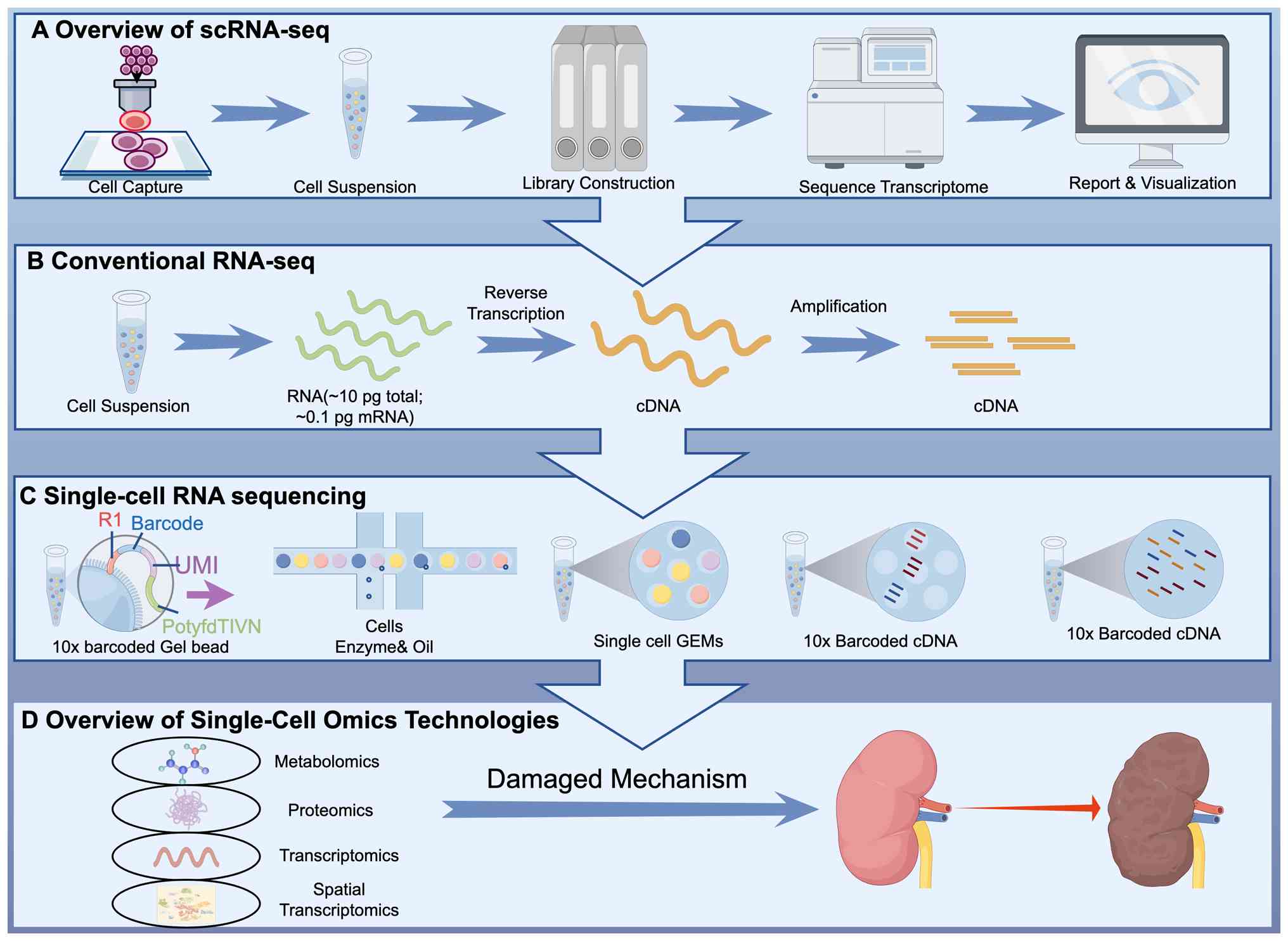 | Figure 1Development and applications of
single-cell omics technologies. (A) The single-cell sequencing
workflow includes cell capture, preparation and separation of
single-cell suspensions, library construction, sequencing and data
analysis. (B) Traditional sequencing technologies involve
sequencing pooled RNA from a population of cells, followed by batch
analysis to obtain average information about the cell population.
(C) Single-cell transcriptomics utilizes microfluidic chips to
encapsulate individual cells alongside gel beads bearing unique
barcodes and UMIs within oil droplets. Within these droplets, mRNA
released from lysed cells binds to primers on the gel beads,
triggering reverse transcription to generate barcode- and
UMI-labelled cDNA, which is subsequently amplified and sequenced.
(D) Metabolomics, proteomics, transcriptomics and spatial omics
technologies are collectively used to advance the exploration of
kidney disease mechanisms. UMIs, unique molecular identifiers. |
Single-cell transcriptomics
Single-cell RNA sequencing (scRNA-seq), as a
cutting-edge branch of transcriptomics, focuses on the types,
quantities, structures and functions of all transcript products
(mRNA and non-coding RNA) within an organism at the single-cell
level under specific time points and conditions. Its core advantage
lies in overcoming the 'averaging' limitations of traditional bulk
transcriptomics, enabling precise analysis of expression patterns
across diverse cell types within complex renal tissue. This makes
scRNA-seq a foundational platform for cell identity identification
and heterogeneity analysis in kidney research, providing essential
molecular clues for subsequent exploration of cellular regulatory
potential, tracking cell fate trajectories, and deciphering
intercellular interactions (5,6).
The primary workflow of scRNA-seq includes sample
collection and processing, single-cell suspension preparation and
isolation, RNA extraction and reverse transcription, library
construction, sequencing and data analysis (7). Among these steps, the preparation
and isolation of single-cell suspensions are critical for enhancing
efficiency and throughput. Iterations of this technology have
significantly advanced kidney research: Early microscopy or laser
capture methods allowed the analysis of only a few cells, failing
to capture kidney cell diversity; subsequent FACS and MACS
technologies enabled preliminary enrichment of kidney cell
subpopulations through automated sorting; and in October 2016, the
10x Genomics Chromium platform emerged. Leveraging microfluidic
chips, barcoding, and unique molecular identifier (UMI) technology
has enabled high-throughput, efficient, and cost-effective
sequencing of kidney cells. The 10x Genomics Chromium platform has
been widely applied in constructing kidney single-cell atlases,
mapping cellular developmental trajectories, studying rare cells,
and exploring immune mechanisms (8) and has become the mainstream
commercial platform for kidney single-cell research (9-11). In 2024, the scGRO-seq technology
developed by Mahat et al (12) employed click chemistry to capture
nascent RNA, enabling precise analysis of real-time gene expression
regulation in kidney cells. This addresses the limitations of
scRNA-seq in studying cellular transcriptional dynamics, providing
a new tool for elucidating transient regulatory mechanisms in
kidney cell function.
SCP
SCP, an innovative branch of proteomics, involves
the systematic investigation of the expression levels,
post-translational modifications, interaction networks, and
functional mechanisms of proteins expressed in individual cells,
tissues, or organisms under specific temporal and conditional
circumstances. It serves as a pivotal tool for deciphering the
molecular foundations of biological activities. Its core value lies
in validating transcriptional regulatory features identified by
scRNA-seq, revealing the functional implementation of cellular
regulatory potential, bridging the 'transcription-translation
disconnect' gap between transcriptomes and proteomes, and
simultaneously enabling the discovery of kidney cell-specific
protein biomarkers and the elucidation of protein interaction
networks. This provides direct evidence for elucidating kidney cell
functional outputs (13,14). Iterative advancements in SCP
technology have further enhanced its applicability in renal
research. In 2018, Budnik et al (15) developed SCP by mass spectrometry
(MS), which integrates isotope labelling and carrier proteomics to
achieve quantitative analysis of more than 1,000 proteins in mouse
embryonic stem cells, laying the technical foundation for renal
cell proteomics research. In 2025, Ye et al (16) introduced the innovative SCP
technology Chip-Tip. By integrating a novel chip with a liquid
chromatography system, it unified cell sample preparation, protein
extraction and MS analysis. This approach increased the number of
identifiable proteins in a single kidney cell from ~2,000 to
5,000-6,500, significantly enhancing the detection sensitivity for
protein profiles in rare kidney cell subpopulations. Subsequently,
the team developed single-cell pulsed stable isotope labelling,
which for the first time enabled precise analysis of protein
turnover rates in individual kidney cells. This breakthrough
provides a novel tool for investigating dynamic protein regulatory
mechanisms during renal cell damage repair, such as the synthesis
and degradation of proteins involved in tubular epithelial cell
repair, which represents a major advance in SCP.
SCMET
Metabolomics, a vital branch of systems biology,
involves the quantitative analysis of the dynamic changes in all
small-molecule metabolites (such as amino acids, carbohydrates,
lipids and nucleotides) within an organism. This approach reveals
the comprehensive metabolic network landscape under specific
physiological or pathological conditions, elucidating the
regulatory mechanisms of life activities (17,18). SCMET, an innovative branch of
metabolomics, primarily employs MS, nuclear magnetic resonance,
combined with microfluidics, laser capture microdissection, and
other techniques to isolate and extract small-molecule metabolites
from individual cells. Its core advantage lies in capturing
metabolic heterogeneity among renal cells, deciphering the
metabolic phenotypes ultimately resulting from cellular regulatory
potential, elucidating the functional output characteristics of
renal cells at the metabolic level, and providing a microscopic
perspective for studying renal physiological metabolic mechanisms
and disease-related metabolic disorders (19,20).
MS, with its high sensitivity, rapid speed and
label-free nature, serves as the core technology for SCMET. Among
these methods, matrix-assisted laser desorption/ionization MS
(MALDI-MS) is the most effective tissue imaging technique and has
been widely applied in renal tissue metabolic imaging studies
(21,22). Previous innovations in SCMET
technology have further expanded its applications in renal
research: Wang et al (23) innovatively combined isotope
labelling with MALDI-MS, overcoming the limitations of traditional
techniques in capturing dynamic metabolic processes. They
successfully traced the metabolic transition from glycolysis to
fatty acid β-oxidation during the differentiation of renal vesicles
into proximal tubules, revealing for the first time the functional
transformation mechanism during renal cell development at the
single-cell metabolic level. In 2024, Qin et al (24) developed the in-depth organic MS
flow cytometry technique. By optimizing lysis strategies and
extending the analysis time, more than 600 metabolites were
successfully identified within individual renal cells. This
achieved precise single-cell metabolite identification and isomer
quantification based on secondary MS, enabling accurate
differentiation of metabolic differences between distinct renal
cell subpopulations (for example, proximal vs. distal convoluted
tubule cells). In 2025, the team integrated organic MS flow
cytometry with stable isotope tracing to establish a
high-throughput, label-free, dynamic SCMET platform. This platform
was successfully applied to identify the polarization subtypes of
tumour-associated macrophages and is equally applicable for
studying the metabolic phenotypes of renal immune cells, providing
a novel tool for deciphering the metabolic regulatory mechanisms of
immune cells during renal inflammation (2).
Spatial transcriptomics
In 2016, the collaborative efforts of Ståhl et
al (25) first introduced
the concept of 'Spatial Transcriptomics (ST)'. The core
breakthrough of this technology lies in preserving the spatial
structural integrity of kidney tissue and addressing the
limitations of scRNA-seq, SCP and SCMET by preserving the spatial
integrity of kidney tissue. This enables simultaneous analysis of
'gene expression and spatial localization', providing a novel tool
for investigating intercellular interactions, spatial organization
patterns, and the spatial regulatory mechanisms underlying cellular
functional outputs in the kidney (26,27). It serves as a critical bridge
connecting single-cell molecular characteristics with kidney tissue
function.
ST technology integrates high-throughput sequencing
with spatial imaging, simultaneously capturing gene expression data
while preserving the spatial positioning of renal cells. This
precisely reveals the spatial distribution patterns of genes within
renal tissue, providing robust support for investigating dynamic
changes in the cellular microenvironment associated with kidney
diseases and elucidating disease mechanisms (12,28-30). Currently employed approaches
primarily fall into three categories: Microarray-based techniques,
bead-based techniques and microfluidic-based techniques. In
microarray-based techniques, spatially barcoded oligonucleotide
primers are fixed onto slides. After kidney tissue sections are
combined with the array, mRNA is captured via poly(A) tails or
probe hybridization, reverse-transcribed into spatially tagged
cDNA, and sequenced to obtain spatial expression data (31,32). In bead-based techniques,
including slide-seq (26) and
HDST (27), beads with unique
DNA barcodes are randomly distributed across kidney tissue
sections. By combining mRNA sequencing with bead localization,
these methods can be used to reconstruct spatial coordinates of
gene expression, enabling higher-resolution spatial mapping of
renal cells. In microfluidic-based techniques, microfluidic chips
are utilized to generate microchannels on the surface of kidney
tissue. Barcoded reagents are injected into specific regions,
enabling precise capture and sequencing of localized mRNA to
increase spatial resolution. In 2020, Liu et al (33) developed DBiT-seq technology,
which uses microfluidic channels to precisely position spatially
deterministic barcodes onto RNA and proteins. This technique
successfully mapped the fine structures of the retinal pigment
epithelium and microvascular endothelium in mouse embryos,
validating its high-resolution advantage (34). This technology can be further
applied to perform detailed spatial transcriptomics analysis of
distinct regions within kidney tissue, including glomeruli, tubules
and interstitial spaces, thereby deciphering the molecular
characteristics of kidney cell spatial organization.
In summary, single-cell omics represents a frontier
branch of systems biology. scRNA-seq can be employed to identify
kidney cell identities and reveal regulatory potential at the
transcriptional level; SCP can be used to validate regulatory
features at the protein level, bridging transcription and function;
SCMET can be used to decipher metabolic phenotypes, clarifying the
metabolic basis of functional outputs; and ST can be used to
supplement spatial information, elucidating cellular spatial
organization patterns and spatially specific interactions. These
four interconnected and complementary technical platforms integrate
multidimensional molecular information from individual cells,
collectively advancing a comprehensive and in-depth understanding
of renal cell identity, regulatory potential, spatial organization
and functional outputs. This propels kidney research from the 'cell
population level' to the 'single-cell precision level,' aiding in
the elucidation of renal physiological functions, disease mechanism
studies, and the identification of therapeutic targets. This drives
the paradigm shift in kidney disease diagnosis and treatment from
'empirical medicine' to 'molecular medicine'. In recent years,
these approaches have demonstrated significant application value in
studies of kidney cell proliferation, development, ageing and
disease states (Fig. 2).
Applications of single-cell omics
technologies in basic renal research
The rapid advancement of single-cell omics
technology has provided revolutionary tools for deeply analysing
the cellular composition and molecular mechanisms of the kidney.
High-resolution kidney cell atlases have been successfully
constructed, offering precise insights into renal physiological
functions and disease development. More importantly, this
technology has been extensively applied to study physiological and
pathological processes such as renal ageing, revealing dynamic
changes in the immune microenvironment during ageing and laying a
solid theoretical foundation for the prevention and treatment of
kidney diseases.
Application of single-cell omics
technology in constructing a renal cell atlas
The rapid growth of single-cell omics technology has
propelled the identification, localization and functional
characterization of renal cell types, leading to the successful
construction of a renal cell atlas. This technology holds profound
significance for deepening the understanding of renal physiology
and disease mechanisms and advancing precision medicine. In 2018,
Park et al (35)
performed single-cell sequencing on kidney cells from healthy mice,
identifying 16 cell types and establishing the first mouse kidney
cell atlas. They discovered a novel transitional cell type in the
collecting duct expressing both principal cell and intercalated
cell marker genes. Abedini et al (36) employed single-cell multi-omics
analysis of 81 human kidney tissue samples from 58 participants.
They precisely defined 44 major cell types and their
subpopulations, including glomeruli, tubules at various segments,
collecting ducts, endothelial cells and stromal cells, at the
molecular level, generating a comprehensive, spatially resolved
single-cell atlas of the human kidney. Li et al (37) combined single-cell
transcriptomics with spatial metabolomics to reveal distinct
transcriptional, epigenetic and metabolic signatures of the same
tubular cell type across cortical, medullary and papillary regions,
establishing a foundation for kidney disease research.
Application of single-cell omics
technologies in renal ageing mechanistic research
The vicious cycle of chronic inflammation triggered
by the accumulation of cellular senescence and immune cell ageing
represents a core mechanism exacerbating renal ageing and
inflammatory injury (38,39).
Single-cell omics technologies (centred on scRNA-Seq) serve as
critical research tools in this field, enabling the precise
identification of abnormal cell subtypes during renal ageing and
the identification of key regulatory factors. These technologies
provide essential technical support for elucidating the mechanisms
underlying renal ageing.
scRNA-Seq plays an irreplaceable role in studying
the vicious 'inflammation-ageing' cycle in the kidney. Lymphocyte
proliferation and tertiary lymphoid tissue (TLT) expansion are
closely linked to senescence-associated T cells and age-associated
B cells (40). By analysing
renal cells from aged mice with unilateral ischaemic-reperfusion
injury, scRNA-seq revealed that senescence-associated T cells
secrete factors that induce age-associated B-cell generation. This
process, coupled with CD153/CD30 signalling, drives TLT expansion
and exacerbates renal inflammation (41). Furthermore, scRNA-seq aided in
identifying GZMK as a key biomarker of renal ageing (42) and elucidated the enrichment
pattern of GZMK+CD8+ T cells in aged mouse
kidneys, along with their mechanism of accelerating renal cell
ageing through IFN-γ secretion (43). In macrophage-related studies,
scRNA-seq further revealed bidirectional regulation between
macrophages and renal ageing (44). Single-cell data from the kidneys
of mice of different ages and aged mice with nephrotoxic serum
nephritis (NTN) revealed that Stat1 upregulation in aged
macrophages suppressed Pcbp1 transcription, activated ferroptosis,
and promoted proinflammatory factor secretion, thereby accelerating
renal interstitial fibrosis (45). Moreover, abnormal expression of
CCR1, CCR2 and Fcγ receptor family members in the kidneys of aged
NTN mice led to renal immune dysregulation and exacerbated renal
injury (46,47). Additionally, scRNA-seq analysis
of clinical data revealed that p21high macrophages mitigate
transplant rejection in aged mice by upregulating Zfp36 expression
and downregulating IL-27 expression, offering novel insights for
kidney transplantation research (48).
In summary, using single-cell omics technologies to
construct renal cell atlases, precisely identify abnormal cell
subpopulations, and reveal key regulatory factors and molecular
biomarkers not only provides core technical support for elucidating
the pathogenesis of renal ageing and related diseases but also lays
the foundation for early disease diagnosis, target screening and
therapeutic optimization.
Applications of single-cell omics
technologies in kidney diseases
Leveraging single-cell resolution as its core
advantage, single-cell omics technologies enable precise analysis
of cellular heterogeneity in kidney tissue, mapping of
intercellular interaction networks, tracking of cellular fate
trajectories, and identification of core regulatory programs. This
technology overcomes the limitations of traditional bulk
sequencing, which cannot capture individual cellular molecular
characteristics. It provides unprecedented technical support for
studying the mechanisms of kidney diseases, discovering novel
non-invasive biomarkers, and screening potential therapeutic
targets. This lays the theoretical foundation for personalized
treatment and accelerates its clinical translation process
(Table I).
 | Table IResearch on kidney diseases based on
single-cell omics technologies. |
Table I
Research on kidney diseases based on
single-cell omics technologies.
| Author/s, year | Types of
disease | Research
subjects | Research
methods | Research
findings | (Refs.) |
|---|
| Hasegawa, 2022 | AKI | AKI mice | scRNA-seq | Between days 6 and
10 following AKI onset, there was a significant increase in the
number of tubular cells expressing inflammatory markers. | (52) |
| Klocke et
al, 2022 | | Urine samples from
32 AKI patients | scRNA-seq | TEC_inj and
TEC_str, which are associated with injury, significantly increased
during the mid-stage of the disease course (6-10 days). | (51) |
| Wagner et
al, 2025 | | Urine of patients
with AKI | scRNA-seq | The ratio of
CD227/CD326+ DTECs correlated with the acute estimated
glomerular filtration rate in patients with AKI. | (53) |
| Li et al,
2025 | | AKI mice | scRNA-seq | In AKI mice,
lactate inhibition of aldehyde dehydrogenase in proximal tubular
epithelial cells suppressed mitochondrial autophagy, exacerbated
mitochondrial dysfunction, and consequently worsened renal
injury. | (54) |
| Polonsky et
al, 2024; Cui et al, 2024 | | AKI mice | scRNA-seq, spatial
transcriptomics | Crlf1, Timp1, Npr3,
ISG15, and TGF-βR1 expression was significantly upregulated in
proximal tubule cells and surrounding fibroblasts. Among these,
ISG15 promoted TGF-βR1 ISGylation and inhibited its ubiquitination,
thereby exacerbating renal ibrosis. | (55,56) |
| Cai et al,
2025 | | AKI mice | scRNA-seq | Differential
expression of S100A8/A9 proteins in renal tubular epithelial cells
promoted renal macrophage polarization and activated the TNF
signalling pathway. | (58) |
| Zhang et al,
2024 | | AKI mice | scRNA-seq and
spatial transcriptomics | Mapping the Dynamic
Profile and Spatial Distribution Characteristics of Renal
Macrophages in the AKI-to-CKD Transition Model. | (57) |
| Yao et al,
2022 | | AKI mice | scRNA-seq | Significant
upregulation of S100A8/A9 expression occurred in the
S100a9hiLy6chiIMs subpopulation of renal macrophages, where high
expression of Ccl2 and Ccl3 genes mediated early inflammation in
AKI. | (59) |
| Qi et al,
2017 | DKD | Patients with
DKD | Single-cell
proteomics | The PKM2 activator
TEPP-46 increased glycolytic flux and upregulated Opa1 expression,
thereby reversing high-glucose-induced toxic glucose metabolite
accumulation and mitochondrial dysfunction, ultimately alleviating
renal injury. | (64) |
| Jiang et al,
2025 | | Patients with
DKD | scRNA-seq | LRPPRC exhibited
low expression levels in damaged glomerular and tubular regions,
with its expression levels negatively correlated with serum
creatinine levels and proteinuria incidence. | (65) |
| Fu et al,
2022; Wang et al, 2025 | | Patients with
DKD | scRNA-seq | Upregulating PPARα
expression effectively reduced M1 macrophage infiltration and
delayed the progression of DKD by inhibiting necrotic apoptosis via
the RIP1/RIP3/MLKL pathway in renal tubular cells. | (67,68) |
| Ji et al,
2024 | | Patients with
DKD | scRNA-seq | The
TGF-β1+Arg1+ macrophage subpopulation
promoted DKD renal fibrosis by activating the TGF-β1/Smad2/3/YAP
signalling pathway, thereby promoting fibroblast activation and
extracellular matrix deposition. | (69) |
| Chen et al,
2024 | | DKD mouse | scRNA-seq and
proteomics technologies | RA reduced the
recruitment of S100a4-positive macrophages, decreased oxidative
stress in NK cells, and alleviated damage to glomerular epithelial
cells. | (70) |
| Park et al,
2025 | | Type 2 diabetic
nephropathy mice | scRNA-seq | CXCL12 secreted by
proximal convoluted tubule cells interacted with glomeruli to
jointly promote T-cell recruitment, thereby driving localized
inflammatory responses in the kidney. | (71) |
| Zheng et al,
2020 | IgA | Patients with
IgAN | scRNA-seq | High expression of
J CHAIN in mesangial cells of IgAN patients promoted IgA1
dimerization and deposition, and enhanced interactions between
mesangial cells and endothelial cells, macrophages, and T
cells. | (74) |
| Zhang et al,
2025 | | IgAN | scRNA-seq Luminex
multiplex immunoassay technology | CX3CR1+
monocytes/macrophages induced mesangial cell injury through
interaction with macrophage migration inhibitory factor and its
receptor CD74 and activated the PI3K/AKT pathway, further
exacerbating mesangial cell damage. | (75) |
| Zambrano et
al, 2022 | | gddY mouse | SMART-seq2 | Genes associated
with endothelial dysfunction, such as Edn1, F8, and Nostrin, along
with MHC I genes mediating endothelial cell immune responses,
showed significantly upregulated expression. Their corresponding
receptors were also overexpressed in PTCs, where binding promotes
the release of inflammatory cytokines and exacerbates the extent of
damage in PTCs during the early stages of IgAN. | (76) |
| Hasegawa et
al, 2025; Chen et al, 2024 | | Patients and mice
with IgAN | scRNA-seq spatial
transcriptomics | Glomerular
endothelial cells express CCL2, CXCL2, and EDNRB, and interact with
macrophages via CXCL12/CXCR4. | (77,78) |
| Li et al,
2024 | LN | Urine samples from
patients with LN | Urine Proximity
Extension Assay proteomics and scRNA-seq | Among patients with
LN, urinary ICAM-2, FABP4, FASLG, IGFBP-2, SELE and TNFSF13B/BAFF
protein levels showed the strongest correlation with clinical
disease activity. | (84) |
| Su et al,
2025 | | Patients with
proliferative LN | scRNA-seq
immunohistochemical analysis | The presence of
CD68+ macrophages in glomeruli of patients with
proliferative LN increased the clinical response rate by 7.92-fold,
serving as a significant predictor of treatment response. | (85) |
| Tang et al,
2022 | | Patients with
LN | scRNA-seq immunopro
teomics | ICx such as prolyl
3-hydroxylase 1, phosphatase and actin regulator 4, and G protein
signal regulator 12 were significantly present in patients with
LN. | (86) |
| Zhang et al,
2022 | | Patients with
LN | Metabolomics based
on UPLC-MS/MS | SM d34:2, DG (18:3
(9Z, 12Z, 15Z)/20:5 (5Z, 8Z, 11Z, 14Z, 17Z)/0:0), nervonic acid,
Cer-NS d27:4, and PC (18:3 (6Z, 9Z, 12Z)/18:3 (6Z, 9Z, 12Z)) were
significantly enriched. | (87) |
| Danaher et
al, 2024 | | cLN | | Precise
identification of 11 immune cell types, clarifying the
subpopulation composition and transcriptomic characteristics of
each immune cell category. | (88) |
| Richoz et
al, 2022 | | LN mice | Spatial
transcriptomics | In LN mouse
kidneys, the MoMac subset highly expressed FcγR receptors,
internalized circulating ICs, and presented antigens. These cells
activated the NF-κB and STAT3 pathways, leading to the secretion of
the proinflammatory factors TNF-α/IL-1β. The TrMac subset secreted
monocyte chemotactic factors and expressed B-cell survival factor
BAFF, recruiting B cells and plasma cells to produce
autoantibodies. | (90) |
| Chen et al,
2024 | | Patients with
LN | scRNA-seq | iPTEC significantly
enriched CD163+ dendritic cells, which activated MHC-II
molecules and upregulated the expression of proinflammatory
cytokines (TNF, IL-1β) and T-cell-associated chemokines (CCL17,
CCL22). This resulted in a highly proinflammatory microenvironment,
exacerbating renal injury. | (91) |
| Gartshteyn et
al, 2024 | | Patients with
LN | scRNA-seq
dataset | SLAM-related
protein expression was significantly elevated in CD4+ T
cells. Upon binding to molecules within the lymphocyte activation
molecule family, they jointly regulated TFH and TPH function,
promoting TPH-dependent B-cell differentiation into plasma cells
and thereby driving autoantibody production. | (92) |
| Wu et al,
2025 | | scRNA-seq | Patients with
LN |
GZMK+CD8+ T cells
promoted ABC differentiation and antibody class switching by
secreting IFN-γ and IL-21, thereby accelerating renal inflammatory
responses. | (93) |
| Yamaguchi et
al, 2024 | | scRNA-seq | LN mice | Podocyte-specific
PI3Kα inhibitors not only suppressed activation of the AKT/mTOR
pathway in LN mouse podocytes but also impaired B-cell and T-cell
function, reducing production of inflammatory cytokines and
autoantibodies. | (94) |
Research advances in single-cell omics
for AKI diagnosis and treatment
AKI is a clinically complex syndrome with high
mortality rates. Its intricate pathogenesis and cellular
heterogeneity significantly impede therapeutic progress (49,50). Single-cell omics technologies
precisely capture novel cellular states and regulatory features
during AKI progression and are pivotal for resolving diagnostic and
therapeutic challenges. They offer novel approaches for early
diagnosis and targeted treatment, establishing themselves as core
technical support in AKI research.
Single-cell omics facilitates AKI
biomarker discovery
Single-cell omics technologies enable
high-throughput sequencing of non-invasive samples to decipher
single-cell molecular characteristics, overcoming the limitations
of traditional biomarkers and establishing non-invasive diagnostic
systems. Klocke et al (51) analysed urine samples from 32
patients with AKI via scRNA-seq and identified four novel
damage-associated states in tubular epithelial cells (TECs). The
urinary TEC transcriptome profile was highly concordant with that
of renal biopsy tissue, confirming its ability to dynamically
reflect renal injury. The study was the first to demonstrate that
urinary single-cell sequencing can replace invasive biopsy.
Hasegawa (52) pioneered
scRNA-seq to demonstrate that the number of inflammation-associated
tubular cells in urine from mice with AKI is correlated with injury
severity. Wagner et al (53) identified a
CD227/CD326+ DTEC subpopulation whose abundance was
correlated with the glomerular filtration rate, positioning it as a
novel non-invasive AKI biomarker. These studies, centred on
single-cell technology, provide a series of highly specific and
sensitive biomarkers for early diagnosis, disease monitoring and
prognosis' assessment.
Single-cell omics advances AKI
mechanistic research
Investigations into the mechanisms of tubular
epithelial cell injury using single-cell omics analyses have
revealed specific core regulatory programs and cellular state
changes associated with injury, confirming the pivotal role of
mitochondrial dysfunction. Li et al (54) utilized scRNA-seq to first
identify a specific regulatory program of ALDH2 lactylation
modification in the proximal tubular epithelial cells of mice with
AKI, which suppressed mitochondrial autophagy, exacerbated
mitochondrial dysfunction, and subsequently drove cellular injury.
Researchers further combined spatial transcriptomics to localize
and identify differentially expressed genes in the proximal
convoluted tubules and surrounding fibroblasts, confirming that
ISG15 regulates TGF-βR1 modification status and participates in the
transition from tubular injury to renal fibrosis (55,56).
In studies of immune-inflammatory-mediated kidney
injury, single-cell omics technologies have clearly resolved
macrophage heterogeneity, core regulatory mechanisms and
intercellular interaction networks for the first time. Zhang et
al (57) employed combined
scRNA-seq and spatial transcriptomics to map the dynamic profiles
and spatial distribution characteristics of renal macrophages in an
AKI-to-CKD progression model. They identified functional
differences between resident and monocyte-derived macrophages and
characterized a novel extracellular matrix (ECM)-remodelling
macrophage subpopulation. This subset communicates with fibroblasts
via the IGF signalling pathway. Spatial transcriptomics further
clarified the spatial distribution patterns of macrophages across
different stages of AKI. Through scRNA-seq, Cai et al
(58) revealed that abnormal
overexpression of S100A8/A9 in renal tubular epithelial cells
paracentrally regulates macrophage polarization, activating TNF
signalling to amplify inflammatory responses. Yao et al
(59) identified
S100a9hiLy6chi-infiltrating macrophages as a distinct
proinflammatory subset via scRNA-seq, whose early infiltration was
associated with tubular apoptosis. The application of single-cell
omics technologies has fundamentally reshaped the understanding of
the role of macrophages in AKI, providing novel therapeutic targets
for preventing and treating AKI and chronic transition. This fully
demonstrates their core value in renal immune-inflammatory
research.
Research advances the diagnosis and
treatment of diabetic kidney disease (DKD) using single-cell
omics
DKD is a form of glomerulosclerosis triggered by
metabolic abnormalities in patients with diabetes and is a major
cause of end-stage renal disease. Previous studies indicated that
30-40% of diabetic patients worldwide develop DKD, with more than
50% of patients with DKD ultimately requiring end-stage renal
replacement therapy, posing a significant threat to human health
(60,61). Single-cell omics technology
overcomes traditional research limitations by providing single-cell
resolution. It revolutionizes DKD research by elucidating the
dynamic states of cell types, core regulatory programs, and
intercellular interaction patterns during DKD progression, offering
core technical support for exploring its pathogenesis and
identifying therapeutic targets (62).
Application of single-cell omics in
decoding DKD metabolic mechanisms
Metabolic abnormalities serve as key drivers of DKD
progression. Renal metabolic dysregulation triggers oxidative
stress and inflammatory responses that impair renal structure and
function, exacerbate fibrosis, and accelerate disease progression
(63). Single-cell omics
precisely deciphers metabolism-related cellular regulatory programs
and fate trajectories in DKD.
Qi et al (64) employed SCP to analyse glomeruli
from patients with DKD at single-cell resolution. They identified
pyruvate kinase M2 as a core protective factor in DKD, whose
expression and activity were significantly reduced in patients and
negatively correlated with renal injury severity. Jiang et
al (65) employed spatial
transcriptomics to reveal low LRPPRC expression in the damaged
glomeruli and tubules of patients with DKD, which was negatively
correlated with serum creatinine levels and proteinuria incidence,
impairing the regulation of mitochondrial energy metabolism. By
combining these findings with those of pseudo-time trajectory
analysis, researchers first revealed that LOH and CT undergo
abnormal differentiation towards podocytes as DKD progresses. These
findings confirm that tubular energy metabolism disorders can
progressively damage glomerular podocytes by regulating cellular
fate trajectories, thereby exacerbating renal injury and driving
disease progression. This approach refines the metabolic pathogenic
mechanism of DKD from the 'metabolic regulation-cell
trajectory-spatial localization' dimension.
Application of single-cell omics in
elucidating DKD immunological mechanisms
Abnormal immune cell function significantly
contributes to worsening renal injury in patients with DKD.
Single-cell omics technologies provide critical support for
identifying DKD immunotherapy strategies and improving patient
outcomes by enabling the elucidation of immune cell heterogeneity,
core regulatory programs and intercellular interaction networks
(66).
Researchers have employed scRNA-seq to analyse
kidneys of mice with DKD and revealed for the first time that
compared with that in the kidneys of normal mice, the proportion of
M2 macrophages in the kidneys of DKD mice decreased to 3.8%, while
the proportion of proinflammatory M1 macrophages increased to
65.3%, confirming the dominance of proinflammatory macrophages in
DKD (67,68). Ji et al (69) employed scRNA-seq to identify a
novel TGF-β1+Arg1+ macrophage subset in
kidneys of mice with DKD. This subset was shown to exacerbate renal
fibrosis by activating the TGF-β1/Smad2/3/YAP pathway, promoting
fibroblast activation and ECM deposition. Chen et al
(70) combined scRNA-seq with
proteomics to reveal that rosmarinic acid mitigates DKD renal
injury by synergistically reducing inflammatory responses and
glomerular epithelial cell damage. This occurs because of the
suppression of S100a4-positive macrophage recruitment and the
alleviation of NK cell oxidative stress, offering a novel
immunomodulatory therapeutic approach for DKD. Furthermore, Park
et al (71) employed
single-cell omics to identify significantly elevated proportions of
NK and T cells in the renal tissues of mice with type 2 DN. They
demonstrated that CXCL12 secreted by proximal tubule cells forms
spatial interactions with glomerular cells, jointly promoting
T-cell recruitment and driving local renal inflammation. This
discovery provides novel therapeutic targets for restoring renal
immune homeostasis in patients with DKD. These studies, which
focused on single-cell omics technology, comprehensively elucidate
the core mechanisms of immune dysregulation in DKD, fully
demonstrating the pivotal value of this technology in DKD
immunology research.
Advances in single-cell omics for IgAN
diagnosis and treatment
IgAN is the most common primary glomerular disease
(72,73). Single-cell omics technology has
revealed core cellular interactions and regulatory programs,
driving breakthroughs in mechanistic research. Key findings include
the following:
Application of single-cell omics in
exploring the immunological mechanisms of IgAN
IgAN is characterized by IgA immune complex
deposition, and single-cell omics technologies have elucidated its
core immunological mechanisms. Zheng et al (74) identified a J CHAIN-upregulated
regulatory program in the mesangial cells of patients with IgAN via
scRNA-seq. This program promotes IgA1 deposition and intercellular
crosstalk, confirming the central regulatory role of mesangial
cells. Zhang et al (75)
combined scRNA-seq with Luminex technology to identify elevated
CX3CL1 and CX3CR1 expression in mesangial cells and
monocytes/macrophages. These molecules interact via MIF/CD74 to
induce mesangial cell injury and activate the PI3K/AKT pathway.
Zambrano et al (76)
employed SMART-seq2 to identify the greatest number of genes that
are differentially expressed in glomerular endothelial cells (GECs)
during early IgAN progression, revealing their inflammatory effects
through multiple molecular interactions with immune cells. Through
combined scRNA-seq and spatial transcriptomics (77,78), researchers have further revealed
that GECs spatially interact with macrophages via axes such as
CXCL12/CXCR4 and that the overexpression of these genes exacerbates
PTC damage. This clarifies the crucial role of 'GEC-PTC'
interactions, offering new insights for early intervention.
LN
Systemic lupus erythematosus (SLE) is a multisystem
autoimmune disease, and LN represents one of its most severe
complications (79). Studies
indicate that ~50% of SLE patients develop LN, with 10-30% of
patients progressing to end-stage kidney disease within 10 years of
onset (80-83). The core pathogenesis of LN
remains unclear, and its high degree of heterogeneity not only
complicates disease classification but also poses significant
challenges for clinical diagnosis, treatment selection and
prognosis assessment (80).
Single-cell omics technology, leveraging its core advantage of
single-cell resolution, has emerged as a pivotal tool for the
thorough exploration of LN heterogeneity, the elucidation of its
pathogenesis, and the development of novel diagnostic and
therapeutic strategies.
Single-cell omics technology
facilitates the identification of LN biomarkers
Non-invasive diagnosis, disease activity monitoring
and treatment response prediction for LN are critical for improving
patient outcomes. Traditional biomarkers suffer from limitations
such as low specificity and difficulty in distinguishing disease
subtypes and stages. By integration with multi-omics technologies,
single-cell omics can be used to precisely identify the cellular
origins of differentially expressed molecules and decipher their
regulatory mechanisms, thereby screening for highly specific and
sensitive non-invasive biomarkers. In a recent study, researchers
innovatively combined urine Proximity Extension Assay proteomics
with renal single-cell transcriptomics to precisely trace the renal
cellular origins of differentially expressed urinary proteins. They
identified six proteins most strongly correlated with LN activity,
establishing novel molecular markers to distinguish active from
patients with inactive LN. Single-cell technology further
elucidated their regulatory mechanisms, enhancing the clinical
reliability of these biomarkers (84). Su et al (85) further addressed the gap in
biomarkers for predicting treatment response in patients with LN.
Using scRNA-seq combined with immunostaining, they reported that
the incorporation of the proportion of CD68+ macrophages
significantly enhanced the prediction efficacy of a model for the
treatment response of patients with proliferative LN, outperforming
traditional models and providing novel cellular markers for precise
prognostic assessment. Tang et al (86) focused on identifying biomarkers
for early LN diagnosis. Through combined scRNA-seq and
immunoproteomic analysis, they discovered three novel circulating
immune complexes, P3H1, PHACTR4 and RGS12, that serve as potential
markers for early diagnosis and disease monitoring in LN. Building
upon single-cell technology research, Zhang et al (87) further integrated
ultrahigh-performance liquid chromatography-tandem MS metabolomics
to identify five serum metabolites specifically enriched in LN.
These metabolites efficiently distinguish SLE from LN, filling a
gap in biomarkers for differentiating between SLE and LN and
providing theoretical support for their clinical application. These
studies demonstrate that single-cell omics technologies, when
integrated with proteomics and metabolomics, can be used to
precisely identify a series of biomarkers suitable for non-invasive
diagnosis, disease monitoring, treatment response prediction, and
differential diagnosis of SLE-LN. The resolution advantages of
single-cell technologies enable the further clarification of
cellular origins, expression regulatory mechanisms, and
associations of these biomarkers with pathological processes.
Single-cell technologies are highly promising tools for the precise
clinical management of LN and have enabled the discovery of LN
biomarkers.
Single-cell omics technologies and
advances in research on the immunological mechanisms of LN
Immune-inflammatory responses serve as the core
driver of renal injury in patients with LN. LN kidney tissue
exhibits extensive immune cell infiltration and abnormal
activation. Complex interactions among diverse immune cells and
renal resident cells collectively constitute the local
proinflammatory microenvironment in LN kidneys, promoting the
progression of renal damage. Single-cell omics technologies have
improved the understanding of the immune pathogenesis of LN at the
single-cell and spatial levels, providing core evidence for the
development of targeted immunomodulatory therapeutic strategies.
Research by Danaher et al (88) laid a crucial foundation for
studying LN immune cell heterogeneity and spatial distribution.
Utilizing scRNA-seq technology, researchers performed single-cell
resolution analysis of renal tissue from patients with paediatric
LN, precisely identifying 11 immune cell types and clarifying the
subpopulation composition and transcriptomic characteristics of
each immune cell type. Further integration with spatial
transcriptomics revealed for the first time the functional
localization specificity of various immune cells within LN kidney
tissue. Myeloid cells (for example, macrophages) predominantly
enriched in the glomerular region exhibited significantly
upregulated expression of genes associated with antigen uptake,
lipid metabolism and inflammatory pathways, directly contributing
to glomerular inflammatory injury. Lymphocytes (for example, B
cells and T cells) predominantly cluster in the tubulointerstitial
region, forming a dense network of cellular interactions with
myeloid dendritic cells and macrophages to jointly alleviate the
dysregulation of the local immune microenvironment in LN kidneys.
The aforementioned study systematically revealed the distribution
patterns and functional characteristics of renal immune cells in
patients with LN through a three-dimensional approach of 'cellular
heterogeneity-spatial localization-functional association', fully
demonstrating the unique advantages of combining single-cell and
spatial transcriptomics in deciphering immune mechanisms in
patients with LN.
Single-cell omics technology serves as a core tool
for analysing the functions of myeloid and lymphoid cells in LN
pathogenesis. Myeloid cells serve as central regulators of renal
immune inflammation in LN. IgG immune complexes in the LN renal
mesangium activate myeloid cells, exacerbating renal injury.
Single-cell omics precisely revealed their subpopulation
heterogeneity and interaction mechanisms, confirming that
macrophages are core regulators capable of amplifying inflammation
(89). Notably, Richoz et
al (90) employed combined
scRNA-seq and spatial transcriptomics to first identify two
macrophage subpopulations, TrMac and MoMac, elucidating their
regulatory programs and synergistic pathogenic roles, thereby
providing novel targets for immune-directed therapies. Abnormal
activation of lymphocyte subsets lies at the core of LN immune
dysregulation. Single-cell omics confirmed that the proportion of
renal T and B cells in patients with LN is positively correlated
with disease severity (91).
Gartshteyn et al (92)
revealed via scRNA-seq that SAP, which is highly expressed in
CD4+ T cells, regulates TFH/TPH function, elucidating
the mechanism underlying the regulation of T-cell imbalance. Wu
et al (93) used
scRNA-seq to demonstrate that GZMK+CD8+ T
cells promote B-cell activation and antibody production, clarifying
the mechanism of abnormal renal B cells. Furthermore, scRNA-seq
confirmed that PIK3CA-mutated podocytes regulate lymphocyte
function (94), broadening the
understanding of LN pathogenesis.
In summary, single-cell technologies have revealed
novel therapeutic targets and immunomodulatory strategies,
providing core support for the precision treatment of LN.
Furthermore, the integration of single-cell omics with multi-omics
approaches has refined the LN biomarker system, offering innovative
tools for non-invasive diagnosis, disease monitoring, and treatment
response prediction. These findings provide crucial foundations for
subsequent mechanistic studies and clinical translation in LN.
Summary and outlook
In recent years, rapid advancements in single-cell
omics technologies have opened new perspectives for kidney disease
research, demonstrating unique advantages in revealing cellular
heterogeneity and elucidating disease mechanisms. However, this
technology faces numerous challenges, including inherent technical
errors, insufficient sensitivity for detecting low-abundance
molecules in rare kidney cells, complex data processing,
inconsistent standards and difficulties in capturing dynamic
cellular changes; moreover, spatial information cannot be
preserved, making deciphering the relationship between cell-cell
interactions and disease progression difficult. The emergence of
multi-omics technologies has effectively addressed the limitations
of single-cell omics through the systematic integration of
multilevel biological information such as transcriptomics and
proteomics. This enables a shift from single-molecule analysis to
multidimensional collaborative interpretation, further refining the
understanding of renal disease pathogenesis and laying a solid
foundation for personalized medicine. However, its application
still faces the following bottlenecks: Inefficient data integration
strategies, a lack of standardized research protocols, and a
pronounced disconnect between basic research and clinical
translation. Moving forwards, efforts should focus on advancing the
high-quality development and clinical application of single-cell
and multi-omics technologies in kidney disease research.
Technologically, this involves optimizing kidney single-cell
suspension preparation, promoting high-sensitivity sequencing, and
integrating single cells with spatial transcriptomics to address
gaps in detection and spatial information. For data processing and
analysis, kidney-specific standardized protocols should be
established, artificial intelligence should be leveraged for
efficient data handling, and disease biomarkers and targets should
be deeply mined. In application translation, multidisciplinary
collaboration should drive the conversion of omics discoveries into
clinical diagnostic and therapeutic tools. As technologies mature
and multidisciplinary integration advances, these approaches hold
promise for overcoming limitations, usher in new breakthroughs for
kidney disease prevention and treatment and propel research towards
precision and individualization.
Availability of data and materials
Not applicable.
Authors' contributions
JX wrote the original manuscript, revised and edited
the draft, and completed the creation of tables and figures. ZC,
SL, XW reviewed and edited the manuscript. MC participated in topic
selection, manuscript review, paper revision, and finalization. All
authors read and approved the final version of the manuscript. Data
authentication is not applicable.
Ethics approval and consent to
participate
Not applicable.
Patient consent for publication
Not applicable.
Competing interests
The authors declare that they have no competing
interests.
Acknowledgements
Not applicable.
Funding
The present study was supported by the National Natural Science
Foundation of China (grant no. 82274487).
References
|
1
|
Francis A, Harhay MN, Ong ACM,
Tummalapalli SL, Ortiz A, Fogo AB, Fliser D, Roy-Chaudhury P,
Fontana M, Nangaku M, et al: Chronic kidney disease and the global
public health agenda: an international consensus. Nat Rev Nephrol.
20:473–485. 2024. View Article : Google Scholar : PubMed/NCBI
|
|
2
|
Zhang Y, Shi M, Li M, Qin S, Miao D and
Bai Y: Dynamic single-cell metabolomics reveals cell-cell
interaction between tumor cells and macrophages. Nat Commun.
16:45822025. View Article : Google Scholar : PubMed/NCBI
|
|
3
|
Sharma K, Zhang G, Hansen J, Bjornstad P,
Lee HJ, Menon R, Hejazi L, Liu JJ, Franzone A, Looker HC, et al:
Endogenous adenine mediates kidney injury in diabetic models and
predicts diabetic kidney disease in patients. J Clin Invest.
133:e1703412023. View Article : Google Scholar : PubMed/NCBI
|
|
4
|
Alaterre E, Ovejero S, Bret C, Dutrieux L,
Sika D, Fernandez Perez R, Espéli M, Fest T, Cogné M, Martin-Subero
JI, et al: Integrative single-cell chromatin and transcriptome
analysis of human plasma cell differentiation. Blood. 144:496–509.
2024. View Article : Google Scholar : PubMed/NCBI
|
|
5
|
Potter SS: Single-cell RNA sequencing for
the study of development, physiology and disease. Nat Rev Nephrol.
14:479–492. 2018. View Article : Google Scholar : PubMed/NCBI
|
|
6
|
Cheng C, Chen W, Jin H and Chen X: A
review of single-cell RNA-Seq annotation, integration, and
cell-cell communication. Cells. 12:19702023. View Article : Google Scholar : PubMed/NCBI
|
|
7
|
Lee J, Hyeon DY and Hwang D: Single-cell
multiomics: Technologies and data analysis methods. Exp Mol Med.
52:1428–1442. 2020. View Article : Google Scholar : PubMed/NCBI
|
|
8
|
Salcher S, Heidegger I, Untergasser G,
Fotakis G, Scheiber A, Martowicz A, Noureen A, Krogsdam A, Schatz
C, Schäfer G, et al: Comparative analysis of 10X Chromium vs BD
Rhapsody whole transcriptome single-cell sequencing technologies in
complex human tissues. Heliyon. 10:e283582024. View Article : Google Scholar
|
|
9
|
Zheng GXY, Terry JM, Belgrader P, Ryvkin
P, Bent ZW, Wilson R, Ziraldo SB, Wheeler TD, McDermott GP, Zhu J,
et al: Massively parallel digital transcriptional profiling of
single cells. Nat Commun. 8:140492017. View Article : Google Scholar : PubMed/NCBI
|
|
10
|
Kim YJ and Takahashi K: Emerging
technologies of single-cell multi-omics. Haematologica.
110:1269–1277. 2025.PubMed/NCBI
|
|
11
|
Wang X, He Y, Zhang Q, Ren X and Zhang Z:
Direct comparative analyses of 10X genomics chromium and
smart-seq2. Genomics Proteomics Bioinformatics. 19:253–266. 2021.
View Article : Google Scholar : PubMed/NCBI
|
|
12
|
Mahat DB, Tippens ND, Martin-Rufino JD,
Waterton SK, Fu J, Blatt SE and Sharp PA: Single-cell nascent RNA
sequencing unveils coordinated global transcription. Nature.
631:216–223. 2024. View Article : Google Scholar : PubMed/NCBI
|
|
13
|
Lundberg E and Borner GHH: Spatial
proteomics: A powerful discovery tool for cell biology. Nat Rev Mol
Cell Biol. 20:285–302. 2019. View Article : Google Scholar : PubMed/NCBI
|
|
14
|
Demichev V, Szyrwiel L, Yu F, Teo GC,
Rosenberger G, Niewienda A, Ludwig D, Decker J, Kaspar-Schoenefeld
S, Lilley KS, et al: dia-PASEF data analysis using FragPipe and
DIA-NN for deep proteomics of low sample amounts. Nat Commun.
13:39442022. View Article : Google Scholar : PubMed/NCBI
|
|
15
|
Budnik B, Levy E, Harmange G and Slavov N:
SCoPE-MS: Mass spectrometry of single mammalian cells quantifies
proteome heterogeneity during cell differentiation. Genome Biol.
19:1612018. View Article : Google Scholar : PubMed/NCBI
|
|
16
|
Ye Z, Sabatier P, Van Der Hoeven L,
Lechner MY, Phlairaharn T, Guzman UH, Liu Z, Huang H, Huang M, Li
X, et al: Enhanced sensitivity and scalability with a Chip-Tip
workflow enables deep single-cell proteomics. Nat Methods.
22:499–509. 2025. View Article : Google Scholar : PubMed/NCBI
|
|
17
|
Vander Heiden MG and DeBerardinis RJ:
Understanding the intersections between metabolism and cancer
biology. Cell. 168:657–669. 2017. View Article : Google Scholar : PubMed/NCBI
|
|
18
|
Petrova B and Guler AT: Recent
developments in single-cell metabolomics by mass spectrometry-a
perspective. J Proteome Res. 24:1493–1518. 2025. View Article : Google Scholar :
|
|
19
|
Tasdogan A, Faubert B, Ramesh V,
Ubellacker JM, Shen B, Solmonson A, Murphy MM, Gu Z, Gu W, Martin
M, et al: Metabolic heterogeneity confers differences in melanoma
metastatic potential. Nature. 577:115–120. 2020. View Article : Google Scholar :
|
|
20
|
Santos AA, Delgado TC, Marques V,
Ramirez-Moncayo C, Alonso C, Vidal-Puig A, Hall Z, Martínez-Chantar
ML and Rodrigues CMP: Spatial metabolomics and its application in
the liver. Hepatology. 79:1158–1179. 2024. View Article : Google Scholar :
|
|
21
|
Zenobi R: Single-cell metabolomics:
Analytical and biological perspectives. Science. 342:12432592013.
View Article : Google Scholar : PubMed/NCBI
|
|
22
|
Zhu X, Xu T, Peng C and Wu S: Advances in
MALDI mass spectrometry imaging single cell and tissues. Front
Chem. 9:7824322022. View Article : Google Scholar : PubMed/NCBI
|
|
23
|
Wang G, Heijs B, Kostidis S, Rietjens RGJ,
Koning M, Yuan L, Tiemeier GL, Mahfouz A, Dumas SJ, Giera M, et al:
Spatial dynamic metabolomics identifies metabolic cell fate
trajectories in human kidney differentiation. Cell Stem Cell.
29:1580–1593.e7. 2022. View Article : Google Scholar : PubMed/NCBI
|
|
24
|
Qin S, Zhang Y, Shi M, Miao D, Lu J, Wen L
and Bai Y: In-depth organic mass cytometry reveals differential
contents of 3-hydroxybutanoic acid at the single-cell level. Nat
Commun. 15:43872024. View Article : Google Scholar : PubMed/NCBI
|
|
25
|
Ståhl PL, Salmén F, Vickovic S, Lundmark
A, Navarro JF, Magnusson J, Giacomello S, Asp M, Westholm JO, Huss
M, et al: Visualization and analysis of gene expression in tissue
sections by spatial transcriptomics. Science. 353:78–82. 2016.
View Article : Google Scholar : PubMed/NCBI
|
|
26
|
Rodriques SG, Stickels RR, Goeva A, Martin
CA, Murray E, Vanderburg CR, Welch J, Chen LM, Chen F and Macosko
EZ: Slide-seq: A scalable technology for measuring genome-wide
expression at high spatial resolution. Science. 363:1463–1467.
2019. View Article : Google Scholar : PubMed/NCBI
|
|
27
|
Vickovic S, Eraslan G, Salmén F,
Klughammer J, Stenbeck L, Schapiro D, Äijö T, Bonneau R,
Bergenstråhle L, Navarro JF, et al: High-definition spatial
transcriptomics for in situ tissue profiling. Nat Methods.
16:987–990. 2019. View Article : Google Scholar : PubMed/NCBI
|
|
28
|
Liu L, Chen A, Li Y, Mulder J, Heyn H and
Xu X: Spatiotemporal omics for biology and medicine. Cell.
187:4488–4519. 2024. View Article : Google Scholar : PubMed/NCBI
|
|
29
|
Sampath Kumar A, Tian L, Bolondi A,
Hernández AA, Stickels R, Kretzmer H, Murray E, Wittler L, Walther
M, Barakat G, et al: Spatiotemporal transcriptomic maps of whole
mouse embryos at the onset of organogenesis. Nat Genet.
55:1176–1185. 2023. View Article : Google Scholar : PubMed/NCBI
|
|
30
|
Moffitt JR, Lundberg E and Heyn H: The
emerging landscape of spatial profiling technologies. Nat Rev
Genet. 23:741–759. 2022. View Article : Google Scholar : PubMed/NCBI
|
|
31
|
Rao A, Barkley D, França GS and Yanai I:
Exploring tissue architecture using spatial transcriptomics.
Nature. 596:211–220. 2021. View Article : Google Scholar : PubMed/NCBI
|
|
32
|
Hsieh WC, Budiarto BR, Wang YF, Lin CY,
Gwo MC, So DK, Tzeng YS and Chen SY: Spatial multi-omics analyses
of the tumor immune microenvironment. J Biomed Sci. 29:962022.
View Article : Google Scholar : PubMed/NCBI
|
|
33
|
Liu Y, Yang M, Deng Y, Su G, Enninful A,
Guo CC, Tebaldi T, Zhang D, Kim D, Bai Z, et al:
High-spatial-resolution multi-omics sequencing via deterministic
barcoding in tissue. Cell. 183:1665–1681.e18. 2020. View Article : Google Scholar : PubMed/NCBI
|
|
34
|
Wu Y, Cheng Y, Wang X, Fan J and Gao Q:
Spatial omics: Navigating to the golden era of cancer research.
Clin Transl Med. 12:e6962022. View Article : Google Scholar : PubMed/NCBI
|
|
35
|
Park J, Shrestha R, Qiu C, Kondo A, Huang
S, Werth M, Li M, Barasch J and Suszták K: Single-cell
transcriptomics of the mouse kidney reveals potential cellular
targets of kidney disease. Science. 360:758–763. 2018. View Article : Google Scholar : PubMed/NCBI
|
|
36
|
Abedini A, Levinsohn J, Klötzer KA,
Dumoulin B, Ma Z, Frederick J, Dhillon P, Balzer MS, Shrestha R,
Liu H, et al: Single-cell multi-omic and spatial profiling of human
kidneys implicates the fibrotic microenvironment in kidney disease
progression. Nat Genet. 56:1712–1724. 2024. View Article : Google Scholar : PubMed/NCBI
|
|
37
|
Li H, Li D, Ledru N, Xuanyuan Q, Wu H,
Asthana A, Byers LN, Tullius SG, Orlando G, Waikar SS and Humphreys
BD: Transcriptomic, epigenomic, and spatial metabolomic cell
profiling redefines regional human kidney anatomy. Cell Metab.
36:1105–1125.e10. 2024. View Article : Google Scholar : PubMed/NCBI
|
|
38
|
López-Otín C, Blasco MA, Partridge L,
Serrano M and Kroemer G: The hallmarks of aging. Cell.
153:1194–1217. 2013. View Article : Google Scholar : PubMed/NCBI
|
|
39
|
Fang Y, Gong AY, Haller ST, Dworkin LD,
Liu Z and Gong R: The ageing kidney: Molecular mechanisms and
clinical implications. Ageing Res Rev. 63:1011512020. View Article : Google Scholar : PubMed/NCBI
|
|
40
|
Antonioli L, Fornai M, Pellegrini C, Masi
S, Puxeddu I and Blandizzi C: Ectopic lymphoid organs and
immune-mediated diseases: Molecular basis for pharmacological
approaches. Trends Mol Med. 26:1021–1033. 2020. View Article : Google Scholar : PubMed/NCBI
|
|
41
|
Sato Y, Oguchi A, Fukushima Y, Masuda K,
Toriu N, Taniguchi K, Yoshikawa T, Cui X, Kondo M, Hosoi T, et al:
CD153/CD30 signaling promotes age-dependent tertiary lymphoid
tissue expansion and kidney injury. J Clin Invest. 132:e1460712022.
View Article : Google Scholar :
|
|
42
|
Mogilenko DA, Shpynov O, Andhey PS, Arthur
L, Swain A, Esaulova E, Brioschi S, Shchukina I, Kerndl M,
Bambouskova M, et al: Comprehensive profiling of an aging immune
system reveals clonal GZMK+ CD8+ T cells as
conserved hallmark of inflammaging. Immunity. 54:99–115.e12. 2021.
View Article : Google Scholar
|
|
43
|
Yu X, Li S, Zhong J, Ji X, Xu H, Chen Q
and Li Q: Single-cell transcriptome sequencing partially revealed
the changes of T cells in the early stage of aging kidney. Mol
Immunol. 173:61–70. 2024. View Article : Google Scholar : PubMed/NCBI
|
|
44
|
Rex N, Melk A and Schmitt R: Cellular
senescence and kidney aging. Clin Sci (Lond). 137:1805–1821. 2023.
View Article : Google Scholar : PubMed/NCBI
|
|
45
|
Wu L, Lin H, Li S, Huang Y, Sun Y, Shu Y,
Luo T, Liang T, Lai W, Rao J, et al: Macrophage iron dyshomeostasis
promotes aging-related renal fibrosis. Aging Cell. 23:e142752024.
View Article : Google Scholar : PubMed/NCBI
|
|
46
|
Medina-Ruiz L, Bartolini R, Mathie H,
Halawa HA, Cunningham M and Graham GJ: CCR1 and CCR2 coexpression
on monocytes is nonredundant and delineates a distinct monocyte
subpopulation. J Immunol. 213:214–225. 2024. View Article : Google Scholar : PubMed/NCBI
|
|
47
|
Kimmel JC, Penland L, Rubinstein ND,
Hendrickson DG, Kelley DR and Rosenthal AZ: Murine single-cell
RNA-seq reveals cell-identity- and tissue-specific trajectories of
aging. Genome Res. 29:2088–2103. 2019. View Article : Google Scholar : PubMed/NCBI
|
|
48
|
Zhu T, Shen Q, Shen L, Wang Y, Zhu B, Ma
L, Feng S, Wang C, Yan S, Li J, et al: Senescence-induced
p21high macrophages contributed to CD8+ T
cells-related immune hyporesponsiveness in kidney transplantation
via Zfp36/IL-27 axis. Cell Discov. 11:382025. View Article : Google Scholar
|
|
49
|
Mehta RL, Cerdá J, Burdmann EA, Tonelli M,
García-García G, Jha V, Susantitaphong P, Rocco M, Vanholder R,
Sever MS, et al: International society of nephrology's 0by25
initiative for acute kidney injury (zero preventable deaths by
2025): A human rights case for nephrology. Lancet. 385:2616–2643.
2015. View Article : Google Scholar : PubMed/NCBI
|
|
50
|
Liu J, Zeng D, Wang Y, Deng F, Wu S and
Deng Z: Identification of druggable targets in acute kidney injury
by proteome- and transcriptome-wide Mendelian randomization and
bioinformatics analysis. Biol Direct. 20:382025. View Article : Google Scholar : PubMed/NCBI
|
|
51
|
Klocke J, Kim SJ, Skopnik CM, Hinze C,
Boltengagen A, Metzke D, Grothgar E, Prskalo L, Wagner L, Freund P,
et al: Urinary single-cell sequencing captures kidney injury and
repair processes in human acute kidney injury. Kidney Int.
102:1359–1370. 2022. View Article : Google Scholar : PubMed/NCBI
|
|
52
|
Hasegawa S: Urinary single-cell
transcriptomics: A promising noninvasive method for assessing acute
kidney injury. Kidney Int. 102:1219–1221. 2022. View Article : Google Scholar : PubMed/NCBI
|
|
53
|
Wagner L, Kujat J, Langhans V, Prskalo L,
Metzke D, Grothgar E, Freund P, Goerlich N, Brand H, Timm S, et al:
Flow-cytometric quantification of urine kidney epithelial cells
specifically reflects tubular damage in acute kidney diseases.
Kidney Int Rep. 10:1260–1273. 2025. View Article : Google Scholar : PubMed/NCBI
|
|
54
|
Li J, Shi X, Xu J, Wang K, Hou F, Luan X
and Chen L: Aldehyde dehydrogenase 2 lactylation aggravates
mitochondrial dysfunction by disrupting PHB2 mediated mitophagy in
acute kidney injury. Adv Sci (Weinh). 12:e24119432025. View Article : Google Scholar :
|
|
55
|
Polonsky M, Gerhardt LMS, Yun J, Koppitch
K, Colón KL, Amrhein H, Wold B, Zheng S, Yuan GC, Thomson M, et al:
Spatial transcriptomics defines injury specific microenvironments
and cellular interactions in kidney regeneration and disease. Nat
Commun. 15:70102024. View Article : Google Scholar : PubMed/NCBI
|
|
56
|
Cui N, Liu C, Tang X, Song L, Xiao Z, Wang
C, Wu Y, Zhou Y, Peng C, Liu Y, et al: ISG15 accelerates acute
kidney injury and the subsequent AKI-to-CKD transition by promoting
TGFβR1 ISGylation. Theranostics. 14:4536–4553. 2024. View Article : Google Scholar
|
|
57
|
Zhang YL, Tang TT, Wang B, Wen Y, Feng Y,
Yin Q, Jiang W, Zhang Y, Li ZL, Wu M, et al: Identification of a
novel ECM remodeling macrophage subset in AKI to CKD transition by
integrative spatial and single-cell analysis. Adv Sci (Weinh).
11:e23097522024. View Article : Google Scholar : PubMed/NCBI
|
|
58
|
Cai X, Li X, Shi J, Tang L, Yang J, Yu R,
Wang Z and Wang D: S100A8/A9 high-expression macrophages mediate
renal tubular epithelial cell damage in acute kidney injury
following acute type A aortic dissection surgery. Front Mol Biosci.
12:15307412025. View Article : Google Scholar : PubMed/NCBI
|
|
59
|
Yao W, Chen Y, Li Z, Ji J, You A, Jin S,
Ma Y, Zhao Y, Wang J, Qu L, et al: Single cell RNA sequencing
identifies a unique inflammatory macrophage subset as a druggable
target for alleviating acute kidney injury. Adv Sci (Weinh).
9:e21036752022. View Article : Google Scholar : PubMed/NCBI
|
|
60
|
Kato M and Natarajan R: Epigenetics and
epigenomics in diabetic kidney disease and metabolic memory. Nat
Rev Nephrol. 15:327–345. 2019. View Article : Google Scholar : PubMed/NCBI
|
|
61
|
Ogurtsova K, Da Rocha Fernandes JD, Huang
Y, Linnenkamp U, Guariguata L, Cho NH, Cavan D, Shaw JE and
Makaroff LE: IDF diabetes atlas: Global estimates for the
prevalence of diabetes for 2015 and 2040. Diabetes Res Clin Pract.
128:40–50. 2017. View Article : Google Scholar : PubMed/NCBI
|
|
62
|
Hu X, Chen S, Ye S, Chen W and Zhou Y: New
insights into the role of immunity and inflammation in diabetic
kidney disease in the omics era. Front Immunol. 15:13428372024.
View Article : Google Scholar : PubMed/NCBI
|
|
63
|
Fan X, Yang M, Lang Y, Lu S, Kong Z, Gao
Y, Shen N, Zhang D and Lv Z: Mitochondrial metabolic reprogramming
in diabetic kidney disease. Cell Death Dis. 15:4422024. View Article : Google Scholar : PubMed/NCBI
|
|
64
|
Qi W, Keenan HA, Li Q, Ishikado A, Kannt
A, Sadowski T, Yorek MA, Wu IH, Lockhart S, Coppey LJ, et al:
Pyruvate kinase M2 activation may protect against the progression
of diabetic glomerular pathology and mitochondrial dysfunction. Nat
Med. 23:753–762. 2017. View Article : Google Scholar : PubMed/NCBI
|
|
65
|
Jiang L, Yu H, Jian J, Sai X, Wang Y,
Zhang Y and Wu X: Landscape analysis of m6A modification regulators
reveals LRPPRC as a key modulator in tubule cells for DKD: A
multi-omics study. Front Pharmacol. 16:15068962025. View Article : Google Scholar : PubMed/NCBI
|
|
66
|
Hou G, Dong Y, Jiang Y, Zhao W, Zhou L,
Cao S and Li W: Immune inflammation and metabolic interactions in
the pathogenesis of diabetic nephropathy. Front Endocrinol
(Lausanne). 16:16025942025. View Article : Google Scholar : PubMed/NCBI
|
|
67
|
Fu J, Sun Z, Wang X, Zhang T, Yuan W,
Salem F, Yu SMW, Zhang W, Lee K and He JC: The single-cell
landscape of kidney immune cells reveals transcriptional
heterogeneity in early diabetic kidney disease. Kidney Int.
102:1291–1304. 2022. View Article : Google Scholar : PubMed/NCBI
|
|
68
|
Wang Y, Feng X, Li Y, Niu S, Li J, Shi H,
Wang G and Wang L: Targeting inflammation and necroptosis in
diabetic kidney disease: A novel approach via PPARα modulation. Int
Immunopharmacol. 154:1145622025. View Article : Google Scholar
|
|
69
|
Ji C, Zhang J, Shi H, Chen B, Xu W, Jin J
and Qian H: Single-cell RNA transcriptomic reveal the mechanism of
MSC derived small extracellular vesicles against DKD fibrosis. J
Nanobiotechnology. 22:3392024. View Article : Google Scholar : PubMed/NCBI
|
|
70
|
Chen J, Zhang Q, Guo J, Gu D, Liu J, Luo
P, Bai Y, Chen J, Zhang X, Nie S, et al: Single-cell
transcriptomics reveals the ameliorative effect of rosmarinic acid
on diabetic nephropathy-induced kidney injury by modulating
oxidative stress and inflammation. Acta Pharm Sin B. 14:1661–1676.
2024. View Article : Google Scholar : PubMed/NCBI
|
|
71
|
Park PG, Hwang J, Kim Y, Hong M, Yun D,
Yoon H, Kang C, Bae S, Kwak SH, Kim YC, et al: Inflammatory milieu
by crosstalk between glomerulus and proximal tubular cells in type
2 diabetes mellitus kidney disease. Diabetes Metab J. 49:1024–1039.
2025. View Article : Google Scholar : PubMed/NCBI
|
|
72
|
Floege J, Rauen T and Tang SCW: Current
treatment of IgA nephropathy. Semin Immunopathol. 43:717–728. 2021.
View Article : Google Scholar : PubMed/NCBI
|
|
73
|
Stamellou E, Seikrit C, Tang SCW, Boor P,
Tesař V, Floege J, Barratt J and Kramann R: IgA nephropathy. Nat
Rev Dis Primers. 9:672023. View Article : Google Scholar : PubMed/NCBI
|
|
74
|
Zheng Y, Lu P, Deng Y, Wen L, Wang Y, Ma
X, Wang Z, Wu L, Hong Q, Duan S, et al: Single-cell transcriptomics
reveal immune mechanisms of the onset and progression of IgA
nephropathy. Cell Rep. 33:1085252020. View Article : Google Scholar : PubMed/NCBI
|
|
75
|
Zhang J, Fang Q, Huang Y, Qu Y, Liu Q, Li
R, Zhou Y, Cui S, Liu R, Wang X, et al: CX3CR1+
monocytes/macrophages promote regional immune injury in mesangial
proliferative glomerulonephritis through crosstalk with activated
mesangial cells. Research (Wash DC). 8:07162025.
|
|
76
|
Zambrano S, He L, Kano T, Sun Y, Charrin
E, Lal M, Betsholtz C, Suzuki Y and Patrakka J: Molecular insights
into the early stage of glomerular injury in IgA nephropathy using
single-cell RNA sequencing. Kidney Int. 101:752–765. 2022.
View Article : Google Scholar
|
|
77
|
Hasegawa K, Kawashima N, Kawabata A,
Sakakura M, Onoda N, Sano T, Urakawa I, Matsubara M and Naito S:
Inflammation-associated molecules in the glomerular-endothelium in
mild IgA-nephropathy patients identified by single-cell and spatial
transcriptome. Commun Biol. 8:9152025. View Article : Google Scholar : PubMed/NCBI
|
|
78
|
Chen L, Chen X, Cai G, Jiang H, Chen X and
Zhang M: An inflammatory cytokine signature predicts IgA
nephropathy severity and progression. MedComm (2020). 5:e7832024.
View Article : Google Scholar : PubMed/NCBI
|
|
79
|
Anders HJ, Saxena R, Zhao M, Parodis I,
Salmon JE and Mohan C: Lupus nephritis. Nat Rev Dis Primers.
6:72020. View Article : Google Scholar : PubMed/NCBI
|
|
80
|
Mok CC: Towards new avenues in the
management of lupus glomerulonephritis. Nat Rev Rheumatol.
12:221–234. 2016. View Article : Google Scholar : PubMed/NCBI
|
|
81
|
Mok CC, Teng YKO, Saxena R and Tanaka Y:
Treatment of lupus nephritis: Consensus, evidence and perspectives.
Nat Rev Rheumatol. 19:227–238. 2023. View Article : Google Scholar : PubMed/NCBI
|
|
82
|
Parikh SV, Almaani S, Brodsky S and Rovin
BH: Update on lupus nephritis: Core curriculum 2020. Am J Kidney
Dis. 76:265–281. 2020. View Article : Google Scholar : PubMed/NCBI
|
|
83
|
Tanaka Y, O'Neill S, Li M, Tsai I and Yang
Y: Systemic lupus erythematosus: Targeted literature review of the
epidemiology, current treatment, and disease burden in the asia
pacific region. Arthritis Care Res (Hoboken). 74:187–198. 2022.
View Article : Google Scholar
|
|
84
|
Li Y, Tang C, Vanarsa K, Thai N, Castillo
J, Lea GAB, Lee KH, Kim S, Pedroza C, Wu T, et al: Proximity
extension assay proteomics and renal single cell transcriptomics
uncover novel urinary biomarkers for active lupus nephritis. J
Autoimmun. 143:1031652024. View Article : Google Scholar : PubMed/NCBI
|
|
85
|
Su C, Cong A, Wu H, Zhou Z, Hu Z, Luo J,
Cui S, Xu D, Zhou Z, Huang Z, et al: Glomerular CD68+
macrophages infiltration at initial biopsy predicts response to
standard immunosuppression in proliferative lupus nephritis:
CD68+ Mø predicts LN treatment response. J Autoimmun.
152:1033922025. View Article : Google Scholar
|
|
86
|
Tang C, Fang M, Tan G, Zhang S, Yang B, Li
Y, Zhang T, Saxena R, Mohan C and Wu T: Discovery of novel
circulating immune complexes in lupus nephritis using
immunoproteomics. Front Immunol. 13:8500152022. View Article : Google Scholar : PubMed/NCBI
|
|
87
|
Zhang Y, Gan L, Tang J, Liu D, Chen G and
Xu B: Metabolic profiling reveals new serum signatures to
discriminate lupus nephritis from systemic lupus erythematosus.
Front Immunol. 13:9673712022. View Article : Google Scholar : PubMed/NCBI
|
|
88
|
Danaher P, Hasle N, Nguyen ED, Roberts JE,
Rosenwasser N, Rickert C, Hsieh EWY, Hayward K, Okamura DM, Alpers
CE, et al: Childhood-onset lupus nephritis is characterized by
complex interactions between kidney stroma and infiltrating immune
cells. Sci Transl Med. 16:eadl16662024. View Article : Google Scholar : PubMed/NCBI
|
|
89
|
Wei S, Shen H, Zhang Y, Liu C, Li S, Yao
J, Jin Z and Yu H: Integrative analysis of single-cell and bulk
transcriptome data reveal the significant role of macrophages in
lupus nephritis. Arthritis Res Ther. 26:842024. View Article : Google Scholar : PubMed/NCBI
|
|
90
|
Richoz N, Tuong ZK, Loudon KW,
Patiño-Martínez E, Ferdinand JR, Portet A, Bashant KR, Thevenon E,
Rucci F, Hoyler T, et al: Distinct pathogenic roles for resident
and monocyte-derived macrophages in lupus nephritis. JCI Insight.
7:e1597512022. View Article : Google Scholar : PubMed/NCBI
|
|
91
|
Chen W, Jin B, Cheng C, Peng H, Zhang X,
Tan W, Tang R, Lian X, Diao H, Luo N, et al: Single-cell profiling
reveals kidney CD163+ dendritic cell participation in
human lupus nephritis. Ann Rheum Dis. 83:608–623. 2024. View Article : Google Scholar : PubMed/NCBI
|
|
92
|
Gartshteyn Y, Geraldino-Pardilla L,
Khalili L, Bukhari S, Lerrer S, Winchester RJ, Askanase AD and Mor
A: SAP-expressing T peripheral helper cells identify systemic lupus
erythematosus patients with lupus nephritis. Front Immunol.
15:13274372024. View Article : Google Scholar : PubMed/NCBI
|
|
93
|
Wu C, Jiang S, Chen Z, Li T, Gu X, Dai M,
Du F, Ye Y, Tang L, Wang M, et al: Single-cell transcriptomics
reveal potent extrafollicular B cell response linked with granzyme
K+ CD8 T cell activation in lupus kidney. Ann Rheum Dis.
84:451–466. 2025. View Article : Google Scholar
|
|
94
|
Yamaguchi J, Isnard P, Robil N, de la
Grange P, Hoguin C, Schmitt A, Hummel A, Megret J, Goudin N, Luka
M, et al: PIK3CA inhibition in models of proliferative
glomerulonephritis and lupus nephritis. J Clin Invest.
134:e1764022024. View Article : Google Scholar : PubMed/NCBI
|