Introduction
Diabetes mellitus has emerged as the predominant
cause of chronic kidney disease worldwide. Up to 40% of diabetic
patients progress to renal dysfunction, with the burden of type 2
diabetes-related chronic kidney disease increasing substantially in
recent decades (1). Diabetic
kidney disease (DKD), a severe microvascular complication of
diabetes, constitutes the main contributor to end-stage renal
disease (ESRD) and imposes a substantial clinical and socioeconomic
burden (2,3). Since DKD progresses to renal
failure, healthcare costs and patient mortality rise markedly,
while effective therapeutic options for DKD remain limited
(4,5). Consequently, elucidating the
mechanism driving DKD progression and identifying potential
therapeutic targets are necessary.
Hyperglycemia triggers molecular perturbations that
amplify oxidative stress and promote the production of inflammatory
cytokines, growth factors, and profibrotic mediators (6,7).
These molecular pathway ultimately lead to renal fibrosis, a major
determinant of DKD progression to ESRD (8,9).
During renal fibrosis, tubular epithelial cells (TECs) are lost via
various forms of cell death, while surviving cells undergo
dedifferentiation, characterized by downregulation of epithelial
markers and upregulation of mesenchymal markers (10,11). Defective tubular repair and
regeneration are now recognized as pivotal contributors to
persistent renal inflammation and fibrotic remodeling (12-14). However, the molecular mechanisms
responsible for TEC injury and subsequent fibrotic progression
remain incompletely understood.
TECs represent the structural and functional core of
renal tubules and are responsible for reabsorbing essential
solutes, a process that demands considerable energy consumption
(15). Tubular reabsorption is
an energy-intensive process and, owing to their high mitochondrial
content, TECs exhibit higher sensitivity to hypoxic conditions than
other renal cell types (16,17). Under physiological conditions,
TECs preferentially rely on fatty acid oxidation (FAO) to generate
ATP efficiently (18,19). Under pathological conditions,
inhibition of the FAO pathway in TEC triggers a metabolic
transition toward glycolysis (19). Although this adaptive glycolytic
response can temporarily mitigate energy imbalance, sustained
glycolysis drives excessive accumulation of intermediates such as
lactate, ultimately aggravating TECs injury (20). Concomitantly, suppression of FAO
results in lipid droplet deposition and activates inflammatory
signaling pathways, mitochondrial dysfunction and
epithelial-mesenchymal transition (EMT), collectively driving the
progression of tubulointerstitial fibrosis (18,21,22). Therefore, restoring balanced
metabolic flux in TECs may represent a promising strategy for
preserving renal function.
Lactate, a key product of glycolysis, plays dual
roles in metabolism and cellular signaling. In patients with type 1
diabetes, strong correlations have been observed between glucose
and lactate concentrations in plasma and urine (23). Accumulating evidence indicates
that glycolysis induced lactate production partially originates
from TECs, as inhibition of the sodium-glucose cotransporter-2
(SGLT2) markedly reduces lactate generation in kidney tissues and
in SGLT2-deficient mice (23).
Beyond its metabolic role, lactate functions as a precursor for
histone lactylation, a newly discovered epigenetic modification
implicated in the modulation of gene transcription (24,25). Research demonstrates that
increased lactate levels drive H3K14la modification, thereby
promoting EMT and contributing to renal tubular fibrosis in DKD
(26). Nevertheless, the overall
function of histone lactylation during the pathogenesis of DKD has
not been fully elucidated.
Collectively, these results indicate that DKD
enhances 6-phosphofructo-2-kinase/fructose-2,6-biphosphatase
(PFKFB) 3 expression and triggers homeodomain-interacting protein
kinase 2 (HIPK2) lactylation at H4K12, uncovering a previously
unrecognized mechanism and a potential target for the prevention of
renal dysfunction in DKD.
Materials and methods
Human specimens
A total of 60 patients were enrolled in this study,
with a male-to-female ratio of 2:3. The participants ranged in age
from 18-65 years. Patient recruitment was conducted between March
2024 and March 2025. Renal biopsy specimens from patients with DKD
were collected from the Department of Nephrology at the Second
Affiliated Hospital of Guangdong Medical University using needle
biopsy. Normal control renal tissues were obtained from
histologically normal, non-tumorous regions of nephrectomy
specimens via wedge biopsy, located at least 5 cm away from the
tumor margin and were confirmed intraoperatively by pathological
examination to be free of malignant cell infiltration (27). Written informed consent was
obtained from all patients. All procedures were approved by the
Medical Ethics Committee of the Second Affiliated Hospital of
Guangdong Medical University (approval no. YSC202400101, 2024) and
were performed in accordance with the Declaration of Helsinki.
Animal models and experimental
design
Male BKS. Cg-Dock7m+/+ Leprdb/J (db/db)
mice, together with their age-matched non-diabetic lean db/m
littermates, were purchased from the Model Animal Research Center
of Nanjing University. A total of 100 male mice (db/m weighting
27±5 g, db/db weighting 41±10 g), were raised to 10 weeks old
housed under specific pathogen-free (SPF) conditions at a constant
temperature of (23±2°C), relative humidity of 50-60%, with a 12-h
light/dark cycle. Animals had free access to standard chow and
sterile acidified drinking water ad libitum. Experimental
protocols were approved by the Animal Ethics Committee of Guangdong
Medical University (approval no. GDY2402166, 2024). Mice in the
treatment groups were administered FX-11 at a dose of 15 mg/kg via
intraperitoneal injection once daily for 10 weeks.
For PFKFB3 knockdown, an adeno-associated virus
(AAV9) encoding a short hairpin RNA targeting PFKFB3 (shPFKFB3
target sequence: GCCTCCAACATCATGGAAGTT; shNC target sequence:
5'-CCTAAGGTTAAGTCGCCCTCG-3', Shanghai GeneChem Co., Ltd.) was
injected directly into the renal pelvis of db/db mice at 10 weeks
of age.
Biochemical analyses
Blood glucose concentrations were measured using a
Roche Diabetes Care glucometer (Roche Diagnostics, Ltd.) from tail
vein blood samples collected at 8 weeks of age. Mice exhibiting a
fasting blood glucose level exceeding 16.7 mM were considered
successfully diabetic. Urinary albumin, urinary creatinine, and
serum creatinine levels were quantified using commercial kits from
Roche Diagnostics, Ltd., following previously established protocols
(28). Starting from 10 weeks of
age, the db/db group received AAV-shPFKFB3 treatment and all groups
were fasted for at least 8 h prior to blood glucose and urine
tests, during which free access to water was permitted. Anesthesia
was induced in an induction chamber with 5% isoflurane delivered in
100% oxygen at a flow rate of 1.5 l/min, and was continued until
the loss of righting and pedal reflexes was confirmed. A blood
volume of ~1.43 ml was obtained from each mouse via cardiac
puncture at 20 weeks of age.
Animal sacrifice
According to the AVMA Guidelines for the Euthanasia
of Animals (2020 Edition) (29),
at the end of the experimental protocol, all animals were humanely
sacrificed. In accordance with the approved animal ethics protocol,
mice were euthanized by overdose of isoflurane anesthesia. Briefly,
mice were placed in an induction chamber and exposed to 5%
isoflurane in oxygen until deep anesthesia was achieved, as
confirmed by the absence of pedal withdrawal and corneal reflexes.
Isoflurane exposure was continued for at least 1 min after
respiratory arrest. Mortality was further confirmed by the absence
of respiration, heartbeat and reflex responses, followed by vital
organ harvest.
Cell culture and in vitro treatments
Human proximal tubular epithelial (HK-2) cells were
cultured in DMEM/F12 medium supplemented with 10% fetal bovine
serum. To model diabetic conditions, cells were exposed to high
glucose (HG; 30 mM), with normal glucose (NG; 5.5 mM) as control.
PFKFB3 silencing was achieved by transfecting HK-2 cells with small
interfering (si)RNA targeting PFKFB3 (siPFKFB3) at a final
concentration of 50 nM using Lipofectamine® RNAiMAX
Transfection Reagent (Thermo Fisher Scientific, Inc.) according to
the manufacturer's instructions. Cells were incubated with the
transfection complex at 37°C for 6 h, after which the medium was
replaced with fresh complete medium. Cells were harvested for
subsequent experiments 48 h after transfection. The siNC
(5'-UUCUCCGAACGUGUCACGUTT-3') was used as a normal control. For
lactate supplementation experiments, cells were treated with 10 mM
sodium L-lactate (cat. no. S108838, Aladdin) in addition to HG
conditions.
Spatial transcriptomics analysis of DKD
patient samples
The spatial transcriptomics (ST) data of human DKD
kidney samples have been deposited in the Gene Expression Omnibus
(GEO) database at the National Center for Biotechnology Information
(accession number: GSE261545; https://www.ncbi.nlm.nih.gov/geo/query/acc.cgi?acc=GSE261545).
For the present study, the ST datasets were retrieved from this GEO
repository (https://www.ncbi.nlm.nih.gov/geo/). Dimensionality
reduction was conducted using the Seurat package (version 4.2.2;
https://satijalab.org/seurat/) within
the R software environment (version 4.3.2; https://cran.r-project.org/bin/windows/base/old/4.3.2/).
Previously established morphology-based clustering annotations were
incorporated into the metadata. SCTransform was applied for data
normalization and scaling using Seurat (version 4.2.2) in the R
environment (version 4.3.2). The first 20 principal components were
then used for Uniform Manifold Approximation and Projection to
achieve dimensionality reduction, generating a comparable number of
clusters for assessment against the morphology-based clusters.
Subsequently, high-resolution spatial plots were generated to
visualize the spatial distribution of these clusters alongside the
expression levels of feature genes. The Loupe file produced by the
10X Space Ranger pipeline (10x Genomics; https://www.10xgenomics.com) was initially opened in
the Loupe Browser (10x Genomics; version 6.5.0; https://www.10xgenomics.com/products/loupe-browser). A
renal pathologist (P.I.) used hematoxylin and eosin
(H&E)-stained (hematoxylin, 5 min; eosin, 2 min at room
temperature) bright-field mosaics (ST slide) as anatomical
references to perform morphology-based clustering for each
spatially resolved spot on the Visium slide (Wuhan Servicebio
Technology Co., Ltd.). This process involved manual delineation of
renal morphological structures and lesions to identify the
clusters.
Gene Ontology (GO) enrichment
analysis
GO enrichment analysis was per for med using the
clusterProfiler R package (version 4.3.2; https://bioconductor.org/packages/release/bioc/html/clusterProfiler.html)
based on the GO database. Differentially expressed genes were
subjected to enrichment analysis, and terms with P<0.05 and FDR
<0.05 were considered significantly enriched. Results were
visualized using R software.
Histological assessment and
immunohistochemistry (IHC)
Kidney tissues were fixed in 10% neutral-buffered
formalin at room temperature for 24 h, dehydrated through a graded
ethanol series, cleared in xylene, and embedded in paraffin.
Paraffin-embedded tissues were sectioned at 3 μm. For
Masson's trichrome staining, sections were stained using a Masson's
Trichrome Staining kit (Wuhan Servicebio Technology Co., Ltd.) at
room temperature for 10 min according to the manufacturer's
instructions. For Sirius Red staining, sections were stained using
a Picro Sirius Red Staining kit (Wuhan Servicebio Technology Co.,
Ltd.) at room temperature for 60 min. For immunohistochemistry,
sections were deparaffinized and rehydrated, followed by
permeabilization with 0.1% Triton X-100 for 10 min. Endogenous
peroxidase activity was quenched with 3% hydrogen peroxide for 10
min. Sections were blocked with 5% bovine serum albumin (BSA) at
room temperature for 30 min. Sections were incubated overnight at
4°C with primary antibodies against PFKFB3 (Proteintech Group,
Inc.; cat. no. 13763-1-AP. 1:100), fibronectin (Abcam; cat. no.
ab2413, 1:200), or α-smooth muscle actin (α-SMA; Abcam; cat. no.
ab5694, 1:200). After washing, sections were incubated with
appropriate horseradish peroxidase-conjugated secondary antibodies
(1:500; Wuhan Servicebio Technology Co., Ltd.) at room temperature
for 1 h. Immunoreactivity was visualized using diaminobenzidine
(DAB), followed by hematoxylin counterstaining for 3 min at room
temperature. Images were acquired using a light microscope at ×200
and ×400 magnification.
Lactate quantification
Lactate levels in kidney tissue homogenates and HK-2
cell culture supernatants were measured using a colorimetric assay
kit (Biovision; Abcam Bio X Cell; cat. no. K627-100) according to
the manufacturer's protocol. Absorbance was read at 450 nm using a
microplate reader, and lactate concentrations were calculated from
a standard curve.
Immunofluorescence (IF)
For immunofluorescence staining, frozen kidney
sections (3 μm) or cultured HK-2 cells were fixed in
ice-cold methanol/acetone for 10 min and washed with
phosphate-buffered saline (PBS). Samples were blocked with 5 % BSA
(Beyotime Biotechnology; cat. no. ST027) in PBS for 1 h at room
temperature. Cells or sections were then incubated overnight at 4°C
with primary antibodies against E-cadherin (Santa Cruz
Biotechnology, Inc.; cat. no. sc-59778), PFKFB3 (Abcam, ab181861),
H4K12la (PTM Bio, PTM-1411RM,), or HIPK2 (Abcam; cat. no.
ab108543), all the primary antibody were diluted to 1:500. After
washing with PBS, samples were incubated with
fluorophore-conjugated secondary antibodies (1:1,000; Invitrogen;
Thermo Fisher Scientific, Inc.) for 1 h at room temperature in the
dark. Nuclei were counterstained with DAPI (1 μg/ml) for 5
min at room temperature. Fluorescence images were captured using a
fluorescence microscope.
Western blotting
Total proteins were extracted from mice kidney
tissues and HK-2 cells using RIPA lysis buffer containing protease
and phosphatase inhibitors (Beyotime Biotechnology; cat. no.
P0013B,). Protein concentrations were determined using a BCA
Protein Assay Kit (Beyotime Biotechnology; cat. no. P0010). Equal
amounts of protein (20 μg per lane) were separated by 10%
SDS-polyacrylamide gel electrophoresis and transferred onto
polyvinylidene difluoride membranes. Following 1 h blocking with 5%
non-fat milk at 25°C and the membranes incubated overnight at 4°C
with primary antibodies against PFKFB3 (1:1,000; Abcam; cat. no.
ab181861,), fibronectin (1:1,000; Abcam ab2413,), α-SMA (1:1,000;
Abcam; cat. no. ab5694,), H4K12la (1:1,000; PTMBio, PTM-1411RM),
HIPK2 (1:1,000; Abcam; cat. no. ab108543,), or β-actin (1:10,000,
loading control; Abcam; cat. no. ab8226,) for 1 h at room
temperature. After incubation with horseradish
peroxidase-conjugated secondary antibodies (1:10,000; Novus
Biologicals; Bio-Techne), protein bands were visualized using an
Enhanced Chemiluminescence (ECL) Detection Kit (Beyotime
Biotechnology; cat. no. P0018FS) and quantified using Image J
(version 1.52a; National Institutes of Health).
RNA extraction and reverse
transcription-quantitative (RT-q) PCR
HK-2 cells were seeded at a density of
3×10⁵ cells/well in 6-well plates and harvested for RNA
extraction. Total RNA was isolated from HK-2 cells and mice kidney
tissues using TRIzol® reagent (Invitrogen; Thermo Fisher
Scientific, Inc.) according to the manufacturer's instructions. RNA
concentration and purity were determined using a Nanodrop
spectrophotometer. Reverse transcription was performed using a
PrimeScript RT Reagent Kit (Takara Bio, Inc.) according to the
manufacturer's protocol. Quantitative PCR was carried out using TB
Green Premix Ex Taq II (Takara Bio, Inc.) on a LightCycler
Real-Time PCR System (Roche Diagnostics Ltd.) according to the
manufacturer's instructions. The PCR cycling conditions were as
follows: Initial denaturation at 95°C for 30 sec, followed by 40
cycles of denaturation at 95°C for 5 sec and annealing/extension at
60°C for 30 sec. Gene expression levels were normalized to β-actin
as an internal control, and relative fold changes were calculated
using the 2-ΔΔCq method (30). Primer sequences are provided in
Table SI. All experiments were
performed in triplicate and repeated at least three independent
times.
CUT&Tag sequencing
HK-2 cells (1×10⁵ cells per sample) were
harvested and subjected to CUT&Tag assay using the Hyperactive
CUT&Tag-seq kit (Vazyme Biotech Co., Ltd.; cat. no. 904).
Briefly, cells were bound to concanavalin A-coated magnetic beads,
followed by sequential incubation with anti-H4K12la antibody
(PTMBio; cat. no. PTM-1411RM), a secondary antibody (goat
anti-rabbit IgG H&L) (Abcam), and a hyperactive pA/G-TN5
transposase (Vazyme Biotech Co., Ltd.). Tagmented DNA fragments
were extracted, amplified by PCR, and purified for library
preparation. Library quality was assessed using a LabChip system,
and sequencing was performed on an Illumina platform. The raw and
processed sequencing data have been uploaded to the NCBI GEO
database under accession number GSE328635 (https://www.ncbi.nlm.nih.gov/geo/query/acc.cgi?acc=GSE328635).
Chromatin immunoprecipitation
(ChIP)-qPCR
ChIP assays were performed using a commercial
Chromatin Immunoprecipitation Kit (MilliporeSigma; cat. no.
17-371). HK-2 cells were seeded at a density of 2×10⁶
cells per 10 cm dish and cultured prior to experiments.
Immunoprecipitation was carried out overnight at 4°C using either
an anti-H4K12la antibody (1:100; PTMBio; cat. no. PTM-1411) or
normal rabbit IgG as a negative control with 25-50 μg of
sheared chromatin per immunoprecipitation reaction and 2-5
μg antibody per IP. A 1-5% aliquot of chromatin was saved as
Input prior to immunoprecipitation. After reversing cross-links and
purifying DNA, enrichment of specific genomic regions was analyzed
by qPCR using promoter-specific primers (Table SII). PCR amplification was
carried out using DreamTaq DNA polymerase (Thermo Fisher
Scientific, Inc.; cat. no. 10342020). The cycling program was: 95°C
for 3 min; 35 cycles of 95°C for 30 sec, 58°C for 30 sec, 72°C for
30 sec; and 72°C for 5 min. PCR products were electrophoresed on a
1.5% agarose gel, stained with ethidium bromide, and imaged using a
ChemiDoc MP system (Bio-Rad Laboratories, Inc.). Washing steps were
performed using the buffers provided in the kit, including
low-salt, high-salt, LiCl, and TE wash buffers. Results are
presented as percentage of input relative to IgG control.
Quantification was performed using the Applied Biosystems 7500
Real-Time PCR System (Thermo Fisher Scientific, Inc.) and SDS
software (v2.4; (Applied Biosystems; Thermo Fisher Scientific,
Inc.).
Glycolytic flux measurement
Extracellular acidification rate (ECAR), a proxy for
glycolytic activity, was measured using a Seahorse XF96
extracellular flux analyzer (Agilent Technologies, Inc.). HK-2
cells were seeded into XF24 V7 cell culture microplates
and incubated for 2 h at 37°C in assay medium containing 10 mM
glucose. Subsequently, oligomycin (an ATP synthase inhibitor) and
2-deoxyglucose (a glycolysis inhibitor) were sequentially injected,
and ECAR was recorded at regular intervals to assess basal
glycolysis, glycolytic capacity, and glycolytic reserve.
Statistical analysis
Data represent mean ± SD from three independent
replicates. Statistical evaluation was carried out using SPSS 15.0
(SPSS, Inc.). Unpaired two-tailed t-tests were applied for pairwise
comparisons, and one-way ANOVA with Bonferroni correction was used
for multiple groups. P<0.05 was considered to indicate a
statistically significant difference.
Results
Elevated expression of PFKFB3 is
associated with renal proximal tubules injury in DKD
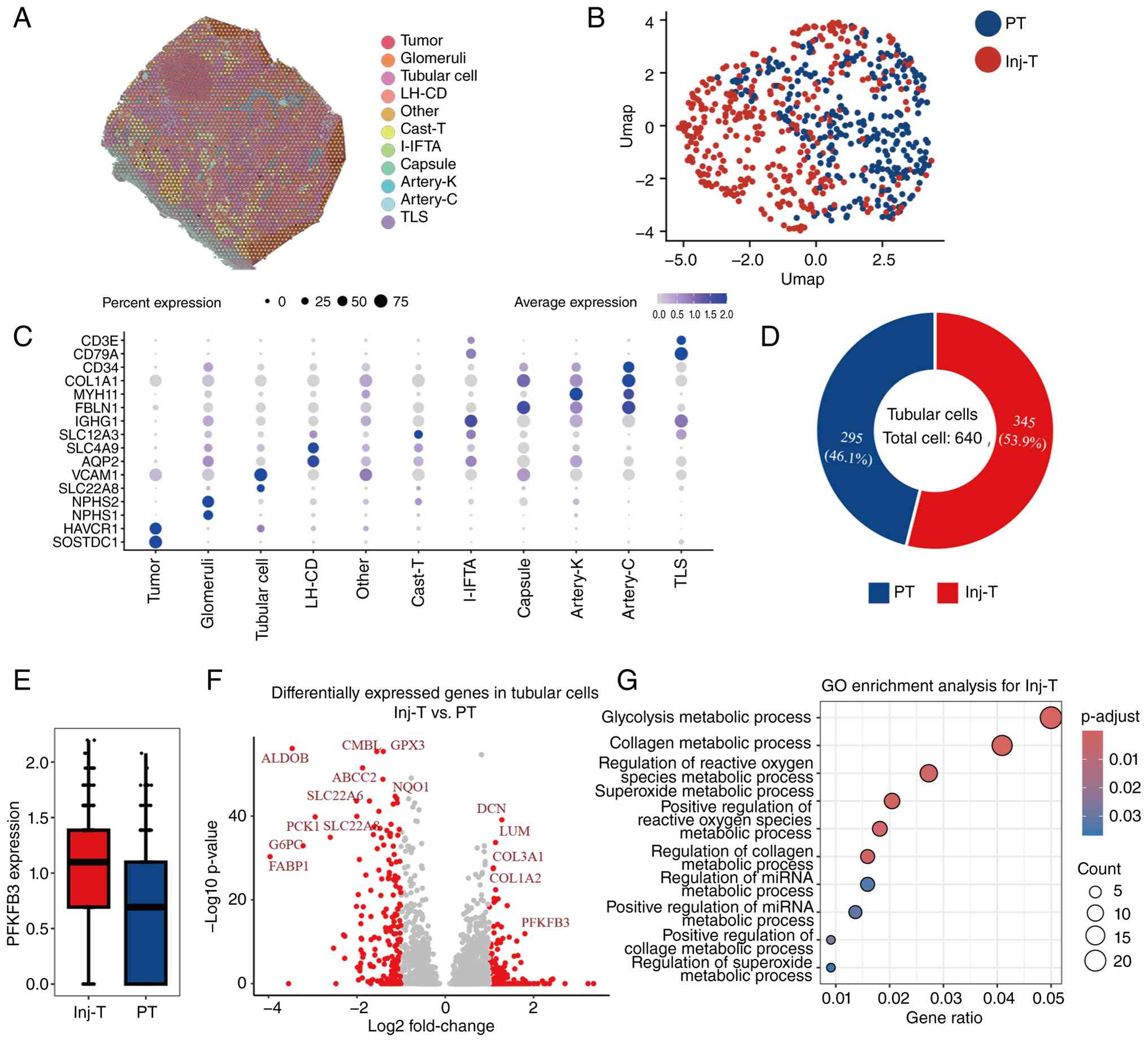
ST profiling was performed on H&E-stained renal
sections from patients with DKD to visualize transcriptional
landscapes within histological context. ST analysis identified a
series of spatial 'spots' that were mapped onto the renal
microanatomy, encompassing glomeruli, tubules, vascular regions and
tubular casts. Reanalysis of a publicly available ST data enabled
improved mapping resolution and the identification of rare cell
populations contributing to local transcriptomic heterogeneity
(Fig. 1A). Distinct clusters
emerged, each displaying marked enrichment of canonical marker
genes corresponding to specific renal structures (Fig. 1C). Further subclustering revealed
that proximal tubular (PT) cells were divided into normal and
injury-associated subtypes, the latter representing 53.9% of the
total tubular cells (Fig. 1B and
D). The expression levels of PFKFB3 in injured PT cells were
markedly increased (Fig. 1E and
F). GO enrichment analysis demonstrated that the
injury-associated PT cluster was markedly enriched for
glycolysis-related pathways, suggesting a metabolic shift
contributing to DKD progression (Fig. 1G).
To further determine whether PFKFB3 expression is
increased in renal tubules during DKD, its transcript and protein
levels were assessed in db/db mice. RT-qPCR revealed a marked
elevation of PFKFB3 mRNA levels in kidney derived from db/db mice,
compared with those derived from db/m group (Fig. 2A). As shown in Fig. 2B, downregulation of E-cadherin
expression, a TEC specific marker critical for maintaining
epithelial integrity, together with elevated PFKFB3 expression,
indicated that PFKFB3 was actively involved in tubular injury
during DKD. In line with the aforementioned results, western blot
and IHC analyses confirmed a significant elevation of PFKFB3
protein expression in the db/db group (Fig. 2C-E).
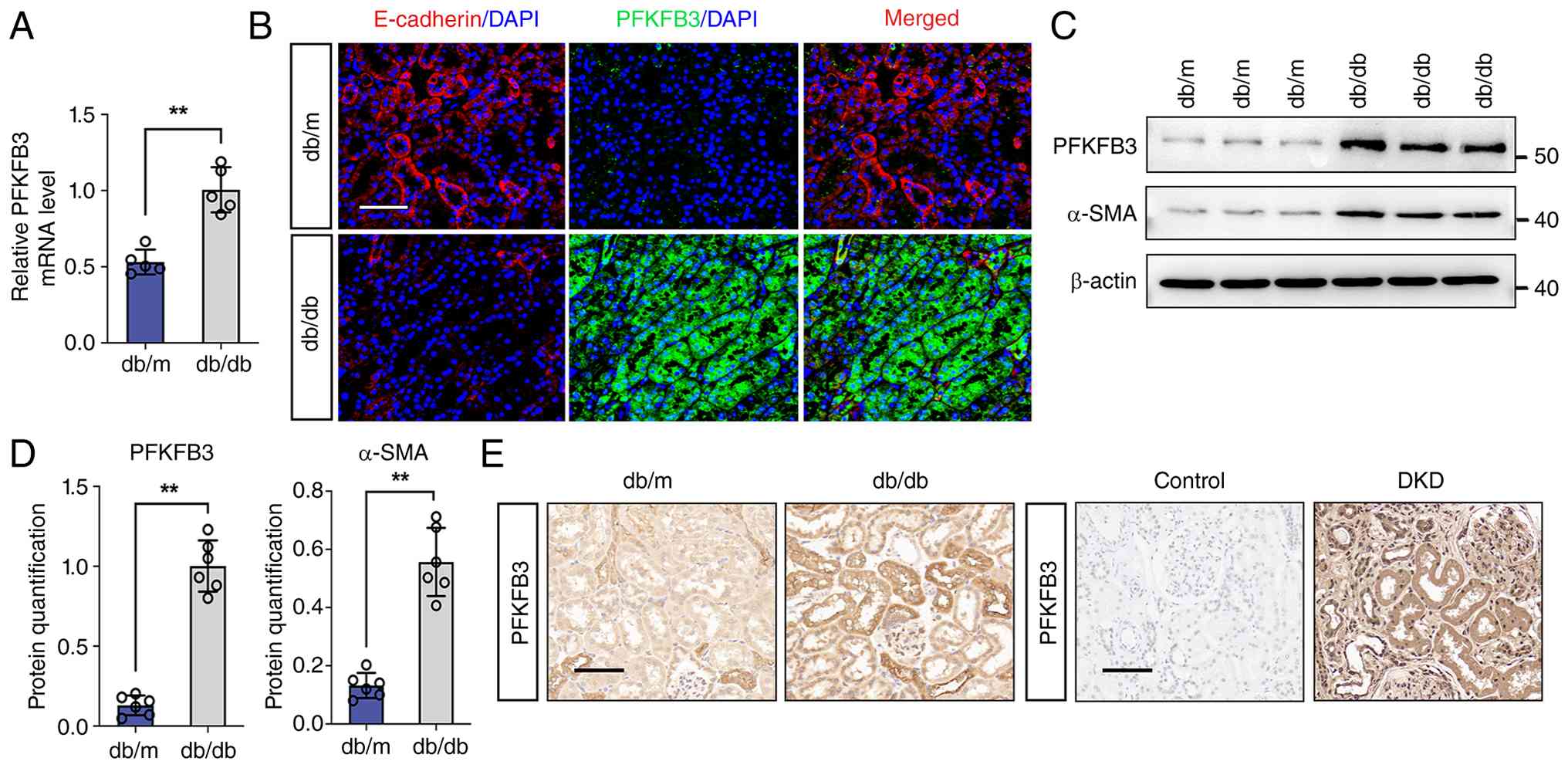 | Figure 2PFKFB3 expression is increased in
renal tubular cells in DKD. (A) RT-qPCR for renal PFKFB3 and HK2 in
db/m and db/db groups (n=5). (B) Immunostained E-cadherin (red) and
PFKFB3 (green), and counterstained with DAPI (blue) by IF staining
in db/m and db/db groups (n=6). Scale bar, 50 μm. Protein
levels of α-SMA and PFKFB3 in db/m and db/db groups by (C) western
blotting and (D) its semi-quantitative analysis (n=6). (E)
Representative images of IHC staining for PFKFB3 (scale bar, 50
μm) in db/m and db/db groups or in Control and DKD group
(n=6). **P<0.01 vs. db/m group by Student's t-test.
PFKFB3, 6-phosphofructo-2-kinase/fructose-2,6-biphosphatase 3; DKD,
diabetic kidney disease; RT-qPCR, reverse
transcription-quantitative PCR; IF, immunofluorescence; α-SMA,
α-smooth muscle actin; IHC, immunohistochemistry. |
Upregulation of PFKFB3 expression is
involved in the development of renal fibrosis in DKD
A kidney-specific PFKFB3 knockdown model was
generated via in situ renal delivery of AAV9-shRNA to db/db
mice to assess the role of PFKFB3 in DKD (Fig. 3A). Silencing of PFKFB3 expression
markedly reduced blood glucose levels, serum creatinine and urinary
albumin-to-creatinine ratio (UACR) at 20 weeks compared with those
noted in the db/db group (Fig. S1A
and B; Fig. 3B).
Histological examination revealed significant renal fibrosis in
db/db mice relative to the db/m group, which was effectively
mitigated by shPFKFB3, as demonstrated by Masson's trichrome and
Sirius Red staining (Fig. 3C).
IHC staining further demonstrated a marked upregulation of
fibronectin (FN) and α-SMA expression in the db/db mice, while
these elevations were apparently reversed following knockdown of
PFKFB3 expression (Fig. 3C). In
line with these results, western blot analysis verified that the
expression levels of FN and α-SMA were increased in db/db mice
compared with those of the db/m mice, and were markedly reduced in
the shPFKFB3-treated group (Fig. 3D
and E). Conversely, PFKFB3 overexpression in db/db mice
exacerbated renal fibrosis (Figs.
S1, S2A and S2B). Increased extracellular matrix accumulation
and interstitial expansion were observed in these mice, accompanied
by upregulation of the expression levels of FN and α-SMA (Fig. S2B-D). Collectively, these
results indicated that inhibition of PFKFB3 expression effectively
attenuates fibrotic progression in DKD.
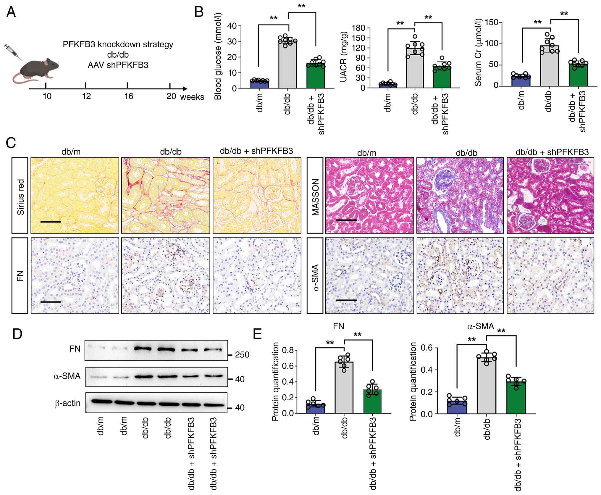 | Figure 3Knockdown of PFKFB3 ameliorates renal
fibrosis in db/db mice. (A) Schematic illustration of AAV9-shPFKFB3
injection in the db/db group; (B). Blood glucose levels, UACR and
Cr levels in db/m, db/db, and db/db + shPFKFB3 group (n=8). (C)
Representative images of Masson, Sirius red staining and IHC
staining (α-SMA and FN level) (scale bar=50 μm) in db/m,
db/db, and db/db + shPFKFB3 groups (n=6). (D and E) Protein levels
of α-SMA and FN in db/m, db/db, and db/db + shPFKFB3 groups by
western blotting, with semi-quantitative analyses (n=6). Data
represent the mean ± SD from three independent experiments.
**P<0.01 vs. db/db group by one-way ANOVA. PFKFB3,
6-phosphofructo-2-kinase/fructose-2,6-biphosphatase 3; AAV,
adeno-associated virus; sh, short hairpin; UACR, urinary albumin
creatinine ratio; Cr, creatinine; IHC, immunohistochemistry; α-SMA,
α-smooth muscle actin; FN, fibronectin. |
PFKFB3-mediated lactate accumulation
contributes to the progression of renal fibrosis in DKD
Among all PFKFB isoforms, PFKFB3 exhibits the
highest kinase-to-phosphatase activity ratio, which preferentially
shunts glucose metabolism toward glycolysis (31,32). Renal lactate levels were
initially assessed, revealing a significant increase in db/db mice
(Fig. S3). Inhibition of PFKFB3
expression markedly decreased lactate production, while its
overexpression further elevated renal lactate accumulation compared
with that noted in db/db mice. To determine whether lactate
reduction could mitigate fibrotic injury, the lactate dehydrogenase
inhibitor FX-11 was administered to db/db mice (Fig. 4A). Treatment with FX-11 led to a
substantial decline in blood glucose and UACR (Fig. 4B). Histological evaluation
revealed that FX-11 markedly alleviated tubulointerstitial
fibrosis, as demonstrated by Sirius Red staining (Fig. 4C). Moreover, FX-11 treatment
markedly decreased FN expression in db/db mice, while IF analysis
revealed lack of notable changes in the db/m group (Fig. 4C). Consistently, western blot
assays confirmed that FX-11 substantially suppressed the protein
levels of FN and α-SMA compared with those of db/db group (Fig. 4D and E). Collectively, these
findings suggested that lactate accumulation contributes to the
progression of renal fibrosis in DKD and that inhibiting lactate
production confers a protective effect against renal injury.
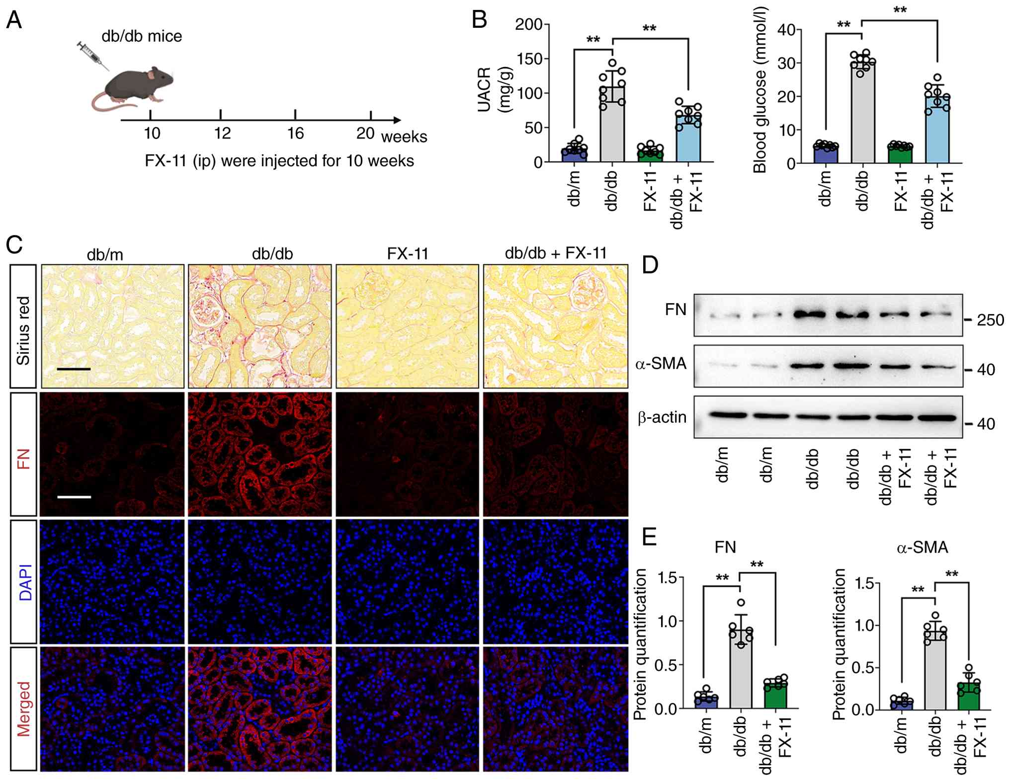 | Figure 4Inhibition of lactate attenuates
renal fibrosis in db/db mice. (A) Illustrative overview of the
FX-11 intervention strategy in db/db mice. (B) Blood glucose levels
and UACR levels in db/m, db/db, db/m + FX-11 and db/db + FX-11
group (n=8). (C) Representative images of Sirius red and IF
staining of FN (green), DAPI (blue) in db/m, db/db, db/m + FX-11
and db/db + FX-11 group (n=6). (D and E) Protein levels of α-SMA
and FN in db/m, db/db, db/m + FX-11 and db/db + FX-11 groups by
western blotting (n=6). Data represent the mean ± SD from three
independent experiments. **P<0.01 vs. db/db group by
one-way ANOVA. UACR, urinary albumin creatinine ratio; IF,
immunofluorescence; FN, fibronectin; α-SMA, α-smooth muscle
actin. |
Identification of potential downstream
genes regulated by of H4K12la
Given that lactate acts as a crucial metabolic
substrate facilitating histone lactylation (33,34), it was hypothesized that this
epigenetic modification may be aberrantly elevated in the kidneys
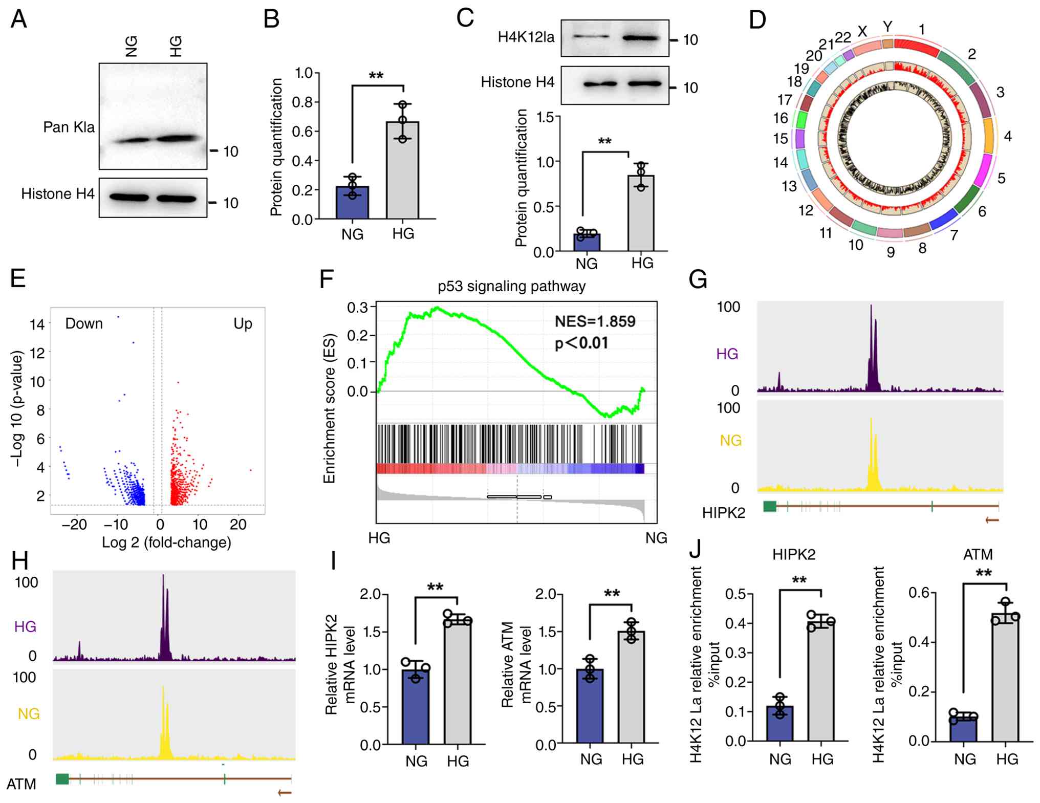
of the DKD mouse models. Western blot analysis indicated that
global histone lactylation was notably increased in TEC following
stimulation with HG conditions compared with those noted under NG
conditions, with a distinct band at ~11 kDa likely corresponding to
histone H4 (Fig. 5A and B).
Subsequent immunoblotting further verified that H4K12la levels were
markedly upregulated in HG-exposed TECs relative to the NG group
(Fig. 5C). Collectively, these
data suggested that lactate accumulation promoted H4K12 lactylation
under diabetic conditions. To characterize the potential downstream
targets of histone lactylation in DKD, CUT&Tag sequencing was
conducted using an anti-H4K12la antibody in TECs cultured under NG
or HG conditions. HG exposure induced a notable increase in H4K12la
enrichment peaks across the genome (Fig. 5D and E). Functional annotation of
these peaks revealed a strong positive association with the p53
signaling pathway (Fig. 5F).
Integrative analysis identified HIPK2 and ATM as putative
lactylation-regulated genes, with genome browser tracks revealing
prominent H4K12la enrichment within their respective promoter
regions (Fig. 5G and H). RT-qPCR
and ChIP-qPCR were performed to confirm the CUT&Tag target
genes, revealing their elevated expression, H4K12la enrichment was
performed at the promoters of HIPK2 and ATM, with both genes
exhibiting substantial H4K12la modification (Fig. 5I and J).
Lactate accumulation is induced by PFKFB3
and facilitates H4K12 histone lactylation in DKD
To elucidate the role of PFKFB3/H4K12la in TEC
function during DKD, renal histone lactylation levels were
initially assessed. Western blot analysis revealed that inhibition
of PFKFB3 expression markedly decreased global histone lactylation
in db/db mice (Fig. S4A and B;
Fig. 6A and B). Knockdown of
PFKFB3 expression in HK-2 cells led to reduced histone lactylation
(Fig. 6C and D).
Immunofluorescence co-staining of H4K12la (red) and E-cadherin
(green) demonstrated markedly elevated H4K12la signals in the renal
tubules of db/db mice, which were attenuated by inhibition of
PFKFB3 expression (Fig. 6E).
Moreover, transfection of HK-2 cells with small interfering
(si)PFKFB3 suppressed the expression levels of H4K12la, α-SMA and
FN (Fig. 6F). Extracellular flux
analysis revealed that PFKFB3 silencing reduced glycolytic capacity
and lactate accumulation compared with those noted in HG group
(Fig. 7A and B). In HK-2 cells,
treatment with siPFKFB3 led to a decrease in the expression levels
of H4K12la, HIPK2, α-smooth muscle actin (α-SMA), and FN compared
with those noted in the HG group. Notably, excess lactate
production reversed this suppression, exacerbating fibrotic
responses and increasing H4K12la and HIPK2 levels relative to
siPFKFB3-treated cells (Fig. 7C and
D). Double immunofluorescence staining further validated the
lactate-induced alterations in the levels of H4K12la and HIPK2 in
HK-2 cells (Fig. 7E).
Collectively, these results indicated that PFKFB3-driven lactate
accumulation promotes tubular fibrosis through H4K12la-mediated
regulation of profibrotic signaling.
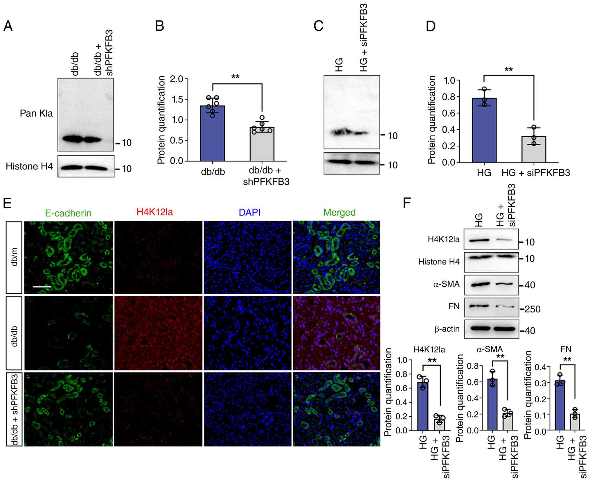 | Figure 6PFKFB3-driven lactate triggers H4K12
lactylation in DKD. (A and B) Protein levels of Pan Kla in db/db
and db/db + shPFKFB3 groups by western blotting and its
semi-quantitative analysis (n=6). (C and D) Protein levels of Pan
Kla in HG and HG + siPFKFB3 groups by western blotting and its
semi-quantitative analysis (n=3). (E) Immunostained E-cadherin
(green) and H4K12la (red), and counterstained with DAPI (blue) by
IF staining in db/m, db/db and db/db + shPFKFB3 groups (n=6). Scale
bar, 50 μm. (F) The protein levels of H4K12la, α-SMA and FN
by western blot in HG and HG + siPFKFB3 groups, and their
semi-quantitative analyses (n=3). Data represent the mean ± SD from
three independent experiments. **P<0.01 vs. (D and F)
HG group or (B) db/db group by Student's t-test. PFKFB3,
6-phosphofructo-2-kinase/fructose-2,6-biphosphatase 3; DKD,
diabetic kidney disease; sh, short hairpin; HG, high glucose; si,
small interfering; IF, immunofluorescence; FN, fibronectin. |
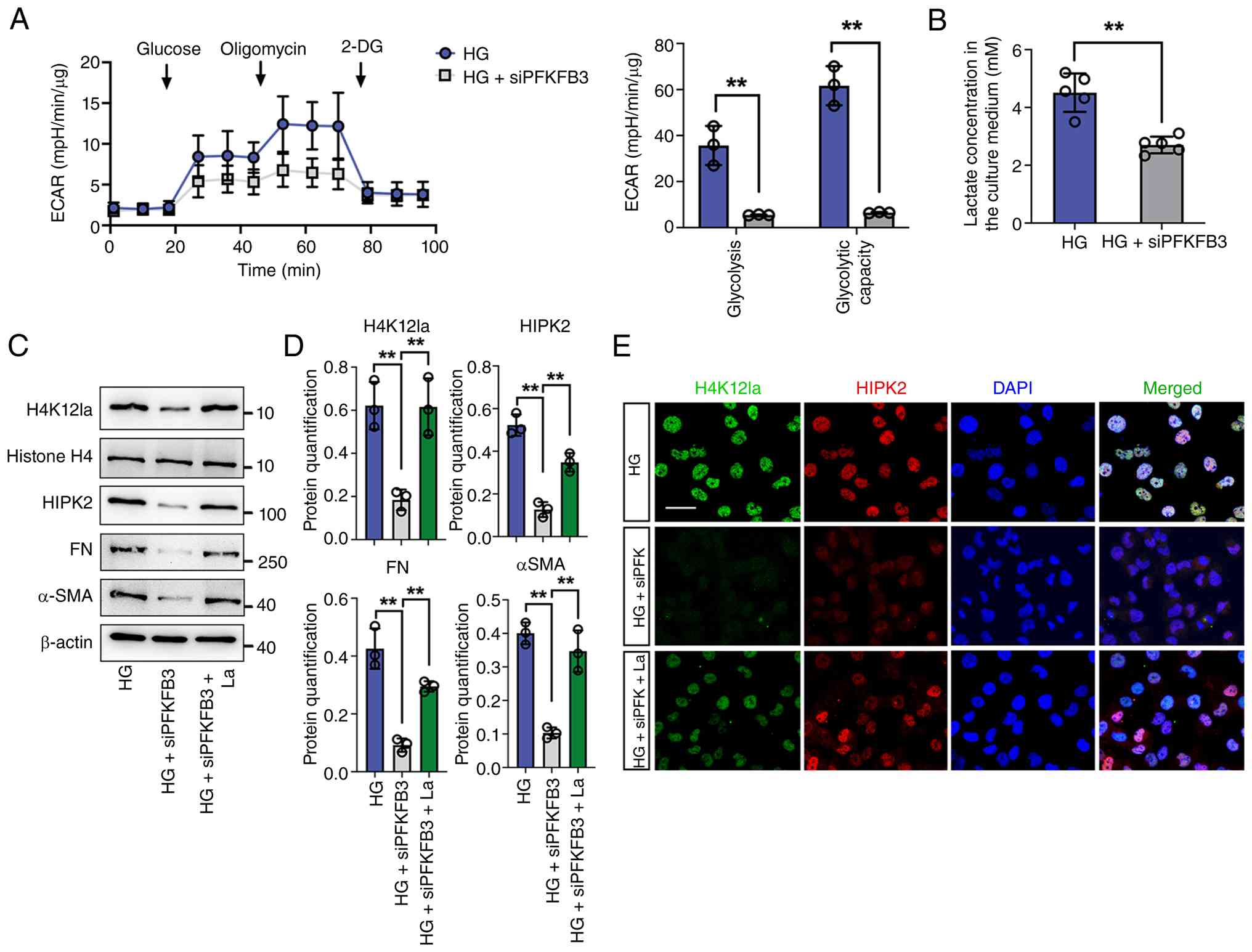 | Figure 7PFKFB3-mediated H4K12la initiates
renal tubule fibrosis in DKD. (A) The ECAR levels after culturing
with glucose followed by oligomycin and 2-DG in HG and HG +
siPFKFB3 groups (n=3). (B) The lactate concentration in HK-2 cells
in the HG and HG + siPFKFB3 groups (n=5). (C and D) Protein levels
of H4K12la, HIPK2, FN and α-SMA in HG, HG + siPFKFB3 and HG +
siPFKFB3 + La groups in HK-2 cells by western blotting, with
semi-quantitative analyses (n=3). (E) Immunostaining for H4K12la
(green) and HIPK2 (red), counterstained with DAPI (blue) in HG, HG
+ siPFKFB3 and HG + siPFKFB3 + La (scale bar, 50 μm). Data
represent the mean ± SD from three independent experiments.
**P<0.01 versus HG group (A and B) by Student's
t-test or HG + siPFKFB3 group (D) by one-way ANOVA. PFKFB3,
6-phosphofructo-2-kinase/fructose-2,6-biphosphatase 3; DKD,
diabetic kidney disease; ECAR, extracellular acidification rate;
HG, high glucose; si, small interfering; FN, fibronectin; α-SMA,
α-smooth muscle actin. |
Discussion
Despite remarkable progress in elucidating the
pathogenesis and diagnostic markers of DKD, this disease is
associated with major complications contributing to morbidity in
diabetic patients (35-37). Growing evidence indicates that
metabolic reprogramming in renal tubules is a key driver of
fibrotic progression in DKD (5,38). In particular, enhanced glycolytic
flux and elevated lactate accumulation have been consistently
observed in both diabetic kidneys and in the circulation (23,39,40). A marked expression of PFKFB3 was
observed, mainly in TECs derived from patients with DKD and mouse
models; the results verified that silencing of PFKFB3 expression
could markedly alleviate renal fibrosis. With regard to its
mechanism of action, deficiency of PFKFB3 expression inhibited
H4K12 lactylation and transcriptional activation of HIPK2.
Collectively, the findings reveal that tubular PFKFB3-driven
glycolytic activation promotes renal fibrosis via lactate-dependent
epigenetic regulation of HIPK2.
The PFKFB family of enzymes plays a pivotal role in
regulating glycolytic flux. Among its isoforms, PFKFB3 possesses
the highest kinase-to-phosphatase activity ratio, thereby
preferentially directing glucose toward glycolysis (41,42). Its catalytic product,
fructose-2,6-bisphosphate, activates phosphofructokinase 1 and
regulates diverse cellular processes including differentiation
(43) and inflammation (44). Under hypoxic conditions,
glycolysis is typically enhanced and the preservation of renal
structure and function relies heavily on the maintenance of
balanced energy metabolism (45). Previous studies have reported
that diabetic kidneys display fibrotic alterations associated with
aberrant glycolytic activation (45,20,21). In endothelial cells,
PFKFB3-driven glycolysis enhances metabolic flux and promotes the
production of metabolites, thereby exacerbating renal inflammation
(46). Similarly,
PFKFB3-mediated glycolytic upregulation promotes lactate
accumulation, triggers osteogenic trans-differentiation of vascular
smooth muscle cells and accelerates vascular calcification in
chronic kidney disease models (47). Collectively, these findings
indicate that dysregulated glycolysis contributes to the
pathogenesis of renal fibrogenesis. In the present study, a
significant upregulation in the expression of PFKFB3 was noted in
kidneys from patients with DKD and in diabetic mice. Genetic
suppression of PFKFB3 mitigated glycolytic activation and
ameliorated renal injury. These results suggested that excessive
PFKFB3-driven glycolysis plays a causal role in promoting renal
fibrosis in DKD.
Elevated glycolytic flux is known to drive excessive
lactate accumulation. In addition to serving as a metabolic
byproduct, lactate acts as a pivotal signaling intermediate that
modulates immune responses and remodels tissue microenvironments
via its influence on cellular signaling and transcriptional
regulation (20,48,49). In diabetic kidneys, metabolic
reprogramming is characterized by increased lactate dehydrogenase A
(LDHA) expression and lactate accumulation, accompanied by reduced
ATP generation. Pharmacological inhibition of LDHA effectively
decreases lactate levels, restores renal energy homeostasis and
mitigates both structural renal damage and albuminuria (40). Clinical and experimental data
further reveal a close correlation between circulating glucose and
lactate levels in circulation and in urine of diabetic individuals.
Proximal tubular epithelial cells represent a major source of this
lactate production, which can be suppressed by sodium-glucose
cotransporter-2 (23). These
findings collectively implicate lactate accumulation as a key
pathogenic contributor to the onset and progression of DKD and its
reduction confers reno-protective effects by limiting fibrotic
remodeling. Lactate-triggered histone lactylation, an emerging
epigenetic modification, acts as a vital molecular bridge
connecting lactate metabolism with gene regulatory networks via
modulating lactate generation, distribution and utilization. This
epigenetic modification induces conformational remodeling of
histone proteins, affects chromatin accessibility and modulates
transcription, with its levels directly governed by lactate spatial
distribution, therefore linking cellular metabolism to epigenetic
control of gene expression (50-52). Notably, H4K12 lactylation was
markedly elevated in diabetic kidneys and in in vitro models
in a lactate-dependent manner. CUT&Tag profiling further
identified prominent H4K12la enrichment at the HIPK2 promoter
region, promoting its transcriptional activation and amplifying
downstream glycolytic signaling. Nevertheless, the mechanistic
crosstalk between the pathogenesis of DKD and HIPK2 activation
remains incompletely elucidated.
While the present study elucidated the role of HIPK2
H4K12 lactylation in regulating glycolysis in vivo and in
vitro, several limitations should be considered. First, the
potential crosstalk between histone lactylation and other
post-translational modifications, such as acetylation remains
unresolved. Histone modifications are dynamically governed by
enzymes responsible for the addition, removal and recognition of
specific markers. A previous study indicated that histone
lactylation is highly reliant on the intracellular level of
lactyl-CoA and the enzymatic activity of acyltransferases including
P300/CBP, which function as major mediators of histone lactylation
modification in macrophages (53). Conversely, deacetylases including
histone deacetylase 1-3 and members of the Sirtuin family can also
catalyze the removal of lactyl groups from histone lysines,
exerting de-lactylation activity (54,55). In the present study, the
contribution of the histone-modifying enzymes to lactylation within
TECs during DKD was not investigated. Further studies employing
quantitative mass spectrometry and site-directed mutagenesis are
warranted to determine the specific enzymes and residues mediating
histone lactylation and to elucidate its biological implications in
renal pathology.
Collectively, the findings of the present study
revealed that PFKFB3-dependent lactate production modulates gene
transcription via histone lactylation. It was further demonstrated
that the upregulation of PFKFB3 expression in tubular epithelial
cells under diabetic kidney disease conditions is essential for the
progression of renal fibrosis and that histone lactylation acting
downstream of PFKFB3 serves as a key determinant of downstream gene
expression. Therefore, these findings suggest that maintaining
metabolic stability via epigenetic regulation in tubular cells
during injury could be an effective strategy for delaying DKD
development.
Supplementary Data
Availability of data and materials
The data generated in the present study may be
requested from the corresponding author.
Authors' contributions
LX conceived and designed the experiments. FX
contributed to clinical study design and collected the data. MX
drafted the manuscript. LF and YZ conceived the study. HW and XG
performed the data analysis. LX revised the manuscript. FX and LF
performed the functional experiments. SG contributed to data
acquisition and critically revised the manuscript. All data were
produced by the authors without the use of a paper mill. The
authors collectively accept responsibility for the accuracy and
integrity of the work. LX and FX confirm the authenticity of all
the raw data. All authors read and approved the final
manuscript.
Ethics approval and consent to
participate
All animal studies were approved by the Ethics
Committee on the Care and Use of Laboratory Animals of Guangdong
Medical University (approval no. GDY2402166).
Patient consent for publication
Not applicable.
Competing interests
The authors declare that they have no competing
interests.
Acknowledgements
Not applicable.
Funding
The present study was supported in part by the Guangdong Natural
Science Foundation General Program (grant no. 2025A1515010621), in
part by Guangdong Medical University Clinical and Basic Science
Innovation Special Project (grant nos. GDMULCJC2024047 and
GDMULCJC2024086), in part by Hundreds Youth Research Projects of
Guangdong Medical University (grant no. GDMUD2024011) and in part
by Medical Science and Technology Research Foundation of Guangdong
Province (grant no. B2025045).
References
|
1
|
Pavkov ME and Miyamoto Y: Diabetes and
kidney disease. IDF Diabetes Atlas. 2023, https://diabetesatlas.org/resources/idf-diabetes-atlas-reports/diabetes-and-kidney-disease/.
Accessed Nov 12, 2025
|
|
2
|
Martinez Leon V, Hilburg R and Susztak K:
Mechanisms of diabetic kidney disease and established and emerging
treatments. Nat Rev Endocrinol. 22:21–35. 2026. View Article : Google Scholar
|
|
3
|
Mentz RJ, Anker SD, Pitt B, Rossing P,
Ruilope LM, Gebel M, Kolkhof P, Lawatscheck R, Rohwedder K and
Bakris GL; on behalf of the FIDELIO-DKD and FIGARO-DKD
investigators: Efficacy and safety of finerenone in patients with
chronic kidney disease and type 2 diabetes by diuretic use: A
FIDELITY analysis. Eur J Heart Fail. 27:764–774. 2025. View Article : Google Scholar : PubMed/NCBI
|
|
4
|
Zhao H and Guo J: Macrophages in focus:
Key drivers and therapeutic opportunities in diabetic kidney
disease. Int J Biol Sci. 21:4647–4662. 2025. View Article : Google Scholar : PubMed/NCBI
|
|
5
|
Naaman SC and Bakris GL: Diabetic
nephropathy: Update on pillars of therapy slowing progression.
Diabetes Care. 46:1574–1586. 2023. View Article : Google Scholar : PubMed/NCBI
|
|
6
|
Mimura I, Chen Z and Natarajan R:
Epigenetic alterations and memory: Key players in the
development/progression of chronic kidney disease promoted by acute
kidney injury and diabetes. Kidney Int. 107:434–456. 2025.
View Article : Google Scholar
|
|
7
|
Mohandes S, Doke T, Hu H, Mukhi D, Dhillon
P and Susztak K: Molecular pathways that drive diabetic kidney
disease. J Clin Invest. 133:e1656542023. View Article : Google Scholar : PubMed/NCBI
|
|
8
|
Fan X, Yang M, Lang Y, Lu S, Kong Z, Gao
Y, Shen N, Zhang D and Lv Z: Mitochondrial metabolic reprogramming
in diabetic kidney disease. Cell Death Dis. 15:4422024. View Article : Google Scholar : PubMed/NCBI
|
|
9
|
Hong Q, Kim H, Cai GY, Chen XM, He JC and
Lee K: Modulation of TGF-β signaling new approaches toward kidney
disease and fibrosis therapy. Int J Biol Sci. 21:1649–1665. 2025.
View Article : Google Scholar
|
|
10
|
Zou Y, Yiu WH, Lok SWY, Ma J, Feng Y, Lai
KN and Tang SCW: Tubular FoxP2 and kidney fibrosis. J Am Soc
Nephrol. 36:544–558. 2025. View Article : Google Scholar
|
|
11
|
Saraswati S, Martínez P, Serrano R, Mejías
D, Graña-Castro O, Díaz RÁ, Flores JM and Blasco MA: Telomere
dysfunction in renal tubular epithelial cells leads to kidney
fibrosis. J Am Soc Nephrol. 36:2348–2363. 2025. View Article : Google Scholar : PubMed/NCBI
|
|
12
|
Yao L, Liang X, Liu Y, Li B, Hong M, Wang
X, Chen B, Liu Z and Wang P: Non-steroidal mineralocorticoid
receptor antagonist finerenone ameliorates mitochondrial
dysfunction via PI3K/Akt/eNOS signaling pathway in diabetic
tubulopathy. Redox Biol. 68:1029462023. View Article : Google Scholar : PubMed/NCBI
|
|
13
|
Wang M, Li Q, Wang S, Zuo L, Hai Y, Yuan
S, Li X, Huang X, Yang C, Yao L, et al: Astragaloside IV protects
renal tubular epithelial cells against oxidative stress-induced
injury by upregulating CPT1A-mediated HSD17B10 lysine succinylation
in diabetic kidney disease. Phytother Res. 38:4519–4540. 2024.
View Article : Google Scholar : PubMed/NCBI
|
|
14
|
Wu YW, Chen JW, Lin LY, Huang JH, Chang CC
and Chang TT: FABP4 inhibition protects renal tubular cells and
ameliorates renal inflammation in diabetic kidney disease. Biochem
Pharmacol. 236:1168992025. View Article : Google Scholar : PubMed/NCBI
|
|
15
|
Hommos MS, Glassock RJ and Rule AD:
Structural and functional changes in human kidneys with healthy
aging. J Am Soc Nephrol. 28:2838–2844. 2017. View Article : Google Scholar : PubMed/NCBI
|
|
16
|
Bao Y, Shan Q, Lu K, Yang Q, Liang Y,
Kuang H, Wang L, Hao M, Peng M, Zhang S and Cao G: Renal tubular
epithelial cell quality control mechanisms as therapeutic targets
in renal fibrosis. J Pharm Anal. 14:1009332024. View Article : Google Scholar : PubMed/NCBI
|
|
17
|
Liu X, Zhang Y, Wang Y, Yang Y, Qiao Z,
Zhan P, Jin H, Xu Q, Tang W, Sun Y, et al: Tubular MYDGF slows
progression of chronic kidney disease by maintaining mitochondrial
homeostasis. Adv Sci (Weinh). 12:e24097562025. View Article : Google Scholar :
|
|
18
|
Huang H, Han Y, Zhang Y, Zeng J, He X,
Cheng J, Wang S, Xiong Y, Yin H, Yuan Q, et al: Deletion of
pyruvate carboxylase in tubular epithelial cell promotes renal
fibrosis by regulating SQOR/cGAS/STING-mediated glycolysis. Adv Sci
(Weinh). 12:e24087532025. View Article : Google Scholar : PubMed/NCBI
|
|
19
|
Nishima N and Tanaka S: Lactate: A missing
link between metabolism and inflammation in CKD progression? Kidney
Int. 106:183–185. 2024. View Article : Google Scholar : PubMed/NCBI
|
|
20
|
Zhu W, Guo S, Sun J, Zhao Y and Liu C:
Lactate and lactylation in cardiovascular diseases: Current
progress and future perspectives. Metabolism. 158:1559572024.
View Article : Google Scholar : PubMed/NCBI
|
|
21
|
Xie W, He Q, Zhang Y, Xu X, Wen P, Cao H,
Zhou Y, Luo J, Yang J and Jiang L: Pyruvate kinase M2 regulates
mitochondrial homeostasis in cisplatin-induced acute kidney injury.
Cell Death Dis. 14:6632023. View Article : Google Scholar : PubMed/NCBI
|
|
22
|
Shao M, Chen D, Wang Q, Guo F, Wei F,
Zhang W, Gan T, Luo Y, Fan X, Du P, et al: Canagliflozin regulates
metabolic reprogramming in diabetic kidney disease by inducing
fasting-like and aestivation-like metabolic patterns. Diabetologia.
67:738–754. 2024. View Article : Google Scholar : PubMed/NCBI
|
|
23
|
Darshi M, Kugathasan L, Maity S, Sridhar
VS, Fernandez R, Limonte CP, Grajeda BI, Saliba A, Zhang G, Drel
VR, et al: Glycolytic lactate in diabetic kidney disease. JCI
Insight. 9:e1688252024. View Article : Google Scholar : PubMed/NCBI
|
|
24
|
Ye Z, Sun Y, Yang S, Li L, Li B, Xia Y,
Yuan T, Yu W, Chen L, Zhou X and Cheng F: Lgals3 promotes calcium
oxalate crystal formation and kidney injury through histone
lactylation-mediated FGFR4 activation. Adv Sci (Weinh).
12:e24139372025. View Article : Google Scholar : PubMed/NCBI
|
|
25
|
Xiang T, Wang X, Huang S, Zhou K, Fei S,
Zhou B, Yue K, Li Q, Xue S, Dai Y, et al: Inhibition of PKM2 by
shikonin impedes TGF-β1 expression by repressing histone
lactylation to alleviate renal fibrosis. Phytomedicine.
136:1563242025. View Article : Google Scholar
|
|
26
|
Zhang X, Chen J, Lin R, Huang Y, Wang Z,
Xu S, Wang L, Chen F, Zhang J, Pan K and Yin Z: Lactate drives
epithelial-mesenchymal transition in diabetic kidney disease via
the H3K14la/KLF5 pathway. Redox Biol. 75:1032462024. View Article : Google Scholar : PubMed/NCBI
|
|
27
|
Arat S, Huynh R, Kumpf S, Qian J, Shoieb
A, Virgen-Slane R, Voigt F, Xie Z and Jakubczak JL: Effects of
donor source on transcriptomic profiles of human kidney tissue.
FASEB J. 37:e228042023. View Article : Google Scholar : PubMed/NCBI
|
|
28
|
Mei H, Jing T, Liu H, Liu Y, Zhu X, Wang J
and Xu L: Ursolic acid alleviates mitotic catastrophe in podocyte
by inhibiting autophagic P62 accumulation in diabetic nephropathy.
Int J Biol Sci. 20:3317–3333. 2024. View Article : Google Scholar : PubMed/NCBI
|
|
29
|
Leary S, Underwood W, Anthony R, Cartner
S, Grandin T, Greenacre C, Gwaltney-Brant S, McCrackin MA, Meyer R,
Miller D, et al: AVMA Guidelines for the Euthanasia of Animals:
2020 Edition. American Veterinary Medical Association; Schaumburg,
IL: 2020
|
|
30
|
Livak KJ and Schmittgen TD: Analysis of
relative gene expression data using real-time quantitative PCR and
the 2(-delta delta C(T)) method. Methods. 25:402–408. 2001.
View Article : Google Scholar
|
|
31
|
Lin XF, Cui XN, Yang J, Jiang YF, Wei TJ,
Xia L, Liao XY, Li F, Wang DD, Li J, et al: SGLT2 inhibitors
ameliorate NAFLD in mice via downregulating PFKFB3, suppressing
glycolysis and modulating macrophage polarization. Acta Pharmacol
Sin. 45:2579–2597. 2024. View Article : Google Scholar : PubMed/NCBI
|
|
32
|
Zhong YX, Zhao HB, Lian MH, Shen JM, Li
CX, Ma HM, Xu D, Chen GQ and Zhang C: SUMOylated hnRNPM suppresses
PFKFB3 phosphorylation to regulate glycolysis and tumorigenesis.
Cancer Lett. 616:2175732025. View Article : Google Scholar : PubMed/NCBI
|
|
33
|
Li Z, Liang Z, Qi H, Luo X, Wang M, Du Z
and Guo W: Lactate shuttling links histone lactylation to adult
hippocampal neurogenesis in mice. Dev Cell. 60:1182–1198.e8. 2025.
View Article : Google Scholar : PubMed/NCBI
|
|
34
|
Zhang J, Wu D, Zeng F, Gu H, Li C, Cata
JP, Guo K, Miao C and Zhang H: Lactate metabolic reprogramming and
histone lactylation modification in sepsis. Int J Biol Sci.
21:5034–5055. 2025. View Article : Google Scholar : PubMed/NCBI
|
|
35
|
Jung CY and Yoo TH: Pathophysiologic
mechanisms and potential biomarkers in diabetic kidney disease.
Diabetes Metab J. 46:181–197. 2022. View Article : Google Scholar : PubMed/NCBI
|
|
36
|
Das S, Devi Rajeswari V, Venkatraman G,
Elumalai R, Dhanasekaran S and Ramanathan G: Current updates on
metabolites and its interlinked pathways as biomarkers for diabetic
kidney disease: A systematic review. Transl Res. 265:71–87. 2024.
View Article : Google Scholar
|
|
37
|
Yang H, Sun J, Sun A, Wei Y, Xie W, Xie P,
Zhang L, Zhao L and Huang Y: Podocyte programmed cell death in
diabetic kidney disease: Molecular mechanisms and therapeutic
prospects. Biomed Pharmacother. 177:1171402024. View Article : Google Scholar : PubMed/NCBI
|
|
38
|
Hu H, Li W, Hao Y, Peng Z, Zou Z and Liang
W: Baicalin ameliorates renal fibrosis by upregulating
CPT1α-mediated fatty acid oxidation in diabetic kidney disease.
Phytomedicine. 122:1551622024. View Article : Google Scholar
|
|
39
|
Yang G, Liu X, Li Y, Li L, Xiang J, Liang
Z, Jiang M and Yang S: TRIM65 as a key regulator of ferroptosis and
glycolysis in lactate-driven renal tubular injury and diabetic
kidney disease. Cell Rep. 44:1160912025. View Article : Google Scholar : PubMed/NCBI
|
|
40
|
Azushima K, Kovalik JP, Yamaji T, Ching J,
Chng TW, Guo J, Liu JJ, Nguyen M, Sakban RB, George SE, et al:
Abnormal lactate metabolism is linked to albuminuria and kidney
injury in diabetic nephropathy. Kidney Int. 104:1135–1149. 2023.
View Article : Google Scholar : PubMed/NCBI
|
|
41
|
Zeng H, Pan T, Zhan M, Hailiwu R, Liu B,
Yang H and Li P: Suppression of PFKFB3-driven glycolysis restrains
endothelial-to-mesenchymal transition and fibrotic response. Signal
Transduct Target Ther. 7:3032022. View Article : Google Scholar : PubMed/NCBI
|
|
42
|
Bartrons R, Rodríguez-García A,
Simon-Molas H, Castaño E, Manzano A and Navarro-Sabaté À: The
potential utility of PFKFB3 as a therapeutic target. Expert Opin
Ther Targets. 22:659–674. 2018. View Article : Google Scholar : PubMed/NCBI
|
|
43
|
Ma J, Li X, Li Q, Sun Z, You Y, Zhang L,
Ji Z, Zhou H, Zhang Q, Wang L, et al: Niacin regulates glucose
metabolism and osteogenic differentiation via the SIRT2-C/EBPβ-AREG
signaling axis. Biomed Pharmacother. 180:1174472024. View Article : Google Scholar
|
|
44
|
Wang TT, Han T, Xiao X, Guo D, Sun X, Liu
Y, Zhao L, Xu H, Li R, Jiang L, et al: SIRT3 deficiency reduces
PFKFB3-driven T-cell glycolysis and promotes arthritic
inflammation. Sci China Life Sci. 68:1755–1769. 2025. View Article : Google Scholar : PubMed/NCBI
|
|
45
|
Jiang A, Liu J, Wang Y and Zhang C:
cGAS-STING signaling pathway promotes hypoxia-induced renal
fibrosis by regulating PFKFB3-mediated glycolysis. Free Radic Biol
Med. 208:516–529. 2023. View Article : Google Scholar : PubMed/NCBI
|
|
46
|
Song C, Wang S, Fu Z, Chi K, Geng X, Liu
C, Cai G, Chen X, Wu D and Hong Q: IGFBP5 promotes diabetic kidney
disease progression by enhancing PFKFB3-mediated endothelial
glycolysis. Cell Death Dis. 13:3402022. View Article : Google Scholar : PubMed/NCBI
|
|
47
|
Li Y, Chen X, Xu X, Chen C, Min M, Liang
D, Ren J and Mao H: OTUB2 contributes to vascular calcification in
chronic kidney disease via the YAP-mediated transcription of
PFKFB3. Theranostics. 15:1185–1204. 2025. View Article : Google Scholar : PubMed/NCBI
|
|
48
|
Chen J, Huang Z, Chen Y, Tian H, Chai P,
Shen Y, Yao Y, Xu S, Ge S and Jia R: Lactate and lactylation in
cancer. Signal Transduct Target Ther. 10:382025. View Article : Google Scholar : PubMed/NCBI
|
|
49
|
Llibre A, Kucuk S, Gope A, Certo M and
Mauro C: Lactate: A key regulator of the immune response. Immunity.
58:535–554. 2025. View Article : Google Scholar : PubMed/NCBI
|
|
50
|
Wang T, Ye Z, Li Z, Jing DS, Fan GX, Liu
MQ, Zhuo QF, Ji SR, Yu XJ, Xu XW and Qin Y: Lactate-induced protein
lactylation: A bridge between epigenetics and metabolic
reprogramming in cancer. Cell Prolif. 56:e134782023. View Article : Google Scholar : PubMed/NCBI
|
|
51
|
Yu X, Yang J, Xu J, Pan H, Wang W, Yu X
and Shi S: Histone lactylation: From tumor lactate metabolism to
epigenetic regulation. Int J Biol Sci. 20:1833–1854. 2024.
View Article : Google Scholar : PubMed/NCBI
|
|
52
|
Li Y, Cao Q, Hu Y, He B, Cao T, Tang Y,
Zhou XP, Lan XP and Liu SQ: Advances in the interaction of
glycolytic reprogramming with lactylation. Biomed Pharmacother.
177:1169822024. View Article : Google Scholar : PubMed/NCBI
|
|
53
|
Li F, Si W, Xia L, Yin D, Wei T, Tao M,
Cui X, Yang J, Hong T and Wei R: Positive feedback regulation
between glycolysis and histone lactylation drives oncogenesis in
pancreatic ductal adenocarcinoma. Mol Cancer. 23:902024. View Article : Google Scholar : PubMed/NCBI
|
|
54
|
Sheng X, Lin H, Cole PA and Zhao Y:
Biochemistry and regulation of histone lysine L-lactylation. Nat
Rev Mol Cell Biol. 27:95–109. 2025. View Article : Google Scholar : PubMed/NCBI
|
|
55
|
Liao Z, Chen B, Yang T, Zhang W and Mei Z:
Lactylation modification in cardio-cerebral diseases: A
state-of-the-art review. Ageing Res Rev. 104:1026312025. View Article : Google Scholar
|