Introduction
Osteoarthritis (OA) is a leading cause of chronic
disability worldwide, yet no disease-modifying therapy has been
approved to date. The increasing prevalence of OA, driven by
population aging and increasing obesity rates, imposes a
substantial socioeconomic burden on health care systems globally.
Vicenti et al (1)
performed an early multi-omics analysis of synovial fluid and
reported that conventional approaches based on radiographic grading
and symptomatic management fail to capture the molecular
heterogeneity of OA, which largely explains the recurrent failures
of clinical trials for structure-modifying drugs. Despite decades
of research, current pharmacological interventions remain limited
to pain relief without altering long-term disease progression,
underscoring the urgent need for a deeper mechanistic understanding
that can guide precision therapeutic strategies.
The traditional view of OA as a simple 'wear and
tear' disease resulting from mechanical overloading has been
progressively supplanted by a more nuanced understanding of OA as a
multifactorial disorder involving metabolic, inflammatory and
genetic components. Iijima et al (2) integrated meta-analysis with
multi-omics data from murine models of age-related knee OA and
demonstrated that pathways associated with oxidative stress,
mitochondrial dysfunction and inflammation converge to drive
cartilage degeneration, thereby challenging the exclusive
biomechanical paradigm. In parallel, Bensa et al (3) systematically reviewed preclinical
evidence on corticosteroid injections and revealed a wide spectrum
of effects ranging from detrimental to disease-modifying,
highlighting that therapeutic responses are highly
context-dependent and likely influenced by underlying molecular
endotypes. Bajpai and Chandra (4) recently reviewed the emerging
anti-inflammatory attributes of curcumin and proposed that
targeting inflammatory cascades represents a promising avenue, yet
they critically noted that the heterogeneous nature of OA
pathogenesis complicates the identification of universally
effective interventions. Collectively, these observations have
shifted the conceptual framework toward recognizing OA as a
systemic metabolic and immune disorder with localized joint
manifestations, thereby necessitating molecular-level
stratification.
The inherent complexity and heterogeneity of OA
demand analytical approaches capable of capturing molecular events
across multiple biological layers. Liu et al (5) recently synthesized advances in
omics technologies and emphasized that while bulk transcriptomics,
proteomics and metabolomics have individually identified
OA-associated pathways, their integration is essential to
disentangle causal relationships from correlative noise. Similarly,
Wei et al (6)
comprehensively reviewed progress in multi-omics studies of OA and
concluded that single-platform approaches are insufficient to
capture the intricate interplay between genes, proteins and
metabolites that drives disease heterogeneity. The advent of
single-cell RNA sequencing (scRNA-seq) has further revolutionized
the field by resolving cell-type-specific transcriptional programs
that bulk analyses average across millions of cells. Gu et
al (7) provided a systematic
overview of scRNA-seq applications in OA and highlighted that this
technology has unveiled previously unrecognized chondrocyte and
synovial cell subsets with distinct metabolic and inflammatory
signatures. Danzeng et al (8) extended this paradigm by
demonstrating that single-cell sequencing can dissect the immune
microenvironment in OA, revealing macrophage and T-cell
heterogeneity that correlates with disease severity. However, a
critical appraisal of these studies reveals that while descriptive
catalogs of cell types have multiplied, functional validation and
integration with metabolite data remain limited, representing a
major knowledge gap.
The present review synthesizes current advances in
multi-omics integration applied to OA, with a specific focus on
reconstructing cell-type-specific gene-metabolite networks and
their implications for precision medicine. This review synthesizes
advances in single-cell transcriptomics, metabolomics, lipidomics
and spatial omics, presenting their collective contributions to a
coherent understanding of OA. While spatial omics technologies have
provided valuable insights into the spatial organization of joint
tissues, their systematic integration with cell-type-specific
gene-metabolite networks remains an ongoing challenge that is
critically discussed in this review. While previous comprehensive
reviews have summarized omics advances in OA and highlighted the
importance of multi-omics integration, they have not systematically
bridged transcriptional and metabolic alterations at cellular
resolution nor comprehensively synthesized the mechanistic links
between lipid metabolic reprogramming and pain generation, which
are gaps that the present review specifically addresses. Recent
systematic reviews by Boffa et al (9) and Sourugeon et al (10) have comprehensively evaluated
cell-based therapies in animal models, demonstrating
disease-modifying effects across multiple cell sources, yet these
studies also highlighted that the mechanisms of action remain
poorly understood due to insufficient molecular characterization.
D'Arrigo et al (11)
further called for standardization in secretome and extracellular
vesicle (EV) research, noting that heterogeneous protocols have
precluded definitive conclusions about therapeutic mechanisms. Tan
et al (12) integrated
single-cell sequencing with genetics and epigenetics to reveal
mesenchymal stem cell (MSC) senescence in OA, providing a molecular
basis for age-related susceptibility. Wang et al (13) recently performed multi-omics
analysis of small EVs and proposed that integrating vesicle cargo
with tissue-level omics can bridge molecular insights and clinical
applications. By critically evaluating these emerging findings, the
present review aims to delineate cell-type-specific gene-metabolite
networks across chondrocytes, synovial fibroblasts and immune
cells; synthesize evidence linking lipid metabolic reprogramming to
OA severity; and discuss how multi-omics-derived molecular
endotypes can inform patient stratification and endotype-driven
clinical trial design.
Omics technologies in OA research: From bulk
to single-cell and spatial resolution
The evolution from conventional bulk omics to
high-resolution single-cell and spatial technologies has
fundamentally reshaped the landscape of OA research. Traditional
platforms provided the first molecular maps of diseased joints but
averaged signals across millions of cells, masking critical
heterogeneity. The subsequent advent of scRNA-seq enabled unbiased
dissection of cell-type-specific transcriptomes, while emerging
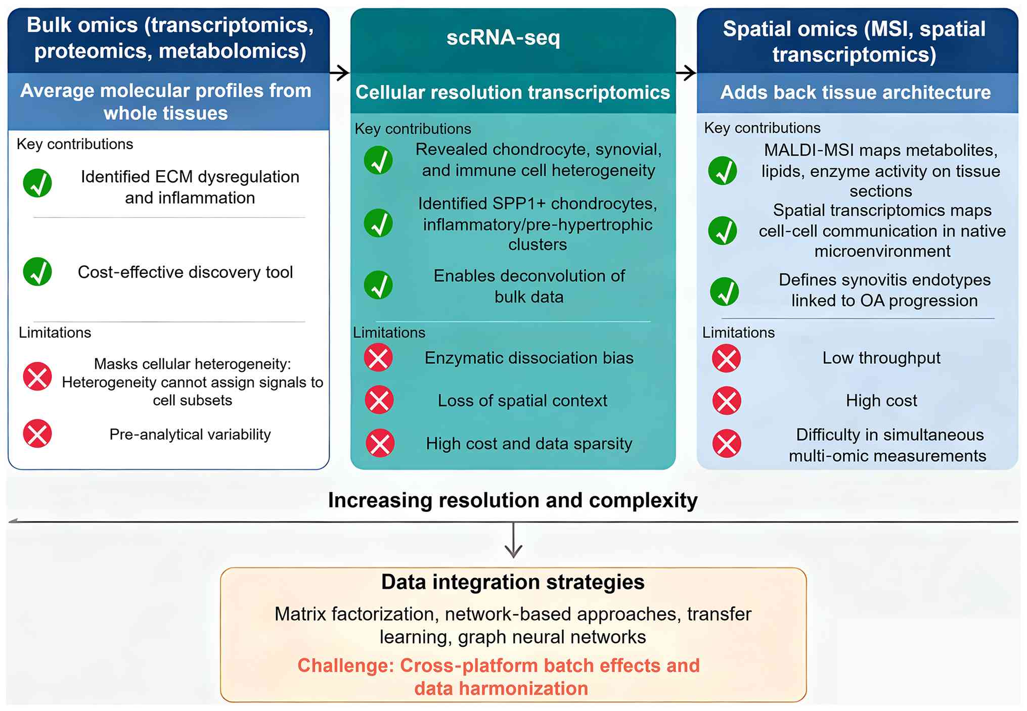
spatial omics technologies now add back the lost tissue context. A
comparative overview of these technological platforms, their
principal contributions to the understanding of OA and their
current methodological limitations is summarized below (Fig. 1).
Traditional bulk omics platforms
Bulk transcriptomics, proteomics and metabolomics
laid the groundwork for understanding OA pathogenesis by profiling
average molecular alterations from whole tissues. In a
comprehensive review, Rai et al (14) examined three decades of OA omics
studies and concluded that while bulk approaches have consistently
identified pathways such as extracellular matrix dysregulation and
inflammation, they cannot resolve whether these signals originate
from distinct cell subsets. Similarly, Van Pevenage et al
(15) performed a systematic
review of metabolomics studies and found that disturbed amino acid
and lipid metabolism are reproducible findings in OA biofluids, but
pre-analytical variability and cohort heterogeneity remain major
barriers to clinical translation. More recently, Liu et al
(5) emphasized that bulk
transcriptomics, when combined with deconvolution algorithms, can
still yield valuable insights, yet the accuracy of such inferences
depends heavily on the quality of single-cell reference data.
Despite their inherent limitations, bulk platforms continue to
serve as cost-effective discovery tools and provide essential
datasets for integrative analyses.
scRNA-seq
The introduction of scRNA-seq has revolutionized OA
research by mapping transcriptional landscapes at cellular
resolution, revealing previously unrecognized heterogeneity within
cartilage, synovium and infrapatellar fat pad. Using scRNA-seq on
human OA cartilage, Ji et al (16) first identified multiple
chondrocyte clusters with distinct metabolic and inflammatory
signatures, setting the stage for cell-level investigations. More
recent work by Fan et al (17) integrated single-cell and bulk
transcriptomics to uncover inflammatory and pre-hypertrophic
chondrocyte subpopulations as key drivers of cartilage
degeneration, and they validated these findings using spatial
transcriptomics. In the synovial compartment, Liao et al
(18) performed comprehensive
scRNA-seq profiling of synovial macrophages and identified a subset
of secreted phosphoprotein 1 (SPP1)+ macrophages that correlates
with OA severity, suggesting that macrophage heterogeneity is
directly linked to disease progression. Using a murine model,
Sebastian et al (19)
demonstrated that post-traumatic OA induces early transcriptomic
changes in immune cells within the synovium, including the
emergence of a novel neutrophil population, highlighting the
dynamic nature of the synovial immune landscape. A major
methodological advance came from Huang et al (20), who developed a computational
strategy to deconvolute bulk synovial transcriptomes using
scRNA-seq references, enabling retrospective analysis of existing
datasets without new experiments. Furthermore, Wang et al
(21) provided a forward-looking
review on how scRNA-seq is shaping orthopedics, emphasizing that
the generation of comprehensive joint cell atlases will be
essential for identifying cell-specific therapeutic targets.
Nevertheless, challenges persist, including enzymatic dissociation
biases and the inevitable loss of spatial context.
Spatial omics: Mass spectrometry imaging
and spatial transcriptomics
While scRNA-seq provides cellular resolution, it
disrupts native tissue architecture, which is particularly
problematic for OA given the zonal organization of cartilage and
synovium. Spatial omics technologies, including mass spectrometry
imaging (MSI) and spatial transcriptomics, have emerged to fill
this gap. A comprehensive review by Fan et al (22) synthesized the current state of
spatial analysis in the OA microenvironment, noting that
matrix-assisted laser desorption ionization-MSI can map
metabolites, lipids and even enzyme activities directly on tissue
sections. In a functional study, Fan et al (23) used MSI to visualize phospholipase
A2 activity across OA joints, demonstrating that active enzyme
localization is spatially restricted to cartilage fissure edges and
adjacent synovium, thereby linking local lipid metabolism to tissue
damage. Lee et al (24)
applied high-resolution N-glycan MSI to subchondral bone and
revealed that complex-type N-glycans accumulate at high mechanical
loading sites, providing a direct molecular bridge between
biomechanical stress and post-translational modifications.
In the synovial membrane, Rocha et al
(25) identified a distinct
lipidomic signature in OA compared to rheumatoid arthritis, with
specific phosphatidylcholines enriched in hyperplastic synovial
areas, suggesting that local lipid alterations drive synovitis
independent of systemic inflammation. More recently, Zhu et
al (26) integrated
spatially resolved proteomic and metabolomic imaging to define
synovitis endotypes associated with OA progression, demonstrating
that spatial multi-omics can stratify patients beyond conventional
histology. For spatial transcriptomics, a review by Xie et
al (27) emphasized that
this technology holds great promise for mapping cell-cell
communication networks within their native microenvironment, such
as ligand-receptor interactions between superficial chondrocytes
and synovial lining cells. Despite these advances, current
limitations include low throughput, high cost and the difficulty of
performing simultaneous multi-omic measurements on the same tissue
section. Critically, while spatial omics technologies have provided
valuable descriptive maps of metabolite and protein distributions
across joint tissues, their integration with cell-type-specific
gene-metabolite networks, including the assignment of spatially
resolved metabolic signals to defined cell populations, remains a
significant technical and analytical challenge that has yet to be
fully resolved (26,27). This limitation is acknowledged
throughout the present review, and the spatial findings discussed
here are presented as complementary evidence rather than as fully
integrated network components.
Data integration strategies and remaining
challenges
Integrating data from bulk, single-cell and spatial
platforms represents a major computational frontier. Kreitmaier
et al (28) discussed
state-of-the-art integration strategies for complex diseases,
including matrix factorization and network-based approaches, and
stressed the importance of tissue-specific context when combining
datasets. A systematic evaluation by Rai et al (14) noted that while integrative
analyses have successfully identified novel OA pathways such as
glycerophospholipid metabolism, cross-platform batch effects and
data sparsity frequently compromise reproducibility. As an example,
Huang et al (29)
combined metabolomics with transcriptomics from the same cartilage
samples and identified glycerophospholipid metabolism as a central
hub, but they acknowledged that the cellular origin of these
signals could not be definitively assigned without single-cell
resolution. More recently, Wei et al (6) provided a comprehensive summary of
multi-omics OA studies and concluded that harmonizing data from
different omics layers requires standardized computational
pipelines and prospective validation in well-phenotyped
cohorts.
An emerging trend is the use of single-cell data as
a reference to deconvolute bulk and spatial data, a strategy that
has yielded insights into synovial macrophage heterogeneity. Liu
et al (5) further
emphasized that recent advances in multi-omics integration,
including the use of transfer learning and graph neural networks,
are beginning to address cross-platform batch effects, but these
methods remain inaccessible to many clinical researchers.
Therefore, while individual omics technologies have matured
considerably, their effective integration into a cohesive and
clinically translatable understanding of OA continues to be an
active area of method development.
Cell-type-specific gene-metabolite networks
in OA
Single-cell transcriptomics integrated with
metabolomics has enabled the reconstruction of cell-type-specific
gene-metabolite networks in OA. These approaches have revealed that
metabolic reprogramming is highly heterogeneous across chondrocyte
subsets, synovial fibroblasts and immune cells, with distinct
pathway alterations driving disease progression in a
cell-type-dependent manner (Fig.
2).
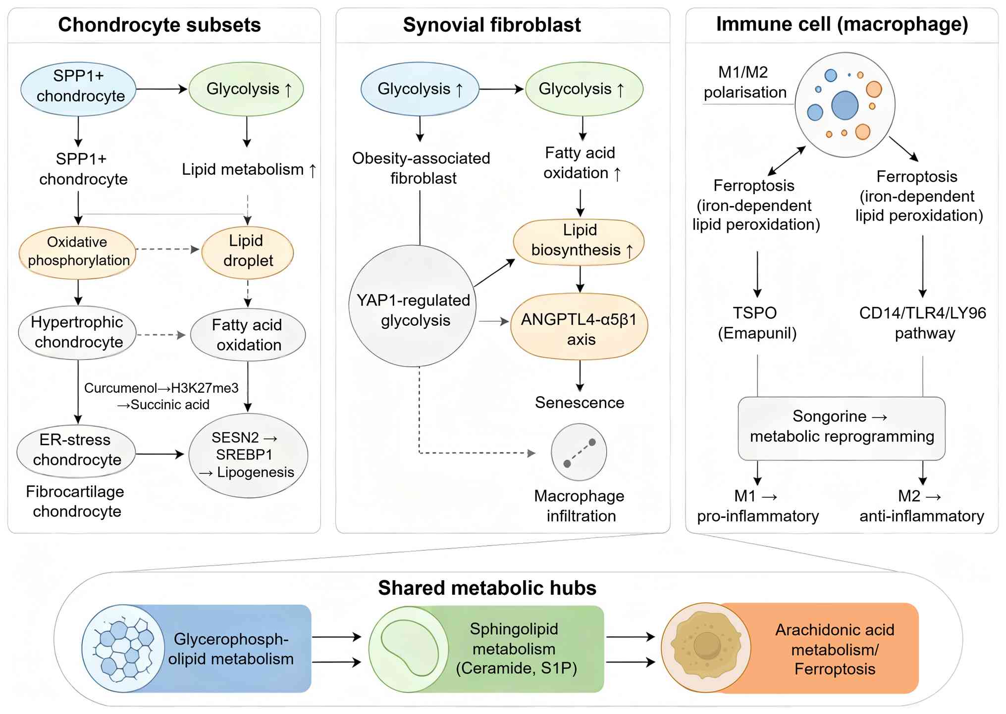 | Figure 2Cell-type-specific gene-metabolite
networks in OA. Chondrocyte subsets (left), synovial fibroblasts
(middle) and immune cells (right) exhibit distinct metabolic
signatures. Shared hubs (bottom) include glycerophospholipid,
sphingolipid and arachidonic acid metabolism. Arrows indicate
pathway activation or crosstalk. The figure was generated with
Figdraw (www.figdraw.com; copyright code:
ORRUT47e0e). ANGPTL4, angiopoietin-like 4; ER, endoplasmic
reticulum; LY96, lymphocyte antigen 96; SPP1, secreted
phosphoprotein 1; S1P, sphingosine-1-phosphate; SESN2, sestrin 2;
SREBP1, sterol regulatory element binding transcription factor 1;
TLR, Toll-like receptor; TSPO, translocator protein 18 kDa; YAP1,
Yes1 associated transcriptional regulator. |
Methodological framework for network
reconstruction
Reconstructing cell-type-specific gene-metabolite
networks requires computational strategies that integrate
single-cell transcriptomic data with metabolomic or lipidomic
profiles. Ojha et al (30) developed a multi-omics integration
framework at cellular resolution to uncover gene-metabolite
mechanisms underlying OA heterogeneity, demonstrating that coupling
scRNA-seq with metabolomics enables the assignment of metabolic
signatures to specific cell populations. This approach addresses a
fundamental limitation of bulk analyses, where tissue-level
averaging obscures cell-type-specific contributions. A critical
methodological advance came from Fan et al (17), who integrated single-cell and
bulk transcriptomics with spatial transcriptomics to identify
inflammatory and prehypertrophic chondrocyte subpopulations as key
drivers of cartilage degeneration, and importantly, validated these
findings across multiple omics layers. However, several challenges
persist. Katsoula et al (31) emphasized that while multi-omics
integration has matured considerably, cross-platform batch effects
and the difficulty of obtaining matched multi-omic data from the
same single cells remain substantial hurdles. Furthermore, the
computational complexity of integrating heterogeneous data types
often requires specialized expertise, limiting accessibility for
many clinical research groups. Taken together, these methodological
advances have established a foundation for dissecting
cell-type-specific metabolic networks, but standardized pipelines
and prospective validation cohorts are urgently needed.
Shared metabolic hubs across cell
types
Despite cell-type-specific metabolic features,
integrative analyses have identified several metabolic hubs that
are consistently altered across multiple joint cell populations in
OA. Wang et al (32)
performed integrated multi-omics analyses and revealed a lipid
metabolic signature characterized by dysregulated
glycerophospholipid and sphingolipid metabolism that spans
chondrocytes, synovial fibroblasts and infiltrating immune cells.
Similarly, He et al (33)
employed integrated machine learning and multi-omics approaches to
demonstrate that immune-metabolic signatures, particularly those
involving arachidonic acid metabolism and ferroptosis-related
pathways, are shared across different cell types in the OA joint.
Notably, Wu et al (34)
integrated bulk and scRNA-seq data and discovered that arachidonic
acid metabolism is consistently upregulated in OA chondrocytes,
suggesting that this pathway represents a convergent metabolic
alteration across cellular compartments. By contrast, Wijesinghe
et al (35) identified
obesity-defined molecular endotypes in the synovium, revealing that
metabolic pathway alterations can vary substantially depending on
systemic metabolic status, with certain gene-metabolite
associations being present only in obese patients with OA. This
finding underscores that shared metabolic hubs may be
context-dependent and modulated by host factors. Collectively,
these observations indicate that while certain lipid metabolic
pathways are broadly dysregulated in OA, their manifestation and
functional consequences are modulated by both cell type and
systemic metabolic context.
Chondrocyte-specific gene-metabolite
associations
A critical synthesis of single-cell transcriptomic
studies reveals both convergent and divergent findings regarding
chondrocyte metabolic heterogeneity (16,36-41) (Table I). Southan et al (36) performed an integrated
transcriptomics and metabolomics analysis of articular cartilage
following mechanical injury and identified that chondrocyte
metabolic responses are characterized by altered amino acid and
lipid metabolism, with specific gene-metabolite pairs
distinguishing injured from healthy cartilage. This study provided
early evidence that chondrocyte metabolic reprogramming is directly
linked to mechanical insult. Using scRNA-seq, Qu et al
(37) identified a unique
SPP1+ chondrocyte subpopulation in human OA cartilage
that exhibits distinct metabolic gene expression signatures,
including enrichment of glycolysis and lipid metabolism pathways,
suggesting that these cells may represent a metabolically active
pathogenic subset. Importantly, the definition of SPP1+
chondrocytes varies across studies: While Qu et al (37) defined this population primarily
by high SPP1 expression coupled with angiogenic and
senescence-associated gene signatures, Kang et al (42) identified SPP1 as one of several
signaling pathways (alongside pleiotrophin, visfatin and TGF-β)
that mediate altered cell-cell communication among chondrocyte
subtypes in OA, without characterizing SPP1+ cells as a
discrete metabolic cluster. Extending these findings, Matta et
al (38) provided a
comprehensive review of chondrocyte populations at single-cell
resolution and highlighted that hypertrophic chondrocytes display a
distinct metabolic profile characterized by altered oxidative
phosphorylation and fatty acid metabolism compared with healthy
articular chondrocytes. Notably, the metabolic signatures
attributed to SPP1+ cells are not entirely consistent:
Qu et al (37) reported
enrichment of glycolysis and lipid metabolism pathways, whereas a
subsequent study associated SPP1 high-expressing cells with energy
metabolism and anabolic-catabolic balance through a Kyoto
Encyclopedia of Genes and Genomes pathway analysis (16). This discrepancy may reflect
differences in dataset composition, analytical pipelines or the
stage of OA progression sampled. Mechanistically, Chen et al
(39) demonstrated that
Curcumenol regulates histone H3K27me3 demethylases to affect
succinic acid metabolism, thereby alleviating cartilage
degeneration in knee OA, revealing a direct link between epigenetic
regulation and chondrocyte-specific metabolic alterations. More
recently, Zhang et al (40) employed single-cell and
transcriptomic profiling to decode endoplasmic reticulum (ER)
stress on chondrocytes driving OA development, showing that ER
stress-responsive chondrocytes exhibit a unique gene-metabolite
network involving lipid droplet formation and ceramide
accumulation. A critical insight from Pan et al (41) was the identification of key
biomarkers related to fibrocartilage chondrocytes, demonstrating
that these cells, which populate repair tissue, show fundamentally
different metabolic gene expression profiles compared with resident
articular chondrocytes. Across these studies, the lack of
standardized cell-type annotation and metabolic pathway scoring
represents a major barrier to direct cross-study comparison. In
aggregate, these findings establish that chondrocyte metabolic
heterogeneity is substantial, with specific subpopulations
exhibiting distinct gene-metabolite networks that may represent
therapeutic opportunities, though harmonized analytical frameworks
are urgently needed to reconcile apparent discrepancies across
datasets.
 | Table IComparative synthesis of chondrocyte
subpopulations and their gene-metabolite signatures in OA. |
Table I
Comparative synthesis of chondrocyte
subpopulations and their gene-metabolite signatures in OA.
| Authors, year | Cell
population | Definition/marker
genes | Key metabolic
signature | Validation
approach | Consistency with
other studies | (Refs.) |
|---|
| Ji et al,
2019 | Seven chondrocyte
subtypes (ProC, preHTC, HTC, FC, EC, RegC, HomC) | Transcriptional
clustering of human OA cartilage | SPP1
high-expressing cells associated with energy metabolism and
anabolic-catabolic balance (KEGG pathway analysis) | scRNA-seq on human
OA cartilage | First comprehensive
scRNA-seq atlas; SPP1+ metabolic associations differ from Qu et
al (37) | (16) |
| Southan et
al, 2020 | Injured vs. healthy
chondrocytes | Mechanical injury
model in articular cartilage | Altered amino acid
and lipid metabolism; specific gene-metabolite pairs distinguish
injured from healthy cartilage | Integrated
transcriptomics + metabolomics | Early evidence
linking mechanical insult to chondrocyte metabolic
reprogramming | (36) |
| Qu et al,
2023 | SPP1+
chondrocytes | High SPP1
expression | Glycolysis and
lipid metabolism enrichment; angiogenic and senescence-associated
gene signatures | Animal model;
spatial heterogeneity of SPP1 confirmed | SPP1 pathway
enrichment also reported by Kang et al (42); metabolic details differ from Ji
et al (16) | (37) |
| Kang et al,
2023 | Multiple
chondrocyte subtypes (not discrete SPP1+ cluster) | SPP1 as signaling
pathway (not cluster marker) | SPP1 pathway
enrichment scores increased in OA; pleiotrophin, visfatin, TGF-β
pathways also altered | Bulk RNA-seq
validation | SPP1 involvement
confirmed; differs from Qu et al (37) in not defining SPP1+ as discrete
metabolic cluster | (42) |
| Chen et al,
2024 | Chondrocytes
(epigenetic regulation) | Curcumenol
regulation of H3K27me3 demethylases KDM6B | Succinic acid
metabolism altered; alleviates cartilage degeneration | In vitro +
in vivo pharmacological intervention | Links epigenetic
regulation to chondrocyte-specific metabolic alterations | (39) |
| Zhang et al,
2025 | ER
stress-responsive chondrocytes | Endoplasmic
reticulum stress gene signatures | Lipid droplet
formation; ceramide accumulation | Single-cell +
transcriptomic profiling | Novel pathway; not
directly comparable to SPP1+ studies; links ER stress to lipid
metabolic reprogramming | (40) |
| Pan et al,
2024 | FCs | Fibrocartilage
markers from scRNA-seq | Fundamentally
different metabolic gene expression vs. resident chondrocytes; six
FC-related biomarkers identified (BCL6, ABCA5, ABCA6, CITED2,
NR1D1, SLC7A8) | Bulk + single-cell
transcriptomics; exploratory risk model | Highlights
heterogeneity across repair vs. resident cells | (41) |
Synovial fibroblast and immune cell
metabolic signatures
The synovial compartment harbors diverse fibroblast
and immune cell populations, each displaying characteristic
gene-metabolite networks. Wijesinghe et al (35) performed multi-omic analysis of
synovium from patients with OA and identified that fibroblast
subsets defined by obesity status exhibit distinct metabolic
signatures, with certain fibroblast clusters showing enrichment of
glycolysis and lipid biosynthesis pathways specifically in obese
individuals. This observation suggests that systemic metabolic
factors directly shape synovial cell metabolism. In a related
study, Acharjee et al (43) conducted cross-species
transcriptomics to identify obesity-associated genes between human
and mouse studies, revealing that synovial fibroblasts from obese
patients with OA upregulate genes involved in fatty acid oxidation
and oxidative stress responses compared with non-obese patients.
Yang et al (44)
demonstrated that targeting Yes1-associated transcriptional
regulator-regulated glycolysis in fibroblast-like synoviocytes
impairs macrophage infiltration and ameliorates diabetic OA
progression, providing functional evidence that fibroblast
metabolic reprogramming directly influences immune cell
recruitment. Regarding immune cells, Wu et al (45) integrated single-cell
transcriptome and multi-omics data to reveal ferroptosis-driven
immune microenvironment remodeling in knee OA, showing that
synovial macrophages undergo iron-dependent lipid peroxidation that
alters their inflammatory phenotype. Furthermore, Wei et al
(46) identified shared M0
macrophage infiltration-related gene signatures in OA and
osteomyelitis, suggesting that metabolic reprogramming of
infiltrating macrophages may represent a common pathogenic
mechanism across inflammatory joint diseases. A recent study by
Deng et al (47) revealed
that senescent synovial intimal fibroblasts aggravate OA by
regulating macrophage polarization and chondrocyte phenotype
through the angiopoietin-like 4-α5β1 axis, demonstrating a direct
metabolic-immune crosstalk mechanism. Collectively, these studies
indicate that synovial fibroblasts and immune cells possess
distinct metabolic signatures that are modulated by systemic
metabolic status and directly contribute to OA pathogenesis through
paracrine and cell-cell interaction mechanisms.
Integration with disease severity and
progression
Linking cell-type-specific gene-metabolite networks
to clinical disease severity and progression represents a critical
translational goal. Komaravolu et al (48) investigated sex-specific effects
of injury and beta-adrenergic activation on metabolic and
inflammatory mediators in a murine model of post-traumatic OA,
revealing that male and female mice exhibit different metabolite
profiles in response to joint injury, suggesting that sex-specific
metabolic networks may influence disease trajectories. This finding
has important implications for precision medicine, as it suggests
that therapeutic targeting of metabolic pathways may need to be
sex-stratified. Huang et al (49) performed multi-omics integrative
analyses and identified two distinct endotypes of hip OA based on
metabolic profiles, with one endotype characterized by altered
amino acid metabolism and the other by lipid metabolism
dysregulation, and importantly, these endotypes correlated with
different rates of radiographic progression. Using spatially
resolved proteomic and metabolomic imaging, Zhu et al
(26) defined synovitis
endotypes associated with OA progression, demonstrating that
patients with specific metabolic signatures in the synovium show
accelerated cartilage loss over 24 months. Tang et al
(50) constructed a single-cell
atlas of human infrapatellar fat pad and synovium and implicated
apolipoprotein E (APOE) signaling in OA pathology, showing that
APOE-expressing cells exhibit metabolic gene signatures that
correlate with pain severity and joint function decline. More
recently, Lu et al (51)
performed an integrated multi-omics analysis to identify a
glutamine metabolism-related gene signature as a diagnostic and
therapeutic target, demonstrating that glutamine metabolism scores
derived from single-cell data correlate with OA severity grades. A
critical contribution reviewed omics-driven insights into OA
pathogenesis and emphasized that while cross-sectional associations
between cell-type-specific networks and disease severity are
increasingly robust, prospective studies linking baseline metabolic
signatures to long-term clinical outcomes remain sparse (52). In summary, existing evidence
supports the notion that cell-type-specific gene-metabolite
networks are associated with OA severity, but prospective
validation and standardization of network reconstruction methods
are required before clinical application.
Lipid metabolism and metabolic reprogramming
in OA pathogenesis
Emerging evidence positions lipid metabolism
dysregulation as a central driver of OA pathogenesis, extending
beyond the traditional view of OA as a purely mechanical disease.
Lipidomic profiling has consistently revealed profound alterations
in glycerophospholipids, sphingolipids and cholesterol metabolism
across joint tissues (Table
II). These metabolic perturbations not only correlate with
disease severity but also actively contribute to chondrocyte
dysfunction, synovial inflammation and pain generation.
| Table IIKey studies on lipid metabolism and
metabolic reprogramming in OA pathogenesis. |
Table II
Key studies on lipid metabolism and
metabolic reprogramming in OA pathogenesis.
| Author/s, year | Study design/sample
source | Key findings | Lipid
pathway/metabolite(s) | Species/model | Technology
platform | (Refs.) |
|---|
| Choi et al,
2019 | Experimental animal
study | Identified CH25H
CYP7B1 RORα axis as a critical catabolic regulator of OA
pathogenesis; genetic ablation of Ch25h or Cyp7b1 in mice protected
against experimental OA | Cholesterol
metabolism | Mouse | Knockout mouse
model | (53) |
| Lee et al,
2025 | Experimental animal
study | Pharmacological
promotion of intracellular cholesterol efflux from chondrocytes
alleviated OA progression, establishing that cholesterol
accumulation within chondrocytes is directly pathogenic | Cholesterol
metabolism | Mouse | Pharmacological
intervention | (54) |
| Kosinska et
al, 2016 | Comparative
lipidomic analysis | Synovial fluid
phospholipid profiles showed remarkably similar alterations between
human and canine OA, including decreased phosphatidylcholine and
increased sphingomyelin |
Glycerophospholipids, sphingolipids | Human, canine | Mass
spectrometry-based lipidomics | (55) |
| Steinmeyer,
2025 | Systematic
narrative review | Concluded that
phospholipids and sphingolipids are not merely passive markers but
active participants in OA pathophysiology, involved in joint
lubrication, inflammation and cartilage degradation | Phospholipids,
sphingolipids | Human, animal
models | Literature
review | (56) |
| Ma et al,
2020 | In vitro
cell study | Myriocin, an
inhibitor of ceramide synthesis, alleviated oleate/palmitate
induced chondrocyte degeneration, confirming that ceramide mediates
lipotoxic chondrocyte injury | Ceramide
(sphingolipid) | Rat
chondrocytes | Pharmacological
inhibition | (57) |
| Cherifi et
al, 2021 | Experimental animal
study | Inhibition of S1P
protected mice against chondrocyte catabolism and OA development,
indicating that different sphingolipid species exert distinct
biological effects | S1P | Mouse | Pharmacological
inhibition | (58) |
| Jacquot et
al, 2022 | Human cohort +
mouse model | Identified
lysophosphatidylcholine (LPC) 16:0 as a chronic joint pain mediator
acting through acid sensing ion channel 3 (ASIC3); LPC16:0 levels
correlated with pain outcomes in OA patients |
Lysophosphatidylcholine (LPC 16:0) | Human, mouse | Lipidomics,
electrophysiology, behavioral assays | (59) |
| Pousinis et
al, 2020 | Mouse model
study | Demonstrated that
specific plasma lipids, including LPCs, were associated with pain
behavior in a mouse OA model, suggesting lipidomic readouts as
surrogate markers of OA pathology | LPC | Mouse | Lipidomics | (60) |
| Cao et al,
2022 | Experimental animal
+ in vitro study | Revealed that
cholesterol induced LRP3 downregulation promotes cartilage
degeneration by targeting Syndecan 4, establishing a mechanistic
axis linking hypercholesterolemia to OA pathogenesis | Cholesterol
metabolism | Mouse, human
chondrocytes | Molecular biology,
knockout models | (61) |
| Zhao et al,
2021 | In vitro
cell study | Showed that
specific phospholipid species activate fibroblast like synoviocytes
through the autotaxin LPA receptor axis, demonstrating cell type
specific lipid actions | Phospholipids,
LPA | Human fibroblast
like synoviocytes | Proteomics,
lipidomics, cell signaling | (62) |
| Zhou et al,
2025 | Mouse model
study | Conducted temporal
profiling of lipid mediators in synovium and tibial plateau during
joint inflammation, revealing dynamic changes in eicosanoids and
specialized pro resolving mediators correlating with disease
progression | Eicosanoids,
oxylipins, PUFAs | Mouse | Lipid mediator
profiling (LC MS/MS) | (63) |
| Lee, 2018 | Rat model
study | Demonstrated that
mesenchymal stem cell injection in monosodium iodoacetate induced
OA rats altered lipid metabolism gene expression, suggesting cell
based therapies exert part of their effects through metabolic
modulation | Lipid metabolism
(global) | Rat | Microarray
analysis | (64) |
| Huang et al,
2021 | Comprehensive
review | Reviewed PPAR
functions in OA; noted PPARγ activation exerts chondroprotective
effects by reducing inflammation and promoting fatty acid
oxidation, while PPARα primarily regulates lipid catabolism | PPAR signaling
(lipid metabolism) | Human, animal
models | Literature
review | (65) |
| Liu et al,
2025 | Experimental animal
+ in vitro study | Demonstrated that
SESN2 maintains cartilage homeostasis by modulating SREBP1 mediated
lipid metabolism; SESN2 deficiency exacerbated OA through
dysregulated lipogenesis | SREBP1 mediated
lipogenesis | Mouse, human
chondrocytes | Transcriptomics,
lipidomics, molecular biology | (66) |
| Sun and Beier,
2020 | In vitro
cell study | Showed that LXR
activation regulates genes involved in lipid homeostasis in
developing chondrocytes, including ABC transporters that mediate
cholesterol efflux | Cholesterol efflux
(LXR ABCA1 axis) | Rat growth plate
chondrocytes | Gene expression
analysis | (67) |
| Park et al,
2018 | Human tissue +
in vitro study | Demonstrated that
suppression of ABCD2 dysregulates lipid metabolism via
dysregulation of the miR 141 ACSL4 axis in human OA, revealing a
non coding RNA mediated regulatory mechanism | Very long chain
fatty acids, ACSL4 | Human
chondrocytes | miRNA profiling,
lipid analysis | (68) |
| Wang et al,
2025 | Human multi omics
study | Integrated multi
omics analyses identified a lipid metabolic signature encompassing
multiple regulators, including ACSL4, that distinguishes patients
with OA from healthy controls |
Glycerophospholipids, sphingolipids,
ACSL4 | Human | Transcriptomics,
metabolomics integration | (32) |
| Kondreddy et
al, 2025 | Mouse model + in
vitro study | Identified the
LPCAT3 ABCA1 axis as a critical regulator of steroid drug dose
sparing effects in OA mice, suggesting that lipid metabolic
regulators can modulate therapeutic responses | LPCAT3 ABCA1 axis
(phospholipid remodeling) | Mouse, human
chondrocytes | Gene silencing,
protein degradation assays | (69) |
Overview of OA-associated lipid
pathways
Integrative lipidomic and transcriptomic analyses
have identified several interconnected lipid metabolic pathways
that are consistently perturbed in OA joints. Cholesterol
metabolism represents a major hub, with the cholesterol
25-hydroxylase (CH25H)-cytochrome P450 family 7 subfamily B member
1 (CYP7B1)-retinoic acid-related orphan receptor alpha (RORα) axis
identified as a critical regulator of OA pathogenesis. Choi et
al (53) demonstrated that
cholesterol metabolite 25-hydroxycholesterol, produced by CH25H, is
converted to 7α,25-dihydroxycholesterol by CYP7B1, which then acts
as a natural ligand for RORα, and genetic ablation of Ch25h or
Cyp7b1 in mice protected against experimental OA. Conversely,
enhancing cholesterol efflux from chondrocytes has emerged as a
protective strategy. Lee et al (54) recently showed that
pharmacological promotion of intracellular cholesterol efflux
alleviated OA progression, suggesting that cholesterol accumulation
within chondrocytes is directly pathogenic. Glycerophospholipid
metabolism is another prominently altered pathway, with multiple
studies reporting dysregulated phosphatidylcholines and
lysophosphatidylcholines (LPCs) in OA synovial fluid and cartilage
(55,56). Sphingolipid metabolism,
particularly involving ceramide and sphingosine-1-phosphate (S1P),
has also been consistently implicated across human and animal
studies (56). A systematic
review by Steinmeyer (56)
consolidated these findings, concluding that phospholipids and
sphingolipids are not merely passive markers but active
participants in OA pathophysiology. Importantly, these pathways are
interconnected: Cholesterol metabolism influences membrane lipid
raft composition, which in turn modulates sphingolipid signaling,
creating a complex metabolic network that requires integrated
investigation.
Key lipid species and their pathogenic
mechanisms
Specific lipid species have been mechanistically
linked to distinct pathogenic processes in OA, including cartilage
degradation, synovial inflammation and pain generation. Among
sphingolipids, ceramide accumulation directly induces chondrocyte
apoptosis and matrix degradation. Ma et al (57) demonstrated that myriocin, an
inhibitor of ceramide synthesis, alleviated
oleate/palmitate-induced chondrocyte degeneration, confirming that
ceramide mediates lipotoxic chondrocyte injury. Conversely, Cherifi
et al (58) showed that
inhibition of S1P protected mice against chondrocyte catabolism and
OA development, indicating that different sphingolipid species
exert distinct biological effects. LPC species have emerged as key
mediators of OA pain. Jacquot et al (59) identified LPC as a chronic joint
pain mediator acting through acid-sensing ion channel 3, providing
a direct molecular link between lipid metabolism and nociception.
This finding was corroborated by Pousinis et al (60), who demonstrated that specific
plasma lipids, including LPC, were associated with pain behavior in
a mouse OA model. For cholesterol metabolism, Cao et al
(61) revealed that
cholesterol-induced low-density lipoprotein receptor-related
protein 3 downregulation promotes cartilage degeneration by
targeting Syndecan-4, establishing a mechanistic axis linking
hypercholesterolemia to OA pathogenesis. The pathogenic effects of
lipids are cell-type specific: While cholesterol accumulation
drives chondrocyte catabolism, specific phospholipid species
activate fibroblast-like synoviocytes through the
autotaxin-lysophosphatidic acid receptor axis, as shown by Zhao
et al (62). Taken
together, these findings indicate that rather than viewing lipids
as a homogeneous group, individual lipid species must be considered
as discrete signaling molecules with specific receptor-mediated
actions.
Cross-species lipidomics and
translational models
Comparative lipidomic studies across species have
proven invaluable for identifying conserved metabolic perturbations
and evaluating potential therapeutic interventions. Kosinska et
al (55) performed a
comparative lipidomic analysis of synovial fluid from human and
canine OA, revealing remarkably similar alterations in phospholipid
profiles, including decreased phosphatidylcholine and increased
sphingomyelin compounds in both species. This cross-species
conservation supports the translational relevance of animal models
for studying OA lipid metabolism. In rodent models, Pousinis et
al (60) established
associations between plasma lipid profiles and pain behavior,
demonstrating that lipidomic readouts can serve as surrogate
markers of OA pathology. More recently, Zhou et al (63) conducted temporal profiling of
lipid mediators in synovium and tibial plateau during joint
inflammation in a collagenase-induced mouse model, revealing
dynamic changes in eicosanoids and specialized pro-resolving
mediators that correlate with disease progression. Importantly,
pharmacological interventions targeting lipid pathways have shown
efficacy across multiple species. Lee (64) demonstrated that MSC injection in
monosodium iodoacetate-induced OA rats altered lipid metabolism
gene expression, suggesting that cell-based therapies may exert
part of their effects through metabolic modulation. However,
species-specific differences exist: While human OA shows pronounced
alterations in sphingolipid metabolism, certain mouse models
exhibit more subtle changes, necessitating careful interpretation
of preclinical data (56). These
cross-species comparisons highlight both conserved pathogenic
mechanisms and species-specific metabolic adaptations that must be
considered when translating findings to human OA.
Regulators of lipid metabolism in OA
Multiple molecular regulators have been identified
that control lipid homeostasis in joint tissues, representing
potential therapeutic targets for OA. The peroxisome
proliferator-activated receptor (PPAR) family plays a central role
in lipid metabolism regulation in OA. Huang et al (65) comprehensively reviewed PPAR
functions in OA, noting that PPARγ activation exerts
chondroprotective effects by reducing inflammation and promoting
fatty acid oxidation, whereas PPARα primarily regulates lipid
catabolism. The transcription factor sterol regulatory
element-binding protein 1 (SREBP1) has emerged as a critical
regulator of chondrocyte lipid metabolism. Liu et al
(66) demonstrated that sestrin
2 (SESN2) maintains cartilage homeostasis by modulating
SREBP1-mediated lipid metabolism during OA progression, and SESN2
deficiency exacerbated OA through dysregulated lipogenesis. Liver X
receptors (LXRs) are additional key regulators. Sun and Beier
(67) showed that LXR activation
regulates genes involved in lipid homeostasis in developing
chondrocytes, including ATP-binding cassette (ABC) transporters
that mediate cholesterol efflux. Epigenetic and post-translational
mechanisms also contribute to lipid metabolism dysregulation. Park
et al (68) demonstrated
that suppression of ABC subfamily D member 2 dysregulates lipid
metabolism via dysregulation of the microRNA-141-Acyl-CoA
synthetase long-chain family member 4 (ACSL4) axis in human OA,
revealing a non-coding RNA-mediated regulatory mechanism. Recently,
Wang et al (32)
performed integrated multi-omics analyses and identified a lipid
metabolic signature encompassing multiple regulators, including
ACSL4, that distinguishes patients with OA from healthy controls.
Notably, Kondreddy et al (69) identified the LPC acyltransferase
3-ABCA1 axis as a critical regulator of steroid drug dose-sparing
effects in OA mice, suggesting that lipid metabolic regulators can
modulate therapeutic responses. A critical appraisal of these
findings reveals that while numerous regulators have been
identified, most studies are descriptive and lack functional
validation in human tissues. Furthermore, the tissue-specific and
cell-type-specific roles of these regulators remain poorly defined,
representing a key knowledge gap for future investigation.
Multi-cellular crosstalk and inter-tissue
communication in OA
The OA joint operates as an integrated organ system
where articular cartilage, subchondral bone, synovium,
infrapatellar fat pad (IPFP) and meniscus engage in dynamic
bidirectional communication. Disruption of these inter-tissue
signalling networks drives disease progression through coordinated
metabolic and inflammatory responses. This section synthesizes how
intercellular communication propagates and amplifies metabolic
reprogramming across tissue compartments, identifying specific
ligand-receptor axes that couple cellular crosstalk to metabolic
dysfunction (Fig. 3).
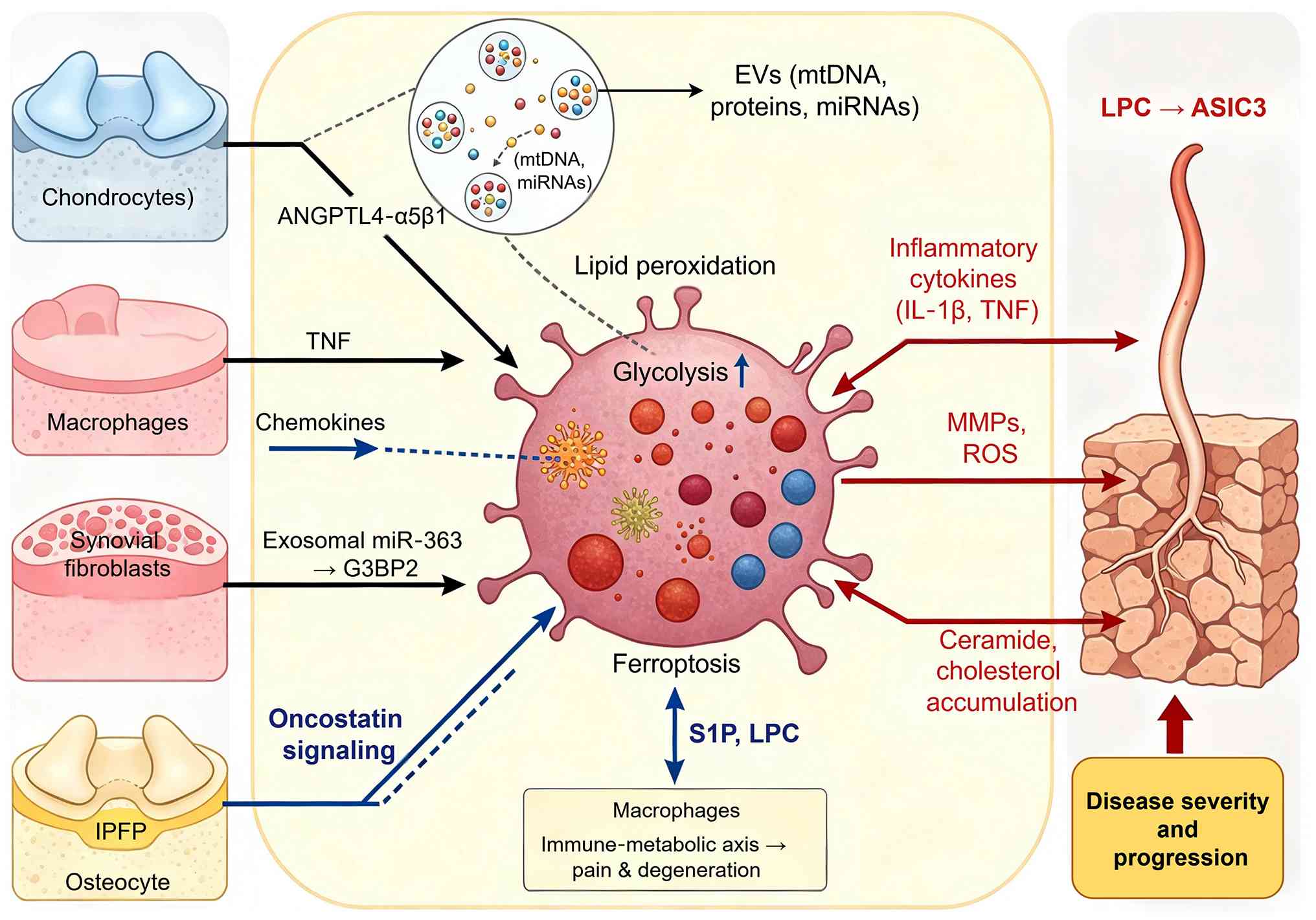 | Figure 3Multi-cellular crosstalk and
inter-tissue communication in the OA joint. The figure was
generated using Figdraw (www.figdraw.com; copyright code: WYWSUb4bf1). OA,
osteoarthritis; FB, fibroblast; Mac, macrophage; Chon, chondrocyte;
EV, extracellular vesicle; IPFP, infrapatellar fat pad; S1P,
sphingosine-1-phosphate; LPC, lysophosphatidylcholine; ASIC3,
acid-sensing ion channel 3; ANGPTL4, angiopoietin-like 4;
miR/miRNA, microRNA; G3BP2, G3BP stress granule assembly factor 2;
ROS, reactive oxygen species; mtDNA, mitochondrial DNA; TNF, tumor
necrosis factor; IL, interleukin. |
The OA joint as an integrated organ
system
Mounting evidence has superseded the traditional
view of isolated cartilage degeneration, establishing OA as a
whole-joint disease where all tissues contribute to and are
affected by pathogenic processes. Pandey and Bhutani (70) provided a comprehensive review of
single-cell resolution profiling of joint tissues, emphasizing that
the molecular connectivity among bone, cartilage and synovium must
be understood collectively. Using a multi-tissue human knee
single-cell atlas, Raut et al (71) identified that OA reduces
regenerative tissue stem cells while increasing inflammatory
pain-associated macrophages, directly demonstrating that cell
populations across different compartments change in a coordinated
manner. The structural basis of this integration was further
clarified by Hu et al (72), who showed that mechanical and
structural properties of articular cartilage and subchondral bone
are tightly coupled in human OA knees. From a therapeutic
perspective, Wang et al (73) reviewed that the Notch signalling
pathway regulates bone and cartilage homeostasis in a highly
integrated fashion, suggesting that pathway modulators could
simultaneously affect multiple tissues. A critical appraisal by
Semenistaja et al (74)
noted that while tissue crosstalk is well accepted, most studies
remain descriptive and lack functional validation of the causal
direction between compartments. Collectively, these findings
establish that the OA joint functions as a mechanically and
biochemically connected system where disruption in one compartment
propagates pathology throughout, but prospective interventional
studies are still needed to prove causality.
Chondrocyte-synoviocyte-immune cell
interaction networks
Direct communication among chondrocytes, synovial
fibroblasts and immune cells creates interaction networks that not
only amplify inflammation but also propagate metabolic alterations
across cell types (Fig. 3).
Using scRNA-seq, Kang et al (42) revealed that cell-cell
communication alterations mediated by interactive signalling
pathways are a hallmark of OA cartilage, with particular enrichment
of ligand-receptor pairs involving inflammatory cytokines. In the
synovial compartment, Wang et al (75) mapped single-cell communication
patterns and their intracellular information flow in OA synovial
fibroblasts, identifying that fibroblast-macrophage crosstalk is
dominated by chemokine and TNF signalling axes. Critically, these
ligand-receptor interactions have downstream metabolic
consequences: TNF signalling induces glycolytic reprogramming in
recipient fibroblasts, while chemokine axes promote lipid
biosynthetic pathway activation in macrophages, creating a
feed-forward loop where metabolic dysfunction begets further
inflammatory signalling (75). A
comprehensive review by Chen et al (76) synthesised the role of crosstalk
between synovial cells and chondrocytes in OA, concluding that
targeting this bidirectional communication holds therapeutic
promise. Critically, a study by Laouteouet et al (77) using advanced co-culture systems
uncovered that monocyte-derived macrophage-synovial fibroblast
crosstalk drives an oncostatin signalling network as a key driver
of synovitis in OA, representing a novel therapeutic target.
Collectively, these observations indicate that the OA joint
operates through interconnected ligand-receptor axes that
simultaneously drive inflammation and metabolic reprogramming, with
therapeutic intervention at any node potentially disrupting this
integrated network.
EVs as intercellular messengers
EVs have emerged as critical mediators of
intercellular communication in OA, transporting proteins, lipids
and nucleic acids between cell types and across tissue
compartments. A comprehensive review by Chen et al (78) highlighted the communication role
of EVs in the OA microenvironment, noting that EV cargo reflects
the pathological state of donor cells and can propagate disease
signals across joint tissues. Kong et al (79) further synthesised evidence on
crosstalk among macrophages, chondrocytes and MSCs via EVs,
proposing that EV-based therapies could simultaneously target
multiple cell types. In a mechanistic study, Liu et al
(80) demonstrated that
osteocyte-derived EVs mediate bone-to-cartilage crosstalk and
promote OA progression, providing direct evidence that EVs from
subchondral bone can drive cartilage degeneration. From the IPFP
perspective, Li et al (81) recently reported that EVs from
IPFP-MSCs trigger OA by transferring mitochondrial DNA, revealing a
previously unrecognised pathogenic mechanism where EVs carry
deleterious cargo. On the therapeutic side, Wu et al
(82) engineered TNF-α
preconditioned IPFP-MSC-derived exosomes that enhanced both yield
and therapeutic efficacy for OA, suggesting that EV engineering can
redirect crosstalk from pathogenic to protective. A critical review
by Clarke et al (83)
noted that while EV research has advanced rapidly, standardisation
of isolation, characterisation and quantification methods remains a
major barrier to clinical translation, and most studies lack direct
comparison of EV efficacy across different cell sources. Therefore,
EVs represent both a mechanistic understanding of cell-cell
communication and a potential therapeutic modality, but
methodological harmonisation and head-to-head comparative studies
are urgently needed.
Immune-metabolic axis in OA
progression
The convergence of immune cell activation and
metabolic reprogramming defines a central feedback loop driving OA
progression, where metabolically reprogrammed cells secrete factors
that further alter the metabolic state of neighboring cells. Yin
et al (84) provided a
systematic review of macrophage polarisation during OA disease
progression, establishing that the M1/M2 balance is dynamically
regulated and represents a tractable therapeutic target. A
comprehensive review by Zou et al (85) further emphasised that macrophage
polarisation is central to both OA pathogenesis and treatment,
linking immune status directly to disease outcomes. The mechanistic
basis for this axis was recently elaborated by He et al
(86), who demonstrated that
Songorine modulates macrophage polarisation and metabolic
reprogramming to alleviate inflammation in OA, showing that small
molecules can simultaneously target immune phenotype and cellular
metabolism.
In the context of synovial macrophages, Yin et
al (87) reported that
Emapunil relieves OA by regulating the CD14/Toll-like receptor
4/lymphocyte antigen 96 pathway through translocator protein 18
kDa, providing a new molecular entry point for immune-metabolic
intervention. A multi-omics integration study by Wu et al
(45) revealed
ferroptosis-driven immune microenvironment remodelling in knee OA,
demonstrating that iron-dependent lipid peroxidation in synovial
macrophages links metabolic stress to inflammatory activation.
These findings collectively establish a model wherein metabolic
perturbations in one cell type are transmitted to neighboring cells
through soluble mediators and EVs, creating self-reinforcing
feedback loops that drive disease progression. From a translational
perspective, Henry and O'Neill (88) critically appraised that metabolic
reprogramming in stromal and immune cells presents therapeutic
possibilities, but cautioned that targeting metabolism may have
off-tissue effects and that most preclinical studies use young,
healthy animals that do not fully recapitulate human OA.
Furthermore, a spatial transcriptomic study by Wang et al
(89) uncovered the spatial and
signalling dynamics of immune infiltration in OA synovium,
revealing that immune-metabolic interactions are highly localised
and vary across synovial subregions. Collectively, these findings
establish the immune-metabolic axis as an integrating hub for
multi-cellular crosstalk in OA, but prospective validation in
well-phenotyped patient cohorts and development of tissue-specific
delivery strategies remain critical challenges.
Molecular subtyping, endotypes, and
precision medicine in OA
The heterogeneity of OA has long hindered the
development of disease-modifying therapies. Emerging evidence has
shifted the paradigm from a one-size-fits-all approach toward
molecularly guided patient stratification. This section synthesizes
current advances in clinical phenotyping, multi-omics-driven
endotyping and precision medicine strategies that collectively aim
to deliver the right treatment to the right patient at the right
time (Table III).
| Table IIIMolecular subtyping, endotypes and
precision medicine studies in OA. |
Table III
Molecular subtyping, endotypes and
precision medicine studies in OA.
| Authors, year | Study type | Omics data
types | Analytical
approach | Key findings | Endotype/subtype
characteristics | (Refs.) |
|---|
| Rockel et
al, 2025 | Original
research | Multi omics
(metabolomics, microRNAs) from three biofluids | Multimodal deep
learning (VAE based clustering) | Identified three
distinct KOA endotypes that predicted 1-year post TKA pain/function
outcomes | Three endotypes
with differential post-surgery prognosis | (97) |
| Huang et al,
2024 | Original
research | Metabolomics,
genomics | Integrative
multi-omics, pathway enrichment | Discovered two hip
OA endotypes with differential amino acid vs. lipid metabolism,
correlating with radiographic progression rates | Endotype 1: Amino
acid metabolism perturbation; Endotype 2: Lipid metabolism
dysregulation | (49) |
| Fan et al,
2024 | Original
research | scRNA-seq, bulk
transcriptomics, spatial transcriptomics | Multi omics
integration, cell-type annotation | Uncovered
inflammatory and pre hypertrophic chondrocyte subpopulations as key
drivers of cartilage degeneration | inflammatory and
pre-hypertrophic chondrocyte subpopulations as cellular level
endotype associated populations | (17) |
| Karsdal et
al, 2025 | Perspective/expert
opinion | Biochemical
markers, clinical data | OARSI symposium
synthesis | Proposed the
inflammatory endotype as a feasible target for clinical
development, with enrichment strategies reducing trial sample
sizes | Inflammatory
endotype (elevated synovial cytokines, acute phase reactants) | (98) |
| Thudium et
al, 2025 | Perspective/expert
opinion | Biochemical
markers | Biomarker panel
analysis | Outlined practical
tools for single-patient endotyping and considerations for
recognizing inflammatory endotype in clinical trials | Inflammatory
molecular endotype as clinically actionable | (99) |
| Hannani et
al, 2025 | Original
research | Biochemical markers
(biomarker panel) | Longitudinal
stability analysis | Demonstrated
molecular endotypes of knee OA remain stable over 12-24 months,
supporting their utility as clinical trial stratification
tools | Structurally stable
molecular endotypes | (102) |
| Mobasheri &
Loeser, 2024 | Perspective/expert
opinion | Clinical
phenotypes, molecular markers | Hierarchical
taxonomy framework | Articulated
clinical phenotypes → molecular endotypes → theratypes as
hierarchical taxonomy for OA therapeutic development | Hierarchical
endotype theratype framework | (91) |
| Ghirardi et
al, 2024 | Perspective/expert
opinion | Chronic joint
diseases | Molecular
taxonomy | Argued defining
theratypes enables repurposing of existing drugs for molecularly
defined patient subsets | Theratype enabled
drug repurposing | (104) |
| Wang et al,
2024 | Original
research | Bulk RNA seq, scRNA
seq | Integrative multi
omics, machine learning | Characterized
Hedgehog pathway features in senescence associated OA, identifying
potential theratype specific targets | Hedgehog pathway
defined theratype | (105) |
| Wu et al,
2024 | Original
research | Multi omics | Integrative
analysis, 7 ML algorithms | Characterized
mitochondrial features in OA; proposed mitochondrial dysfunction
defines a distinct theratype amenable to metabolic
interventions | Mitochondrial
dysfunction theratype | (106) |
| Ojha et al,
2025 | Original
research | Metabolomics (bone
marrow), scRNA-seq | Multi omics
integration at cell type resolution | Reconstructed gene
metabolite networks assigned to specific cell populations;
demonstrated cell-type-specific networks can nominate
theratype-specific drug targets | Cell type specific
theratype targets | (30) |
| Lu et al,
2026 | Original
research | Multi-omics (blood
based) | Integrated multi
omics, gene signature analysis | Identified
glutamine metabolism related gene signature as diagnostic marker
and therapeutic target; signature correlated with OA severity
grades | Glutamine
metabolism defined theratype | (51) |
| Hao et al,
2025 | Original
research | Multi omics | Integrative
analysis | Identified OA
associated chondrocyte subpopulations and key gene regulating
drugs, providing direct bridge from molecular subtyping to drug
repurposing | Chondrocyte
subpopulation specific theratypes | (107) |
Clinical phenotypes vs. molecular
endotypes
Traditional OA classification has relied on
clinical features such as joint involvement, pain patterns and
radiographic severity. However, the heterogeneity of OA has long
hindered the development of disease-modifying therapies, prompting
a shift toward molecularly guided patient stratification. A
hierarchical taxonomy has been proposed to organize this
complexity: Clinical phenotypes (observable patient
characteristics), molecular endotypes (distinct underlying
biological mechanisms) and theratypes (endotypes with validated
differential treatment responses) (90,91). This framework provides a
structured path from molecular discovery to precision therapeutics.
However, it must be acknowledged that while endotype discovery has
advanced considerably, the validation of theratypes, demonstrating
that a molecularly defined patient subgroup responds differently to
a specific therapy, has not yet been achieved in OA. This critical
gap is emphasized throughout the present section.
Mobasheri et al (90,92) distinguished clinical phenotypes,
observable patient characteristics, from molecular endotypes, which
are defined by distinct underlying biological mechanisms. Henrotin
(93) emphasized that
biochemical markers reflecting cartilage degradation, synovial
inflammation and bone turnover can help stratify patients beyond
conventional imaging. Hannani et al (94) provided a systematic review of
validated biomarkers and proposed that combinations of soluble
markers define discrete molecular endotypes with prognostic value.
By contrast, Pattappa et al (95) critically noted that
terminological inconsistency between preclinical and clinical
studies hampers translation, as basic research often uses
'endotype' differently from clinical investigators. Karalilova
et al (96) recently
demonstrated that ultrasound-detected synovitis phenotypes
correlate with distinct biomarker profiles, suggesting that
imaging-based phenotyping can approximate underlying endotypes. A
critical appraisal of this literature reveals that while numerous
candidate biomarkers exist, few have been prospectively validated
for endotype assignment and most studies remain cross-sectional
rather than longitudinal.
Multi-omics-driven molecular
classification
Integrative multi-omics approaches have enabled
unbiased discovery of OA molecular subtypes. Rockel et al
(97) employed deep
learning-based clustering of multi-omic data from three biofluids
and identified three distinct knee OA endotypes that predicted
post-arthroplasty pain outcomes, demonstrating the clinical utility
of data-driven classification. Huang et al (49) performed multi-omics integrative
analyses of hip OA and discovered two endotypes characterized by
differential amino acid vs. lipid metabolism perturbations, which
correlated with radiographic progression rates. As discussed in
Section 2, Fan et al (17) integrated single-cell and bulk
transcriptomics with spatial data to uncover inflammatory and
prehypertrophic chondrocyte subpopulations. Similarly, the lipid
metabolic signature identified by Wang et al (32) supports the feasibility of
metabolic endotyping using peripheral biofluids. Welsing et
al (52) provided a
comprehensive review of omics-driven insights and concluded that
while molecular classification has advanced considerably,
standardization of computational pipelines and prospective
validation remain critical gaps. Collectively, these studies
establish that multi-omics integration yields reproducible
molecular subtypes, but head-to-head comparisons across cohorts and
platforms are lacking.
Endotype-driven trial design and targeted
therapies
The identification of molecular endotypes has
direct implications for clinical trial design and therapeutic
development. Karsdal et al (98) synthesized discussions from the
Osteoarthritis Research Society International clinical trials
symposium and proposed that the inflammatory endotype, which is
characterized by elevated synovial cytokines and acute-phase
reactants, represents a feasible target for clinical development,
with enrichment strategies potentially reducing trial sample sizes.
Thudium et al (99)
further elaborated that diagnosing, treating and monitoring the
inflammatory endotype requires a panel of validated biochemical
markers, including C-reactive protein and matrix
metalloproteinase-derived fragments. Kim et al (100) comprehensively reviewed the OA
drug development pipeline and emphasized that most failed
disease-modifying osteoarthritis drug (DMOAD) trials did not employ
molecular stratification, potentially masking efficacy in
responsive subgroups. Jenei-Lanzl et al (101) critically appraised emerging
concepts in DMOAD development and argued that endotype-driven
enrichment should become standard practice, though they cautioned
that regulatory frameworks for biomarker-guided trial design are
still evolving. Hannani et al (102) provided longitudinal evidence
that molecular endotypes of patients with knee OA remain stable
over 12-24 months, supporting their utility as stratification tools
for clinical trials. Oo (103)
comprehensively reviewed prospects for DMOADs and concluded that
endotype-driven approaches are necessary but not sufficient;
concomitant advances in imaging and patient-reported outcomes are
equally essential. A critical synthesis of this evidence indicates
that while the conceptual framework for endotype-driven trials is
well established, prospective implementation in phase 3 studies
remains limited.
From endotypes to theratypes and target
identification
Translating molecular endotypes into actionable
therapeutic strategies requires the concept of 'theratypes',
endotypes with validated treatment responses. Mobasheri and Loeser
(91) articulated this
framework, proposing that clinical phenotypes, molecular endotypes
and theratypes form a hierarchical taxonomy for OA therapeutic
development. Ghirardi et al (104) extended this concept to chronic
joint diseases generally, arguing that defining theratypes enables
repurposing of existing drugs for molecularly defined patient
subsets.
Several studies have identified potential
theratype-specific targets through multi-omics integration
(30,51,105-107). Wang et al (105) employed integrative multi-omics
and machine learning to characterize Hedgehog pathway features in
senescence-associated OA, identifying potential theratype-specific
targets. Wu et al (106)
similarly characterized mitochondrial features in OA through
multi-omics integration, proposing that mitochondrial dysfunction
defines a distinct theratype amenable to metabolic interventions.
Ojha et al (30)
developed a multi-omics integration framework at cellular
resolution to uncover gene-metabolite mechanisms underlying OA
heterogeneity, demonstrating that cell-type-specific networks can
nominate theratype-specific drug targets. Lu et al (51) identified a glutamine
metabolism-related gene signature as both a diagnostic marker and
therapeutic target, exemplifying how multi-omics can directly
inform target discovery. Hao et al (107) identified OA-associated
chondrocyte subpopulations and key gene-regulating drugs through
multi-omics analysis, providing a direct bridge from molecular
subtyping to drug repurposing.
Critically, however, none of these studies have
demonstrated differential treatment response in endotype-defined
patient subgroups, the defining feature of a true theratype. As
noted by Winthrop et al (108), the broader rheumatology
community still faces substantial unmet needs in translating
molecular taxonomy into clinical practice. The path from endotype
to theratype requires iterative integration of multi-omics
discovery, biomarker development, and crucially, endotype-enriched
clinical trials that prospectively validate differential treatment
responses. This represents the central translational gap in the
field.
Integrative bioinformatics, machine learning
and clinical translation
Over the past five years, the exponential growth of
multi-omics data in OA has driven the development of advanced
computational strategies for meaningful integration and
interpretation. Machine learning and artificial intelligence (AI)
have become indispensable tools for pattern recognition, molecular
subtyping and predictive modeling across imaging and omics domains,
as summarized in Table IV.
These approaches have enabled translational genomic discoveries,
including the identification of diagnostic and prognostic
biomarkers, yet persistent roadblocks ranging from batch effects
and data heterogeneity to poor model generalizability and lack of
prospective validation still hinder clinical implementation.
| Table IVSummary of integrative
bioinformatics, machine learning and translational genomics studies
in OA. |
Table IV
Summary of integrative
bioinformatics, machine learning and translational genomics studies
in OA.
| Authors, year |
Method/algorithm | Data source/omics
layer | Application
domain | Main findings/key
outcome | Key
limitation(s) | (Refs.) |
|---|
| He et al,
2025 | Seven ML algorithms
(LASSO, RF, GBM, XGBoost, decision tree, bagging) + WGCNA | Bulk and single
cell RNA seq transcriptomics | Immune metabolic
signature discovery | Identified 13 hub
immune metabolism genes (including CX3CR1, ADIPOQ, IL17RA, SPP1);
stratified patients into two subgroups with distinct immune/drug
response profiles | In vitro
validation only; no prospective clinical validation | (33) |
| Jamshidi et
al, 2026 | ML/DL prognostic
model (LightGBM, XGBoost, neural network) | Serum
metabolomics | Early prediction of
knee structural OA progression | Metabolomic
signatures outperformed clinical variables alone; interpretable
model with SHAP analysis; six metabolite panel achieved excellent
predictive performance (AUC 0.89 in validation) | Cross cohort
heterogeneity; no external validation in diverse populations | (109) |
| Kang et al,
2025 | LightGBM + SHAP
explainability | Plasma proteomics
(1,463 proteins, n=45,307, UK Biobank) | Future OA risk
prediction | COL9A1 and CRTAC1
predicted incident OA (HR 1.54 and 1.65); 5-10 year prediction AUC
~0.72-0.82; protein trajectories deviate >10 years pre
onset | UK Biobank
primarily European ancestry; lacks cross population validation | (114) |
| Lu et al,
2026 | WGCNA + LASSO + ML
pipeline | Multi tissue
transcriptomics (cartilage, synovium, bone, blood) + plasma
metabolomics | Diagnostic
biomarker and therapeutic target discovery | Identified 3 gene
signature (F13A1, IRS2, RELA); AUC up to 0.966; plasma creatine as
severity predictor; drug repurposing candidates nominated | Small validation
cohort (n=62); cross sectional design | (51) |
| Yan et al,
2025 | LASSO, RF +
Mendelian randomization + scRNA seq | GWAS, eQTL,
transcriptomics, scRNA seq, in vitro/in vivo
experiments | Diagnostic
biomarker discovery | FGF1 identified as
diagnostic biomarker (AUC 1.000/0.790/0.761); MR supports causal
link (OR 1.04, 95% CI 1.002-1.081); validated via RAS MAPK
pathway | MR effect size
modest; functional validation in animal models only | (113) |
| Pang et al
2024 | Multiple ML methods
+ WGCNA + two sample MR | Transcriptomics
(GSE89408, GSE143514) + GWAS (n=24,955 cases, 378,169
controls) | Immune related
biomarker discovery | Identified three
immune biomarkers (FCER1G, HLA DMB, HLA DPA1); MR supports causal
effects (OR 1.118, 1.057 and 1.030) | Expression data
from bulk tissues; cell type specificity undetermined | (115) |
| Liao et al,
2026 | Multi omics
integration (genetic correlation, transcriptomics, proteomics) | GWAS + eQTL +
pQTL | Shared genetic
architecture of OA and metabolic traits | Uncovered shared
genetic mechanisms between OA and metabolic traits; identified
novel OA MT genes, proteins and pathways; lipid metabolism genes
represent druggable targets | Functional
validation lacking; tissue specificity remains undetermined | (116) |
| Tu et al,
2025 | WGCNA + machine
learning + scRNA seq + flow cytometry | Multi tissue
transcriptomics (4 joint tissues) + scRNA seq (217,983 cells) +
peripheral blood | Age specific blood
biomarkers | Identified 5 gene
blood panel (MAPK1, MAP3K8, ING1, LDLR, NUP153) with AUC 0.966; age
specific performance (elderly AUC 0.8 vs. younger 0.7); B-cell
remodeling in elderly OA | Cross sectional
design; causal direction not established | (117) |
| Rafiei et
al, 2025 | Random forest
regression | Self reported
patient information + functional measures (GLA:D registry) | Personalized pain
prediction after exercise/education | Concise 6 variable
model correctly predicted pain change in 58% of cases (vs. 51%
using average values); R2 0.31-0.32 | Modest predictive
performance; guidance needed on clinically acceptable accuracy
threshold | (110) |
Computational strategies for multi-omics
integration
Integrating heterogeneous omics data requires
sophisticated bioinformatic pipelines that can handle
dimensionality, batch effects and biological complexity. As
summarized in Table IV and
discussed in Section 6, unsupervised learning approaches such as
those employed by Rockel et al (97) have demonstrated utility in
patient stratification through multi-omic clustering. Similarly,
integrative multi-omics analyses have enabled the discovery of
molecular endotypes in OA (33,49). At the cellular resolution, Ojha
et al (30) developed a
framework that assigns metabolic signatures to specific cell
populations, addressing the limitation of tissue-level averaging.
In a critical appraisal, Liu et al (5) noted that while transfer learning
and graph neural networks are beginning to address cross-platform
batch effects, these methods remain inaccessible to many clinical
researchers. Taken together, these computational strategies have
established proof of concept for multi-omics integration, but
standardized pipelines and prospective validation cohorts are
urgently needed before clinical deployment.
Machine learning (ML) and AI
applications
ML has become an essential tool for integrating and
interpreting multi-omics data in OA research, enabling pattern
recognition, molecular subtyping and predictive modeling. As
detailed in Section 6, Rockel et al (97) employed deep learning-based
clustering for OA endotyping, while He et al (33) applied multiple ML algorithms to
identify immune-metabolic signatures. Beyond these, Jamshidi et
al (109) developed an
interpretable metabolomic-driven ML model for early prediction of
knee structural OA progression, demonstrating that serum metabolite
signatures outperform clinical variables alone, with a
six-metabolite panel achieving excellent predictive performance in
validation cohorts. Rafiei et al (110) developed personalized prediction
models for changes in knee pain among patients participating in
supervised exercise and education, showing that ML can inform
patient-specific treatment responses. These studies collectively
illustrate that ML approaches, when applied directly to multi-omics
data, can generate clinically relevant insights ranging from
molecular endotyping to prognostic prediction.
A critical appraisal reveals that while ML models
achieve promising performance in controlled research settings,
studies combining ML with multi-omics data in OA remain relatively
limited. Most published ML applications in OA focus on radiographic
image analysis rather than molecular data integration. Furthermore,
the translation of ML-driven omics models to diverse clinical
settings is hampered by data heterogeneity, lack of
interpretability, cross-cohort generalizability and insufficient
prospective validation (52,111). As Russell et al
(112) noted, until ML systems
consistently outperform conventional approaches in well-designed
prospective trials, they should be viewed as decision support tools
rather than replacements for clinical judgment.
Translational genomics and multi-omics
biomarker discovery
Multi-omics integration coupled with translational
genomics has accelerated the discovery of diagnostic and prognostic
biomarkers for OA. Lu et al (51) identified a glutamine
metabolism-related gene signature as both a diagnostic marker and
therapeutic target through integrated multi-omics analysis, with
the signature correlating with OA severity grades. Yan et al
(113) performed multi-omics
integration to identify fibroblast growth factor 1 as a diagnostic
biomarker for OA, validating their findings using Mendelian
randomization, transcriptomics and in vitro experiments.
Kang et al (114)
demonstrated that plasma proteomic profiles predict individual
future OA risk, with a panel of 12 proteins achieving high
predictive accuracy years before radiographic onset. Pang et
al (115) employed
Mendelian randomization and transcriptome analysis to identify
immune-related biomarkers for OA, establishing causal relationships
between specific cytokines and disease risk. Liao et al
(116) unraveled the shared
genetic architecture of OA and metabolic traits through multi-omics
insights, revealing that lipid metabolism genes contribute to both
conditions and may represent druggable targets. Tu et al
(117) performed multi-omics
profiling of blood samples and identified age-specific biomarkers,
demonstrating that aging drives B-cell remodeling in OA and that
biomarker performance varies across age strata. A critical
appraisal of these biomarker studies reveals that while discovery
phases are increasingly robust, few candidates have been
prospectively validated in independent cohorts, and even fewer have
been integrated into clinical decision tools. Furthermore, as noted
by Welsing et al (52),
cross-sectional associations between molecular signatures and
disease severity are abundant, but longitudinal studies linking
baseline biomarkers to long-term clinical outcomes remain
sparse.
Bottlenecks and future directions for
clinical implementation
Despite substantial progress, multiple roadblocks
hinder the clinical translation of integrative bioinformatics and
AI tools for OA. Glinkowski et al (118) reviewed the clinical
performance, limitations and translational readiness of AI in
orthopaedics, concluding that most models lack external validation,
regulatory clearance, and integration into clinical workflows. Luo
et al (119) highlighted
the translational gap between algorithmic development and
real-world implementation, noting that issues of data privacy,
model interpretability, and health economics are rarely addressed
in published studies. Awasthi et al (120) discussed future prospects for AI
in OA diagnosis and treatment, emphasizing that prospective
randomized trials comparing AI-guided vs. standard care are needed
before widespread adoption. Kim et al (121) identified key challenges
including data standardization, algorithm generalizability and
clinician acceptance as critical barriers to AI adoption in
orthopaedic practice. Fairley et al (122) conducted a systematic scoping
review of generative AI in OA and found that while large language
models and image generation tools show promise for patient
education and synthetic data creation, their current applications
remain experimental without proven clinical utility. A critical
synthesis indicates that the path forward requires not only
technical improvements in model robustness and interpretability but
also concerted efforts in prospective validation, regulatory
science and health economic assessment. As Russell et al
(112) forcefully argued, until
AI systems consistently outperform expert clinicians in
well-designed prospective trials, they should be viewed as decision
support tools rather than replacements for clinical judgment.
Therefore, the successful translation of integrative bioinformatics
and AI into OA precision medicine will depend on interdisciplinary
collaboration, rigorous validation frameworks and a clear focus on
patient-centered outcomes.
Future perspectives
The multi-omics landscape of OA has advanced
considerably, yet several frontiers remain underexplored. Emerging
technologies and conceptual shifts promise to address current
limitations and accelerate clinical translation. However, a
critical appraisal of feasibility, cost and scalability is
essential to distinguish realistically achievable advances from
longer-term aspirations. This section prioritizes opportunities
according to their anticipated timeframes for clinical
translation.
Realistically achievable in the next 5
years
The application of AI and machine learning
represents another transformative direction. Sharma (111) recently synthesized how omics
data integration with AI can enhance patient stratification, though
prospective validation in independent cohorts remains scarce. Ou
et al (123)
comprehensively reviewed AI applications across OA clinical,
imaging and omics domains, concluding that deep learning excels at
pattern recognition but generalizability across diverse populations
is unproven. Given the rapid pace of computational method
development and the increasing availability of public multi-omics
datasets, AI-driven endotyping and biomarker discovery are
realistically achievable within 5 years, provided that prospective
validation cohorts are established.
Drug repurposing based on multi-omics-defined
molecular subtypes also represents a near-term opportunity.
Kuswanto and Baker (124)
systematically evaluated drug repurposing opportunities for OA,
arguing that molecularly defined patient subgroups may respond to
existing agents targeting metabolic pathways. Maroun et al
(125) identified
senescence-regulatory factors as both circulating biomarkers and
therapeutic targets, suggesting that cellular senescence represents
a tractable node for intervention. Repurposing of approved drugs
for endotype-defined patient subsets is feasible within 5 years, as
it bypasses de novo drug development timelines and leverages
existing safety data, though endotype-enriched trial designs remain
to be validated.
Medium-term opportunities with
significant barriers
The integration of multi-omics with human-relevant
experimental models holds substantial promise but faces
considerable technical and standardization challenges.
Osteochondral organoids offer a direct platform for functional
validation of the cell type-specific gene-metabolite networks
identified from patient multi-omics datasets. This integrated
paradigm, which combines multi-omics discovery with organoid-based
functional testing, enables researchers to interrogate whether
specific gene-metabolite perturbations observed in patient tissues,
such as dysregulated glycerophospholipid metabolism in inflammatory
chondrocyte subpopulations, are sufficient to drive pathological
phenotypes when recapitulated in a controlled 3D microenvironment
(17,32). Furthermore, osteochondral
organoids allow for systematic manipulation of candidate metabolic
regulators, such as ACSL4 or SREBP1, while monitoring downstream
metabolomic and phenotypic outcomes (32,66). Synovium-cartilage organoid
co-culture systems further enable validation of cell-cell crosstalk
mechanisms identified in single-cell transcriptomic studies
(3). Such functional validation
is essential because multi-omics data, while powerful for
hypothesis generation, cannot distinguish causal drivers from
passive correlates. Complementing this, microphysiological systems
now allow co-culture of multiple joint tissues under controlled
mechanical and biochemical conditions, facilitating causal testing
of cell-type-specific metabolic hypotheses (126). However, these platforms remain
technically challenging to standardize, have limited throughput and
lack validation against human disease progression. Widespread
adoption within 5 years is unlikely; rather, they will serve as
specialized mechanistic tools in research settings.
Long-term challenges
The emerging concept of 'pre-OA' offers
opportunities for early intervention. Multi-omics evidence has
begun to distinguish pre-OA from mild OA at the molecular level.
Del Río (127) proposed a
multidimensional definition of pre-OA that incorporates molecular,
imaging and clinical parameters, arguing that subclinical detection
is essential for disease modification. Lipidomic profiling has
provided particularly compelling evidence in this regard. Eichner
et al (128)
systematically quantified 91 lipid species from 6 major classes
(phosphatidylcholine, phosphatidylethanolamine, phosphatidylserine,
phosphatidylinositol, phosphatidic acid and sphingomyelin) in serum
and synovial fluid from 44 joint-healthy donors and 58 early OA or
late OA patients, demonstrating that significant lipidomic
alterations are already detectable at an average Outerbridge score
of <2, before radiologic detection is possible. Notably, nearly
10% of phospholipid species were elevated exclusively in OA serum,
indicating a systemic metabolic response that parallels the local
lipid metabolic response to OA (128). Single-cell transcriptomics has
further enabled the identification of early cell-state transitions,
including pre-hypertrophic chondrocyte populations that precede
overt cartilage degeneration (17). Spatial multi-omics has revealed
distinct synovitis stages, namely quiescent, microvasculopathic,
pre-fibrotic and post-fibrotic, providing a molecular trajectory of
disease progression from subclinical inflammation to established
pathology (26).
Longitudinal cohort studies have substantiated the
utility of molecular screening for early OA detection. A nested
case-control study within the UK Biobank (n=30, 490 incident OA
cases) demonstrated that systemic metabolic perturbations,
particularly involving energy metabolism (lactate, citrate, amino
acids), chronic low-grade inflammation (glycoprotein acetyls) and
adverse lipid composition (apolipoprotein B, very low density
lipoprotein cholesterol), are detectable a median of 7.3 years
before OA diagnosis (129).
Large-scale proteomic profiling of 45,307 UK Biobank participants
further revealed that plasma protein trajectories, particularly
collagen type IX alpha 1 chain and cartilage acidic protein 1,
begin to deviate from normal more than a decade before OA onset
(114). Similarly, a
case-cohort study within the OA Initiative (603 participants, 237
incident knee OA cases, 8-year follow-up) demonstrated that
blood-based metabolites reflective of the gut microbiome, including
3-(3-hydroxyphenyl) propanoic acid, N,N-dimethylglycine and butyric
acid, are associated with incident knee OA risk (130). The APPROACH cohort further
demonstrated that biomarker-based molecular endotypes remain stable
over 6-, 12- and 24-month follow-ups, with more than half of knee
OA participants exhibiting a longitudinally stable endotype
(average overall stability, 55%; Fleiss' Kappa, 0.53; 95% CI
0.46-0.60), supporting the applicability of biomarker-based
subtyping in clinical trial settings (102). Despite these promising
findings, pre-OA detection requires prospective longitudinal
cohorts with deep phenotyping, as exemplified by the OPTIMA-C study
protocol (131), and validation
of biomarkers across diverse populations. This remains a long-term
goal beyond 5-10 years, as it requires large-scale, expensive
longitudinal studies with extended follow-up.
The critical barrier of cost and
scalability
A fundamental roadblock that underpins many of the
challenges discussed above is the prohibitive cost and limited
scalability of current multi-omics technologies. Spatial
transcriptomics and single-cell metabolomics, while offering
unprecedented resolution, remain prohibitively expensive for
application to large patient cohorts. The cost of generating
spatially resolved omics data from a single tissue section can
exceed thousands of dollars, making it infeasible for routine
clinical application or large-scale biomarker discovery studies.
Similarly, comprehensive multi-omics profiling combining genomics,
transcriptomics, proteomics and metabolomics from the same patient
sample is currently confined to small, well-funded research
cohorts. As noted by several comprehensive reviews, the lack of
cost-effective, high-throughput platforms for multi-omics analysis
represents a major translational roadblock that limits both
discovery-phase sample sizes and clinical validation studies
(5,14,52). Furthermore, the computational
infrastructure required to store, process and integrate these
massive datasets is not readily available in most clinical
settings. Acknowledging these cost and scalability barriers is
essential for setting realistic expectations for clinical
translation.
Off-target risks and delivery limitations
of lipid modulators
A critical barrier to translating lipid-targeted
strategies into clinical practice is that most currently discussed
therapeutic targets remain at preclinical cell or animal model
stages, with limited data on off-target effects and tissue-specific
delivery in human joints (52).
Systemic lipid-modulating agents repurposed for OA, including
statins and PPAR agonists, carry well-documented off-target risks.
Statins have been associated with increased OA risk in large-scale
observational studies (odds ratio, 1.099; 95% CI 1.002-1.206), with
higher doses conferring greater risk, and Mendelian randomization
has revealed population-specific effects on skeletal outcomes
(2,6). PPAR agonists present cardiovascular
safety concerns with limited clinical evidence for OA-specific
efficacy (5). For novel
multi-omics-identified targets such as SREBP1 and ACSL4 (32,66,68), systemic inhibition could
theoretically disrupt hepatic lipid metabolism and essential fatty
acid homeostasis in other tissues, yet the off-target landscape
remains largely uncharacterized. Beyond molecular risks, the unique
anatomical features of the joint, including rapid synovial fluid
turnover and dense negatively charged cartilage extracellular
matrix, pose formidable barriers to drug delivery, limiting
therapeutic accumulation at target cells (14,15). Lipid-based nanocarriers have been
developed to address these limitations, but they introduce their
own safety challenges, including potential inflammatory responses
and unresolved issues of long-term biocompatibility, scalable
manufacturing and regulatory classification (14). Emerging strategies such as
microenvironment-responsive delivery systems and
chondrocyte-biomimetic nanoparticles remain largely at preclinical
stages (12,13). In summary, the translation of
multi-omics-identified lipid metabolic targets requires systematic
evaluation of off-target risks, development of delivery strategies
overcoming joint-specific barriers and rigorous safety assessment
of nanocarrier systems themselves (14).
In summary, the roadmap to precision medicine in OA
requires prioritized, realistic goals: Short-term (5 years):
AI-driven endotyping and drug repurposing based on existing omics
data, with prospective validation in well-phenotyped cohorts;
medium-term (5-10 years): Integration of organoid and
microphysiological systems for mechanistic validation of
omics-derived hypotheses, alongside development of cost-reduced
multi-omics platforms; and long-term (>10 years): Pre-OA
detection strategies and endotype-enriched clinical trials of novel
therapeutics. Achieving these goals will demand interdisciplinary
collaboration among omics scientists, computational biologists,
bioengineers and clinician-researchers, as well as sustained
investment in scalable technologies and prospective longitudinal
cohorts.
Conclusions
Integrating single cell transcriptomics,
metabolomics and lipidomics has uncovered cell type-specific
gene-metabolite networks in OA. Dysregulated glycerophospholipid
and sphingolipid metabolism across chondrocytes, synovial
fibroblasts and immune cells correlates with disease severity.
These insights enable molecular endotyping and support the
conceptual framework for precision medicine. However, the
validation of theratypes, demonstrating differential treatment
responses in endotype-defined patient subgroups, has not yet been
achieved in OA. Prospective, endotype-enriched clinical trials
represent the critical next step for translating multi-omics
discoveries into clinical practice.
Availability of data and materials
Not applicable.
Authors' contributions
YY and RZ contributed equally to conceptualization,
literature review and drafting the original manuscript. YZ and DY
performed the literature search and figure preparation. JX and CZ
critically revised the manuscript for important intellectual
content and gave final approval. All authors agree to be
accountable for all aspects of the work and have read and approved
the final version of the manuscript. Data authentication is not
applicable.
Ethics approval and consent to
participate
Not applicable.
Patient consent for publication
Not applicable.
Competing interests
The authors declare that they have no competing
interests.
Use of artificial intelligence tools
During the preparation of this work, artificial
intelligence tools (DeepSeek-V3.2; https://chat.deepseek.com/) were used to improve the
readability and language of the manuscript or to generate images,
and subsequently, the authors revised and edited the content
produced by the artificial intelligence tools as necessary, taking
full responsibility for the ultimate content of the present
manuscript.
Abbreviations:
|
OA
|
osteoarthritis
|
|
scRNA-seq
|
single-cell RNA sequencing
|
|
MSI
|
mass spectrometry imaging
|
|
CH25H
|
cholesterol 25-hydroxylase
|
|
CYP7B1
|
cytochrome P450 family 7 subfamily B
member 1
|
|
RORα
|
retinoic acid-related orphan receptor
α
|
|
LPC
|
lysophosphatidylcholine
|
|
ASIC3
|
acid-sensing ion channel 3
|
|
PPAR
|
peroxisome proliferator-activated
receptor
|
|
LXR
|
liver X receptor
|
|
SREBP1
|
sterol regulatory element-binding
protein 1
|
|
SESN2
|
Sestrin 2
|
|
ABCD2
|
ATP-binding cassette subfamily D
member 2
|
|
ACSL4
|
acyl-CoA synthetase long-chain family
member 4
|
|
LPCAT3
|
LPC acyltransferase 3
|
|
ABCA1
|
ATP-binding cassette subfamily A
member 1
|
|
IPFP
|
infrapatellar fat pad
|
|
EV
|
extracellular vesicle
|
|
DMOAD
|
disease-modifying osteoarthritis
drug
|
|
OARSI
|
Osteoarthritis Research Society
International
|
|
AI
|
artificial intelligence
|
Acknowledgements
Not applicable.
Funding
No funding was received.
References
|
1
|
Vicenti G, Bizzoca D, Carrozzo M, Solarino
G and Moretti B: Multi-omics analysis of synovial fluid: A
promising approach in the study of osteoarthritis. J Biol Regul
Homeost Agents. 32(Suppl 1): S9–S13. 2018.
|
|
2
|
Iijima H, Gilmer G, Wang K, Sivakumar S,
Evans C, Matsui Y and Ambrosio F: Meta-analysis integrated with
multi-omics data analysis to elucidate pathogenic mechanisms of
age-related knee osteoarthritis in mice. J Gerontol A Biol Sci Med
Sci. 77:1321–1334. 2022. View Article : Google Scholar : PubMed/NCBI
|
|
3
|
Bensa A, Salerno M, Boffa A, de Girolamo
L, Laver L, Magalon J, Sánchez M, Tischer T and Filardo G:
Corticosteroid injections for the treatment of osteoarthritis
present a wide spectrum of effects ranging from detrimental to
disease-modifying: A systematic review of preclinical evidence by
the ESSKA orthobiologic initiative. Knee Surg Sports Traumatol
Arthrosc. 32:2725–2745. 2024. View Article : Google Scholar : PubMed/NCBI
|
|
4
|
Bajpai P and Chandra P: Emerging
anti-inflammatory attributes of curcumin: A novel paradigm and
ameliorative attributes for the treatment of osteoarthritis. Curr
Rheumatol Rev. 21:228–238. 2025. View Article : Google Scholar
|
|
5
|
Liu Y, Molchanov V, Brass D and Yang T:
Recent advances in omics and the integration of multi-omics in
osteoarthritis research. Arthritis Res Ther. 27:1002025. View Article : Google Scholar : PubMed/NCBI
|
|
6
|
Wei Y, Qian H, Zhang X, Wang J, Yan H,
Xiao N, Zeng S, Chen B, Yang Q, Lu H, et al: Progress in
multi-omics studies of osteoarthritis. Biomark Res. 13:262025.
View Article : Google Scholar : PubMed/NCBI
|
|
7
|
Gu Y, Hu Y, Zhang H, Wang S, Xu K and Su
J: Single-cell RNA sequencing in osteoarthritis. Cell Prolif.
56:e135172023. View Article : Google Scholar : PubMed/NCBI
|
|
8
|
Danzeng L, Sun Y, He Z, Hou X and Li L:
Single-cell sequencing reveals the immune microenvironment in
osteoarthritis: From heterogeneity to therapeutic targets. Int
Immunopharmacol. 165:1155212025. View Article : Google Scholar : PubMed/NCBI
|
|
9
|
Boffa A, Perucca Orfei C, Sourugeon Y,
Laver L, Magalon J, Sánchez M, Tischer T, de Girolamo L and Filardo
G: Cell-based therapies have disease-modifying effects on
osteoarthritis in animal models. A systematic review by the ESSKA
orthobiologic initiative. Part 2: Bone marrow-derived cell-based
injectable therapies. Knee Surg Sports Traumatol Arthrosc.
31:3230–3242. 2023. View Article : Google Scholar : PubMed/NCBI
|
|
10
|
Sourugeon Y, Boffa A, Perucca Orfei C, de
Girolamo L, Magalon J, Sánchez M, Tischer T, Filardo G and Laver L:
Cell-based therapies have disease-modifying effects on
osteoarthritis in animal models: A systematic review by the ESSKA
orthobiologic initiative. Part 3: Umbilical cord, placenta, and
other sources for cell-based injectable therapies. Knee Surg Sports
Traumatol Arthrosc. 33:1695–1708. 2025. View Article : Google Scholar :
|
|
11
|
D'Arrigo D, Salerno M, De Marziani L,
Boffa A and Filardo G: A call for standardization for secretome and
extracellular vesicles in osteoarthritis: Results show
disease-modifying potential, but protocols are too heterogeneous-a
systematic review. Hum Cell. 37:1243–1275. 2024. View Article : Google Scholar : PubMed/NCBI
|
|
12
|
Tan D, Huang Z, Zhao Z, Chen X, Liu J,
Wang D, Deng Z and Li W: Single-cell sequencing, genetics, and
epigenetics reveal mesenchymal stem cell senescence in
osteoarthritis (review). Int J Mol Med. 53:22024. View Article : Google Scholar
|
|
13
|
Wang T, Ng CY, Ng BZJ, Toh WS and Hui JHP:
Multi-omics analysis of small extracellular vesicles in
osteoarthritis: Bridging the gap between molecular insights and
clinical applications. Burns Trauma. 13:tkaf0232025. View Article : Google Scholar : PubMed/NCBI
|
|
14
|
Rai MF, Collins KH, Lang A, Maerz T,
Geurts J, Ruiz-Romero C, June RK, Ramos Y, Rice SJ, Ali SA, et al:
Three decades of advancements in osteoarthritis research: Insights
from transcriptomic, proteomic, and metabolomic studies.
Osteoarthritis Cartilage. 32:385–397. 2024. View Article : Google Scholar
|
|
15
|
Van Pevenage PM, Birchmier JT and June RK:
Utilizing metabolomics to identify potential biomarkers and
perturbed metabolic pathways in osteoarthritis: A systematic
review. Semin Arthritis Rheum. 59:1521632023. View Article : Google Scholar : PubMed/NCBI
|
|
16
|
Ji Q, Zheng Y, Zhang G, Hu Y, Fan X, Hou
Y, Wen L, Li L, Xu Y, Wang Y and Tang F: Single-cell RNA-seq
analysis reveals the progression of human osteoarthritis. Ann Rheum
Dis. 78:100–110. 2019. View Article : Google Scholar :
|
|
17
|
Fan Y, Bian X, Meng X, Li L, Fu L, Zhang
Y, Wang L, Zhang Y, Gao D, Guo X, et al: Unveiling inflammatory and
prehypertrophic cell populations as key contributors to knee
cartilage degeneration in osteoarthritis using multi-omics data
integration. Ann Rheum Dis. 83:926–944. 2024. View Article : Google Scholar : PubMed/NCBI
|
|
18
|
Liao S, Yang M, Li D, Wu Y, Sun H, Lu J,
Liu X, Deng T, Wang Y, Xie N, et al: Comprehensive bulk and
single-cell transcriptome profiling give useful insights into the
characteristics of osteoarthritis associated synovial macrophages.
Front Immunol. 13:10784142022. View Article : Google Scholar
|
|
19
|
Sebastian A, Hum NR, McCool JL, Wilson SP,
Murugesh DK, Martin KA, Rios-Arce ND, Amiri B, Christiansen BA and
Loots GG: Single-cell RNA-Seq reveals changes in immune landscape
in post-traumatic osteoarthritis. Front Immunol. 13:9380752022.
View Article : Google Scholar : PubMed/NCBI
|
|
20
|
Huang ZY, Luo ZY, Cai YR, Chou CH, Yao ML,
Pei FX, Kraus VB and Zhou ZK: Single cell transcriptomics in human
osteoarthritis synovium and in silico deconvoluted bulk RNA
sequencing. Osteoarthritis Cartilage. 30:475–480. 2022. View Article : Google Scholar : PubMed/NCBI
|
|
21
|
Wang Q, Huang M and Guo JJ: From cells to
clinic: Single-cell transcriptomics shaping the future of
orthopedics. J Orthop Translat. 53:1–11. 2025. View Article : Google Scholar : PubMed/NCBI
|
|
22
|
Fan X, Sun AR, Young RSE, Afara IO,
Hamilton BR, Ong LJY, Crawford R and Prasadam I: Spatial analysis
of the osteoarthritis microenvironment: Techniques, insights, and
applications. Bone Res. 12:72024. View Article : Google Scholar : PubMed/NCBI
|
|
23
|
Fan X, Young RSE, Sun AR, Hamilton BR,
Nedunchezhiyan U, Crawford R, Blanksby SJ and Prasadam I:
Functional mass spectrometry imaging maps phospholipase-A2 enzyme
activity during osteoarthritis progression. Theranostics.
13:4636–4649. 2023. View Article : Google Scholar : PubMed/NCBI
|
|
24
|
Lee YR, Briggs MT, Kuliwaba JS, Jagiello
J, Anderson PH and Hoffmann P: High-resolution N-glycan MALDI mass
spectrometry imaging of subchondral bone tissue microarrays in
patients with knee osteoarthritis. Anal Chem. 95:12640–12647. 2023.
View Article : Google Scholar : PubMed/NCBI
|
|
25
|
Rocha B, Cillero-Pastor B, Ruiz-Romero C,
Paine MRL, Cañete JD, Heeren RMA and Blanco FJ: Identification of a
distinct lipidomic profile in the osteoarthritic synovial membrane
by mass spectrometry imaging. Osteoarthritis Cartilage. 29:750–761.
2021. View Article : Google Scholar : PubMed/NCBI
|
|
26
|
Zhu L, Diao X, Yuan C, Lau CML, Wang J, Wu
W, Mobasheri A, Houard X, Wen C and Cai Z: Integrative spatially
resolved proteomic and metabolomic imaging reveals synovitis
endotypes implicated in osteoarthritis progression. Theranostics.
15:9729–9741. 2025. View Article : Google Scholar : PubMed/NCBI
|
|
27
|
Xie P, Sun AR, Gao F, Li L, Crawford R and
Prasadam I: Resolving microenvironment complexity and cellular
heterogeneity in osteoarthritis via spatial transcriptomics.
Osteoarthritis Cartilage. 34:332–342. 2026. View Article : Google Scholar
|
|
28
|
Kreitmaier P, Katsoula G and Zeggini E:
Insights from multi-omics integration in complex disease primary
tissues. Trends Genet. 39:46–58. 2023. View Article : Google Scholar
|
|
29
|
Huang Z, He Z, Kong Y, Liu Z and Gong L:
Insight into osteoarthritis through integrative analysis of
metabolomics and transcriptomics. Clin Chim Acta. 510:323–329.
2020. View Article : Google Scholar : PubMed/NCBI
|
|
30
|
Ojha A, Chatterjee P, Kippner LE, Stevens
HY, Boden SD, Drissi H, Mautner K, Yeago C, Roy K and Sinha S:
Multi-omics integration at cell type resolution uncovers
gene-metabolite mechanisms underlying osteoarthritis heterogeneity.
bioRxiv [Preprint]: 2025.09.26.678886. 2025.
|
|
31
|
Katsoula G, Kreitmaier P and Zeggini E:
Insights into the molecular landscape of osteoarthritis in human
tissues. Curr Opin Rheumatol. 34:79–90. 2022. View Article : Google Scholar
|
|
32
|
Wang Y, Zeng T, Tang D, Cui H, Wan Y and
Tang H: Integrated multi-omics analyses reveal lipid metabolic
signature in osteoarthritis. J Mol Biol. 437:1688882025. View Article : Google Scholar
|
|
33
|
He H, Zhao X, Zhang B, Zhao S and Wu Y:
Integrated multi-omics and machine learning reveals
immune-metabolic signatures in osteoarthritis: From bulk RNA-seq to
single-cell resolution. Front Immunol. 16:15999302025. View Article : Google Scholar : PubMed/NCBI
|
|
34
|
Wu K, Zhong Z, Chen L, Luo H, Jiang A, Tao
L and Jiang Y: Integrative analysis of bulk and single-cell RNA
sequencing data reveals increased arachidonic acid metabolism in
osteoarthritic chondrocytes. Front Med (Lausanne). 12:15520292025.
View Article : Google Scholar : PubMed/NCBI
|
|
35
|
Wijesinghe SN, Badoume A, Nanus DE,
Sharma-Oates A, Farah H, Certo M, Alnajjar F, Davis ET, Mauro C,
Lindsay MA and Jones SW: Obesity defined molecular endotypes in the
synovium of patients with osteoarthritis provides a rationale for
therapeutic targeting of fibroblast subsets. Clin Transl Med.
13:e12322023. View Article : Google Scholar : PubMed/NCBI
|
|
36
|
Southan J, McHugh E, Walker H and Ismail
HM: Metabolic signature of articular cartilage following mechanical
injury: An integrated transcriptomics and metabolomics analysis.
Front Mol Biosci. 7:5929052020. View Article : Google Scholar
|
|
37
|
Qu Y, Wang Y, Wang S, Yu X, He Y, Lu R,
Chen S, Meng C, Xu H, Pei W, et al: A comprehensive analysis of
single-cell RNA transcriptome reveals unique SPP1+ chondrocytes in
human osteoarthritis. Comput Biol Med. 160:1069262023. View Article : Google Scholar : PubMed/NCBI
|
|
38
|
Matta C, Takács R, Dvir-Ginzberg M,
Richardson SM, Pelttari K, Pattappa G, Risbud MV and Mobasheri A:
Insights into chondrocyte populations in cartilaginous tissues at
the single-cell level. Nat Rev Rheumatol. 21:465–477. 2025.
View Article : Google Scholar : PubMed/NCBI
|
|
39
|
Chen W, Xiao J, Zhou Y, Liu W, Jian J,
Yang J, Chen B, Ye Z, Liu J, Xu X, et al: Curcumenol regulates
Histone H3K27me3 demethylases KDM6B affecting Succinic acid
metabolism to alleviate cartilage degeneration in knee
osteoarthritis. Phytomedicine. 133:1559222024. View Article : Google Scholar : PubMed/NCBI
|
|
40
|
Zhang Z, Cheng D, Dang J, Wang X, Fan H
and Liu D: Decoding endoplasmic reticulum stress on chondrocyte
driving osteoarthritis development through integrating single-cell
and transcriptomic profiling. Int J Med Sci. 22:3906–3923. 2025.
View Article : Google Scholar : PubMed/NCBI
|
|
41
|
Pan B, Yao P, Ma J, Lin X, Zhou L and Lin
C, Zhang Y, Lin B and Lin C: Identification of key biomarkers
related to fibrocartilage chondrocytes for osteoarthritis based on
bulk, single-cell transcriptomic data. Front Immunol.
15:14823612024. View Article : Google Scholar : PubMed/NCBI
|
|
42
|
Kang X, Zhang K, Wang Y, Zhao Y and Lu Y:
Single-cell RNA sequencing analysis of human chondrocytes reveals
cell-cell communication alterations mediated by interactive
signaling pathways in osteoarthritis. Front Cell Dev Biol.
11:10992872023. View Article : Google Scholar : PubMed/NCBI
|
|
43
|
Acharjee A, Wijesinghe SN, Russ D, Gkoutos
G and Jones SW: Cross-species transcriptomics identifies obesity
associated genes between human and mouse studies. J Transl Med.
22:5922024. View Article : Google Scholar : PubMed/NCBI
|
|
44
|
Yang J, Li S, Li Z, Yao L, Liu M, Tong KL,
Xu Q, Yu B, Peng R, Gui T, et al: Targeting YAP1-regulated
glycolysis in fibroblast-like synoviocytes impairs macrophage
infiltration to ameliorate diabetic osteoarthritis progression. Adv
Sci (Weinh). 11:e23046172024. View Article : Google Scholar :
|
|
45
|
Wu Y, Liu J, Yu W, Wang X, Li J and Zeng
W: Single-cell transcriptome and multi-omics integration reveal
ferroptosis-driven immune microenvironment remodeling in knee
osteoarthritis. Front Immunol. 16:16083782025. View Article : Google Scholar : PubMed/NCBI
|
|
46
|
Wei M, Shi X, Tang W, Lv Q, Wu Y and Xu Y:
Shared M0 macrophage infiltration-related gene signatures in
osteoarthritis and osteomyelitis by integrating bulk and
single-cell RNA transcriptome. Eur J Med Res. 30:11112025.
View Article : Google Scholar : PubMed/NCBI
|
|
47
|
Deng M, Jiang Y, Chen Z, Chen K, Cao N,
Huang Y, Quan Z and Chen C: Senescent synovial intimal fibroblasts
aggravate osteoarthritis by regulating macrophage polarization and
chondrocyte phenotype through ANGPTL4-α5β1 axis. Adv Sci (Weinh).
13:e180562026. View Article : Google Scholar
|
|
48
|
Komaravolu RK, Mehta-D'souza P, Conner T,
Allen M, Lumry J, Batushansky A, Pezant NP, Montgomery CG and
Griffin TM: Sex-specific effects of injury and beta-adrenergic
activation on metabolic and inflammatory mediators in a murine
model of post-traumatic osteoarthritis. Osteoarthritis Cartilage.
32:1097–1112. 2024. View Article : Google Scholar : PubMed/NCBI
|
|
49
|
Huang J, Liu M, Zhang H, Sun G, Furey A,
Rahman P and Zhai G: Multi-omics integrative analyses identified
two endotypes of hip osteoarthritis. Metabolites. 14:4802024.
View Article : Google Scholar : PubMed/NCBI
|
|
50
|
Tang S, Yao L, Ruan J, Kang J, Cao Y, Nie
X, Lan W, Zhu Z, Han W, Liu Y, et al: Single-cell atlas of human
infrapatellar fat pad and synovium implicates APOE signaling in
osteoarthritis pathology. Sci Transl Med. 16:eadf45902024.
View Article : Google Scholar : PubMed/NCBI
|
|
51
|
Lu Y, Liu J, Wang S, Gan J, Cao W and Wu
B: Identification and validation of a glutamine metabolism-related
gene signature as a diagnostic and therapeutic target in
osteoarthritis through integrated multi-omics analysis. PeerJ.
14:e207012026. View Article : Google Scholar : PubMed/NCBI
|
|
52
|
Welsing PMJ, El Bouhaddani S, Zhu L,
Nijhof NC, Mastbergen SC, Wen C, Bacardit J, Ruiz-Romero C, Blanco
FJ and Mobasheri A: Omics-driven insights into the molecular
pathways driving osteoarthritis pathogenesis. Connect Tissue Res.
66:323–330. 2025. View Article : Google Scholar : PubMed/NCBI
|
|
53
|
Choi WS, Lee G, Song WH, Koh JT, Yang J,
Kwak JS, Kim HE, Kim SK, Son YO, Nam H, et al: The
CH25H-CYP7B1-RORα axis of cholesterol metabolism regulates
osteoarthritis. Nature. 566:254–258. 2019. View Article : Google Scholar : PubMed/NCBI
|
|
54
|
Lee G, Yang J, Kim SJ, Tran TT, Lee SY,
Park KH, Kwon SH, Chung KH, Koh JT, Huh YH, et al: Enhancement of
intracellular cholesterol efflux in chondrocytes leading to
alleviation of osteoarthritis progression. Arthritis Rheumatol.
77:151–162. 2025. View Article : Google Scholar :
|
|
55
|
Kosinska MK, Mastbergen SC, Liebisch G,
Wilhelm J, Dettmeyer RB, Ishaque B, Rickert M, Schmitz G, Lafeber
FP and Steinmeyer J: Comparative lipidomic analysis of synovial
fluid in human and canine osteoarthritis. Osteoarthritis Cartilage.
24:1470–1478. 2016. View Article : Google Scholar : PubMed/NCBI
|
|
56
|
Steinmeyer J: Phospholipids and
sphingolipids in osteoarthritis. Biomolecules. 15:2502025.
View Article : Google Scholar : PubMed/NCBI
|
|
57
|
Ma H, Liu CM, Shao SQ, Song YY, Zhou J,
Cao LN, Yin HQ and Yin SL: Myriocin alleviates Oleic/Palmitate
induced chondrocyte degeneration via the suppression of ceramide.
Eur Rev Med Pharmacol Sci. 24:12938–12947. 2020.PubMed/NCBI
|
|
58
|
Cherifi C, Latourte A, Vettorazzi S,
Tuckermann J, Provot S, Ea HK, Ledoux A, Casas J, Cuvillier O,
Richette P, et al: Inhibition of sphingosine 1-phosphate protects
mice against chondrocyte catabolism and osteoarthritis.
Osteoarthritis Cartilage. 29:1335–1345. 2021. View Article : Google Scholar : PubMed/NCBI
|
|
59
|
Jacquot F, Khoury S, Labrum B, Delanoe K,
Pidoux L, Barbier J, Delay L, Bayle A, Aissouni Y, Barriere DA, et
al: Lysophosphatidylcholine 16:0 mediates chronic joint pain
associated to rheumatic diseases through acid-sensing ion channel
3. Pain. 163:1999–2013. 2022. View Article : Google Scholar : PubMed/NCBI
|
|
60
|
Pousinis P, Gowler PRW, Burston JJ, Ortori
CA, Chapman V and Barrett DA: Lipidomic identification of plasma
lipids associated with pain behaviour and pathology in a mouse
model of osteoarthritis. Metabolomics. 16:322020. View Article : Google Scholar : PubMed/NCBI
|
|
61
|
Cao C, Shi Y, Zhang X, Li Q, Zhang J, Zhao
F, Meng Q, Dai W, Liu Z, Yan W, et al: Cholesterol-induced LRP3
downregulation promotes cartilage degeneration in osteoarthritis by
targeting Syndecan-4. Nat Commun. 13:71392022. View Article : Google Scholar : PubMed/NCBI
|
|
62
|
Zhao Y, Hasse S, Vaillancourt M, Zhao C,
Davis L, Boilard E, Fortin P, Di Battista J, Poubelle PE and
Bourgoin SG: Phospholipase A1 member A activates fibroblast-like
synoviocytes through the autotaxin-lysophosphatidic acid receptor
axis. Int J Mol Sci. 22:126852021. View Article : Google Scholar : PubMed/NCBI
|
|
63
|
Zhou Q, Ghorasaini M, Cornelis FMF, Hayer
S, Giera M, Monteagudo S and Lories RJ: Temporal profiling of lipid
mediators in synovium and tibial plateau during joint inflammation
in a collagenase-induced mouse model of osteoarthritis.
Osteoarthritis Cartilage. 33:1307–1315. 2025. View Article : Google Scholar : PubMed/NCBI
|
|
64
|
Lee JC: Microarray analysis after adipose
derived mesenchymal stem cells injection in monosodium
iodoacetate-induced osteoarthritis rats. Genes Genomics. 40:25–37.
2018. View Article : Google Scholar : PubMed/NCBI
|
|
65
|
Huang G, Jiang W, Xie W, Lu W, Zhu W and
Deng Z: Role of peroxisome proliferator-activated receptors in
osteoarthritis (review). Mol Med Rep. 23:1592021. View Article : Google Scholar
|
|
66
|
Liu Z, Jiang H, Zhang Z, Jiang R, Wang R,
Li J, Li W, Tan G, Fei Y, Lv Z, et al: SESN2 maintains cartilage
homeostasis by SREBP1-mediated lipid metabolism during
osteoarthritis progression. iScience. 28:1130972025. View Article : Google Scholar : PubMed/NCBI
|
|
67
|
Sun MM and Beier F: Liver X receptor
activation regulates genes involved in lipid homeostasis in
developing chondrocytes. Osteoarthr Cartil Open. 2:1000302020.
View Article : Google Scholar : PubMed/NCBI
|
|
68
|
Park S, Oh J, Kim YI, Choe SK, Chun CH and
Jin EJ: Suppression of ABCD2 dysregulates lipid metabolism via
dysregulation of miR-141:ACSL4 in human osteoarthritis. Cell
Biochem Funct. 36:366–376. 2018. View Article : Google Scholar : PubMed/NCBI
|
|
69
|
Kondreddy V, Magisetty J, Kathirvel M,
Helal M, Kumar P, Jala R and Das K: LPCAT3-ABCA1 axis regulates the
dose-sparing effects of steroid drugs in osteoarthritis in mice.
Int J Biol Macromol. 330:1481402025. View Article : Google Scholar : PubMed/NCBI
|
|
70
|
Pandey A and Bhutani N: Profiling joint
tissues at single-cell resolution: Advances and insights. Nat Rev
Rheumatol. 20:7–20. 2024. View Article : Google Scholar
|
|
71
|
Raut RD, Chakraborty AK, Neogi T, Albro M,
Snyder B, Schaer TP, Zhang C, Grinstaff MW and Bais MV: A
multi-tissue human knee single-cell atlas identifies that
osteoarthritis reduces regenerative tissue stem cells while
increasing inflammatory pain macrophages. Commun Biol. 8:11462025.
View Article : Google Scholar : PubMed/NCBI
|
|
72
|
Hu YJ, Yu YE, Cooper HJ, Shah RP, Geller
JA, Lu XL, Shane E, Bathon J, Lane NE and Guo XE: Mechanical and
structural properties of articular cartilage and subchondral bone
in human osteoarthritic knees. J Bone Miner Res. 39:1120–1131.
2024. View Article : Google Scholar : PubMed/NCBI
|
|
73
|
Wang H, Li M, Wang X, Han J and Zhang XA:
The Notch signaling pathway in regulating bone and cartilage
homeostasis: Novel insights into the pathogenesis and therapeutics
of osteoarthritis. Cell Commun Signal. 24:352025. View Article : Google Scholar : PubMed/NCBI
|
|
74
|
Semenistaja S, Skuja S, Kadisa A and Groma
V: Healthy and osteoarthritis-affected joints facing the cellular
crosstalk. Int J Mol Sci. 24:41202023. View Article : Google Scholar : PubMed/NCBI
|
|
75
|
Wang J, Liu C, Wang T, Li S, Bai Y, Pan F,
Wang J, Han J, Luo R, Wan X, et al: Single-cell communication
patterns and their intracellular information flow in synovial
fibroblastic osteoarthritis and rheumatoid arthritis. Immunol Lett.
263:1–13. 2023. View Article : Google Scholar : PubMed/NCBI
|
|
76
|
Chen B, Sun Y, Xu G, Jiang J, Zhang W, Wu
C, Xue P and Cui Z: Role of crosstalk between synovial cells and
chondrocytes in osteoarthritis (review). Exp Ther Med. 27:2012024.
View Article : Google Scholar : PubMed/NCBI
|
|
77
|
Laouteouet D, Bortolotti O, Marineche L,
Hannemann N, Chambon M, Glasson Y, Dumé AS, De Lima J, Fournial M,
Touchet D, et al: Monocyte-derived macrophages-synovial fibroblasts
crosstalk unravels oncostatin signaling network as a driver of
synovitis in osteoarthritis. Arthritis Rheumatol. 78:105–118. 2026.
View Article : Google Scholar :
|
|
78
|
Chen P, Zeng L, Wang T, He J, Xiong S,
Chen G, Wang Q, Chen H and Xie J: The communication role of
extracellular vesicles in the osteoarthritis microenvironment.
Front Immunol. 16:15498332025. View Article : Google Scholar : PubMed/NCBI
|
|
79
|
Kong H, Zheng J, Han J, Meng X, Li X, Wang
X and Zhang XA: The crosstalk among macrophages, chondrocytes and
mesenchymal stem cells in osteoarthritis: The role of extracellular
vesicles. Stem Cell Res Ther. 16:5532025. View Article : Google Scholar : PubMed/NCBI
|
|
80
|
Liu N, Ma Y, Gong W, Shao X, Shi T, Li L,
Wu W, Chen X, Shi Y, Zhang P, et al: Osteocyte-derived
extracellular vesicles mediate the bone-to-cartilage crosstalk and
promote osteoarthritis progression. Nat Commun. 16:47462025.
View Article : Google Scholar : PubMed/NCBI
|
|
81
|
Li S, Yan Z, Zhi X, Zheng W, Zhang Z, Dai
Z, Chen W, Lu H, Feng Z, Cheng T, et al: Extracellular vesicles
from IPFP-MSCs trigger osteoarthritis by transferring mtDNA. Bioact
Mater. 58:252–273. 2026.PubMed/NCBI
|
|
82
|
Wu J, Wu J, Xiang W, Gong Y, Feng D, Fang
S, Wu Y, Liu Z, Li Y, Chen R, et al: Engineering exosomes derived
from TNF-α preconditioned IPFP-MSCs enhance both yield and
therapeutic efficacy for osteoarthritis. J Nanobiotechnology.
22:5552024. View Article : Google Scholar
|
|
83
|
Clarke EJ, Chabronova A and Peffers MJ:
Extracellular vesicles in cartilage homeostasis, osteoarthritis,
and biomarker discovery. Connect Tissue Res. 66:428–434. 2025.
View Article : Google Scholar : PubMed/NCBI
|
|
84
|
Yin X, Wang Q, Tang Y, Wang T, Zhang Y and
Yu T: Research progress on macrophage polarization during
osteoarthritis disease progression: A review. J Orthop Surg Res.
19:5842024. View Article : Google Scholar : PubMed/NCBI
|
|
85
|
Zou X, Xu H and Qian W: Macrophage
polarization in the osteoarthritis pathogenesis and treatment.
Orthop Surg. 17:22–35. 2025. View Article : Google Scholar :
|
|
86
|
He XX, Huang YJ, Hu CL, Xu QQ and Wei QJ:
Songorine modulates macrophage polarization and metabolic
reprogramming to alleviate inflammation in osteoarthritis. Front
Immunol. 15:13449492024. View Article : Google Scholar : PubMed/NCBI
|
|
87
|
Yin J, Huang J, Wu J, Gao L, Xu H, Zhu J,
Mai H, Luo J, Yao Z, Li Y, et al: Emapunil relieves osteoarthritis
by regulating the CD14/TLR4/LY96 pathway in synovial macrophages
through translocator protein 18 kDa. Osteoarthritis Cartilage.
33:1107–1120. 2025. View Article : Google Scholar : PubMed/NCBI
|
|
88
|
Henry ÓC and O'Neill LAJ: Metabolic
reprogramming in stromal and immune cells in rheumatoid arthritis
and osteoarthritis: Therapeutic possibilities. Eur J Immunol.
55:e2024513812025. View Article : Google Scholar : PubMed/NCBI
|
|
89
|
Wang C, Zeng Z, Wang T, Xie Z, Zhang J,
Dong W, Zhang F and Peng W: Unraveling the spatial and signaling
dynamics and splicing kinetics of immune infiltration in
osteoarthritis synovium. Front Immunol. 16:15210382025. View Article : Google Scholar : PubMed/NCBI
|
|
90
|
Mobasheri A, Saarakkala S, Finnilä M,
Karsdal MA, Bay-Jensen AC and van Spil WE: Recent advances in
understanding the phenotypes of osteoarthritis. F1000Res. 8:F1000
Faculty Rev-20912019. View Article : Google Scholar : PubMed/NCBI
|
|
91
|
Mobasheri A and Loeser R: Clinical
phenotypes, molecular endotypes and theratypes in OA therapeutic
development. Nat Rev Rheumatol. 20:525–526. 2024. View Article : Google Scholar : PubMed/NCBI
|
|
92
|
Mobasheri A, van Spil WE, Budd E,
Uzieliene I, Bernotiene E, Bay-Jensen AC, Larkin J, Levesque MC,
Gualillo O and Henrotin Y: Molecular taxonomy of osteoarthritis for
patient stratification, disease management and drug development:
Biochemical markers associated with emerging clinical phenotypes
and molecular endotypes. Curr Opin Rheumatol. 31:80–89. 2019.
View Article : Google Scholar
|
|
93
|
Henrotin Y: Osteoarthritis in year 2021:
Biochemical markers. Osteoarthritis Cartilage. 30:237–248. 2022.
View Article : Google Scholar
|
|
94
|
Hannani MT, Thudium CS, Karsdal MA, Ladel
C, Mobasheri A, Uebelhoer M, Larkin J, Bacardit J, Struglics A and
Bay-Jensen AC: From biochemical markers to molecular endotypes of
osteoarthritis: A review on validated biomarkers. Expert Rev Mol
Diagn. 24:23–38. 2024. View Article : Google Scholar : PubMed/NCBI
|
|
95
|
Pattappa G, Karlsson NG,
Steinecker-Frohnwieser B, Mobasheri A, Bernotiene E, Zaucke F,
Roesch G, Uzieliene I, Meulenbelt I, Rios JL, et al: Towards
stratification in osteoarthritis: A review of the scientific
terminology used in published basic research. BMC Rheumatol.
9:1092025. View Article : Google Scholar : PubMed/NCBI
|
|
96
|
Karalilova R, Popova V, Batalov K, Kolev
D, Kodzhaahmed L, Petrova-Stoyankova D, Tepeliev N, Kostova T,
Mekenyan L and Batalov Z: Ultrasound phenotype-based approach to
treatment choice in osteoarthritis. Life (Basel).
15:11402025.PubMed/NCBI
|
|
97
|
Rockel JS, Sharma D, Espin-Garcia O,
Hueniken K, Sandhu A, Pastrello C, Sundararajan K, Potla P, Fine N,
Lively SS, et al: Deep learning-based clustering for endotyping and
post-arthroplasty response classification using knee osteoarthritis
multiomic data. Ann Rheum Dis. 84:844–855. 2025. View Article : Google Scholar : PubMed/NCBI
|
|
98
|
Karsdal MA, Rovati LC, Tambiah J,
Kubassova O, Ladel C, Berenbaum F, Bay-Jensen AC, Mclean L, Loeser
R, Mobasheri A and Kraus VB: The inflammatory endotype in
osteoarthritis: Reflections from the 2024 OARSI clinical trials
symposium (CTS) with a special emphasis on feasibility for clinical
development. Osteoarthr Cartil Open. 7:1005722025. View Article : Google Scholar : PubMed/NCBI
|
|
99
|
Thudium CS, Hannani MT, Collins JE, Roemer
FW, Kraus VB, Bihlet AR, Karsdal MA and Bay-Jensen AC: Diagnosing,
treating and monitoring the inflammatory endotype in osteoarthritis
clinical trials. Expert Rev Mol Diagn. 25:709–722. 2025. View Article : Google Scholar : PubMed/NCBI
|
|
100
|
Kim H, Seo J, Lee Y, Park K, Perry TA,
Arden NK, Mobasheri A and Choi H: The current state of the
osteoarthritis drug development pipeline: A comprehensive narrative
review of the present challenges and future opportunities. Ther Adv
Musculoskelet Dis. 14:1759720X2210859522022. View Article : Google Scholar : PubMed/NCBI
|
|
101
|
Jenei-Lanzl Z, Maurer S, Brenner RE,
Zaucke F, Fuchs M and Riegger J: Emerging concepts and challenges
in the development of disease-modifying osteoarthritis drugs-a more
refined perspective. Arch Pharm Res. 48:467–494. 2025. View Article : Google Scholar : PubMed/NCBI
|
|
102
|
Hannani MT, Thudium CS, Gellhorn AC,
Larkin J, Karsdal MA, Lisowska-Petersen Z, Frederiksen P, Bager CL,
Ladel C, Struglics A, et al: Longitudinal stability of molecular
endotypes of knee osteoarthritis patients. Osteoarthritis
Cartilage. 33:166–175. 2025. View Article : Google Scholar
|
|
103
|
Oo WM: Prospects of disease-modifying
osteoarthritis drugs. Rheum Dis Clin North Am. 50:483–518. 2024.
View Article : Google Scholar : PubMed/NCBI
|
|
104
|
Ghirardi GM, Delrosso CA, Nerviani A and
Boutet MA: Molecular portrait of chronic joint diseases: Defining
endotypes toward personalized medicine. Joint Bone Spine.
91:1056922024. View Article : Google Scholar : PubMed/NCBI
|
|
105
|
Wang T, Li Z, Zhao S, Liu Y, Guo W,
Alarcòn Rodrìguez R, Wu Y and Wei R: Characterizing hedgehog
pathway features in senescence associated osteoarthritis through
Integrative multi-omics and machine learning analysis. Front Genet.
15:12554552024. View Article : Google Scholar : PubMed/NCBI
|
|
106
|
Wu Y, Hu H, Wang T, Guo W, Zhao S and Wei
R: Characterizing mitochondrial features in osteoarthritis through
integrative multi-omics and machine learning analysis. Front
Immunol. 15:14143012024. View Article : Google Scholar : PubMed/NCBI
|
|
107
|
Hao T, Pei Z, Hu S, Zhao Z, He W, Wang J,
Jiang L, Ariben J, Wu L, Yang X, et al: Identification of
osteoarthritis-associated chondrocyte subpopulations and key
gene-regulating drugs based on multi-omics analysis. Sci Rep.
15:124482025. View Article : Google Scholar : PubMed/NCBI
|
|
108
|
Winthrop KL, Mease P, Kerschbaumer A, Voll
RE, Breedveld FC, Smolen JS, Gottenberg JE, Baraliakos X, Kiener
HP, Aletaha D, et al: Unmet need in rheumatology: Reports from the
Advances in Targeted Therapies meeting, 2023. Ann Rheum Dis.
83:409–416. 2024. View Article : Google Scholar
|
|
109
|
Jamshidi A, Zhai G, Liu M, Zhang W, Jones
G, Cicuttini FM, Pelletier JP and Martel-Pelletier J: An
interpretable metabolomic-driven machine learning model for early
prediction of knee structural OA progression. Rheumatology
(Oxford). 65:keaf6862026. View Article : Google Scholar
|
|
110
|
Rafiei M, Das S, Bakhtiari M, Roos EM,
Skou ST, Grønne DT, Baumbach J and Baumbach L: Personalized
predictions for changes in knee pain among patients with
osteoarthritis participating in supervised exercise and education:
Prognostic model study. JMIR Rehabil Assist Technol. 12:e601622025.
View Article : Google Scholar : PubMed/NCBI
|
|
111
|
Sharma D: Artificial intelligence, machine
learning and omic data integration in osteoarthritis.
Osteoarthritis Cartilage. 34:311–320. 2026. View Article : Google Scholar
|
|
112
|
Russell J, Rosen J and Vella-Baldacchino
M: Artificial intelligence fails to outperform orthopaedic
surgeons: A systematic review. J Exp Orthop. 12:e705482025.
View Article : Google Scholar : PubMed/NCBI
|
|
113
|
Yan Y, Wang C, Zhang M, Jiang X and Cheng
W: Multi-omics integration identifies FGF1 as a diagnostic
biomarker and RAS-MAPK-driven pathogenic factor in osteoarthritis.
J Inflamm Res. 18:17803–17824. 2025. View Article : Google Scholar : PubMed/NCBI
|
|
114
|
Kang Z, Zhang J, Liu W, Zhu C, Zhu Y, Li
P, Li K, Tong Q and Dai SM: Plasma proteomic profiles predict
individual future osteoarthritis risk. Arthritis Rheumatol.
77:981–995. 2025. View Article : Google Scholar : PubMed/NCBI
|
|
115
|
Pang WW, Cai YS, Cao C, Zhang FR, Zeng Q,
Liu DY, Wang N, Qu XC, Chen XD, Deng HW and Tan LJ: Mendelian
randomization and transcriptome analysis identified immune-related
biomarkers for osteoarthritis. Front Immunol. 15:13344792024.
View Article : Google Scholar : PubMed/NCBI
|
|
116
|
Liao B, Mu Y, Luo M, Liu Y, Yang Q, Yang
H, Ye J, Wu Y, Cheng F, Sun X and Nie M: Unraveling the shared
genetic architecture of osteoarthritis and metabolic traits through
multi-omics insights. Osteoarthritis Cartilage. 34:460–474. 2026.
View Article : Google Scholar
|
|
117
|
Tu B, Fang R, Lu P, Liu M, Yang S, Hu D,
Ruan R and Ning R: Multi-omic profiling reveals age-specific blood
biomarkers and aging-driven B cell remodeling in osteoarthritis.
Int J Surg. 111:7814–7828. 2025. View Article : Google Scholar : PubMed/NCBI
|
|
118
|
Glinkowski WM, Spalińska A, Wołk A and
Wołk K: Artificial intelligence in orthopaedics: Clinical
performance, limitations, and translational readiness-a review. J
Clin Med. 15:17512026. View Article : Google Scholar : PubMed/NCBI
|
|
119
|
Luo G, Tan S, Luo L and Hu K: Artificial
intelligence and multimodal imaging in orthopaedics: From
technological advances to clinical translation. Front Med
(Lausanne). 12:17282482026. View Article : Google Scholar : PubMed/NCBI
|
|
120
|
Awasthi S, Tiwari PC, Awasthi S, Dwivedi A
and Srivastava S: Artificial intelligence in osteoarthritis
diagnosis and treatment: Advancements, challenges, and future
prospects. Hand Surg Rehabil. Jan 31–2026.Epub ahead of print.
View Article : Google Scholar : PubMed/NCBI
|
|
121
|
Kim SY, Choi BS, Han HS and Ro DH:
Artificial intelligence in orthopedics: Current applications,
challenges, and future directions. Knee Surg Relat Res. 38:142026.
View Article : Google Scholar : PubMed/NCBI
|
|
122
|
Fairley JL, Kapoor M and Sharma D:
Generative artificial intelligence in osteoarthritis: A systematic
scoping review of current applications and future directions.
Osteoarthritis Cartilage. 34:784–793. 2026. View Article : Google Scholar : PubMed/NCBI
|
|
123
|
Ou J, Zhang J, Alswadeh M, Zhu Z, Tang J,
Sang H and Lu K: Advancing osteoarthritis research: The role of AI
in clinical, imaging and omics fields. Bone Res. 13:482025.
View Article : Google Scholar : PubMed/NCBI
|
|
124
|
Kuswanto W and Baker MC: Repurposing drugs
for the treatment of osteoarthritis. Osteoarthritis Cartilage.
32:886–895. 2024. View Article : Google Scholar : PubMed/NCBI
|
|
125
|
Maroun G, Fissoun C, Villaverde M,
Brondello JM and Pers YM: Senescence-regulatory factors as novel
circulating biomarkers and therapeutic targets in regenerative
medicine for osteoarthritis. Joint Bone Spine. 91:1056402024.
View Article : Google Scholar
|
|
126
|
Han M, Lin W, Cao Y, Murray P, Wilm B,
McWilliam SJ, Curran J and Xie R: Microphysiological systems for
comorbidity studies: Chronic kidney disease and osteoarthritis. Adv
Healthc Mater. 14:e25005502025. View Article : Google Scholar : PubMed/NCBI
|
|
127
|
Del Río E: A multidimensional definition
of pre-osteoarthritis: Toward 21st-century subclinical detection
and targeted intervention. Int J Mol Sci. 26:114472025. View Article : Google Scholar : PubMed/NCBI
|
|
128
|
Eichner G, Liebisch G, Hild C, Rickert M
and Steinmeyer J: Serum phospholipids and sphingolipids are linked
to early-stage osteoarthritis by lipidomic profiling. Arthritis Res
Ther. 27:692025. View Article : Google Scholar : PubMed/NCBI
|
|
129
|
Saha P, Defernez M, Gall GL,
Cardenas-Canto P, Dainty J, Wortley GM, Yates M, Davidson R, Clark
IM, Traka M, et al: OA04 Metabolomic predictors of osteoarthritis
onset in the UK Biobank. Rheumatology. 65(Suppl 2):
keag121.0042026. View Article : Google Scholar
|
|
130
|
Xu S, Driban JB, Prakash PS, McAlindon TE,
Eaton CB and Lu B: Metabolomic signatures of dietary patterns and
incident radiographic knee osteoarthritis: A case-cohort study from
the osteoarthritis initiative. J Rheumatol. 53:443–449. 2026.
View Article : Google Scholar
|
|
131
|
Tay MRJ, Kim JM, Ong PL, Khin LW, Wong CJ,
Kong KH, Tan BY, Lee ES, Sim SZ, Lim WS, et al: Targeting
osteosarcopenia and multimorbidity for frailty prevention through
identification and deep phenotyping methods in healthy ageing and
high-burden disease cohorts (OPTIMA-C): A longitudinal
observational cohort study protocol for neuromusculoskeletal muscle
health. BMJ Open. 15:e0942792025. View Article : Google Scholar
|