Introduction
Cardiovascular diseases (CVDs) are the leading cause
of death worldwide. In 2023, the global prevalence of CVDs reached
626 million, representing a 19.7% increase compared with 2019,
while cardiovascular deaths totaled 19.2 million and accounted for
32% of global mortality (1). If
this trend persists, annual CVD-related deaths are projected to
approach 23.3 million by 2030 (2). A growing body of evidence indicates
that inflammation is a shared pathological basis for numerous CVDs
(3-8). Among innate immune cells,
neutrophils are among the earliest first-line responders recruited
to sites of cardiovascular injury. They arrive within hours of
tissue damage and establish the conditions for subsequent
inflammatory cell infiltration and tissue remodeling (9). Although neutrophils were
historically regarded as short-lived effector cells with limited
functions, they are now recognized as active participants in
cardiovascular pathological processes through multiple mechanisms,
among which the release of neutrophil extracellular traps (NETs) is
particularly important (10).
NETs are web-like structures composed of decondensed chromatin and
granular proteins and were initially identified as an antimicrobial
defense mechanism capable of capturing and killing pathogens.
However, accumulating evidence over the past decade has
demonstrated that NETs can also form under sterile inflammatory
conditions and play critical roles in cardiovascular
pathophysiology by promoting inflammation, thrombosis and fibrosis.
Aberrant NET release has been implicated in nearly all major CVDs;
nevertheless, the cellular interactions, molecular mechanisms and
therapeutic potential associated with NETs remain incompletely
understood.
Therefore, the present review systematically
integrated the interaction networks between NETs and multiple cell
types, including macrophages, monocytes, endothelial cells,
fibroblasts and platelets. It further examined the specific
mechanisms through which NETs mediate pathological injury in
different CVDs and evaluates the clinical translational potential
of NET-targeted therapeutic strategies within a
'target-drug-translational status' framework. The present review
aims to deepen understanding of the roles of NETs in CVDs, provide
further insights into the mechanisms underlying CVD development and
progression and identify potential avenues for translating
therapeutic strategies into clinical practice.
NETs: An overview
Definition and composition of NETs
In 2004, Brinkmann et al (11) first demonstrated the existence of
NETs and showed that they contain neutrophil elastase (NE),
cathepsin G, myeloperoxidase (MPO), lactoferrin, gelatinase,
histones H1, H2A, H2B, H3 and H4 and H2A-H2B-DNA complexes.
Subsequent research has generated extensive proteomic data on NETs.
Dwyer et al (12) found
that NETs induced by both nonmucoid and mucoid Pseudomonas
aeruginosa strains contain a conserved group of proteins,
irrespective of the inducing factor, which they termed the 'core
NET signature'. This conserved protein set includes
nucleosome-associated proteins (such as histones), structural
proteins (such as coronin-1, β-actin and α-actinin), enzymes (such
as NE, transaldolase, thymidine kinase, glyceraldehyde-3-phosphate
dehydrogenase and α-enolase) and antimicrobial proteins (such as
MPO, lysozyme C, lactoferrin and azurocidin).
However, NETs are not structurally uniform and their
protein composition varies according to the inducing stimulus and
tissue microenvironment. Proteomic analyses have identified
stimulus-specific NET signatures. For example, phorbol 12-myristate
13-acetate (PMA)-induced NETs are enriched in heat shock proteins,
whereas lipopolysaccharide (LPS)-induced NETs contain higher levels
of lysosomal proteins, such as LAMP2 (13).
Mechanisms of NET formation
NETs are released by activated neutrophils through a
process termed NETosis. The currently recognized pathways of NET
formation are broadly classified as NADPH oxidase (NOX)-dependent
NETosis, also known as suicidal NETosis and NOX-independent
NETosis, also known as vital NETosis. Vital NETosis can be further
divided into nuclear DNA-derived and mitochondrial DNA-derived NET
(mtNET) subtypes. The specific mechanisms are shown in Fig. 1.
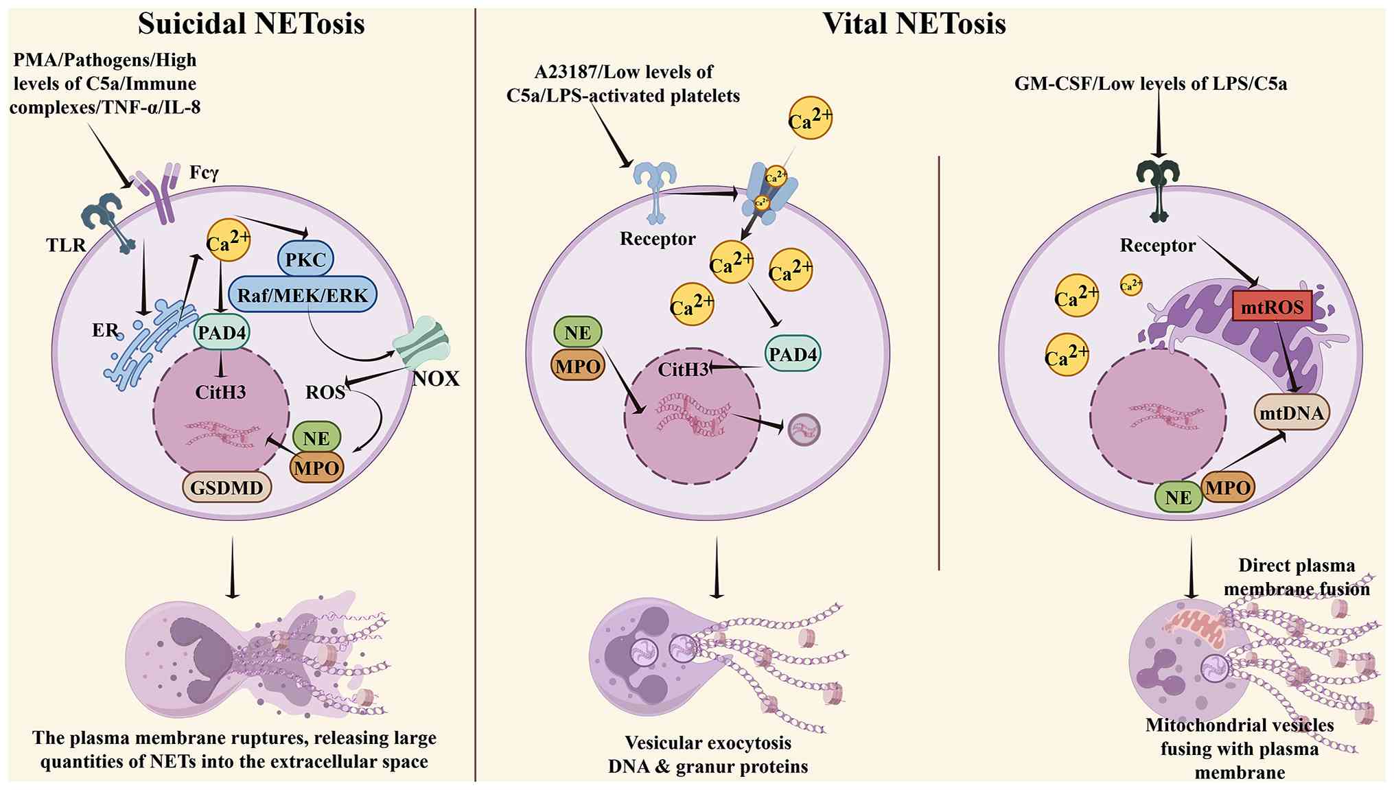 | Figure 1The two major pathways of neutrophil
extracellular trap formation. i) Suicidal NETosis: High
concentrations of LPS, PMA, pathogens and inflammatory mediators
stimulate TLRs, Fcγ and complement receptors on the cell membrane,
triggering endoplasmic reticulum calcium release and extracellular
Ca2+ influx through store-operated calcium channels.
This activates PKC, which subsequently phosphorylates the NOX
complex via the Raf/MEK/ERK pathway, resulting in the generation of
large amounts of membrane-derived ROS. Elevated intracellular
calcium activates PAD4, which catalyzes histone arginine
citrullination and initiates chromatin decondensation.
Concurrently, NOX-derived ROS induce degranulation of azurophilic
granules, releasing MPO and NE. NE cleaves histones, while MPO
synergistically amplifies oxidative signaling, completing full
chromatin decondensation. Downstream calcium signaling and ROS
activate GSDMD, which forms pores in the plasma membrane. The
nuclear envelope disintegrates, decondensed chromatin mixes with
granular proteins and the cell ultimately lyses, releasing NETs.
ii) Vital NETosis: Upon stimulation with A23187, low concentrations
of C5a, Staphylococcus aureus, or low-dose LPS presented by
activated platelets, rapid extracellular Ca2+ influx
directly activates PAD4, driving nuclear chromatin decondensation.
The nuclear envelope buds to form vesicles that release nuclear
DNA-derived vital NETs via exocytosis. Alternatively, under
stimulation with GM-CSF and low-dose LPS, calcium signaling induces
opening of the mitochondrial permeability transition pore,
generating mtROS and releasing mtDNA. After oxidative modification,
mtDNA associates with granular proteins such as MPO and NE and is
released as mtDNA-type NETs through vesicle secretion or fusion of
mitochondria with the plasma membrane. NETs, neutrophil
extracellular traps; LPS, lipopolysaccharide; PMA, phorbol
12-myristate 13-acetate; TLRs, Toll-like receptors; TNF, tumor
necrosis factor; IL, interleukin; PKC, protein kinase C; PAD4,
peptidylarginine deiminase 4; NOX, NADPH oxidase; Raf, rapidly
accelerated fibrosarcoma; MEK, mitogen-activated protein kinase
kinase; ERK, extracellular signal-regulated kinase; ROS, reactive
oxygen species; MPO, myeloperoxidase; NE, neutrophil elastase;
GSDMD, gasdermin D; mtROS, mitochondrial ROS; GM-CSF,
granulocyte-macrophage colony-stimulating factor; mtDNA,
mitochondrial DNA. Image created with Figdraw.com,
with permission. |
i) NOX-dependent NETosis (classical pathway). Also
known as suicidal NETosis, this process typically occurs within 2-4
h and culminates in the complete rupture of the neutrophil nuclear
envelope and plasma membrane, accompanied by cell death. It can be
initiated by high concentrations of LPS, pathogens such as
Staphylococcus aureus, PMA, high concentrations of complement
component C5a, immune complexes and inflammatory mediators such as
tumor necrosis factor (TNF)-α and interleukin (IL)-8. These stimuli
activate neutrophil Toll-like receptors (TLRs), Fcγ receptors and
complement receptors (14).
Following receptor activation, calcium is initially released from
endoplasmic reticulum stores, followed by substantial extracellular
Ca2+ influx through store-operated calcium channels. The
resulting increase in cytosolic calcium activates protein kinase C
(PKC), which subsequently initiates the Raf-MEK-ERK signaling
pathway (15). This pathway
phosphorylates and activates the NOX complex, resulting in the
sustained generation of large amounts of reactive oxygen species
(ROS), which further increase intracellular calcium levels
(16). Calcium-dependent
peptidylarginine deiminase 4 (PAD4) is subsequently activated and
catalyzes the citrullination of arginine residues in the tails of
histones H1, H2A, H3 and H4. Citrullination neutralizes the
positive charges of histones, weakens their electrostatic
interactions with DNA and initiates chromatin decondensation
(17). Concurrently, NOX-derived
ROS promote the release of MPO and NE from azurophilic granules
into the cytoplasm, after which these enzymes translocate to the
nucleus. NE cleaves histones through its proteolytic activity,
whereas MPO enhances oxidative signaling and facilitates chromatin
relaxation; together, these processes promote complete chromatin
decondensation. Downstream ROS and calcium signaling activate
gasdermin D (GSDMD), which oligomerizes and forms membrane pores.
The nuclear envelope subsequently disintegrates, allowing
decondensed chromatin to mix with granule proteins. Finally, the
plasma membrane ruptures, releasing large quantities of NETs into
the extracellular space (18).
ii) NOX-independent NETosis. Also referred to as
vital NETosis, this pathway occurs rapidly, typically within 15-60
min. The plasma membrane remains intact throughout the process,
allowing neutrophils to survive and retain their phagocytic and
chemotactic functions. This pathway does not depend on plasma
membrane-associated NOX-derived ROS, although certain subtypes
require mitochondrial ROS (mtROS). It can be divided into two
principal categories: i) Nuclear DNA-derived vital NETs: Classical
inducers include the calcium ionophore A23187, low concentrations
of C5a, S. aureus and low-dose LPS presented by activated
platelets. Following stimulation, rapid and substantial
extracellular Ca2+ influx directly activates PAD4, which
catalyzes histone citrullination and promotes nuclear chromatin
decondensation (14).
Decondensed chromatin and granule proteins are packaged into
vesicles that bud from the nuclear envelope and are released into
the extracellular space through exocytosis. Once outside the cell,
granule proteins bind to DNA to form web-like NETs. The resulting
enucleated neutrophils retain phagocytic vacuoles and remain
capable of phagocytosis (19).
Notably, stimulation with Candida albicans can induce
PAD4-independent vital NETosis, in which chromatin decondensation
is mediated solely by NE-dependent histone cleavage without
requiring histone citrullination (20). ii) Mitochondrial DNA
(mtDNA)-derived vital NETs: When neutrophils are primed with
granulocyte-macrophage colony-stimulating factor (GM-CSF) and
subsequently stimulated with low-dose LPS or C5a, increased calcium
signaling induces opening of the mitochondrial permeability
transition pore, resulting in mitochondrial damage, mtROS
production and the release of mtDNA (14). After oxidative modification,
mtDNA associates with granule proteins, including MPO and NE, to
form complexes that are released through mitochondrial vesicle
secretion or local fusion of mitochondria with the plasma membrane
(21). This process generates
mtNETs with mtDNA as their structural backbone, while the
neutrophil remains intact and does not undergo lysis.
NETs in the context of cardiovascular
disease
Although NETs were initially identified in the
context of microbial infection, numerous stimuli relevant to
cardiovascular pathology can also induce NETosis. Within the
cardiovascular system, NET-inducing stimuli predominantly include
lipid metabolites, damage-associated molecular patterns (DAMPs) and
intercellular signaling, rather than direct stimulation by
exogenous pathogens. First, among lipid-related triggers, oxidized
low-density lipoprotein (oxLDL) and endogenous cholesterol crystals
can engage TLR4 and the receptor for advanced glycation end
products on the neutrophil surface, thereby promoting sustained
neutrophil activation and acting as major chemical initiators of
NETosis in lipid-associated CVDs (22,23). Metabolic abnormalities, including
hyperglycemia and elevated levels of free fatty acids, can also
function as persistent stimuli and induce sustained NET release
from perivascular neutrophils (14,24). Second, DAMPs released following
tissue injury contribute substantially to NET formation. After
myocardial infarction, ischemic stroke, or organ
ischemia-reperfusion injury, necrotic endothelial and parenchymal
cells release high-mobility group box 1 (HMGB1), cell-free mtDNA
and extracellular histones (25). These molecules can establish a
positive-feedback loop that continuously stimulates circulating
neutrophils to generate additional NETs. Third, intercellular
signaling associated with vascular injury can induce NETosis.
P-selectin expressed on activated platelets binds to P-selectin
glycoprotein ligand-1 (PSGL-1) on neutrophils, thereby promoting
NET formation (26). The
complement activation product C5a regulates NETosis in a
concentration-dependent manner: Low concentrations of C5a primarily
induce calcium influx and mtNET formation, whereas high
concentrations activate the NOX complex and promote suicidal
NETosis (14). Furthermore,
cardiovascular risk factors, including monosodium urate crystals,
tobacco smoke and fine particulate matter with an aerodynamic
diameter of ≤2.5 µm (PM2.5), can chronically stimulate
neutrophils within the vascular wall, thereby sustaining
persistent, low-grade NET release (14).
NETs induced in the cardiovascular milieu are
enriched with a variety of disease-specific functional molecules
that form the molecular basis of their prothrombotic and
vascular-damaging effects. First, NETs contain procoagulant
proteins: NET fibers display abundant tissue factor (TF) and von
Willebrand factor (vWF) on their surfaces. TF directly activates
the extrinsic coagulation pathway, whereas vWF captures circulating
platelets; moreover, NET-bound NE can degrade tissue factor pathway
inhibitor, thereby disrupting the anticoagulant balance and
promoting immunothrombosis (27,28). Second, NETs contain
matrix-degrading proteases: matrix metalloproteinase (MMP)-9 is
abundantly associated with plaque-derived NETs and degrades
collagen within the arterial fibrous cap, resulting in cap thinning
and increased plaque vulnerability to rupture, thereby
precipitating acute cardiovascular and cerebrovascular events
(29). Third, NETs contain
inflammatory chemotactic mediators: NET-associated chemokines such
as IL-8 continuously recruit peripheral neutrophils to vascular
lesions, thereby perpetuating chronic inflammation (30). At the clinical level, plasma
MPO-DNA complexes, circulating citrullinated histone H3 (CitH3) and
cell-free mtDNA are specific biomarkers of NET formation in
cardiovascular disease; their levels are markedly elevated in
patients with coronary artery disease, ischemic stroke and deep
vein thrombosis and can be used to assess disease severity
(31). However, direct
comparative proteomic analyses of NETs across different CVDs remain
scarce. Defining disease-specific NET signatures will not only
deepen mechanistic understanding but also facilitate the
development of targeted diagnostic biomarkers and selective
therapeutic strategies.
NETs as a universal cellular interaction
network in cardiovascular pathology: Inflammation, thrombosis and
fibrosis
NETs are not merely antimicrobial structures; they
also function as multifunctional signaling platforms that
coordinate crosstalk among diverse cell types in the cardiovascular
system. Through their interactions with macrophages, monocytes,
endothelial cells, fibroblasts and platelets, NETs establish an
integrated cellular network that drives three interrelated
pathological processes: inflammation, thrombosis and fibrosis.
NETs and inflammation
At sites of cardiovascular injury, NETs shape and
amplify the local inflammatory microenvironment by acting on
macrophages, monocytes and endothelial cells. These effects occur
at three interconnected levels: Induction of pro-inflammatory
macrophage polarization, chemotactic recruitment of monocytes and
maintenance of their pro-inflammatory phenotype and
pro-inflammatory activation of endothelial cells (Fig. 2).
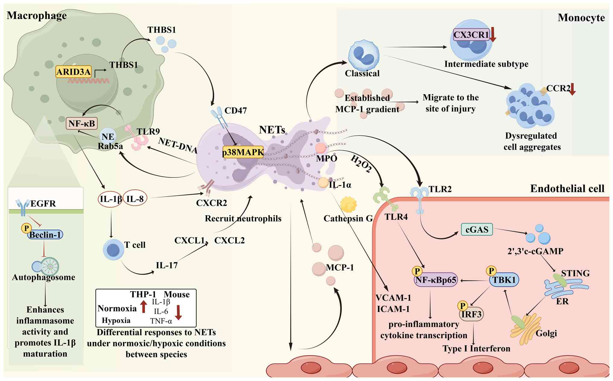 | Figure 2Mechanisms by which NETs establish
and amplify the inflammatory microenvironment at sites of
cardiovascular injury. NETs form a multicellular pro-inflammatory
network through interactions with macrophages, monocytes and
endothelial cells. i) NETs drive pro-inflammatory macrophage
polarization via the TLR9/NF-κB and NE/Rab5a-NF-κB pathways and
establish inflammatory amplification loops through IL-1β/IL-17 and
IL-8/CXCR2. M1 macrophages, in turn, induce NETosis through the
ARID3A/THBS1/CD47/p38 MAPK axis, creating a bidirectional
positive-feedback loop. ii) NETs upregulate endothelial MCP-1,
which promotes monocyte chemotaxis and maintains their
pro-inflammatory phenotype; MCP-1 reciprocally enhances NET
release, establishing a NETs-MCP-1 loop (3). NETs carry IL-1α and activate
endothelial cells via the TLR4/NF-κB and cGAS-STING pathways,
upregulating adhesion molecules and releasing pro-inflammatory
cytokines, thereby collectively amplifying local inflammation.
NETs, neutrophil extracellular traps; TLR, Toll-like receptor; IL,
interleukin; CXCL, C-X-C motif chemokine ligand; CXCR, C-X-C motif
chemokine receptor; ARID3A, AT-rich interaction domain 3A; THBS1,
thrombospondin 1; CD47, cluster of differentiation 47; p38 MAPK,
p38 mitogen-activated protein kinase; NE, neutrophil elastase;
NF-κB, nuclear factor kappa-light-chain-enhancer of activated B
cells; EGFR, epidermal growth factor receptor; TNF, tumor necrosis
factor; MPO, myeloperoxidase; CCR, C-C chemokine receptor; ER,
endoplasmic reticulum; TBK1, TANK-binding kinase 1; IRF3,
interferon regulatory factor 3; VCAM, vascular cell adhesion
molecule; ICAM, intercellular adhesion molecule; MCP, monocyte
chemoattractant protein; cGAMP, cyclic GMP-AMP; cGAS-STING, cyclic
GMP-AMP synthase-stimulator of interferon genes. Image created with
Figdraw.com, with permission. |
NETs drive pro-inflammatory macrophage
polarization and inflammation amplification
Macrophages are heterogeneous innate myeloid cells
that perform diverse functions in host defense, regulation of
inflammation, tissue repair and maintenance of homeostasis
(32-34). They are often broadly classified
into pro-inflammatory M1 and anti-inflammatory M2 phenotypes and
play central roles in both the initiation and resolution of
inflammatory responses (35).
The pro-inflammatory interaction between NETs and macrophages is
bidirectional and forms a positive-feedback loop. A recent study
has shown that pro-inflammatory M1 macrophages can directly induce
NETosis through the transcription factor ARID3A (36). Following its nuclear
translocation in M1 macrophages under inflammatory conditions,
ARID3A binds directly to the THBS1 promoter and activates its
transcription, thereby promoting substantial THBS1 secretion.
Secreted THBS1 then engages the CD47 receptor on neutrophils,
activates the p38 MAPK signaling pathway and specifically induces
NET release (36). These
findings indicate that NET-activated macrophages can, in turn,
promote further NET formation, creating a self-reinforcing
inflammatory amplification circuit. NETs complete this
positive-feedback loop by driving pro-inflammatory macrophage
polarization and amplifying inflammation through several
synergistic pathways. At the molecular level, NET-associated DNA is
recognized by TLR9 in macrophages, activating the NF-κB signaling
cascade and inducing the transcription of pro-inflammatory
cytokines, including IL-1β, IL-8 and TNF-α (22,30). Following internalization by
macrophages, NET-associated NE further activates the intracellular
Rab5a-NF-κB pathway, thereby sustaining NF-κB-dependent
inflammatory gene expression (37). These mechanisms establish two
positive-feedback circuits. First, NET-induced IL-1β secretion
increases T cell-derived IL-17 production, which induces C-X-C
motif chemokine ligand (CXCL) 1 and CXCL2 expression, recruits
additional neutrophils and sustains a self-reinforcing cycle of NET
release and macrophage activation (22). Second, macrophage-derived IL-8
induces further NET release through C-X-C motif chemokine receptor
(CXCR) 2 on neutrophils; the resulting NETs then stimulate
additional IL-8 production, forming a bidirectional IL-8-CXCR2
inflammatory amplification loop (30). Moreover, NETs inhibit
autophagosome formation in macrophages through the EGFR-Beclin-1
pathway. The resulting impairment of autophagy further enhances
inflammasome activity and promotes IL-1β maturation, thereby
reinforcing the pro-inflammatory response at the level of
post-translational processing (38).
Notably, the functional consequences of
NET-macrophage interactions are not uniformly pro-inflammatory but
are modulated by the tissue microenvironment. For example, the
effects of NETs vary between human and murine macrophages and
according to oxygen availability. Under normoxic conditions, NETs
markedly upregulate the pro-inflammatory cytokines IL-1β, TNF-α and
IL-6 in human THP-1-derived macrophages (39). By contrast, NETs strongly
suppress pro-inflammatory cytokine expression in murine macrophages
under both normoxic and hypoxic conditions (39). The mechanistic basis for these
species- and context-dependent effects remains incompletely
understood and may reflect differences in macrophage origin,
differentiation state, or oxygen-sensing pathways.
NETs regulate monocyte chemotactic
recruitment and maintenance of pro-inflammatory phenotype
Monocytes are highly plastic innate immune cells.
The monocyte chemoattractant protein (MCP)-1/CCR2 chemokine axis is
a central mechanism governing monocyte recruitment to inflammatory
sites. NETs regulate monocyte recruitment and phenotype through
several interconnected mechanisms. First, NETs stimulate vascular
endothelial cells to markedly increase MCP-1 transcription and
secretion, establishing a local chemotactic gradient that directs
circulating monocytes toward sites of injury. In turn, MCP-1 can
prime neutrophils, promoting robust NET release upon secondary
stimulation. This establishes a bidirectional NET-MCP-1
inflammatory amplification loop that sustains local inflammatory
infiltration (40). Second, NETs
drive the transition of monocytes from the classical to the
intermediate subset while downregulating the repair-associated
molecule CX3CR1. These effects maintain monocytes in a
pro-inflammatory state, impair their tissue-reparative capacity and
prolong local inflammatory injury (41). Furthermore, NETs can downregulate
CCR2 expression on the monocyte surface, thereby inhibiting
MCP-1-mediated directional migration. This disrupts the homeostatic
regulation of immune-cell recruitment and shifts inflammatory
infiltration from regulated chemotaxis toward dysregulated
aggregation (40).
NETs activate a pro-inflammatory
endothelial cell phenotype
Endothelial cells form the innermost barrier of the
vessel wall and endothelial dysfunction is a critical step in the
initiation of inflammation. NETs can carry mature IL-1α and,
together with cathepsin G, activate endothelial cells, markedly
increasing the expression of leukocyte adhesion molecules such as
vascular cell adhesion molecule (VCAM)-1 and intercellular adhesion
molecule (ICAM)-1 and thereby facilitating leukocyte adhesion and
transendothelial infiltration (42). NETs can also activate the
endothelial TLR4/NF-κB signaling axis through an MPO/H2O2-dependent
pathway, thereby driving endothelial cells toward a broadly
pro-inflammatory phenotype (43). At the level of intracellular DNA
sensing, the cyclic GMP-AMP synthase (cGAS)-stimulator of
interferon genes (STING) pathway serves as a central axis through
which NETs amplify endothelial inflammation. NETs activate
cGAS-STING signaling through TLR2 on the endothelial cell surface.
Sequence-independent binding of double-stranded DNA activates cGAS,
which catalyzes the synthesis of the second messenger 2′,3′-cyclic
GMP-AMP (cGAMP). cGAMP subsequently binds to endoplasmic
reticulum-localized STING, inducing its oligomerization and
trafficking to the Golgi apparatus, where STING recruits and
activates TBK1 kinase (44-46). Activated TBK1 phosphorylates both
IRF3 and the NF-κB p65 subunit (44,45). IRF3 signaling drives robust type
I interferon secretion, whereas NF-κB activation induces multiple
pro-inflammatory cytokines; together, these responses amplify the
local inflammatory response (44,45).
NETs and thrombosis
NETs drive immunothrombosis by inducing procoagulant
activity in endothelial cells and activating platelets, thereby
forming a key interface at which inflammation and thrombosis
reinforce each other in cardiovascular disease. In this
pathological sequence, NETs first shift endothelial cells from an
anticoagulant to a procoagulant phenotype, providing the initiating
signal for subsequent platelet adhesion and activation. Activated
platelets and NETs then establish a bidirectional amplification
loop that markedly augments the initial thrombotic signal and
ultimately promotes the formation of stable immunothrombi.
NETs induce a procoagulant endothelial
cell phenotype
Under homeostatic conditions, endothelial cells
possess intrinsic anticoagulant properties; however, endothelial
injury markedly increases the risk of local thrombosis (47). NETs act synergistically with
IL-1α and cathepsin G to stimulate endothelial cells, increase TF
transcription and procoagulant activity and thereby directly
enhance the local propensity for coagulation (42). The cGAS-STING pathway also
mediates endothelial procoagulant activity. In addition to inducing
pro-inflammatory cytokines, activated NF-κB promotes TF
transcription and downregulates endothelial anticoagulant
molecules. Together, these effects establish a procoagulant
endothelial phenotype (47).
NETs-platelet bidirectional
amplification loop
Platelets are central mediators at the interface
between inflammation and thrombosis and their bidirectional
interactions with NETs continuously amplify the local
immunothrombotic microenvironment. Platelet-driven NETosis occurs
through several mechanisms. P-selectin on the platelet surface
induces NET release by binding to PSGL-1 on neutrophils (48). TLR4-activated platelets can
adhere directly to neutrophils and induce NETosis, whereas the
interaction between platelet integrin αIIbβ3 and neutrophil SLC44A2
triggers intracellular calcium mobilization and promotes
NOX-dependent NET release. Soluble mediators released from
degranulating platelets, including thromboxane, PF4, vWF and HMGB1,
can also act as upstream triggers of NETosis (49,50).
A mutually reinforcing positive-feedback loop
operates between platelets and NETs. S100A8/A9 can induce platelet
pyroptosis and the release of oxidized mitochondrial DNA, which
further promotes NET formation. In turn, S100A8/A9 released from
NETs can induce additional platelet pyroptosis, perpetuating the
local inflammatory-thrombotic response (51). One study has further delineated
this bidirectional mechanism, showing that double-stranded DNA
(dsDNA), a structural component of NETs, is a central effector of
platelet hyperactivation (52).
A functional platelet-intrinsic cGAS-STING signaling axis
specifically recognizes intracellular dsDNA and initiates a
distinct signaling cascade that enhances thrombotic potential.
Following uptake by platelets, NET-derived dsDNA activates the
cytosolic DNA sensor cGAS, which catalyzes the production of the
second messenger cGAMP. cGAMP binds to endoplasmic
reticulum-localized STING and induces its oligomerization and
trafficking, leading to activation of the downstream NLRP3
inflammasome. Activated NLRP3 promotes caspase-1 cleavage and the
maturation and release of IL-1β. IL-1β subsequently modulates
outside-in signaling through platelet integrin αIIbβ3, increasing
the phosphorylation of Src, Syk and PLCγ2 and promoting
intracellular calcium influx. These events ultimately enhance
platelet aggregation, granule release, spreading and clot
retraction (52).
NETs and fibrosis
NETs promote pathological fibrosis in cardiovascular
tissues through three principal mechanisms: Direct activation of
fibroblasts, induction of monocyte-to-fibroblast-like
transdifferentiation and promotion of endothelial-to-mesenchymal
transition (EndoMT). The pro-fibrotic matrix deposition and
tissue-invasive capacity conferred on monocytes by NETs provide an
important link between inflammation and fibrosis.
NETs directly activate
fibroblasts
Fibroblasts are the principal stromal cells
responsible for maintaining tissue homeostasis. Under physiological
conditions, they synthesize and secrete extracellular matrix (ECM)
and contribute to tissue development and normal structural repair.
Following tissue injury, local stress signals and the inflammatory
microenvironment activate quiescent fibroblasts, inducing
proliferation, directed migration and extensive synthesis of matrix
components and thereby initiating the tissue-repair program
(53). Transforming growth
factor-β (TGF-β) is a central regulator of matrix remodeling.
Downstream TGF-β signaling can activate several cascades, including
SMAD, MAPK and RhoA, which regulate the transcription and
modification of type I and type III collagens. Persistent
overactivation of this pathway causes abnormal collagen
accumulation and promotes pathological tissue fibrosis (54-57). Studies have shown that NETs can
directly promote the pro-fibrotic activation of fibroblasts by
targeting the TGF-β signaling axis. Under hypoxic conditions, NETs
markedly increase TGF-β transcript levels in fibroblasts.
Concurrently, NETs directly induce fibroblast-to-myofibroblast
transition, enhance fibroblast migration and increase the levels of
type I collagen, phosphorylated SMAD2, phosphorylated p38 and RhoA.
These changes collectively activate the SMAD, MAPK and RhoA
pro-fibrotic pathways and promote excessive matrix deposition
(58,59). In addition to TGF-β signaling,
NET-associated histones and MPO can induce substantial ROS
production, further stimulating fibroblast activation and
proliferation and exacerbating dysregulated ECM remodeling
(60).
As well as directly activating the fibroblast
fibrotic program, NETs can indirectly amplify matrix remodeling by
reshaping the local inflammatory microenvironment. NET release is
accompanied by the release of various pro-inflammatory mediators,
including TNF-α and IL-6, which coordinately regulate fibroblast
function. TNF-α primarily promotes fibroblast proliferation,
whereas IL-6 both induces cell proliferation and increases ECM
synthesis (61-64). A recent study has also shown that
NETs can promote fibrosis by inducing pyroptosis in cardiac
fibroblasts (65). NETs directly
increase the levels of NLRP3, caspase-1 and GSDMD-N, triggering
canonical pyroptosis characterized by plasma membrane rupture and
the release of intracellular contents. Simultaneously, NETs
upregulate type I collagen and α-SMA expression. This pyroptotic
response is accompanied by substantial secretion of IL-1β and IL-6
(65). However, the specific
upstream and downstream molecular pathways mediating this process
remain unclear.
NETs induce
monocyte-to-fibroblast-like transdifferentiation and tissue
invasion
NETs not only promote fibrosis indirectly through
inflammatory signaling but also directly regulate monocyte
differentiation and effector functions, converting monocytes into
active participants in ECM degradation and deposition. NETs induce
the transdifferentiation of monocytes into fibroblast-like cells
through TLR4 signaling, leading to extensive collagen deposition
and directly contributing to pathological fibrosis (40). During tissue invasion,
NET-derived DNA is recognized by monocytes and upregulates the
alarmin S100A9. S100A9 then activates TLR4 signaling in monocytes
in an autocrine manner and induces substantial MMP-9 synthesis,
thereby enhancing monocyte tissue invasiveness and exacerbating ECM
degradation and disruption of tissue architecture (66). This mechanism directly links the
inflammatory regulatory functions of NETs to subsequent matrix
remodeling and provides an important cellular basis for their role
in bridging early inflammation and later fibrosis. Monocytes thus
function as both responders to inflammatory signals and architects
of the fibrotic microenvironment.
NETs drive EndoMT
Endothelial cells can undergo EndoMT, during which
they lose endothelial-specific markers such as VE-cadherin and
concomitantly upregulate mesenchymal markers, including α-smooth
muscle actin, vimentin and type I collagen. These changes confer
myofibroblast-like properties and enable the synthesis and
secretion of large amounts of matrix proteins, thereby promoting
abnormal tissue fibrosis (67).
Low levels of NETs can be rapidly cleared by endothelial cells
through the receptor for advanced glycation end products without
inducing pathological changes. However, when NETs accumulate
extensively in the microenvironment, NET-bound elastase cleaves
VE-cadherin and disrupts the structural integrity of the
endothelial monolayer while simultaneously activating β-catenin
signaling. Together, these effects induce a sustained EndoMT
phenotype and promote excessive ECM deposition (68).
An integrative perspective: NETs as a
central hub of the inflammation-thrombosis-fibrosis axis
Growing evidence indicates that inflammation,
thrombosis and fibrosis are not isolated pathological events in
cardiovascular disease but instead form an interconnected network
of mutually reinforcing and sequential processes. NETs serve as a
central integrative node within this triad.
NETs establish a bidirectional amplification loop
between inflammation and thrombosis. NET-associated IL-1α and
cathepsin G, together with the activation of endothelial TLR2 and
downstream cGAS-STING signaling, synergistically upregulate TF and
adhesion molecules, shifting the endothelium toward a
pro-inflammatory and procoagulant phenotype (46,60). Activated platelets, in turn,
potently induce NETosis through multiple pathways, including
P-selectin/PSGL-1, TLR4 and αIIbβ3-SLC44A2. Conversely, NET
components such as S100A8/A9 and dsDNA activate platelets;
S100A8/A9 induces platelet pyroptosis, whereas dsDNA elicits
cGAS-STING-dependent platelet activation. Together, these
reciprocal interactions form a bidirectional platelet-NET
amplification loop that continuously reinforces the local
immunothrombotic microenvironment (48-51). During acute events such as
myocardial infarction and plaque erosion, this
inflammation-thrombosis axis is particularly prominent. NET-rich
thrombi not only exacerbate ischemic tissue injury but also promote
further recruitment of inflammatory cells.
NETs also link early inflammation to subsequent
fibrosis. NET-recruited monocytes can transdifferentiate into
fibroblast-like cells and directly contribute to ECM production
(40). At the endothelial level,
NET-associated elastase cleaves VE-cadherin and activates β-catenin
signaling, inducing EndoMT and the acquisition of a
myofibroblast-like phenotype (68). Furthermore, NETs directly
activate cardiac fibroblasts through the TGF-β/SMAD, MAPK and RhoA
signaling axes, promoting collagen deposition and myofibroblast
differentiation (58,59).
A bidirectional relationship also exists between
thrombosis and fibrosis. The thrombotic network formed by NETs and
fibrin provides a structural scaffold for fibroblast migration and
matrix deposition (69,70). Meanwhile, thrombin and other
coagulation proteases generated through the NET-driven coagulation
cascade can activate protease-activated receptors on the fibroblast
surface, further reinforcing the fibrotic response and promoting
the gradual progression of thrombotic lesions toward fibrosis
(71).
Collectively, these findings support a unified model
in which NETs act as a central hub that integrates and propagates
the inflammation-thrombosis-fibrosis cascade in cardiovascular
pathology. This framework has potential therapeutic implications:
targeting NETs may attenuate all three arms of the cascade and
therefore offer broader clinical benefits than inhibiting a single
pathological component.
NETs in cardiac diseases
NETs contribute to a broad spectrum of CVDs and
participate in the pathogenesis of multiple cardiac conditions.
Building on the general inflammation-thrombosis-fibrosis framework
aforementioned, this section examines the roles of NETs in acute
myocardial infarction (AMI), atrial fibrillation (AF), viral
myocarditis (VMC), endocarditis and heart failure (HF), with an
emphasis on disease-specific mechanisms.
AMI
In AMI, NETs are not merely markers of sterile
inflammation but also constitute an important mechanistic link
between the initial inflammatory burst and subsequent reparative
fibrosis. Clinical studies have consistently demonstrated that
coronary thrombi and infarcted myocardial tissue from patients with
ST-segment elevation myocardial infarction (STEMI) are enriched in
NETs and that a greater NET burden is closely associated with
microvascular obstruction, adverse left ventricular remodeling and
worse clinical outcomes (72-75).
The intense sterile inflammatory response triggered
by myocardial infarction makes NET-monocyte interactions a major
determinant of repair quality. NETs stimulate vascular endothelial
cells to secrete large amounts of MCP-1. High local concentrations
of MCP-1, in turn, prime neutrophils for NETosis, establishing a
positive-feedback loop between NETs and MCP-1 in the infarct zone
that directs monocyte infiltration (40). Following NET stimulation,
infiltrating monocytes transdifferentiate into fibroblast-like
cells through TLR4 signaling, directly participate in collagen
deposition and become an important cellular source of
post-infarction fibrosis (75).
These findings suggest that NETs function as a molecular switch in
AMI, redirecting the acute defensive response toward pro-fibrotic
remodeling.
Studies have further clarified the upstream triggers
and downstream amplification mechanisms of NETosis. Zhang et
al (76) found that
post-infarction hypoxia induces high S100A12 expression in
neutrophils through HIF1α. S100A12 then binds to ANXA5, triggering
calcium influx and activating the NOX4-ROS-ERK pathway to drive
'suicidal' NETosis, thereby aggravating cardiac dysfunction. In
parallel, dsDNA released from NETs activates the cGAS-STING-NLRP3
pathway in platelets, enhancing platelet activation and thrombus
formation and further worsening myocardial ischemia (52). Together, these studies delineate
a pathogenic sequence in which the S100A12-ANXA5-Ca2+
axis initiates NETosis, whereas the dsDNA-cGAS-platelet pathway
amplifies thrombotic injury following NET release. These mechanisms
operate sequentially as upstream and downstream components of the
same pathogenic cascade. Notably, during the later stages of
infarct healing, moderate NET levels may facilitate the clearance
of necrotic tissue by promoting Mertk+ reparative macrophage
polarization (77), suggesting
that NETs exert stage-dependent effects in AMI. Therefore,
transient suppression of excessive NETosis during the early phase,
while preserving the physiological functions of NETs during tissue
repair, may represent a more rational therapeutic strategy.
However, the longitudinal dynamics of NET activity in humans
following myocardial infarction remain poorly characterized.
Identifying the signals that terminate NETosis and define the
repair window is therefore an important priority for translational
research.
AF
AF is the most common sustained tachyarrhythmia and
atrial remodeling is central to its initiation and maintenance.
Early histological studies showed that approximately two-thirds of
patients with 'lone AF' exhibited localized atrial inflammation
that met the criteria for myocarditis (78). NETosis has been shown to
participate in the inflammatory response associated with AF
(79), making NETs an emerging
focus of AF pathophysiology research.
Emerging evidence indicates that NETs are not merely
an inflammatory epiphenomenon in AF but instead function as a
central hub driving atrial remodeling. He et al (80) found that elevated angiotensin II
(Ang II) levels in AF increase AKT phosphorylation in neutrophils
through the AT1 receptor. However, because the mTOR pathway is
simultaneously inhibited, Ang II does not induce autophagy and is
insufficient to initiate NETosis alone; instead, it synergizes with
inflammatory stimuli to enhance NET release. Meanwhile, mtDNA and
HMGB1 released following rapid pacing-induced mitochondrial damage
in cardiomyocytes can act directly as DAMPs and trigger NETosis.
Meng et al (81) further
showed that chronic stress induces high S100A8/A9 expression in
atrial tissue. S100A8/A9 recruits neutrophils and activates the
TLR4-NLRP3 inflammatory pathway, providing another upstream trigger
of NETosis. Downstream, NETs exert distinct effects on
cardiomyocytes and fibroblasts. In cardiomyocytes, NETs induce
autophagic apoptosis by upregulating p62/Beclin-1/LC3B signaling
and cause irreversible mitochondrial damage through mitochondrial
membrane depolarization and a burst of mtROS. In fibroblasts, NETs
activate Smad/p-p38 MAPK signaling, promote
fibroblast-to-myofibroblast transition and exacerbate atrial
fibrosis and electrical remodeling (80). Collectively, these findings
delineate a self-sustaining pathogenic circuit in AF: Ang
II-mediated synergistic signaling and the S100A8/A9-TLR4 axis
initiate NETosis; DAMPs such as mtDNA and HMGB1 sustain NET
formation; and NET-induced mitochondrial damage promotes the
release of additional mtDNA. These three components establish a
positive-feedback loop that can persist independently of the
initial stressor, thereby driving the perpetuation and recurrence
of AF.
However, some gaps remain in AF research. Although
NETs induce mitochondrial damage and mtDNA release, direct
experimental evidence that the classical mtDNA-cGAS-STING cytosolic
DNA-sensing pathway is activated in AF and amplifies the
inflammatory cycle is currently lacking.
VMC
VMC is myocardial inflammation caused by viral
infection and may progress to dilated cardiomyopathy or HF. In a
mouse model of coxsackievirus B3 (CVB3)-induced VMC, NETs were
detected in both serum and myocardial tissue and NET degradation
markedly attenuated the severity of inflammation (82). The attenuation of inflammation
following NET degradation provides causal evidence that NETs
participate in the inflammatory pathology of VMC.
Recent research using the CVB3 myocarditis model has
identified a previously unrecognized mechanism through which CVB3
infection regulates NETosis. CVB3 infection markedly upregulates
calpain small subunit 1 (CAPNS1) expression in neutrophils. CAPNS1
binds to the catalytic subunit calpain-1, which then directly
cleaves the nuclear envelope protein nesprin-1. This disrupts
nuclear envelope integrity, permits chromatin release and thereby
drives NETosis (83). However,
the specific mechanisms through which NETs contribute to
VMC-associated inflammation remain unexplored. Subsequent studies
showed that CVB3 infection induces the mitochondrial accumulation
of calpain-1, leading sequentially to ATP5A1 cleavage, excessive
mitochondrial ROS production, mitochondrial dysfunction, NLRP3
inflammasome activation and cardiomyocyte pyroptosis (84). These findings raise the
possibility that NETs promote VMC-associated inflammation by
disrupting mitochondrial function and activating NLRP3, a
hypothesis that warrants further investigation.
Importantly, NETs may exert context-dependent
protective and pathological effects in VMC. NETs can restrict the
intercellular spread of viruses; therefore, indiscriminate NET
depletion may increase the viral load and promote tissue
dissemination. This trade-off represents a broader challenge for
NET-targeted anti-inflammatory strategies in infectious diseases.
Future interventions should selectively suppress the pathological
effects of NETs without compromising host defense.
Endocarditis
Endocarditis is inflammation of the inner lining of
the heart and its valves and may present acutely or chronically. It
is broadly categorized as infective endocarditis (IE) or non-IE. IE
is a severe, potentially fatal cardiac disease characterized by
direct pathogen invasion of the heart valves, resulting in valvular
damage, vegetation formation and inflammation. Li et al
(85) detected NETs in the
peripheral blood of patients with IE and found that elevated NET
levels were markedly associated with poor prognosis. During IE
pathogenesis, NETs do not function as host bactericidal effectors;
instead, pathogens 'hijack' them to create a central platform for
vegetation formation and immune evasion. This process may occur
through both classical and nonclassical pathways. In the classical
pathway, Streptococcus mutans bridges to platelet Fcγ
receptors via specific IgG, thereby upregulating platelet
P-selectin expression through the Src family kinase/Syk/PI3K/p38
MAPK signaling pathway (86).
Activated platelets subsequently bind to PSGL-1 on the neutrophil
surface via P-selectin, providing one of the key signals that
trigger NETosis. Simultaneously, bacteria stimulate neutrophil ROS
production and induce histone H3 citrullination via TLR2/FcγR
signaling. Both signals are indispensable and act together to drive
NET release (86). The
nonclassical pathway is mediated directly by EsxA, an effector
protein secreted through the type VII secretion system of
Streptococcus gordonii. EsxA can induce NETosis at a
distance and in a dose-dependent manner, independently of
platelets, thereby enabling bacteria to evade phagocytosis during
the early stages of bloodstream infection (87). Through their DNA scaffolds and
histone components, the released NETs physically entrap and
immobilize bacteria-platelet aggregates, promoting the formation of
multilayered, biofilm-like vegetations. They also activate the
coagulation cascade by promoting thrombin generation and enhance
platelet aggregation, thereby establishing a self-amplifying
bacteria-platelet-NET feedback loop that drives continued
vegetation growth (88).
Notably, in the aforementioned studies, NETs exhibited no direct
bactericidal activity against the pathogens tested. Instead, they
encased the bacteria and consolidated free-floating organisms into
biofilm-like vegetations that were difficult to eradicate.
Regarding non-IE, a case report involving patients
with systemic lupus erythematosus suggested that Libman-Sacks
endocarditis may be associated with NETs (89). However, mechanistic evidence
supporting this association remains extremely limited and currently
consists only of isolated case observations. Systematic studies are
therefore needed to determine whether NETs have a causal role in
non-IE.
HF
HF is a clinical syndrome resulting from structural
and functional abnormalities of the heart and is among the leading
causes of death worldwide (90).
Persistent low-grade inflammation is recognized as a central
mechanism underlying the pathogenesis and progression of HF
(91) and NETs are emerging as
important mediators linking immune-mediated inflammation to
myocardial energy failure. Clinical studies have shown that serum
NET levels are markedly elevated in patients with HF and are
associated with poor prognosis (92,93).
Studies have identified NETs as active drivers of HF
progression rather than merely inflammatory biomarkers. Tang et
al (94) found that elevated
Ang II levels in HF downregulate the transcription factor KLF2 in
neutrophils and relieve suppression of the HIF1α pathway, thereby
promoting neutrophil infiltration into the heart and NET release.
Zhang et al (95) further
showed that HMGB1 released following pressure overload-induced
myocardial injury acts as a DAMP, directly recruiting neutrophils
and inducing NETosis. Furthermore, Mang et al (96) demonstrated that vWF/ADAMTS13
imbalance in HF results in excessive vWF accumulation, which
activates NET formation through SLC44A2 on the neutrophil surface.
Zhao et al (97) found
that downregulation of the endogenous anti-inflammatory protein
DEL-1 relieves inhibition of p38 signaling in neutrophils,
providing another upstream mechanism for initiating NETosis.
Downstream, NET-associated NE activates TLR4-p38 signaling in
cardiomyocytes, suppresses peroxisome pro liferator-activated
receptor gamma coactivator 1-alpha (PGC-1α)-mediated mitochondrial
biogenesis and causes mitochondrial respiratory dysfunction and
cardiomyocyte apoptosis (96).
NETs also serve as procoagulant scaffolds that induce
immunothrombosis in the myocardial microvasculature, resulting in
episodes of ischemia, hypoxia and microcirculatory dysfunction
(94). In addition, NETs promote
macrophage infiltration and exacerbate cardiac inflammation
(95,97). Together, these pathways define
the mechanistic framework through which NETs contribute to HF.
Neurohormonal activation mediated by Ang II, the release of
damage-associated signals such as HMGB1 and coagulation-factor
imbalance involving VWF initiate NETosis. NET-induced mitochondrial
injury and microthrombosis then promote further DAMP release and
aggravate ischemia. These mechanisms establish a self-sustaining
positive-feedback loop that persists independently of the initial
stressor and drives the progression and deterioration of HF.
Notably, empagliflozin can disrupt the HMGB1-NET axis, suggesting
that the clinical benefits of SGLT2 inhibitors may be mediated
partly through NET-related pathways. This possibility warrants
further validation (Fig. 3).
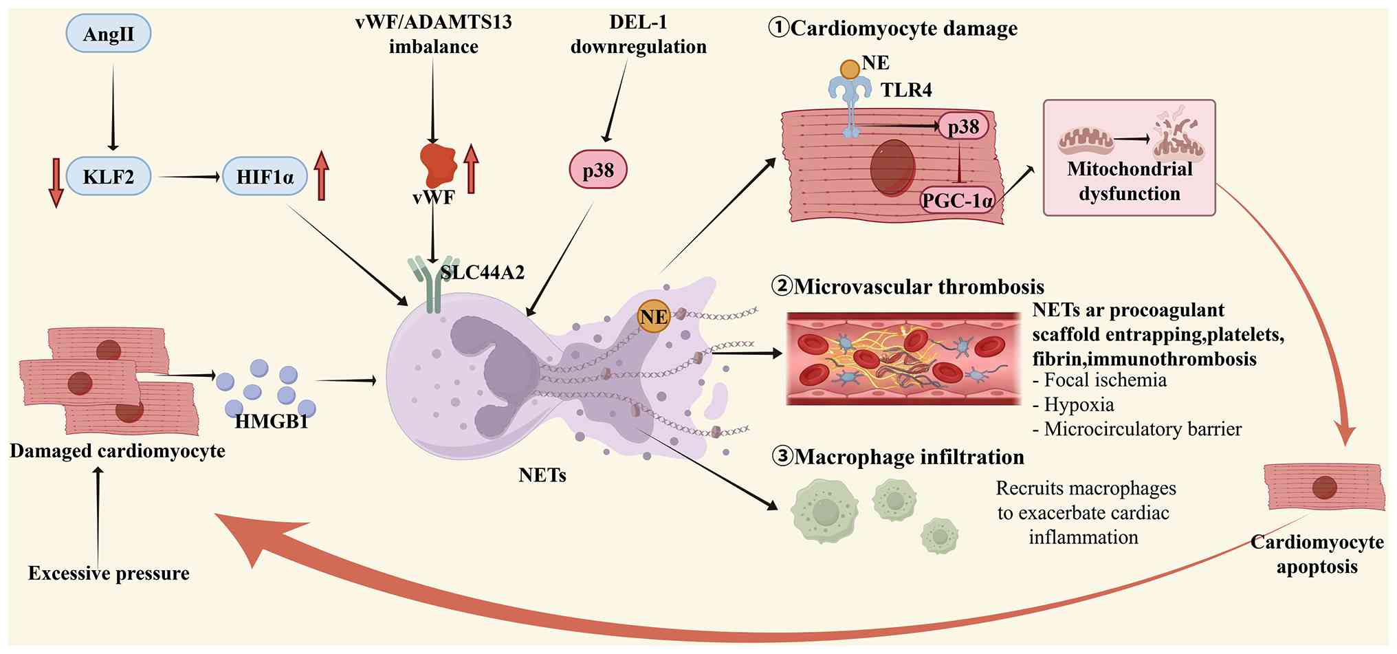 | Figure 3Mechanisms by which NETs drive the
progression of heart failure. i) Upstream initiation: Ang II
downregulates KLF2, thereby relieving the suppression of HIF1α;
myocardial injury releases HMGB1; an imbalance in vWF/ADAMTS13
activates NETosis via SLC44A2; and downregulation of DEL-1 relieves
the inhibition of p38 signaling. Together, these pathways induce
NET formation. ii) Downstream effects: The NET component NE
inhibits PGC-1α through the TLR4-p38 pathway, impairing
mitochondrial biogenesis and function in cardiomyocytes and leading
to myocardial apoptosis. NETs also serve as a procoagulant
scaffold, triggering microvascular immunothrombosis and causing
microcirculatory disturbance. Meanwhile, they promote macrophage
infiltration, aggravating cardiac inflammation (3). Positive-feedback loop:
Mitochondrial damage leads to cardiomyocyte apoptosis and the
release of DAMPs, while microthrombosis exacerbates ischemia. These
events, in turn, reactivate the upstream NETosis pathways,
establishing a self-sustaining pro-inflammatory cycle that drives
the self-perpetuation and deterioration of heart failure. NETs,
neutrophil extracellular traps; Ang II, angiotensin II; KLF2,
Krüppel-like factor 2; HIF1α, hypoxia-inducible factor 1-alpha;
ADAMTS13, a disintegrin and metalloproteinase with thrombospondin
type 1 motif, member 13; SLC44A2, solute carrier family 44 member
2; DEL-1, developmental endothelial locus-1; HMGB1, high-mobility
group box 1;vWF, von Willebrand factor; NE, neutrophil elastase;
PGC-1α; TLR, Toll-like receptor. Image created with Figdraw.com, with permission. |
NETs in vascular diseases
As well as cardiac diseases, NETs also contribute
substantially to vascular pathologies. Building on the general
cellular mechanisms linking inflammation, thrombosis and fibrosis
aforementioned, this section examines the disease-specific roles of
NETs in atherosclerosis (AS), abdominal aortic aneurysm (AAA),
hypertension and aortic valve stenosis.
AS
AS is the most common underlying pathology leading
to myocardial infarction, stroke and peripheral vascular disease
and is fundamentally a lipid-driven chronic inflammatory disease
affecting the intima of large- and medium-sized arteries (98). In 2012, Megens et al
(99) first directly visualized
NET deposition in human and murine AS lesions. Subsequent studies
have shown that serum NET levels in patients with coronary artery
disease are positively correlated with the severity of coronary
stenosis (100). Evidence
accumulated in recent years indicates that the mechanisms through
which NETs contribute to AS can be organized into three stages:
NETosis induction, impaired NET clearance and downstream effector
activity (Fig. 4).
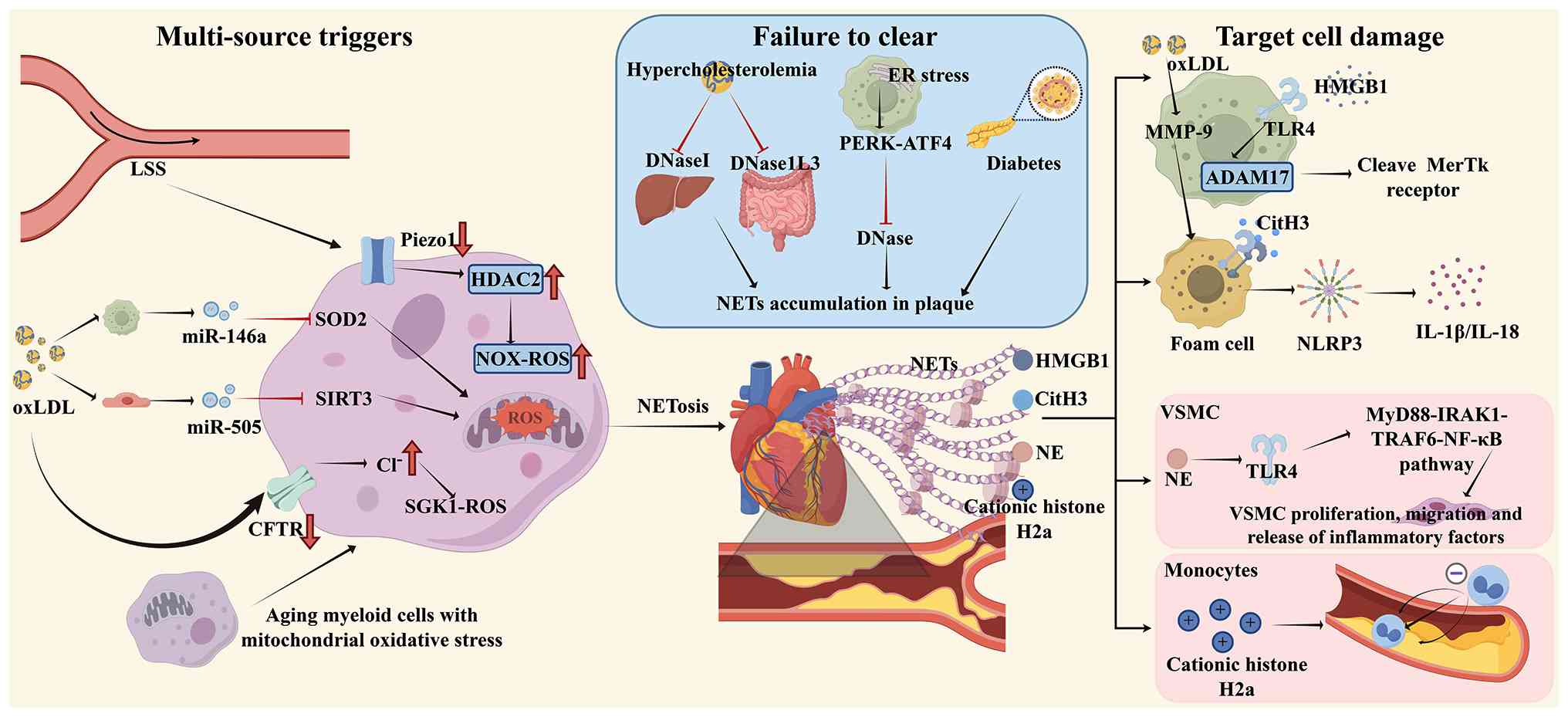 | Figure 4The 'trigger-clearance-effector'
mechanism by which NETs drive the progression of atherosclerosis.
i) Trigger: Low shear stress downregulates Piezo1 and upregulates
HDAC2, thereby promoting NOX-dependent ROS generation; oxLDL
induces NETosis via exosomal miR-146a/miR-505 and the CFTR/SGK1
pathway; and aging-related mitochondrial oxidative stress further
enhances NET release. ii) Clearance: Hypercholesterolemia,
endoplasmic reticulum stress (PERK-ATF4) in plaque macrophages and
diabetes synergistically suppress DNase I/DNase1L3-mediated NET
degradation, resulting in persistent NET accumulation within
plaques. iii) Effector: NETs enhance macrophage oxLDL uptake and
MMP-9 expression, impair efferocytosis through
HMGB1/TLR4/ADAM17-mediated MerTK cleavage and expand the necrotic
core; NE cleaves TLR4 to drive VSMC proliferation and migration;
histone H2A captures monocytes; and CitH3 activates the NLRP3
inflammasome, collectively exacerbating plaque inflammation and
instability. NETs, neutrophil extracellular traps; HDAC2, histone
deacetylase 2; NOX, NADPH oxidase; ROS, reactive oxygen species;
ox-LDL, oxidized low-density lipoprotein; miR. micro RNA; CFTR,
cystic fibrosis transmembrane conductance regulator; SGK1, serum
and glucocorticoid inducible protein kinase 1; MMP, matrix
metalloproteinase; HMGB1, high-mobility group box 1; TLR, Toll-like
receptor; NE, neutrophil elastase; CitH3, citrullinated histone H3;
LSS, low shear stress; DNase, deoxyribonuclease; ER, endoplasmic
reticulum; VSMC, vascular smooth muscle cell; SOD2, histone
deacetylase 2; SIRT3, sirtuin 3; PERK, protein kinase RNA-like
endoplasmic reticulum kinase; ATF4, activating transcription factor
4; ADAM17, a disintegrin and metalloproteinase domain-containing
protein 17; NLRP3, NLR family pyrin domain containing 3; IL,
interleukin; MyD88, myeloid differentiation primary response 88;
IRAK1, interleukin-1 receptor-associated kinase 1; TRAF6, TNF
receptor-associated factor 6; NF-κB, nuclear factor
kappa-light-chain-enhancer of activated B cells. Image created with
Figdraw.com, with permission. |
The initiating signals for NETosis are highly
dependent on the pathological context. At the hemodynamic level,
low shear stress at sites such as vascular bifurcations
downregulates the mechanosensitive ion channel Piezo1, thereby
reducing Ca2+ influx and upregulating histone
deacetylase 2. These changes promote NOX-mediated ROS generation
and directly induce NETosis (101), helping to explain the
predilection of AS for vascular bifurcations. At the metabolic
level, oxLDL can induce NETosis through several pathways. First,
oxLDL-stimulated macrophages release exosomes enriched in miR-146a
that induce NETosis at a distance by targeting and suppressing
neutrophil SOD2, resulting in a burst of mitochondrial ROS
(102). Second, oxLDL
stimulates vascular endothelial cells to secrete exosomes enriched
in miR-505. Following uptake by neutrophils, these exosomes target
and suppress the mitochondrial deacetylase SIRT3, causing a burst
of mitochondrial ROS and NET release (103). Finally, oxLDL downregulates the
cystic fibrosis transmembrane conductance regulator in neutrophils,
increasing intracellular Cl− concentrations and
activating the serum and glucocorticoid inducible protein kinase 1
(SGK1)-ROS pathway to promote NETosis (104). Aging represents another
important contributing factor. Myeloid cells from aged mice exhibit
markedly greater mitochondrial oxidative stress (mitoOS) and
7-ketocholesterol induces NET release more potently in aged
neutrophils, whereas mitoOS scavenging effectively inhibits this
process (105). Thus, NETosis
in AS arises from the convergence of mechanical, metabolic and
age-related stimuli.
The pathological effects of NETs depend not only on
their rate of formation but, more importantly, on the efficiency of
their clearance. Under homeostatic conditions, deoxyribonuclease
(DNase) I and DNase1L3 rapidly degrade NETs and restore
homeostasis. However, AS-associated metabolic abnormalities can
severely impair this clearance system. Hypercholesterolemia
systemically suppresses the NET-induced DNase feedback response. In
ApoE−/− mice, severe hypercholesterolemia
impairs the secretory responses of hepatic DNase I and intestinal
DNase1L3, delaying NET clearance and preventing the resolution of
inflammation (106). Within
plaques, macrophages are the principal source of DNase. However,
lipid loading-induced endoplasmic reticulum stress activates the
PERK-ATF4 signaling axis, which markedly inhibits macrophage DNase
secretion and promotes persistent NET accumulation (107). Diabetes further aggravates this
clearance defect. In STZ-induced diabetic
Ldlr−/− mice, NETs persist within plaques and
fail to regress spontaneously following lipid lowering, unlike
those in nondiabetic mice, thereby sustaining the inflammatory
microenvironment (108).
Importantly, these clearance defects do not occur in isolation.
Hypercholesterolemia frequently coexists with diabetes and advanced
age and their synergistic effects can lead to widespread disruption
of the NET clearance system.
Once NETs persist within plaques, they disrupt the
functions of multiple cell types and establish a self-reinforcing
pathological cycle. Their effects on macrophages are multifaceted.
NETs directly increase macrophage uptake of oxLDL and promote MMP-9
expression and secretion, thereby exacerbating plaque instability
(39). A recent study has
further shown that NET-associated HMGB1 activates ADAM17 through
TLR4. ADAM17 then cleaves the efferocytosis receptor MerTK on the
macrophage surface, impairing apoptotic-cell clearance and
consequently promoting expansion of the necrotic core (107). In vascular smooth muscle cells
(VSMCs), NE cleaves TLR4 and activates the downstream
MyD88-IRAK1-TRAF6-NF-κB pathway, promoting VSMC proliferation,
migration and inflammatory cytokine release. These effects drive
neointimal hyperplasia and contribute to plaque inflammation,
growth and instability (109).
In infection-associated AS, cationic histone H2A on the NET surface
captures negatively charged monocytes through receptor-independent
electrostatic attraction, facilitating their infiltration into
plaques and markedly accelerating plaque progression (110). In foam cells, NET-associated
CitH3 directly binds to and activates the NLRP3 inflammasome,
promoting IL-1β and IL-18 release (111). Collectively, these findings
demonstrate that NETs affect virtually all major cell types within
plaques through diverse molecular mechanisms.
Overall, NET-driven AS can be conceptualized as a
systemic pathological process involving multisource induction,
multistep failure of clearance and multicellular effector activity.
However, most existing studies rely on endpoint analyses and the
specific contributions of NETs to plaque initiation, progression,
regression and rupture have not been longitudinally characterized.
Furthermore, the functional differences among NETs induced by
distinct stimuli have not been systematically compared. LSS-induced
NETosis may primarily determine lesion localization, whereas
metabolic and aging-related factors may be more important in
sustaining plaque progression. Defining these spatiotemporal
regulatory differences will be essential for developing
stage-specific NET-targeted interventions.
Hypertension
Hypertension is one of the most common chronic
diseases and is characterized by persistently elevated arterial
blood pressure. Its pathogenesis involves inflammation-driven
oxidative stress, endothelial dysfunction, VSMC phenotypic
switching and ECM remodeling (6). Serum levels of the NET markers
cfDNA and MPO are markedly elevated in patients with hypertension
(112), whereas the Ang
II-induced increase in blood pressure is markedly attenuated in
NET-deficient mice (113),
providing functional evidence for the involvement of NETs in
hypertension.
Studies have delineated a pathogenic cascade
extending from upstream stimuli and NET release to downstream
effects and target-organ damage. Under hypertensive conditions,
vascular mechanical stretch and neurohumoral activation constitute
the two principal triggers of NETosis. Krishnan et al
(113) demonstrated that cyclic
mechanical stretch of the vessel wall activates the
mechanosensitive calcium channel TRPV4 on the neutrophil membrane,
triggering calcium influx and robust NET release. A stretch
magnitude of 10%, simulating hypertension, induced markedly greater
NETosis than 5% stretch, which simulated normotension.
Concurrently, Chrysanthopoulou et al (114) showed that Ang II directly
induces NET formation through the AT1 receptor in a process
requiring NOX-derived ROS production, autophagy activation and
PAD4-mediated histone citrullination. These pathways do not operate
independently but converge on ROS as a common mediator. In addition
to causing direct cellular damage, ROS generate isolevuglandins
(isoLGs). IsoLGs covalently modify lysine residues in histones and
disrupt higher-order nucleosome structures, thereby promoting
chromatin decondensation and providing the structural basis for NET
release (115).
Following NET release, NET-associated histones,
particularly CitH3 and TF increase blood pressure through two
distinct mechanisms: Vascular functional dysregulation and
structural remodeling. At the functional level, CitH3 directly
activates endothelial cells, upregulates adhesion molecules such as
ICAM-1, VCAM-1 and E-selectin and impairs acetylcholine-induced
endothelium-dependent vasodilation (113). At the vascular structural
level, Fang et al (116)
showed that NETs activate PI3K-Akt signaling in VSMCs, leading to
downregulation of the cyclin-dependent kinase inhibitor CDKN1b.
This relieves G1/S checkpoint arrest and promotes
cell-cycle progression. The response is accompanied by marked
upregulation of thymidine kinase 1 (TK1), a rate-limiting enzyme in
DNA synthesis that directly promotes VSMC proliferation. Moreover,
NET-stimulated VSMCs can transmit proliferative signals
horizontally to neighboring VSMCs not directly exposed to NETs by
releasing TK1-containing exosomes, thereby extending the spatial
range of vascular remodeling (116).
The pathological effects of NETs ultimately
converge on fibrosis and functional decompensation in hypertensive
target organs. At the cardiac level, Tu et al (117) first linked NETs to ferroptosis,
demonstrating that NETs activate NF-κB signaling, upregulate the
pro-ferroptotic protein ACSL4 and simultaneously downregulate
ferritin heavy chain FTH1 and the antioxidant enzyme GPX4. These
changes induce iron-dependent lipid peroxidation and ferroptotic
cardiomyocyte death. This process not only directly promotes
cardiomyocyte loss but also upregulates pro-fibrotic markers,
including α-smooth muscle actin, fibronectin and collagen II,
through pro-inflammatory and pro-fibrotic signaling, thereby
driving myocardial interstitial fibrosis and cardiac
dysfunction.
AAA
AAA is a degenerative vascular disease
characterized by chronic vessel-wall inflammation, ECM degradation
and disrupted vascular smooth muscle cell homeostasis (118). Multiple clinical studies have
shown that circulating and aortic tissue levels of NET-specific
markers, including CitH3, cell-free DNA, nucleosomes, MPO and NE,
are markedly higher in patients with AAA than in healthy controls.
The concentrations of these markers are positively correlated with
aneurysm diameter, expansion rate and rupture risk, suggesting that
NET burden may reflect AAA severity and adverse prognosis (119,120).
NET formation is tightly regulated by upstream
signaling mechanisms. Phosphoinositide 3-kinase γ promotes
noncanonical pyroptosis, specifically activation of the
caspase-11/GSDMD pathway, through cAMP/PKA signaling, thereby
enhancing NET release and aggravating AAA (121). Studies have identified another
important upstream mechanism involving the gut microbiota. Patients
with AAA exhibit marked gut dysbiosis characterized by a reduced
abundance of the butyrate-producing bacterium Roseburia
intestinalis and a concomitant decrease in butyrate levels
(122). Reduced butyrate
availability diminishes its inhibitory effect on neutrophil NOX2
expression, resulting in excessive NET release. Supplementation
with R. intestinalis or butyrate markedly reduces neutrophil
infiltration and NET deposition in the aortic wall and attenuates
AAA progression (122). These
findings link the gut microbiota-metabolite axis to NET formation
and provide a rationale for microbiota-targeted interventions in
AAA.
NETs promote AAA formation and progression through
multiple convergent mechanisms. First, NETs directly induce
ferroptosis, a form of programmed cell death driven by
iron-dependent lipid peroxidation, in VSMCs. NETs inhibit PI3K/AKT
signaling in VSMCs, downregulating SLC7A11 and GPX4 while
upregulating ACSL4 and TFR1, thereby triggering lipid peroxidation
and ferroptosis (123).
Additionally, NETs also destabilize the dimeric form of the
mitochondrial glutathione transporter SLC25A11, leading to
mitochondrial glutathione (mitoGSH) depletion and further inducing
ferroptosis in SMCs (124).
Second, NETs suppress Hippo signaling through TLR9, thereby
increasing YAP nuclear translocation and driving VSMC phenotypic
switching from a contractile state to a synthetic and
pro-inflammatory phenotype. This transition is characterized by
downregulation of contractile marker genes, including ACTA2, MYH11
and CNN1 and upregulation of pro-inflammatory and matrix
metalloproteinase genes, including MMP2, MMP9, IL-1β and TNF-α,
thereby exacerbating arterial-wall inflammation and matrix
degradation (125). Third, NETs
activate p38/JNK signaling and induce VSMC apoptosis, aggravating
medial VSMC depletion and elastic fiber fragmentation and promoting
AAA rupture (126).
Furthermore, the DNA component of NETs forms complexes with
cathelicidin-related antimicrobial peptide (CRAMP/LL-37), which
recruit and activate plasmacytoid dendritic cells (pDCs), induce
robust type I interferon production and subsequently activate T
cells to amplify the inflammatory cascade within the aortic wall
(127).
In summary, NETs act through multiple mechanisms to
drive AAA formation and progression, including the induction of SMC
ferroptosis, phenotypic switching and apoptosis and the activation
of pDC-mediated immune amplification. Potential therapeutic
approaches therefore include inhibiting upstream NET formation,
modulating gut microbial metabolites to reduce NET generation and
targeting downstream effector mechanisms, such as ferroptosis, YAP
signaling and pDC activation. These strategies may have
translational potential for the treatment of AAA.
Aortic valve stenosis
Aortic valve stenosis is the most common valvular
heart disease, with a prevalence exceeding 2% among individuals
older than 60 years and a two-year mortality rate of ~50% among
those with severe stenosis (128). The disease is primarily driven
by progressive inflammatory and fibrocalcific processes involving
interactions among circulating inflammatory mediators, valvular
endothelial cells and valvular interstitial cells (129,130).
At the tissue level, abundant NET-specific markers,
including CitH3 and cfDNA, have been detected in human stenotic
aortic valve tissue (131,132), confirming the presence of NETs
in valvular pathology. oxLDL is considered an important mediator of
inflammation in aortic valve stenosis (133) and Awasthi et al
(134) further showed that
oxLDL induces NET formation through the TLR-PKC-IRAK-MAPK pathway
and NADPH oxidase activation, establishing a molecular basis for
the functional relationship between oxLDL and NETs. Building on
these observations, evidence increasingly implicates NF-κB as a key
downstream effector of NETs. NF-κB is a well-established
pro-inflammatory signaling hub and contributes to the calcification
associated with aortic valve stenosis (135). As NETs can activate NF-κB
through multiple pathways, the present study proposed that NETs
within stenotic aortic valves may convert the initial inflammatory
signal induced by oxLDL into an osteogenic differentiation program
in valvular interstitial cells through NF-κB signaling. NETs may
simultaneously promote ECM remodeling through NET-associated
elastase and cathepsins, thereby providing a collagen scaffold for
calcific nodule formation. However, this hypothesis currently lacks
direct functional validation. No studies have yet used NET
inhibitors or PAD4-deficient animals in aortic valve stenosis
models to evaluate changes in valvular NF-κB activity,
calcification markers and valve function.
Potential NETs-targeted therapeutic
strategies in cardiovascular disease
Given the central role of NETs in cardiovascular
disease, therapeutic targeting of NETs has attracted increasing
attention. Investigated approaches span multiple intervention
points, including inhibition of upstream NETosis signaling,
enzymatic degradation of preformed NETs and neutralization of
downstream NET components. This section categorizes current
NET-targeted interventions according to their mechanisms of action
and evaluates the available preclinical evidence, clinical
translational status and inherent limitations (Table I).
 | Table ITherapeutic interventions targeting
NETs. |
Table I
Therapeutic interventions targeting
NETs.
| Authors, year | Intervention
strategy | Target | Compound | Mechanism | Model | Key therapeutic
outcomes | Evidence level | Translational
status | Key
limitations | (Refs.) |
|---|
| Mangold et
al, 2022 | NETs
degradation | NETs-DNA
backbone | DNase-I | Enzymatic cleavage
of DNA matrix. |
ApoE−/− mouse model
of atherosclerosis (high-fat diet). | Reduced
atherosclerotic plaque size by ~3-fold; decreased plasma IL-1β and
IL-6 levels. | Preclinical (in
vivo). | Approved (Cystic
fibrosis). | (1) Releases toxic histones; (2) Inactivated by plasma G-actin;
(3) Poor lesion
specificity. | (73) |
| Morita et
al, 2004 | Targeting NETs
components | NETs-DNA
backbone | TREX1 | Intracellular
degradation of internalized NETs via TREX1 (DNase III) in
macrophages; DNase 1L3 mediates extracellular NET degradation in
DCs. | Primary human
monocyte-derived macrophages (HMDMs) and dendritic cells (MDDCs);
NETs from primary human neutrophils. | TREX1 silencing led
to accumulation of undigested NETs in macrophages; DC-secreted
DNase1L3 degraded NETs extracellularly. | Preclinical (in
vivo). | Preclinical
(Theoretical target). | No pharmacological
agonist available; pure theoretical target. | (141) |
| Iba et al,
2015; Wildhagen et al, 2014 | Histone
neutralization | Histones |
Unfractionated/LMWH/Non-anticoagulant
heparin | Charge-dependent
electrostatic binding to neutralize histone cytotoxicity. | Mouse (C57BL/6)
models of ConA-induced hepatitis, CLP and LPS challenge; Rat
(Wistar) models of histone H3-induced organ injury; EA.hy926 cells
and primary endothelial/leukocytes. | Attenuates
histone-mediated cytotoxicity and organ damage. | Preclinical (in
vivo). | Approved
(Anticoagulant, repurposing). | The effective dose
is far beyond the anticoagulation threshold; broad charge effects;
poor tissue penetration; stimulus-specific efficacy. | (142, 143) |
| Osada et al,
2017 | | | TM alfa | Promotes APC
generation to proteolytically degrade histones. | EA.hy926
endothelial cells. | Reduces
histone-induced thrombin generation and endothelial death. | In
vitro. | Approved
(DIC). | Limited
cardiovascular-specific data. | (145) |
| Abrams et
al, 2013 | | | CRP | Binds directly to
histones, blocking their integration into cell membranes and
subsequent Ca2+ influx; competes with phospholipids for
histone binding. | Mouse models;
EA.hy926 cells; Serum from trauma/sepsis patients. | Reduces
histone-mediated endothelial injury. | Preclinical and
Ex vivo (Patient serum). | Biomarker/Drug
candidate. | CRP has
context-dependent pro-/anti-inflammatory effects. | (146) |
| Li et al,
2025 | Neutrophil elastase
inhibition | NE | Sivelestat | Selective NE
inhibition | LPS-induced sepsis
mouse; MI/R models | Preserves
endothelial glycocalyx; reduces myocardial injury | Preclinical (in
vivo). | Approved
(ARDS) | Protease redundancy
may (PR3, CatG, MMPs) compensate; PK challenges. | (147) |
| Bidouard et
al, 2003 | | | SSR69071 | NE inhibition | Rabbit coronary
ischemia-reperfusion model | Reduces infarct
area | Preclinical (in
vivo). | Preclinical | Intravenous
delivery required; limited cardiovascular data. | (149) |
| Delbosc et
al, 2016 | Inhibition of
NETosis | | AZD9668 | NE inhibition | Elastase-induced
AAA rat model (with P. gingivalis enhancement) | Suppressed
Pg-enhanced AAA diameter growth; promoted healing process (smooth
muscle cell colonization and collagen deposition) within the
intraluminal thrombus; reduced calcification, MPO and cell-free DNA
release. | Preclinical (in
vivo). | Phase II
(Respiratory diseases) | Repurposing
potential but cardiovascular trials lacking. | (150) |
| Knight et
al, 2014 | PAD4
inhibition | PAD4 | Cl-amidine | Irreversible
pan-PAD inhibition |
ApoE−/− mouse
carotid thrombosis; AS models | Reduces NETosis,
atherosclerosis and arterial thrombosis | Preclinical (in
vivo). | Preclinical (Tool
compound) | Narrow therapeutic
time window; interferes with physiological NETosis; no reliable
pharmacodynamic biomarker. | (151) |
| Du et al,
2020 | | | GSK484 | Reversible
selective PAD4 inhibition | Mouse MI model (LAD
ligation) | Reduces infarct
size, neutrophil infiltration, CitH3 expression and cardiomyocyte
apoptosis | Preclinical (in
vivo). | Preclinical | Same as
aforementioned; optimal dosing window undefined. | (153) |
| Heger et al,
2023 | | | JBI-589 | Oral selective PAD4
inhibition | Collagen-induced
arthritis (DBA/1J mice) | Preserves LV
diastolic function; reduces CitH3 and thrombotic inflammation | Preclinical (in
vivo). | Preclinical | Long-term safety
unknown; cardiovascular data limited. | (154) |
| Li et al,
2024 | ROS inhibition | NADPH
oxidase/ROS | Colchicine | Inhibits ROS
production and Ca2+ influx; suppresses NETosis | Mouse model of
permanent LAD ligation-induced AMI. | Improves cardiac
function and remodeling; reduces post-AMI inflammation | Preclinical (in
vivo). | Approved (CVDs: CAD
and post-MI), | NETs-specific
benefit not demonstrated in clinical trials; anti-NETs effect
inferred | (155) |
| Shirakawa et
al, 2022 | | | HOCl (hypochlorous
acid)/Hydroxyl radicals (•OH) | H2 | Neutralizes HOCl
produced during oxidative burst, reducing DNA damage (γ-H2AX);
suppresses CXCR4+ aged neutrophil subset and neutrophil
aggregation; may also inhibit PAD4 activity in NOX-independent
NETosis. | Scavenges hydroxyl
radicals and HOCl; suppresses ROS. | Inhibits
PMA-induced NETosis; reduces DNA damage in neutrophils | Preclinical (in
vivo). | Medical gas.
Molecular target unclear; no direct CVDs patient evidence | (157) |
| Khan et al,
2019 | Others Indirect
inhibition via inflammation/metabolism | DNA/Chromatin
(intercalation) | Anthracyclines
(e.g., doxorubicin, daunorubicin) | Intercalates into
DNA, blocking chromatin decondensation and transcriptional firing;
inhibits both Nox-dependent and Nox-independent NETosis without
affecting ROS production or inducing apoptosis. | Primary human
neutrophils. | Dose-dependent
NETosis inhibition | In
vitro. | Approved
(Chemotherapy) | Intrinsic
cardiotoxicity prevents repurposing; requires cardio-safe
derivatives. | (158) |
| Kindberg et
al, 2024 | Cytokine
targeting | IL-6 receptor | Tocilizumab | Reduces circulating
NET markers (dsDNA, MPO-DNA, H3Cit) in STEMI patients; mechanism
may involve downregulation of NETosis-related genes (histones,
HDAC) and reduced neutrophil mobilization; exact mechanism remains
unclear. | STEMI patients
(ASSAIL-MI trial). | Reduced NET markers
at 24h and 3-7 days; NET reduction partially mediated tocilizumab's
beneficial effect on myocardial salvage index (MSI); higher NET
levels associated with larger infarct size and more microvascular
obstruction. | Clinical. | Approved
(Rheumatoid arthritis, CRS). | Mechanism of NETs
inhibition unclear; lacks robust cardiovascular outcome data. | (159) |
| Awasthi et
al, 2019 | Immunometabolic
Intervention | Lactate
dehydrogenase | sodium oxamate
dehydrogenase, | Inhibits lactate
LPS-induced reducing lactate accumulation; lactate itself acts as a
signaling molecule promoting NETosis via Warburg effect (PKM2
dimerization). | Mouse model of
formation. sepsis (C57BL/6, 10 mg/kg LPS, i.p.); primary human
polymorphonuclear neutrophils (PMNs). | Suppresses NET
(in vivo). | Preclinical | Preclinical. may
disrupt cardiac | Broad LDH
inhibition lactate metabolism; off-target risks unassessed. | (160) |
| Burczyk et
al, 2022 | Hif-1α/Nrf2
pathway | 4-OI | Downregulates
Hif-1α; upregulates Nrf2/HO-1 pathway. | Bone marrow
neutrophils (C57BL/6J). | Suppresses
LPS-induced NETosis with minimal effect on basal metabolism. | In
vitro. | Preclinical. | Non-specific
alkylation of metabolic enzymes (e.g., GAPDH); long-term cardiac
effects unknown. | | (161) |
NETs degradation strategies
DNA forms the structural backbone of NETs, making
enzymatic degradation the most direct approach for dismantling
preformed NET structures.
DNase I is the most extensively investigated enzyme
for NET degradation. Across multiple cardiovascular disease models,
DNase I attenuates NET-mediated pathological changes. For example,
in myocardial infarction models, DNase I dissolves NET-rich
microthrombi, thereby improving microvascular perfusion and
reducing infarct size (73).
Recombinant human DNase I is clinically approved for cystic
fibrosis, in which it reduces sputum viscosity when administered by
aerosol inhalation, bronchoscopic instillation, or other routes
(136,137). However, no relevant clinical
studies have evaluated DNase I in cardiovascular disease and its
translation to cardiovascular indications faces several challenges.
First, NETs consist of a DNA backbone decorated with histones and
granular proteins. DNase I cleaves DNA but does not neutralize
histones. DNA degradation may therefore release substantial
quantities of positively charged histones into the circulation.
Extracellular histones are highly cytotoxic and can directly damage
endothelial cells, activate platelets, promote microthrombus
formation and trigger further sterile inflammation (138). Second, blood contains G-actin,
a high-affinity inhibitor of DNase I. Plasma G-actin concentrations
are sufficient to eliminate most of the activity of administered
DNase I within minutes. Although modified DNase I variants,
including actin-resistant mutants, are under investigation, none
have entered cardiovascular clinical trials (139). Third, NETs in cardiovascular
disease are predominantly localized within vascular-wall plaques,
thrombi and the microcirculation. Neither intravenous nor
intra-arterial administration achieves efficient lesion-specific
accumulation of DNase I. Instead, DNase I nonspecifically degrades
circulating NET structures, potentially disrupting the
physiological roles of NETs in host defense and vascular
homeostasis. Consequently, the translational potential of DNase I
monotherapy in cardiovascular disease remains limited. Potential
strategies for overcoming these limitations include combining DNase
I with histone-neutralizing agents, developing actin-resistant
DNase I variants and using nanocarriers for lesion-targeted
delivery.
Other nucleases, including three-prime repair
exonuclease 1 (TREX1), contribute to the physiological clearance of
NETs by macrophages (140).
TREX1-deficient mice develop inflammatory myocarditis that
gradually progresses to dilated cardiomyopathy and circulatory
failure, possibly because of impaired NET degradation (141). However, the therapeutic
potential of pharmacologically enhancing TREX1 activity remains
unexplored and is currently theoretical.
Targeting NETs components
Histone neutralization
Histones are the most abundant protein components
of NETs and exert direct cytotoxic effects on endothelial cells,
cardiomyocytes and other cell types (138). Several strategies have
therefore been developed to sequester or neutralize extracellular
histones.
Unfractionated heparin, low-molecular-weight
heparin and their non-anticoagulant derivatives neutralize histones
through electrostatic interactions mediated by their negative
charge, thereby mitigating cytotoxicity and organ injury. However,
the effective doses required substantially exceed those used for
clinical anticoagulation, representing a major barrier to clinical
translation (142,143). Although non-anticoagulant
heparin derivatives may reduce bleeding risk, their efficacy in
bacterial sepsis is markedly lower than that in sterile
inflammation, suggesting that stimulus-dependent activity limits
their generalizability. Moreover, the broad, charge-based binding
of heparin may interfere with host-defense peptides, whereas its
macromolecular structure limits penetration into the vessel wall
and the neutralization of tissue-bound histones. Heparin also does
not target other harmful NET components, including the DNA backbone
and elastase (144). Targeting
histones alone may carry the additional immunological risk of
suppressing beneficial DAMP signaling and the therapeutic window
appears narrow: Prophylactic administration is effective, whereas
delayed treatment provides minimal benefit. Thus, heparin-based
histone neutralization alone has limited translational potential.
Further development should focus on histone-specific antibodies,
tissue-penetrant small-molecule inhibitors and biomarker-guided
patient stratification according to the underlying inflammatory
stimulus.
Thrombomodulin alpha promotes the generation of
activated protein C (APC), which proteolytically degrades
extracellular histones and attenuates histone-induced toxicity
(145). This mechanism may
contribute to the protective effects of thrombomodulin signaling in
sepsis-associated organ injury, although cardiovascular-specific
evidence remains limited.
C-reactive protein (CRP) can form complexes with
histones, preventing their incorporation into cell membranes and
reducing histone-induced calcium influx and cellular injury
(146). However, CRP has both
pro-inflammatory and anti-inflammatory properties and its net
effect on NET-mediated pathology remains context-dependent.
NE inhibitors
NE is a key mediator of NET formation and a major
effector of NET-mediated tissue injury. Sivelestat is a selective
NE inhibitor used clinically to treat acute respiratory distress
syndrome (147). Preclinical
studies have demonstrated that sivelestat protects against
myocardial ischemia-reperfusion injury and preserves endothelial
glycocalyx integrity (148).
However, large-scale clinical trials in cardiovascular disease
remain lacking. Second-generation NE inhibitors, including SSR69071
and AZD9668, have shown efficacy in preclinical models of
myocardial ischemia-reperfusion injury and AAA (149,150). Their clinical development has
been constrained by pharmacokinetic limitations and the need for
intravenous administration.
A major limitation of NE inhibition is functional
redundancy. NETs contain multiple proteases, including proteinase
3, cathepsin G and MMPs, that may compensate for NE blockade and
thereby limit the efficacy of single-target therapy.
Inhibition of NETosis
PAD4 inhibitors
PAD4-mediated histone citrullination is a key step
in chromatin decondensation during NETosis, making PAD4 a major
therapeutic target. Cl-amidine is a first-generation broad-spectrum
PAD inhibitor. It has been extensively used in cardiovascular
animal models, including models of AS and myocardial infarction, to
suppress NET formation and attenuate tissue injury (151). It is also widely used as a
reference compound for investigating the PAD4-NET pathway (152). GSK484 is a more selective PAD4
inhibitor that reduces neutrophil infiltration, inflammatory
cytokine release and NET formation and attenuates cardiomyocyte
apoptosis in myocardial infarction models (153). JBI-589 is another selective,
orally bioavailable PAD4 inhibitor that reduces myocardial NET
deposition and pro-fibrotic cytokine levels (154).
The principal translational challenge facing PAD4
inhibitors is uncertainty regarding the therapeutic window. NETs
are not uniformly detrimental throughout the course of
cardiovascular disease. Inhibition of excessive NETosis is
particularly important during the early phase of AMI-associated
ischemia-reperfusion injury, spanning hours to days. During later
stages of infarct healing, however, moderate NET levels may
contribute to necrotic tissue clearance and repair. Current
evidence does not permit precise determination of the optimal
timing or duration of PAD4 inhibition during cardiovascular events
and pharmacodynamic biomarkers to guide treatment duration are
lacking. Furthermore, long-term systemic PAD4 inhibition may
compromise physiological NETosis-mediated host defense. Future
clinical trials could consider circulating CitH3 as a surrogate
endpoint when designing time-sequenced dosing regimens.
ROS inhibitors
Given the central role of ROS in classical NETosis,
targeting oxidative stress represents another potential therapeutic
strategy. Colchicine, a widely used anti-inflammatory drug, has
been mechanistically shown to reduce NADPH oxidase-derived ROS
production and calcium influx, thereby inhibiting NET formation
(155). Supported by evidence
from landmark randomized controlled trials, including COLCOT and
LoDoCo2, low-dose colchicine (0.5 mg/day) administered as an
adjunct to standard therapy markedly reduces the risk of
cardiovascular events in patients with chronic coronary artery
disease and those with AMI, with an overall favorable tolerability
profile (156). Colchicine is
therefore the only NET-related anti-inflammatory drug to date with
clinical evidence of cardiovascular benefit, providing proof of
principle for the clinical feasibility of NET-targeted strategies.
However, clinical trials have not established that its
cardiovascular benefits are attributable to NET inhibition.
Molecular hydrogen (H2) is a safe
antioxidant that inhibits PMA-induced NET formation by reducing ROS
levels (157). However, its
precise molecular targets in neutrophils remain unclear and direct
evidence of NET inhibition in patients with cardiovascular disease
is lacking.
Others
Anthracyclines, including doxorubicin and
epirubicin, are widely used chemotherapeutic agents that inhibit
NETosis while preserving ROS production and neutrophil viability
(158). However, their inherent
cardiotoxicity precludes their repurposing for cardiovascular
indications. The cardioprotective agent dexrazoxane does not
interfere with anthracycline-mediated NET inhibition, suggesting
that the anti-NET and cardiotoxic effects of anthracyclines may be
separable (158). Future
studies could integrate high-resolution mass spectrometry, nuclear
magnetic resonance spectroscopy and X-ray crystallography to
characterize the atomic-level interactions of doxorubicin with two
key molecular targets: Topoisomerase IIβ, which mediates
cardiotoxicity and the as-yet-unidentified target responsible for
NET inhibition. Comparing their molecular interaction fingerprints
may identify a toxicophore that is essential for cardiotoxicity but
dispensable for NET inhibition. Structure-based drug design using
bioisosteric replacement, the introduction of metabolic soft spots,
or steric modifications could then selectively eliminate this
toxicophore and potentially yield a first generation of
anthracycline-derived NET inhibitors with reduced
cardiotoxicity.
Indirect inhibition of NETs via
inflammatory and metabolic pathways
Cytokine-targeted therapy
Tocilizumab is a monoclonal antibody targeting the
IL-6 receptor. In a clinical study, tocilizumab reduced circulating
levels of NET markers, including CitH3 and dsDNA, in patients with
STEMI and attenuated myocardial injury (159). However, the mechanism
underlying this effect remains unexplored. More robust clinical
evidence and mechanistic studies are therefore required before
tocilizumab can be considered a NET-targeted therapy for
cardiovascular disease.
Immunometabolic interventions
Metabolic pathways are increasingly recognized as
important regulators of NET formation. Glycolysis plays a central
role in both NOX-dependent and NOX-independent NETosis. The lactate
dehydrogenase inhibitor sodium oxamate reduces LPS-induced NET
formation and lactate accumulation, highlighting the importance of
glycolytic metabolism in NETosis (160). Furthermore, 4-octyl itaconate
(4-OI), a cell-permeable derivative of the immunometabolite
itaconate, markedly inhibits LPS-induced NET formation by
downregulating HIF-1α and, in part, through the Nrf2/HO-1 pathway
(161). These findings identify
4-OI as an emerging immunometabolic modulator with NET-targeting
potential.
Although sodium oxamate and 4-OI reduce NET
formation in experimental models, their clinical translation raises
potential concerns regarding off-target effects. As a
broad-spectrum LDH inhibitor, sodium oxamate may disrupt systemic
lactate metabolism, including that of the myocardium; however, this
possibility has not been evaluated in cardiovascular models. 4-OI
can nonspecifically alkylate metabolic enzymes such as GAPDH and
its long-term effects on mitochondria-rich tissues, including the
myocardium, remain unclear (161). Moreover, no direct evidence is
available regarding the effects of either agent on the acid-base
balance of the plaque microenvironment. Future studies should
prioritize evaluating the metabolic toxicity of these inhibitors in
cardiovascular cells and developing targeted delivery strategies to
minimize off-target effects.
Conclusion
In summary, the present review synthesized the
multidimensional regulatory network of NETs in cardiovascular
disease. Initially characterized as antimicrobial defense
structures, NETs are now recognized as a central hub connecting
inflammation, thrombosis and fibrosis. In major CVDs, including
AMI, AF, HF, AS, hypertension and AAA, NETs drive disease
initiation and progression through diverse effector mechanisms.
These include disrupting mitochondrial function, inducing cellular
phenotypic switching, activating the coagulation cascade, releasing
matrix-degrading proteases and triggering ferroptosis and
pyroptosis. Cardiovascular microenvironmental stimuli, including
oxLDL, mechanical stretch, Ang II and DAMPs, interact with
regulatory mechanisms such as gut microbiota-associated metabolic
dysregulation, cGAS-STING-mediated cytosolic DNA sensing and the
S100A8/A9 positive-feedback loop. Together, these factors establish
disease-specific networks that initiate and sustain NETosis.
Accordingly, NET-targeted strategies now encompass DNase I-mediated
degradation, histone neutralization, NE inhibition, blockade of
PAD4 and ROS signaling, immunometabolic modulation and
cytokine-targeted interventions. Although low-dose colchicine has
entered cardiovascular clinical practice, most NET-directed
approaches remain limited by the modest efficacy of single-target
interventions, poorly defined therapeutic windows, off-target
effects and insufficient cardiovascular-specific clinical
evidence.
Despite substantial advances, several important
questions must be resolved before NET-targeted therapies can be
translated into clinical practice. First, NET structure and
function are highly heterogeneous. Single-cell multi-omics and
spatial proteomics should be used to characterize NET molecular
signatures across CVDs, disease stages and tissue regions. Such
studies may distinguish pathogenic NET components from relatively
benign or reparative signals and provide a molecular basis for
selective neutralization. Second, NETosis exerts stage- and
context-dependent pathogenic and reparative effects. During the
inflammatory clearance and proliferative repair phases of AMI, NETs
may perform opposing functions. This duality is even more
pronounced in infectious conditions such as IE and VMC, in which
indiscriminate NET inhibition may facilitate pathogen
dissemination. Future studies should establish conditional animal
models that permit temporal control of NETosis, characterize the
kinetics of NET formation and degradation and define the transition
from global NET clearance to precise temporal regulation.
Interventions should also be stratified according to sterile or
infectious etiology, with priority given to targets that suppress
pathological NET activity without compromising host defense. Third,
the release of toxic histones following DNase I-mediated
degradation, functional redundancy among NET-associated proteases
and the off-target effects of metabolic modulators indicate that
single-node interventions may be insufficient because of the
diverse activities and compensatory mechanisms of NET components.
Multitarget combination strategies, such as DNase I combined with
histone-specific antibodies, PAD4 inhibitors combined with SGLT2
inhibitors, or simultaneous inhibition of the upstream
S100A8/A9-TLR4 axis and downstream cGAS-STING pathway, may provide
synergistic efficacy. Lesion-targeted delivery using nanocarriers,
antibody-drug conjugates, or localized perfusion may further reduce
the risks of infection and impaired immune surveillance associated
with prolonged systemic NETosis inhibition. However, most
NET-targeted interventions remain at the preclinical stage and
cardiovascular-specific evidence is limited. Clinical trials
incorporating NET-specific markers, such as MPO-DNA and CitH3, as
pharmacodynamic surrogate endpoints are needed to evaluate the
cardiovascular benefits of PAD4 inhibitors, DNase I and
immunometabolic modulators and to define dose-biomarker-outcome
relationships. Finally, identification of the microbiome-metabolite
axis, exemplified by the Roseburia
intestinalis-butyrate-NOX2 pathway, has expanded the
therapeutic landscape of NET regulation. Integrating AI-assisted
multimodal data analysis with pharmacogenetic approaches may enable
precision cardiovascular therapy based on individualized NET
molecular signatures and risk stratification. Such advances could
ultimately translate NETs from mechanistic mediators identified in
basic research into clinically actionable therapeutic targets that
can be monitored and modulated.
Availability of data and materials
Not applicable.
Authors' contributions
ZW was responsible for writing and revising the
manuscript, creating the figures and constructing the table. MC
contributed to the figure editing and revision process. WS
contributed to the figure editing and revision process. YJ
contributed to literature search and study screening. XS
contributed to constructing the table. XC contributed to literature
search and study screening. YY and JZ offered funding support and
played a role in the conception, design, editing and supervision of
the manuscript. Data authentication is not applicable. All authors
read and approved the final manuscript.
Ethics approval and consent to
participate
Not applicable.
Patient consent for publication
Not applicable.
Competing interests
The authors declare that they have no competing
interests.
Acknowledgments
Not applicable.
Funding
The present study was supported by the National Natural Science
Foundation of China (grant no. 82574965), the National Natural
Science Foundation of China Youth Fund Project (grant no. 82204965)
and Traditional Chinese medicine atherosclerosis inheritance and
innovation team (grant no. 4042502037).
References
|
1
|
Global Burden of Cardiovascular Diseases
and Risks 2023 Collaborators: Global, Regional, and National Burden
of Cardiovascular Diseases and Risk Factorsin 204 Countries and
Territories, 1990-2023. J Am Coll Cardiol. 86:2167–2243. 2025.
View Article : Google Scholar
|
|
2
|
Writing Group Members; Mozaffarian D,
Benjamin EJ, Go AS, Arnett DK, Blaha MJ, Cushman M, Das SR, de
Ferranti S, Després JP, et al: Executive summary: Heart disease and
stroke statistics-2016 Update: A report from the american heart
association. Circulation. 133:447–454. 2016. View Article : Google Scholar : PubMed/NCBI
|
|
3
|
Caldarelli M, Franza L, Cutrupi S,
Menegolo M, Franceschi F, Gasbarrini A, Gambassi G and Cianci R:
Inflammasomes in cardiovascular diseases: Current knowledge and
future perspectives. Int J Mol Sci. 26:54392025. View Article : Google Scholar : PubMed/NCBI
|
|
4
|
Borim PA, Gatto M, Mota GAF, Meirelles
ALB, Dos Santos ACC, Pagan LU, Ojopi EPB, Rodrigues EA, Souza LM,
Damatto FC, et al: Nlrc4 Inflammasome expression after acute
myocardial infarction in rats. Int J Mol Sci. 26:36972025.
View Article : Google Scholar : PubMed/NCBI
|
|
5
|
Sun B, Xun Z, Zhou Z, Zhang N, Piao M, Li
C, Li J, Li S, Zhang L, Chen X, et al: Single-cell transcriptomic
analysis deciphers the inflammatory microenvironment characterized
by CXCL9+ fibroblasts and ACKR1+ endothelial cells in
immune-related myocarditis. J Transl Med. 23:5552025. View Article : Google Scholar
|
|
6
|
Aboukhater D, Morad B, Nasrallah N, Nasser
SA, Sahebkar A, Kobeissy F, Boudaka A and Eid AH: Inflammation and
hypertension: Underlying mechanisms and emerging understandings. J
Cell Physiol. 238:1148–1159. 2023. View Article : Google Scholar : PubMed/NCBI
|
|
7
|
Tezen O, Hayıroğlu Mİ, Pay L, Yumurtaş AÇ,
Keskin K, Çetin T, Dereli Ş, Kadığoğlu H, Yücedağ FF, Arter E and
Dayı ŞÜ: The role of systemic immune-inflammatory index in
predicting contrast-induced nephropathy in non-ST-segment elevation
myocardial infarction cases. Biomark Med. 18:937–944. 2024.
View Article : Google Scholar : PubMed/NCBI
|
|
8
|
Hayıroğlu Mİ and Altay S: Overlap between
nutritional indices in patients with acute coronary syndrome: A
focus on albumin. Balkan Med J. 41:324–325. 2024.
|
|
9
|
Lian Y, Lai X, Wu C, Wang L, Shang J,
Zhang H, Jia S, Xing W and Liu H: The roles of neutrophils in
cardiovascular diseases. Front Cardiovasc Med. 12:15261702025.
View Article : Google Scholar : PubMed/NCBI
|
|
10
|
Döring Y, Libby P and Soehnlein O:
Neutrophil extracellular traps participate in cardiovascular
diseases: Recent experimental and clinical insights. Circ Res.
126:1228–1241. 2020. View Article : Google Scholar : PubMed/NCBI
|
|
11
|
Brinkmann V, Reichard U, Goosmann C,
Fauler B, Uhlemann Y, Weiss DS, Weinrauch Y and Zychlinsky A:
Neutrophil extracellular traps kill bacteria. Science.
303:1532–1535. 2004. View Article : Google Scholar : PubMed/NCBI
|
|
12
|
Dwyer M, Shan Q, D'Ortona S, Maurer R,
Mitchell R, Olesen H, Thiel S, Huebner J and Gadjeva M: Cystic
fibrosis sputum DNA has NETosis characteristics and neutrophil
extracellular trap release is regulated by macrophage
migration-inhibitory factor. J Innate Immun. 6:765–779. 2014.
View Article : Google Scholar : PubMed/NCBI
|
|
13
|
Petretto A, Bruschi M, Pratesi F, Croia C,
Candiano G, Ghiggeri G and Migliorini P: Neutrophil extracellular
traps (NET) induced by different stimuli: A comparative proteomic
analysis. PLoS One. 14:e02189462019. View Article : Google Scholar : PubMed/NCBI
|
|
14
|
Wang H, Kim SJ, Lei Y, Wang S, Wang H,
Huang H, Zhang H and Tsung A: Neutrophil extracellular traps in
homeostasis and disease. Signal Transduct Target Ther. 9:2352024.
View Article : Google Scholar : PubMed/NCBI
|
|
15
|
Hakkim A, Fuchs TA, Martinez NE, Hess S,
Prinz H, Zychlinsky A and Waldmann H: Activation of the Raf-MEK-ERK
pathway is required for neutrophil extracellular trap formation.
Nat Chem Biol. 7:75–77. 2011. View Article : Google Scholar
|
|
16
|
Vorobjeva N, Dagil Y, Pashenkov M, Pinegin
B and Chernyak B: Protein kinase C isoforms mediate the formation
of neutrophil extracellular traps. Int Immunopharmacol.
114:1094482023. View Article : Google Scholar
|
|
17
|
Hidalgo A, Libby P, Soehnlein O, Aramburu
IV, Papayannopoulos V and Silvestre-Roig C: Neutrophil
extracellular traps: From physiology to pathology. Cardiovasc Res.
118:2737–2753. 2022. View Article : Google Scholar :
|
|
18
|
Vorobjeva NV and Chernyak BV: NETosis:
Molecular mechanisms, role in physiology and pathology.
Biochemistry (Mosc). 85:1178–1190. 2020. View Article : Google Scholar : PubMed/NCBI
|
|
19
|
Herre M, Cedervall J, Mackman N and Olsson
AK: Neutrophil extracellular traps in the pathology of cancer and
other inflammatory diseases. Physiol Rev. 103:277–312. 2023.
View Article : Google Scholar
|
|
20
|
Liang C, Lian N and Li M: The emerging
role of neutrophil extracellular traps in fungal infection. Front
Cell Infect Microbiol. 12:9008952022. View Article : Google Scholar : PubMed/NCBI
|
|
21
|
Douda DN, Khan MA, Grasemann H and
Palaniyar N: SK3 channel and mitochondrial ROS mediate NADPH
oxidaseindependent NETosis induced by calcium influx. Proc Natl
Acad Sci USA. 112:2817–2822. 2015. View Article : Google Scholar
|
|
22
|
Warnatsch A, Ioannou M, Wang Q and
Papayannopoulos V: Inflammation. Neutrophil extracellular traps
license macrophages for cytokine production in atherosclerosis.
Science. 349:316–320. 2015. View Article : Google Scholar : PubMed/NCBI
|
|
23
|
Sorci-Thomas MG and Thomas MJ:
Microdomains, inflammation, and atherosclerosis. Circ Res.
118:679–691. 2016. View Article : Google Scholar : PubMed/NCBI
|
|
24
|
Wang L, Zhou X, Yin Y, Mai Y, Wang D and
Zhang X: Hyperglycemia induces neutrophil extracellular traps
formation through an NADPH Oxidase-dependent pathway in diabetic
retinopathy. Front Immunol. 9:30762019. View Article : Google Scholar : PubMed/NCBI
|
|
25
|
Kim SW and Lee JK: Role of HMGB1 in the
interplay between NETosis and Thrombosis in ischemic stroke: A
review. Cells. 9:17942020. View Article : Google Scholar : PubMed/NCBI
|
|
26
|
McDonald B, Davis RP, Kim SJ, Tse M, Esmon
CT, Kolaczkowska E and Jenne CN: Platelets and neutrophil
extracellular traps collaborate to promote intravascular
coagulation during sepsis in mice. Blood. 129:1357–1367. 2017.
View Article : Google Scholar : PubMed/NCBI
|
|
27
|
Iba T, Levi M and Levy JH: Intracellular
communication and immunothrombosis in sepsis. J Thromb Haemost.
20:2475–2484. 2022. View Article : Google Scholar : PubMed/NCBI
|
|
28
|
Patel P, Michael JV, Naik UP and McKenzie
SE: Platelet FcγRIIA in immunity and thrombosis: Adaptive
immunothrombosis. J Thromb Haemost. 19:1149–1160. 2021. View Article : Google Scholar : PubMed/NCBI
|
|
29
|
Molinaro R, Yu M, Sausen G, Bichsel CA,
Corbo C, Folco EJ, Lee GY, Liu Y, Tesmenitsky Y, Shvartz E, et al:
Targeted delivery of protein arginine deiminase-4 inhibitors to
limit arterial intimal NETosis and preserve endothelial integrity.
Cardiovasc Res. 117:2652–2663. 2021.PubMed/NCBI
|
|
30
|
An Z, Li J, Yu J, Wang X, Gao H, Zhang W,
Wei Z, Zhang J, Zhang Y, Zhao J and Liang X: Neutrophil
extracellular traps induced by IL-8 aggravate atherosclerosis via
activation NF-κB signaling in macrophages. Cell Cycle.
18:2928–2938. 2019. View Article : Google Scholar : PubMed/NCBI
|
|
31
|
Dabravolski SA, Bukrinsky MI, Utkina AS,
Ravani AL, Sukhorukov VN and Orekhov AN: Exploring neutrophil
extracellular traps in cardiovascular pathologies: The impact of
lipid profiles, PAD4, and radiation. Biocell. 49:931–959. 2025.
View Article : Google Scholar : PubMed/NCBI
|
|
32
|
Ross EA, Devitt A and Johnson JR:
Macrophages: The Good, the bad, and the gluttony. Front Immunol.
12:7081862021. View Article : Google Scholar : PubMed/NCBI
|
|
33
|
Davies LC, Jenkins SJ, Allen JE and Taylor
PR: Tissue-resident macrophages. Nat Immunol. 14:986–995. 2013.
View Article : Google Scholar : PubMed/NCBI
|
|
34
|
Wynn TA and Vannella KM: Macrophages in
tissue repair, regeneration, and fibrosis. Immunity. 44:450–462.
2016. View Article : Google Scholar : PubMed/NCBI
|
|
35
|
Yunna C, Mengru H, Lei W and Weidong C:
Macrophage M1/M2 polarization. Eur J Pharmacol. 877:1730902020.
View Article : Google Scholar : PubMed/NCBI
|
|
36
|
Tian H, Xiong Y, Zhan J, Lu Z, Zhang Y,
Leng Y, Huang Q and Xia Z: Inhibition of Macrophage ARID3A
alleviates myocardial ischemia-reperfusion injury after heart
transplantation by reducing THBS1/CD47 Signaling-mediated
neutrophil extracellular traps formation. Adv Sci (Weinh).
12:e099522025. View Article : Google Scholar : PubMed/NCBI
|
|
37
|
Ye H, Yang Q, Guo H, Wang X, Cheng L, Han
B, Hong M, Ma F, Li M, Wu X, et al: Internalisation of neutrophils
extracellular traps by macrophages aggravate rheumatoid arthritis
via Rab5a. RMD Open. 10:e0038472024. View Article : Google Scholar : PubMed/NCBI
|
|
38
|
Sano M, Maejima Y, Nakagama S,
Shiheido-Watanabe Y, Tamura N, Hirao K, Hirao K, Isobe M and Sasano
T: Neutrophil extracellular traps-mediated Beclin-1 suppression
aggravates atherosclerosis by inhibiting macrophage autophagy.
Front Cell Dev Biol. 10:8761472022. View Article : Google Scholar : PubMed/NCBI
|
|
39
|
Conforti A, Wahlers T and Paunel-Görgülü
A: Neutrophil extracellular traps modulate inflammatory markers and
uptake of oxidized LDL by human and murine macrophages. PLoS One.
16:e02598942021. View Article : Google Scholar : PubMed/NCBI
|
|
40
|
Hofbauer TM, Ondracek AS, Mangold A,
Scherz T, Nechvile J, Seidl V, Brostjan C and Lang IM: Neutrophil
extracellular traps induce MCP-1 at the culprit site in ST-Segment
elevation myocardial infarction. Front Cell Dev Biol. 8:5641692020.
View Article : Google Scholar : PubMed/NCBI
|
|
41
|
Mangold A, Hofbauer TM, Ondracek AS,
Artner T, Scherz T, Speidl WS, Krychtiuk KA, Sadushi-Kolici R,
Jakowitsch J and Lang IM: Neutrophil extracellular traps and
monocyte subsets at the culprit lesion site of myocardial
infarction patients. Sci Rep. 9:163042019. View Article : Google Scholar : PubMed/NCBI
|
|
42
|
Folco EJ, Mawson TL, Vromman A,
Bernardes-Souza B, Franck G, Persson O, Nakamura M, Newton G,
Luscinskas FW and Libby P: Neutrophil extracellular traps induce
endothelial cell activation and tissue factor production through
Interleukin-1α and cathepsin G. Arterioscler Thromb Vasc Biol.
38:1901–1912. 2018. View Article : Google Scholar : PubMed/NCBI
|
|
43
|
Aldabbous L, Abdul-Salam V, McKinnon T,
Duluc L, Pepke-Zaba J, Southwood M, Ainscough AJ, Hadinnapola C,
Wilkins MR, Toshner M and Wojciak-Stothard B: Neutrophil
extracellular traps promote angiogenesis: Evidence from vascular
pathology in pulmonary hypertension. Arterioscler Thromb Vasc Biol.
36:2078–2087. 2016. View Article : Google Scholar : PubMed/NCBI
|
|
44
|
An Y, Geng K, Wang HY, Wan SR, Ma XM, Long
Y, Xu Y and Jiang ZZ: Hyperglycemia-induced STING signaling
activation leads to aortic endothelial injury in diabetes. Cell
Commun Signal. 21:3652023. View Article : Google Scholar : PubMed/NCBI
|
|
45
|
Mou J, Chen Y, Zhu X, Xu B, Wang M, Xie J,
Lin T, Gu Q, Wu Q, Che Z, et al: Emerging role of the cGAS-STING
pathway in cardiovascular diseases: Biologic function, mechanisms
and targeted therapy. Mol Med. 31:2182025. View Article : Google Scholar : PubMed/NCBI
|
|
46
|
Zhu S, Yu Y, Qu M, Qiu Z, Zhang H, Miao C
and Guo K: Neutrophil extracellular traps contribute to
immunothrombosis formation via the STING pathway in
sepsis-associated lung injury. Cell Death Discov. 9:3152023.
View Article : Google Scholar : PubMed/NCBI
|
|
47
|
Badimon L and Vilahur G: Thrombosis
formation on atherosclerotic lesions and plaque rupture. J Intern
Med. 276:618–632. 2014. View Article : Google Scholar : PubMed/NCBI
|
|
48
|
Etulain J, Martinod K, Wong SL, Cifuni SM,
Schattner M and Wagner DD: P-selectin promotes neutrophil
extracellular trap formation in mice. Blood. 126:242–246. 2015.
View Article : Google Scholar : PubMed/NCBI
|
|
49
|
Constantinescu-Bercu A, Grassi L, Frontini
M, Salles-Crawley II, Woollard K and Crawley JT: Activated αIIbβ3
on platelets mediates flow-dependent NETosis via SLC44A2. Elife.
9:e533532020. View Article : Google Scholar
|
|
50
|
Mussbacher M, Salzmann M, Brostjan C,
Hoesel B, Schoergenhofer C, Datler H, Hohensinner P, Basílio J,
Petzelbauer P, Assinger A and Schmid JA: Cell Type-Specific roles
of NF-κB linking inflammation and thrombosis. Front Immunol.
10:852019. View Article : Google Scholar
|
|
51
|
Su M, Chen C, Li S, Li M, Zeng Z, Zhang Y,
Xia L, Li X, Zheng D, Lin Q, et al: Gasdermin D-dependent platelet
pyroptosis exacerbates NET formation and inflammation in severe
sepsis. Nat Cardiovasc Res. 1:732–747. 2022. View Article : Google Scholar : PubMed/NCBI
|
|
52
|
Zhang W, Zhang Y, Han L, Bo T, Qi Z, Zhong
H, Xu H, Hu L, Chen S and Zhang S: Double-stranded DNA enhances
platelet activation, thrombosis, and myocardial injury via cyclic
GMP-AMP synthase. Cardiovasc Res. 121:353–366. 2025. View Article : Google Scholar
|
|
53
|
Tallquist MD: Cardiac fibroblast
diversity. Annu Rev Physiol. 82:63–78. 2020. View Article : Google Scholar : PubMed/NCBI
|
|
54
|
Ong CH, Tham CL, Harith HH, Firdaus N and
Israf DA: TGF-β-induced fibrosis: A review on the underlying
mechanism and potential therapeutic strategies. Eur J Pharmacol.
911:1745102021. View Article : Google Scholar
|
|
55
|
Schlittler M, Pramstaller PP, Rossini A
and De Bortoli M: Myocardial fibrosis in hypertrophic
cardiomyopathy: A perspective from fibroblasts. Int J Mol Sci.
24:148452023. View Article : Google Scholar : PubMed/NCBI
|
|
56
|
Frangogiannis N: Transforming growth
factor-β in tissue fibrosis. J Exp Med. 217:e201901032020.
View Article : Google Scholar
|
|
57
|
Kuwahara F, Kai H, Tokuda K, Kai M,
Takeshita A, Egashira K and Imaizumi T: Transforming growth
factor-beta function blocking prevents myocardial fibrosis and
diastolic dysfunction in pressure-overloaded rats. Circulation.
106:130–135. 2002. View Article : Google Scholar : PubMed/NCBI
|
|
58
|
Eghbalzadeh K, Georgi L, Louis T, Zhao H,
Keser U, Weber C, Mollenhauer M, Conforti A, Wahlers T and
Paunel-Görgülü A: Compromised Anti-inflammatory action of
neutrophil extracellular traps in PAD4-deficient mice contributes
to aggravated acute inflammation after myocardial infarction. Front
Immunol. 10:23132019. View Article : Google Scholar : PubMed/NCBI
|
|
59
|
He L, Liu R, Yue H, Zhu G, Fu L, Chen H,
Guo Y and Qin C: NETs promote pathogenic cardiac fibrosis and
participate in ventricular aneurysm formation after ischemia injury
through the facilitation of perivascular fibrosis. Biochem Biophys
Res Commun. 583:154–161. 2021. View Article : Google Scholar : PubMed/NCBI
|
|
60
|
Hara A and Tallquist MD: Fibroblast and
immune cell Cross-Talk in cardiac fibrosis. Curr Cardiol Rep.
25:485–493. 2023. View Article : Google Scholar : PubMed/NCBI
|
|
61
|
Venkatachalam K, Venkatesan B, Valente AJ,
Melby PC, Nandish S, Reusch JE, Clark RA and Chandrasekar B: WISP1,
a pro-mitogenic, pro-survival factor, mediates tumor necrosis
factor-alpha (TNF-alpha)-stimulated cardiac fibroblast
proliferation but inhibits TNF-alpha-induced cardiomyocyte death. J
Biol Chem. 284:14414–14427. 2009. View Article : Google Scholar : PubMed/NCBI
|
|
62
|
Leicht M, Briest W and Zimmer HG:
Regulation of norepinephrine-induced proliferation in cardiac
fibroblasts by interleukin-6 and p42/p44 mitogen activated protein
kinase. Mol Cell Biochem. 243:65–72. 2003. View Article : Google Scholar : PubMed/NCBI
|
|
63
|
Siwik DA, Chang DL and Colucci WS:
Interleukin-1beta and tumor necrosis factor-alpha decrease collagen
synthesis and increase matrix metalloproteinase activity in cardiac
fibroblasts in vitro. Circ Res. 86:1259–1265. 2000. View Article : Google Scholar : PubMed/NCBI
|
|
64
|
Mir SA, Chatterjee A, Mitra A, Pathak K,
Mahata SK and Sarkar S: Inhibition of signal transducer and
activator of transcription 3 (STAT3) attenuates interleukin-6
(IL-6)-induced collagen synthesis and resultant hypertrophy in rat
heart. J Biol Chem. 287:2666–2677. 2012. View Article : Google Scholar
|
|
65
|
Xu Y, Ren Y, Zou W, Wang M, Ye X and Shen
W: Neutrophil extracellular traps aggravate uremic cardiomyopathy
by inducing myocardial fibroblast pyroptosis. Sci Rep.
15:175092025. View Article : Google Scholar : PubMed/NCBI
|
|
66
|
Akiyama M, Zeisbrich M, Ibrahim N, Ohtsuki
S, Berry GJ, Hwang PH, Goronzy JJ and Weyand CM: Neutrophil
extracellular traps induce Tissue-invasive monocytes in
granulomatosis with polyangiitis. Front Immunol. 10:26172019.
View Article : Google Scholar : PubMed/NCBI
|
|
67
|
Piera-Velazquez S, Li Z and Jimenez SA:
Role of endothelial-mesenchymal transition (EndoMT) in the
pathogenesis of fibrotic disorders. Am J Pathol. 179:1074–1080.
2011. View Article : Google Scholar : PubMed/NCBI
|
|
68
|
Pieterse E, Rother N, Garsen M, Hofstra
JM, Satchell SC, Hoffmann M, Loeven MA, Knaapen HK, van der Heijden
OWH, Berden JHM, et al: Neutrophil Extracellular traps drive
Endothelial-to-Mesenchymal transition. Arterioscler Thromb Vasc
Biol. 37:1371–1379. 2017. View Article : Google Scholar : PubMed/NCBI
|
|
69
|
Fuchs TA, Brill A, Duerschmied D,
Schatzberg D, Monestier M, Myers DD Jr, Wrobleski SK, Wakefield TW,
Hartwig JH and Wagner DD: Extracellular DNA traps promote
thrombosis. Proc Natl Acad Sci USA. 107:15880–15885. 2010.
View Article : Google Scholar : PubMed/NCBI
|
|
70
|
Zhang YS, Davoudi F, Walch P, Manbachi A,
Luo X, Dell'Erba V, Miri AK, Albadawi H, Arneri A, Li X, et al:
Bioprinted thrombosis-on-a-chip. Lab Chip. 16:4097–4105. 2016.
View Article : Google Scholar : PubMed/NCBI
|
|
71
|
Borensztajn K, Stiekema J, Nijmeijer S,
Reitsma PH, Peppelenbosch MP and Spek CA: Factor Xa stimulates
proinflammatory and profibrotic responses in fibroblasts via
protease-activated receptor-2 activation. Am J Pathol. 172:309–320.
2008. View Article : Google Scholar : PubMed/NCBI
|
|
72
|
Mangold A, Alias S, Scherz T, Hofbauer M,
Jakowitsch J, Panzenböck A, Simon D, Laimer D, Bangert C,
Kammerlander A, et al: Coronary neutrophil extracellular trap
burden and deoxyribonuclease activity in ST-elevation acute
coronary syndrome are predictors of ST-segment resolution and
infarct size. Circ Res. 116:1182–1192. 2015. View Article : Google Scholar
|
|
73
|
Mangold A, Ondracek AS, Hofbauer TM,
Scherz T, Artner T, Panagiotides N, Beitzke D, Ruzicka G, Nistler
S, Wohlschläger-Krenn E, et al: Culprit site extracellular DNA and
microvascular obstruction in ST-elevation myocardial infarction.
Cardiovasc Res. 118:2006–2017. 2022. View Article : Google Scholar
|
|
74
|
Helseth R, Shetelig C, Andersen GØ,
Langseth MS, Limalanathan S, Opstad TB, Arnesen H, Hoffmann P,
Eritsland J and Seljeflot I: Neutrophil extracellular trap
components associate with infarct size, ventricular function, and
clinical outcome in STEMI. Mediators Inflamm. 2019:78164912019.
View Article : Google Scholar : PubMed/NCBI
|
|
75
|
Hofbauer TM, Mangold A, Scherz T, Seidl V,
Panzenböck A, Ondracek AS, Müller J, Schneider M, Binder T, Hell L
and Lang IM: Neutrophil extracellular traps and fibrocytes in
ST-segment elevation myocardial infarction. Basic Res Cardiol.
114:332019. View Article : Google Scholar : PubMed/NCBI
|
|
76
|
Zhang X, Song H, Liu D, Cai Y, Liu Z,
Zhang X, Zhao X, Zhang Y, Jing Q, Yan C and Han Y: S100A12 triggers
NETosis to aggravate myocardial infarction injury via the Annexin
A5-calcium axis. Nat Commun. 16:17462025. View Article : Google Scholar : PubMed/NCBI
|
|
77
|
Wei X, Zou S, Xie Z, Wang Z, Huang N, Cen
Z, Hao Y, Zhang C, Chen Z, Zhao F, et al: EDIL3 deficiency
ameliorates adverse cardiac remodelling by neutrophil extracellular
traps (NET)-mediated macrophage polarization. Cardiovasc Res.
118:2179–2195. 2022. View Article : Google Scholar
|
|
78
|
Frustaci A, Chimenti C, Bellocci F,
Morgante E, Russo MA and Maseri A: Histological substrate of atrial
biopsies in patients with lone atrial fibrillation. Circulation.
96:1180–1184. 1997. View Article : Google Scholar : PubMed/NCBI
|
|
79
|
Arroyo AB, de Los Reyes-García AM,
Rivera-Caravaca JM, Valledor P, García-Barberá N, Roldán V, Vicente
V, Martínez C and González-Conejero R: MiR-146a regulates
neutrophil extracellular trap formation that predicts adverse
cardiovascular events in patients with atrial fibrillation.
Arterioscler Thromb Vasc Biol. 38:892–902. 2018. View Article : Google Scholar : PubMed/NCBI
|
|
80
|
He L, Liu R, Yue H, Zhang X, Pan X, Sun Y,
Shi J, Zhu G, Qin C and Guo Y: Interaction between neutrophil
extracellular traps and cardiomyocytes contributes to atrial
fibrillation progression. Signal Transduct Target Ther. 8:2792023.
View Article : Google Scholar : PubMed/NCBI
|
|
81
|
Meng S, Huang T, Zhou Z, Yu L and Wang H:
Chronic mild stress exacerbates atrial fibrillation and neutrophil
extracellular traps formation through S100A8/A9 signaling. Signal
Transduct Target Ther. 10:1082025. View Article : Google Scholar : PubMed/NCBI
|
|
82
|
Carai P, González LF, Van Bruggen S,
Spalart V, De Giorgio D, Geuens N, Martinod K, Jones EAV and
Heymans S: Neutrophil inhibition improves acute inflammation in a
murine model of viral myocarditis. Cardiovasc Res. 118:3331–3345.
2023. View Article : Google Scholar :
|
|
83
|
Wang Y, Chen J, Yu S, Yu Y, Yu Y, Shi H,
Liu X, Chen Z, Li M, Chen R and Ge J: Neutrophil CAPNS1 regulates
cardiac inflammation and injury by promoting NETosis in
CVB3-induced myocarditis. Free Radic Biol Med. 248:500–515. 2026.
View Article : Google Scholar : PubMed/NCBI
|
|
84
|
Liu X, Li M, Chen Z, Yu Y, Shi H, Yu Y,
Wang Y, Chen R and Ge J: Mitochondrial calpain-1 activates NLRP3
inflammasome by cleaving ATP5A1 and inducing mitochondrial ROS in
CVB3-induced myocarditis. Basic Res Cardio. 117:402022. View Article : Google Scholar
|
|
85
|
Li K, Song LL, Ji DJ, Zhao HQ and Wang HP:
Correlation between the level of peripheral blood neutrophil
extracellular trap nets and T cell expression and prognosis in
patients with infective endocarditis. J Mod Lab Med. 37:183–187.
2022.In China.
|
|
86
|
Jung CJ, Yeh CY, Hsu RB, Lee CM, Shun CT
and Chia JS: Endocarditis pathogen promotes vegetation formation by
inducing intravascular neutrophil extracellular traps through
activated platelets. Circulation. 131:571–581. 2015. View Article : Google Scholar
|
|
87
|
Hsu CC, Chuang YC, Hsu RB, Chen JW, Chia
JS, Shih YH, Kuo YM and Jung CJ: Streptococcus gordonii type VII
secretion system substrate EsxA induces neutrophil extracellular
trap formation in infective endocarditis. Sci Rep. 15:437082025.
View Article : Google Scholar : PubMed/NCBI
|
|
88
|
Hsu CC, Hsu RB, Ohniwa RL, Chen JW, Yuan
CT, Chia JS and Jung CJ: Neutrophil extracellular traps enhance
Staphylococcus aureus vegetation formation through interaction with
platelets in infective endocarditis. Thromb Haemost. 119:786–796.
2019. View Article : Google Scholar : PubMed/NCBI
|
|
89
|
Appelgren D, Dahle C, Knopf J, Bilyy R,
Vovk V, Sundgren PC, Bengtsson AA, Wetterö J, Muñoz LE, Herrmann M,
et al: Active NET formation in Libman-Sacks endocarditis without
antiphospholipid antibodies: A dramatic onset of systemic lupus
erythematosus. Autoimmunity. 51:310–318. 2018. View Article : Google Scholar : PubMed/NCBI
|
|
90
|
Ziaeian B and Fonarow GC: Epidemiology and
aetiology of heart failure. Nat Rev Cardiol. 13:368–378. 2016.
View Article : Google Scholar : PubMed/NCBI
|
|
91
|
Mann DL: Inflammatory mediators and the
failing heart: Past, present, and the foreseeable future. Circ Res.
91:988–998. 2002. View Article : Google Scholar : PubMed/NCBI
|
|
92
|
Ichimura S, Misaka T, Ogawara R, Tomita Y,
Anzai F, Sato Y, Miura S, Yokokawa T, Sato T, Oikawa M, et al:
Neutrophil extracellular traps in myocardial tissue drive cardiac
dysfunction and adverse outcomes in patients with heart failure
with dilated cardiomyopathy. Circ Heart Fail. 17:e0110572024.
View Article : Google Scholar : PubMed/NCBI
|
|
93
|
Liu F and Zhai Q: Expression level of
neutrophil extracellular traps in peripheral blood of patients with
chronic heart failure complicated with venous thrombosis and its
clinical significance. J Cardiothorac Surg. 19:1292024. View Article : Google Scholar : PubMed/NCBI
|
|
94
|
Tang X, Wang P, Zhang R, Watanabe I, Chang
E, Vinayachandran V, Nayak L, Lapping S, Liao S, Madera A, et al:
KLF2 regulates neutrophil activation and thrombosis in cardiac
hypertrophy and heart failure progression. J Clin Invest.
132:e1471912022. View Article : Google Scholar :
|
|
95
|
Zhang XL, Wang TY, Chen Z, Wang HW, Yin Y,
Wang L, Wang Y, Xu B and Xu W: HMGB1-promoted neutrophil
extracellular traps contribute to cardiac diastolic dysfunction in
mice. J Am Heart Assoc. 11:e0238002022. View Article : Google Scholar : PubMed/NCBI
|
|
96
|
Mang G, Chen J, Sun P, Ma R, Du J, Wang X,
Cui J, Yang M, Tong Z, Yan X, et al: Von Willebrand factor
exacerbates heart failure through formation of neutrophil
extracellular traps. Eur Heart J. 45:3853–3867. 2024. View Article : Google Scholar : PubMed/NCBI
|
|
97
|
Zhao M, Zheng Z, Yin Z, Zhang J, Peng S,
Liu J, Pan W, Wei C, Xu Y, Qin JJ, et al: DEL-1 deficiency
aggravates pressure overload-induced heart failure by promoting
neutrophil infiltration and neutrophil extracellular traps
formation. Biochem Pharmacol. 218:1159122023. View Article : Google Scholar : PubMed/NCBI
|
|
98
|
Libby P: Inflammation in atherosclerosis.
Nature. 420:868–874. 2002. View Article : Google Scholar : PubMed/NCBI
|
|
99
|
Megens RT, Vijayan S, Lievens D, Döring Y,
van Zandvoort MA, Grommes J, Weber C and Soehnlein O: Presence of
luminal neutrophil extracellular traps in atherosclerosis. Thromb
Haemost. 107:597–598. 2012. View Article : Google Scholar : PubMed/NCBI
|
|
100
|
Wang Y, Yang M, Xu Y, Yan S, Jin E and Li
X: Neutrophil extracellular trap burden correlates with the
stenosis of coronary atherosclerosis. PeerJ. 11:e154712023.
View Article : Google Scholar : PubMed/NCBI
|
|
101
|
Zhu Y, Wang T, Yang Y, Wang Z, Chen X,
Wang L, Niu R, Sun Z, Zhang C, Luo Y, et al: Low shear stress
exacerbates atherosclerosis by inducing the generation of
neutrophil extracellular traps via Piezo1-mediated
mechanosensation. Atherosclerosis. 391:1174732024. View Article : Google Scholar : PubMed/NCBI
|
|
102
|
Zhang YG, Song Y, Guo XL, Miao RY, Fu YQ,
Miao CF and Zhang C: Exosomes derived from oxLDL-stimulated
macrophages induce neutrophil extracellular traps to drive
atherosclerosis. Cell Cycle. 18:2674–2684. 2019. View Article : Google Scholar : PubMed/NCBI
|
|
103
|
Chen L, Hu L, Li Q, Ma J and Li H:
Exosome-encapsulated miR-505 from ox-LDL-treated vascular
endothelial cells aggravates atherosclerosis by inducing NET
formation. Acta Biochim Biophys Sin (Shanghai). 51:1233–1241. 2019.
View Article : Google Scholar : PubMed/NCBI
|
|
104
|
Han H, Liu C, Li M, Wang J, Liu YS, Zhou
Y, Li ZC, Hu R, Li ZH, Wang RM, et al: Increased intracellular
Cl− concentration mediates neutrophil extracellular
traps formation in atherosclerotic cardiovascular diseases. Acta
Pharmacol Sin. 43:2848–2861. 2022. View Article : Google Scholar : PubMed/NCBI
|
|
105
|
Wang Y, Wang W, Wang N, Tall AR and Tabas
I: Mitochondrial oxidative stress promotes atherosclerosis and
neutrophil extracellular traps in aged mice. Arterioscler Thromb
Vasc Biol. 37:e99–e107. 2017. View Article : Google Scholar : PubMed/NCBI
|
|
106
|
Dhawan UK, Bhattacharya P, Narayanan S,
Manickam V, Aggarwal A and Subramanian M: Hypercholesterolemia
impairs clearance of neutrophil extracellular traps and promotes
inflammation and atherosclerotic plaque progression. Arterioscler
Thromb Vasc Biol. 41:2598–2615. 2021. View Article : Google Scholar : PubMed/NCBI
|
|
107
|
Dhawan UK, Vartak T, Englert H, Russo S,
Vasconcellos LRC, Singhal A, Chakraborty R, Bhagat KK, McDonnell C,
Connolly M, et al: Macrophage DNases limit neutrophil extracellular
trap-mediated defective efferocytosis in atherosclerosis. Circ Res.
137:1255–1275. 2025. View Article : Google Scholar : PubMed/NCBI
|
|
108
|
Josefs T, Barrett TJ, Brown EJ, Quezada A,
Wu X, Voisin M, Amengual J and Fisher EA: Neutrophil extracellular
traps promote macrophage inflammation and impair atherosclerosis
resolution in diabetic mice. JCI Insight. 5:e1347962020. View Article : Google Scholar : PubMed/NCBI
|
|
109
|
Yang M, Chen Q, Mei L, Wen G, An W, Zhou
X, Niu K, Liu C, Ren M, Sun K, et al: Neutrophil elastase promotes
neointimal hyperplasia by targeting toll-like receptor 4
(TLR4)-NF-κB signalling. Br J Pharmacol. 178:4048–4068. 2021.
View Article : Google Scholar : PubMed/NCBI
|
|
110
|
Schumski A, Ortega-Gómez A, Wichapong K,
Winter C, Lemnitzer P, Viola JR, Pinilla-Vera M, Folco E,
Solis-Mezarino V, Völker-Albert M, et al: Endotoxinemia accelerates
atherosclerosis through electrostatic Charge-mediated monocyte
adhesion. Circulation. 143:254–266. 2021. View Article : Google Scholar
|
|
111
|
Ma X, Zhao X, Yang Y, Yan J, Shi X, Wu H,
Liu Y and Dai M: Paeonol inhibits NETs-mediated foam cell
inflammation through the CitH3/NLRP3/caspase-1 signaling pathway in
atherosclerosis. Int Immunopharmacol. 151:1143402025. View Article : Google Scholar : PubMed/NCBI
|
|
112
|
Charkiewicz AE, Garley M, Ratajczak-Wrona
W, Nowak K, Jabłońska E, Maślach D and Omeljaniuk WJ: Profile of
new vascular damage biomarkers in middle-aged men with arterial
hypertension. Adv Med Sci. 66:185–191. 2021. View Article : Google Scholar : PubMed/NCBI
|
|
113
|
Krishnan J, Hennen EM, Ao M, Kirabo A,
Ahmad T, de la Visitación N and Patrick DM: NETosis drives blood
pressure elevation and vascular dysfunction in hypertension. Circ
Res. 134:1483–1494. 2024. View Article : Google Scholar : PubMed/NCBI
|
|
114
|
Chrysanthopoulou A, Gkaliagkousi E,
Lazaridis A, Arelaki S, Pateinakis P, Ntinopoulou M, Mitsios A,
Antoniadou C, Argyriou C, Georgiadis GS, et al: Angiotensin II
triggers release of neutrophil extracellular traps, linking
thromboinflammation with essential hypertension. JCI Insight.
6:e1486682021. View Article : Google Scholar : PubMed/NCBI
|
|
115
|
Krishnan J, de la Visitación N, Hennen EM,
Amarnath V, Harrison DG and Patrick DM: IsoLGs (Isolevuglandins)
drive neutrophil migration in hypertension and are essential for
the formation of neutrophil extracellular traps. Hypertension.
79:1644–1655. 2022. View Article : Google Scholar : PubMed/NCBI
|
|
116
|
Fang X, Ma L, Wang Y, Ren F, Yu Y, Yuan Z,
Wei H, Zhang H and Sun Y: Neutrophil extracellular traps accelerate
vascular smooth muscle cell proliferation via Akt/CDKN1b/TK1
accompanying with the occurrence of hypertension. J Hypertens.
40:2045–2057. 2022. View Article : Google Scholar : PubMed/NCBI
|
|
117
|
Tu Q, Gong X, Yuan X, Tang Y, Huang R,
Jiang Q and Li W: NETs drive myocardial fibrosis in hypertension
via an NF-κB/ferroptosis axis. Front Immunol. 16:17127862026.
View Article : Google Scholar
|
|
118
|
Nordon IM, Hinchliffe RJ, Loftus IM and
Thompson MM: Pathophysiology and epidemiology of abdominal aortic
aneurysms. Nat Rev Cardio. 8:92–102. 2011. View Article : Google Scholar
|
|
119
|
Meher AK, Spinosa M, Davis JP, Pope N,
Laubach VE, Su G, Serbulea V, Leitinger N, Ailawadi G and Upchurch
GR Jr: Novel role of IL (Interleukin)-1β in neutrophil
extracellular trap formation and abdominal aortic aneurysms.
Arterioscler Thromb Vasc Biol. 38:843–853. 2018. View Article : Google Scholar : PubMed/NCBI
|
|
120
|
Wang H, Zhang R, Jia X, Gao S, Gao T, Fan
K, Li Y, Wang S, Qiao M, Yan S, et al: Highly sensitive magnetic
particle imaging of abdominal aortic aneurysm NETosis with
anti-Ly6G iron oxide nanoparticles. Cell Death Discov. 10:3952024.
View Article : Google Scholar : PubMed/NCBI
|
|
121
|
Xiong Y, Liu S, Liu Y, Zhao J, Sun J, Li
Y, Pan B and Wang W: PI3Kγ promotes neutrophil extracellular trap
formation by noncanonical pyroptosis in abdominal aortic aneurysm.
JCI Insight. 9:e1832372024. View Article : Google Scholar
|
|
122
|
Tian Z, Zhang Y, Zheng Z, Zhang M, Zhang
T, Jin J, Zhang X, Yao G, Kong D, Zhang C, et al: Gut microbiome
dysbiosis contributes to abdominal aortic aneurysm by promoting
neutrophil extracellular trap formation. Cell Host Microbe.
30:1450–1463. 2022. View Article : Google Scholar : PubMed/NCBI
|
|
123
|
Chen L, Liu Y, Wang Z, Zhang L, Xu Y, Li
Y, Zhang L, Wang G, Yang S and Xue G: Mesenchymal stem cell-derived
extracellular vesicles protect against abdominal aortic aneurysm
formation by inhibiting NET-induced ferroptosis. Exp Mol Med.
55:939–951. 2023. View Article : Google Scholar : PubMed/NCBI
|
|
124
|
Qi Y, Chen L, Ding S, Shen X, Wang Z, Qi H
and Yang S: Neutrophil extracellular trap-induced ferroptosis
promotes abdominal aortic aneurysm formation via SLC25A11-mediated
depletion of mitochondrial glutathione. Free Radic Biol Med.
221:215–224. 2024. View Article : Google Scholar : PubMed/NCBI
|
|
125
|
Yang S, Chen L, Wang Z, Chen J, Ni Q, Guo
X, Liu W, Lv L and Xue G: Neutrophil extracellular traps induce
abdominal aortic aneurysm formation by promoting the synthetic and
proinflammatory smooth muscle cell phenotype via Hippo-YAP pathway.
Transl Res. 255:85–96. 2023. View Article : Google Scholar
|
|
126
|
Wei M, Wang X, Song Y, Zhu D, Qi D, Jiao
S, Xie G, Liu Y, Yu B, Du J, et al: Inhibition of peptidyl arginine
deiminase 4-Dependent neutrophil extracellular trap formation
reduces angiotensin II-Induced abdominal aortic aneurysm rupture in
mice. Front Cardiovasc Med. 8:6766122021. View Article : Google Scholar : PubMed/NCBI
|
|
127
|
Yan H, Zhou HF, Akk A, Hu Y, Springer LE,
Ennis TL and Pham CTN: Neutrophil proteases promote experimental
abdominal aortic aneurysm via extracellular trap release and
plasmacytoid dendritic cell activation. Arterioscler Thromb Vasc
Biol. 36:1660–1669. 2016. View Article : Google Scholar : PubMed/NCBI
|
|
128
|
Goody PR, Hosen MR, Christmann D, Niepmann
ST, Zietzer A, Adam M, Bönner F, Zimmer S, Nickenig G and Jansen F:
Aortic valve stenosis: From basic mechanisms to novel therapeutic
targets. Arterioscler Thromb Vasc Biol. 40:885–900. 2020.
View Article : Google Scholar : PubMed/NCBI
|
|
129
|
Dweck MR, Khaw HJ, Sng GK, Luo EL, Baird
A, Williams MC, Makiello P, Mirsadraee S, Joshi NV, van Beek EJ, et
al: Aortic stenosis, atherosclerosis, and skeletal bone: Is there a
common link with calcification and inflammation? Eur Heart J.
34:1567–1574. 2013. View Article : Google Scholar : PubMed/NCBI
|
|
130
|
Aikawa E and Libby P: A Rock and a hard
place: Chiseling away at the multiple mechanisms of aortic
stenosis. Circulation. 135:1951–1955. 2017. View Article : Google Scholar : PubMed/NCBI
|
|
131
|
Kopytek M, Kolasa-Trela R, Ząbczyk M,
Undas A and Natorska J: NETosis is associated with the severity of
aortic stenosis: Links with inflammation. Int J Cardiol.
286:121–126. 2019. View Article : Google Scholar : PubMed/NCBI
|
|
132
|
Mangold A, Ondracek AS, Hofbauer TM,
Artner T, Nechvile J, Panagiotides NG, Mirna M, Hammerer M, Fejzic
D, Hoppe U, et al: Deoxyribonuclease is prognostic in patients
undergoing transcatheter aortic valve replacement. Eur J Clin
Invest. 51:e135952021. View Article : Google Scholar : PubMed/NCBI
|
|
133
|
Mohty D, Pibarot P, Després JP, Côté C,
Arsenault B, Cartier A, Cosnay P, Couture C and Mathieu P:
Association between plasma LDL particle size, valvular accumulation
of oxidized LDL, and inflammation in patients with aortic stenosis.
Arterioscler Thromb Vasc Biol. 28:187–193. 2008. View Article : Google Scholar
|
|
134
|
Awasthi D, Nagarkoti S, Kumar A, Dubey M,
Singh AK, Pathak P, Chandra T, Barthwal MK and Dikshit M: Oxidized
LDL induced extracellular trap formation in human neutrophils via
TLR-PKC-IRAK-MAPK and NADPH-oxidase activation. Free Radic Biol
Med. 93:190–203. 2016. View Article : Google Scholar : PubMed/NCBI
|
|
135
|
Akahori H, Tsujino T, Masuyama T and
Ishihara M: Mechanisms of aortic stenosis. J Cardiol. 71:215–220.
2018. View Article : Google Scholar
|
|
136
|
Cotter AB, Costa C, Alhajj N, da Silva JP,
Grenha A, Mukherjee S, O'Reilly NJ and Padrela L: Inhalable dry
powder formulations of the commercialised form of Deoxyribonuclease
I-a dried alternative for the treatment of cystic fibrosis. Eur J
Pharm Sci. 220:1074632026. View Article : Google Scholar
|
|
137
|
Roe T, Talbot T, Terrington I, Johal J,
Kemp I, Saeed K, Webb E, Cusack R, Grocott MPW and Dushianthan A:
Physiology and pathophysiology of mucus and mucolytic use in
critically ill patients. Crit Care. 29:682025. View Article : Google Scholar : PubMed/NCBI
|
|
138
|
Saffarzadeh M, Juenemann C, Queisser MA,
Lochnit G, Barreto G, Galuska SP, Lohmeyer J and Preissner KT:
Neutrophil extracellular traps directly induce epithelial and
endothelial cell death: A predominant role of histones. PLoS One.
7:e323662012. View Article : Google Scholar : PubMed/NCBI
|
|
139
|
Ulmer JS, Herzka A, Toy KJ, Baker DL,
Dodge AH, Sinicropi D, Shak S and Lazarus RA: Engineering
actin-resistant human DNase I for treatment of cystic fibrosis.
Proc Natl Acad Sci USA. 93:8225–8229. 1996. View Article : Google Scholar : PubMed/NCBI
|
|
140
|
Lazzaretto B and Fadeel B: Intra- and
extracellular degradation of neutrophil extracellular traps by
macrophages and dendritic cells. J Immunol. 203:2276–2290. 2019.
View Article : Google Scholar : PubMed/NCBI
|
|
141
|
Morita M, Stamp G, Robins P, Dulic A,
Rosewell I, Hrivnak G, Daly G, Lindahl T and Barnes DE:
Gene-targeted mice lacking the Trex1 (DNase III) 3′->5'DNA
exonuclease develop inflammatory myocarditis. Mol Cell Biol.
24:6719–6727. 2004. View Article : Google Scholar : PubMed/NCBI
|
|
142
|
Iba T, Hashiguchi N, Nagaoka I, Tabe Y,
Kadota K and Sato K: Heparins attenuated histone-mediated
cytotoxicity in vitro and improved the survival in a rat model of
histone-induced organ dysfunction. Intensive Care Med Exp.
3:362015. View Article : Google Scholar : PubMed/NCBI
|
|
143
|
Wildhagen KC, García de Frutos P,
Reutelingsperger CP, Schrijver R, Aresté C, Ortega-Gómez A, Deckers
NM, Hemker HC, Soehnlein O and Nicolaes GA: Nonanticoagulant
heparin prevents histone-mediated cytotoxicity in vitro and
improves survival in sepsis. Blood. 123:1098–1101. 2014. View Article : Google Scholar
|
|
144
|
Guzmán MA, Simpson L and Li RHL:
Non-anticoagulant heparin attenuates histone-mediated
platelet-leukocyte aggregation and neutrophil extracellular trap
formation in a canine whole blood model. Front Vet Sci.
13:17784532026. View Article : Google Scholar : PubMed/NCBI
|
|
145
|
Osada K, Minami T, Arioka T, Sakai T,
Tawara S, Kawasaki K, Fareed J and Matsuzaki O: Thrombomodulin alfa
attenuates the procoagulant effect and cytotoxicity of
extracellular histones through the promotion of protein C
activation. Thromb Res. 160:51–57. 2017. View Article : Google Scholar : PubMed/NCBI
|
|
146
|
Abrams ST, Zhang N, Dart C, Wang SS,
Thachil J, Guan Y, Wang G and Toh CH: Human CRP defends against the
toxicity of circulating histones. J Immunol. 191:2495–2502. 2013.
View Article : Google Scholar : PubMed/NCBI
|
|
147
|
Li Y, Zhao J, Wei J, Zhang Y, Zhang H, Li
Y, Liao T, Hu Y, Yuan B, Zhang X, et al: Neutrophil elastase
inhibitor (Sivelestat) in the treatment of acute respiratory
distress syndrome induced by COVID-19: A multicenter retrospective
cohort study. Respir Res. 26:282025. View Article : Google Scholar : PubMed/NCBI
|
|
148
|
Fukuta T, Okada H, Takemura G, Suzuki K,
Takada C, Tomita H, Suzuki A, Oda K, Uchida A, Matsuo S, et al:
Neutrophil elastase inhibition ameliorates Endotoxin-induced
myocardial injury accompanying degradation of cardiac capillary
glycocalyx. Shock. 54:386–393. 2020. View Article : Google Scholar :
|
|
149
|
Bidouard JP, Duval N, Kapui Z, Herbert JM,
O'Connor SE and Janiak P: SSR69071, an elastase inhibitor, reduces
myocardial infarct size following ischemia-reperfusion injury. Eur
J Pharmacol. 461:49–52. 2003. View Article : Google Scholar : PubMed/NCBI
|
|
150
|
Delbosc S, Rouer M, Alsac JM, Louedec L,
Philippe M, Meilhac O, Whatling C and Michel JB: Elastase inhibitor
AZD9668 treatment prevented progression of experimental abdominal
aortic aneurysms. J Vasc Surg. 63:486–492. 2016. View Article : Google Scholar
|
|
151
|
Knight JS, Luo W, O'Dell AA, Yalavarthi S,
Zhao W, Subramanian V, Guo C, Grenn RC, Thompson PR, Eitzman DT and
Kaplan MJ: Peptidylarginine deiminase inhibition reduces vascular
damage and modulates innate immune responses in murine models of
atherosclerosis. Circ Res. 114:947–956. 2014. View Article : Google Scholar : PubMed/NCBI
|
|
152
|
Zou P, Su XD, Tian W, Xie XY and Yang K:
Peptidylarginine deiminase 4-mediated citrullination in human
disease: Molecular mechanisms and therapeutic targeting. Front
Pharmacol. 17:18540782026. View Article : Google Scholar : PubMed/NCBI
|
|
153
|
Du M, Yang W, Schmull S, Gu J and Xue S:
Inhibition of peptidyl arginine deiminase-4 protects against
myocardial infarction induced cardiac dysfunction. Int
Immunopharmacol. 78:1060552020. View Article : Google Scholar
|
|
154
|
Heger LA, Schommer N, Fukui S, Van Bruggen
S, Sheehy CE, Chu L, Rajagopal S, Sivanandhan D, Ewenstein B and
Wagner DD: Inhibition of protein arginine deiminase 4 prevents
inflammation-mediated heart failure in arthritis. Life Sci
Alliance. 6:e2023020552023. View Article : Google Scholar : PubMed/NCBI
|
|
155
|
Li YW, Chen SX, Yang Y, Zhang ZH, Zhou WB,
Huang YN, Huang ZQ, He JQ, Chen TF, Wang JF, et al: Colchicine
inhibits NETs and alleviates cardiac remodeling after acute
myocardial infarction. Cardiovasc Drugs Ther. 38:31–41. 2024.
View Article : Google Scholar
|
|
156
|
Ding H, Cheng CK, Jiang M, Cheng X and
Huang Y: Colchicine in cardiovascular medicine: Current
perspectives on mechanisms and clinical therapeutics. Pharmacol
Ther. 285:1090532026. View Article : Google Scholar : PubMed/NCBI
|
|
157
|
Shirakawa K, Kobayashi E, Ichihara G,
Kitakata H, Katsumata Y, Sugai K, Hakamata Y and Sano M: H2
Inhibits the formation of neutrophil extracellular traps. JACC
Basic Transl Sci. 7:146–161. 2022. View Article : Google Scholar : PubMed/NCBI
|
|
158
|
Khan MA, D'Ovidio A, Tran H and Palaniyar
N: Anthracyclines suppress both NADPH Oxidase-dependent and
-independent NETosis in human neutrophils. Cancers (Basel).
11:13282019. View Article : Google Scholar
|
|
159
|
Kindberg KM, Broch K, Andersen GØ,
Anstensrud AK, Åkra S, Woxholt S, Tøllefsen IM, Ueland T, Amundsen
BH, Kløw NE, et al: Neutrophil Extracellular traps in ST-Segment
elevation myocardial infarction: Reduced by tocilizumab and
associated with infarct size. JACC Adv. 3:1011932024. View Article : Google Scholar : PubMed/NCBI
|
|
160
|
Awasthi D, Nagarkoti S, Sadaf S, Chandra
T, Kumar S and Dikshit M: Glycolysis dependent lactate formation in
neutrophils: A metabolic link between NOX-dependent and independent
NETosis. Biochim Biophys Acta Mol Basis Dis. 1865:1655422019.
View Article : Google Scholar : PubMed/NCBI
|
|
161
|
Burczyk G, Cichon I and Kolaczkowska E:
Itaconate suppresses formation of neutrophil extracellular traps
(NETs): Involvement of Hypoxia-Inducible factor 1α (Hif-1α) and
heme oxygenase (HO-1). Front Immunol. 13:8646382022. View Article : Google Scholar
|

















