Introduction
Chemotherapeutic agents that directly target the DNA
can do so in a variety of ways including alkylation, intercalation,
groove binding and disruption of enzymes that govern its topology.
The manner, in which these agents interact, has significant
biological implications that may affect their therapeutic outcome.
Agents that associate non-covalently with the nucleic acid either
by groove binding or intercalation, have been shown to be more
selective towards specific DNA sequences (1). Such agents can serve as carrier
structures to deliver alkylating agents and hence improve potency
but with reduced side effects. Minor groove binders have better
ability to discriminate between DNA sequences than intercalators.
DNA intercalators tend to target major groove regions of the
oligonucleotide that are rich in GC sequences (2). They are also generally more cytotoxic
partly due to their influence on topoisomerase enzyme activity
(2). Intercalators have planar
structures and can slide in between adjoining DNA bases (3). This enables relatively strong binding
by stacking interaction between adjacent base pairs through
electrostatic forces (4). The
strong attraction to the DNA polymer give intercalators potent
biological effect making them an important class of
chemotherapeutic agent (4). The
binding interaction can also be enhanced by increasing the
chromophore size and introducing specific structures that can slow
down the dissociation kinetics which will allow the drug longer
time within the DNA vicinity hence improving drug potency (4).
Amongst the many molecular configurations of MGBs,
the arc-shaped conformer has the best structural feature that can
insert itself snugly into the DNA minor groove forming hydrogen
bonds between the available hydrogen bond donors and acceptors
(5). A good MGB has positive
charge on one or both ends of its structure which draws it to the
negative electrostatic charges present on the floor of the minor
groove region of the DNA (1).
Schiff base compounds
(R1R2C=N−R3 where R is
aryl/alkyl), also known as azomethines can form complexes with
transition metals and consequently have wide applications in
corrosion science (5,6). They were reported to have
anti-bacterial (7–9) and anti tumor properties (10). Studies on Schiff base interaction
with the nucleic acid is limited with most work focusing on metal
complex of this compound (11,12).
Their mode of interaction with DNA varies depending on the
structure, with some compounds showing good intercalative ability
with significant anti-tumor property (12). A number of these Schiff base metal
complexes were able to penetrate into the groove region (13) while others caused DNA strand
cleavage (14).
Schiff bases can be synthesized from aromatic amines
and carbonyl compounds through nucleophilic addition reaction
forming a hemiaminal via tetrahedral mechanism. This is further
dehydrated to form an imine derivative (15–17).
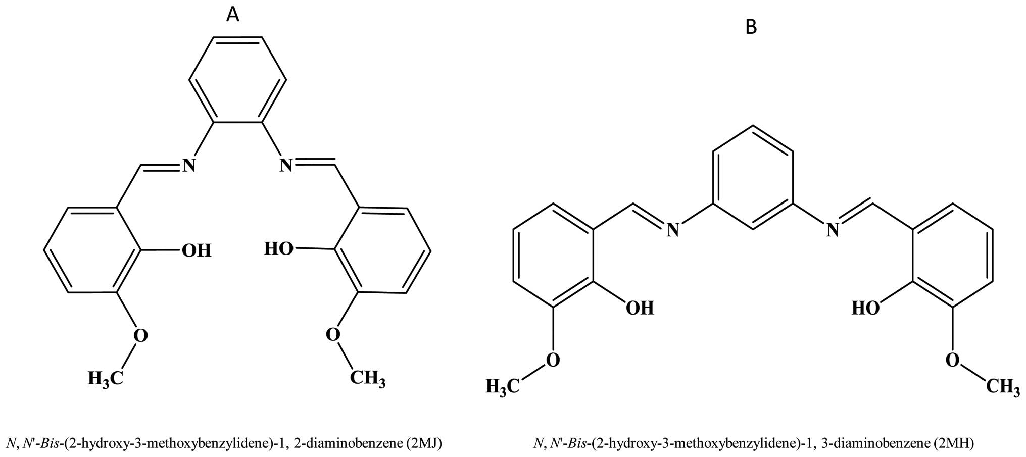
We have synthesized two bis-Schiff base compounds (2MJ) and (2MH)
and solved their crystal structures (Fig. 1A and B) (15–19).
The rigid C=N structure, the flat aromatic moiety and the presence
of imine moieties in both compounds allow a wide variety of
structural analogues to be prepared.
In this study we analyze the suitability of these
two bis-Schiff base compounds as potential MGBs. We probed the
nature of their binding characteristics so as to understand
structural features that are important for good binding efficiency
between diaminobenzene Schiff base and DNA. With this in mind,
these two Schiff base structural analogues, which differ in their
2-hydroxy-3-methoxybenzylidene binding position, were employed in
this study. 2MJ (Fig. 1A) has its
2-hydroxy-3-methoxybenzylidene substructure located at the 1 and 2
positions of its diaminobenzene unit, while 2MH (Fig. 1B), has this moiety located at the 1
and 3 positions making its structure wider than the former.
Materials and methods
Calf thymus DNA, MgCl2, NaCl, phosphate
buffer, EtBr, dimethyl sulfoxide (DMSO) solvent, trypsin and
Hoechst 33258 were all purchased from Sigma-Aldrich (St. Louis, MO,
USA). For the cytotoxicity work; human breast ductal carcinoma
(T-74D), human colon carcinoma (HCT-116), human hepatocellular
carcinoma (HepG2) cells were all sourced from ATCC (American Type
Cell Culture). Fetal bovine serum, McCoy’s 5A, minimum essential
medium (MEM) and RPMI-1641 medium were acquired from Life
Technologies (CA, USA).
3-[4,5-dimethylthiazol-2-yl]-2,5-diphenyltetrazolium bromide (MTT)
was purchased from PhytoTechnology Laboratories (KS, USA). 2MJ and
2MH were synthesized in our laboratory according to previous
published methods (18–20).
Standard buffer solution containing 0.15 M NaCl,
0.50 mM MgCl2, and 10.00 mM phosphate buffer (pH 7.3)
was used throughout (20)]. Schiff
base compounds were separately dissolved in ethanol forming 1000
μM stock solutions. Stock solution of calf thymus DNA was
prepared by dissolving 4.57 mg of the calf thymus in 1.50 ml of the
standard buffer solution.
The procedure was performed according to previous
published work (21). Briefly, 30
μl of each drug was added to 500 μl buffer and the
absorption was measured. Thirty μl were taken from 2MJ and
diluted to 500 μl using the same standard buffer. UV
absorbance was measured at 200–550 nm wavelengths. The DNA stock
solution was titrated against the respective drug solution. The
absorbance measurement was taken after each addition of DNA in
order to calculate the intrinsic binding constant for each compound
with the nucleic acid. The volumes of DNA added to 2MJ solution
were 2, 4, 6, 8, 15, 20, 30, 50, 80 and 130 μl to give
effective DNA concentration of 0.003, 0.006, 0.009, 0.012, 0.022,
0.03, 0.045, 0.075, 0.12, 0.195 mg/ml, respectively. The DNA was
added until no apparent decrease in absorption reading was
observed. This procedure was repeated for 2MH, using the 2, 4, 6,
8, 10, 12, 14, 16, 22 and 30 μl of the DNA stock solution to
give 0.003, 0.006, 0.009, 0.012, 0.015, 0.018, 0.021, 0.024, 0.033,
0.045 mg/ml solution. The UV absorbance values were measured on a
USA Perkin-Elmer Lambda 45 spectrometer.
The drug binding fraction α, and the equilibrium
distribution at each titration position is calculated according to
the following formula:
A=Cb/C=(1−Cf/C)=(Afº−A)/(Afº−Abº).
Afº and Abº are the measured
absorption for the free and fully bound drug at the monitoring
wavelength. r=α.C/CDNA and Cf=
(1−α).C, where CDNA is the total concentration of
DNA or oligonucleotide titrant at each point. The binding constant
value K, was determined by plotting a scatchard plot of r/Cf vs r
(22).
Competitive binding assay was carried out according
to literature (23–26). The fluorescence displacement assay
was performed using a Perkin-Elmer LS45 luminescence spectrometer.
Emission spectra were measured at 600 nm, using the excitation
wavelength 525 nm. The temperature was fixed at 22°C throughout the
work. Ethedium bromide and Hoechst 33258 were separately mixed with
the calf thymus DNA prior to the addition of the test compounds.
Calf thymus (30 μl) was made up to 2.0 ml using the standard
buffer forming a 0.046 mg/ml prior to its fluorescence intensity
measurement. Ethedium bromide (30 μl) was then added to it
and the fluorescence intensity was re-measured.
Viscometer experiments were performed using an
Ubbelohde viscometer (Cannon, USA). The temperature was maintained
at room temperature (25°C) with the aid of a water bath. Calf
thymus DNA solution (10 ml of 240 μg/ml) was placed in the
viscometer and allowed to pass through the small capillary tube.
The time taken for the sample to pass through was measured by using
a digital stop watch. This procedure was repeated but with the
addition of varying concentration of 2MJ and 2MH to the calf thymus
DNA. A volume of 1.3, 2.6, 4.0, 6.6, 9.33 and 13.33 μl
containing 30 mg/ml of the indivi dual compounds were added to 10
ml of the 240 μg/ml calf thymus DNA to give compound-DNA
ratio of 1:1, 1:2, 1:3, 1:5, 1:7 and 1:10. Ethedium bromide and
Hoechst 33258 were used as positive control representing
intercalation and minor groove binding compounds, respectively. The
time required for each mixture to pass was recorded. The procedure
was performed in triplicate for each sample ratio.
Compound 2MJ, with concentrations 1.25, 2.5, 6.25,
12.5, 25, 37.5, 62.5, 87.5, 125, 162.5, 200, 250, 287.5 μM,
was then titrated against the calf thymus DNA and ethedium bromide
mixture and the fluorescence intensity reading was taken after each
addition until the initial fluorescence intensity value was halved.
This process was repeated for 2MH using concentrations 0.75, 1.5,
3.75, 7.46, 14.85, 22.33, 36.58, 50.72, 71.42, 91.54, 111.1 and
136.36 μM of the drug, employing Hoechst 33258 as the
competitive substrate in the manner described previously (21,27).
The emission intensity was measured at 490 nm using excitation
wavelength of 360 nm.
All the cell lines used for the cytotocity
evaluation were between passages 5–9. The cells were cultured in
their respective growth medium supplemented with 10% fetal bovine
serum and 1% penicillin-streptomycin. The colon HCT-116 cells were
cultured in McCoy’s 5A medium while the liver HepG2 cells were
grown in MEM medium. The breast T-47D cells were developed in
RPMI-1641 medium. MTT viability assay was performed with slight
modifications as described by Mosmann (28). In brief, cells were seeded at 5,000
cell density per well for each 96-well plates in 180 μl
medium. 2MJ was dissolved in 100% DMSO as the main stock solution.
After an overnight incubation, 2MJ reagent was added into each well
to make the final concentration 100, 50, 25, 12.5, 6.25, 3.12 and
1.56 μg/ml. The untreated cells received only DMSO as a
negative control. DMSO was serially diluted at concentrations
ranging from 1%–0.03%. All cells were treated for 48 h. The
experiment was repeated twice with four replicates for each
concentration. MTT was first prepared as a stock solution in 5
mg/ml of phosphate buffer saline solution. At the end of the
treatment period (48 h), 20 μl of MTT solution was added to
each well. After 4 h incubation at 37°C, the medium was removed and
200 μl of DMSO was added to the well to dissolve the
formazon crystal. After 1 min of shaking, the optical density was
recorded using a plate reader (Multiskan Ascent) at 570 nm for
absorbance and 650 nm as reference filter. This procedure was
repeated for 2MH.
All molecular docking simulations were performed by
using a PC under Red Hat Linux 9.0 operating system. X-ray
crystallographic structure between Hoechst 33258 and a synthetic
B-DNA dodecamer of sequence C-G-C-G-A-A-T-T-C-G-C-G (pdb: 8BNA) was
used as a model (29). The ligand
within the DNA was substituted with 2MJ and 2MH crystal structure
data (15–19). All the water molecules and
heteroatoms were removed from the DNA pdb file. Polar hydrogens
were added using Insight II (30)
and the charges were assigned from AMBER force fields library using
Insight II program (Accelrys Inc.). Each atom was then assigned a
solvation parameter based on the affinity of the atom for solvent,
and a fragmental volume based on the amount of water that the atom
excludes from solvating the surrounding atoms (31). This step was achieved by using
ADDSOL utility. Grid parameter file (GPF) was then prepared. Each
grid point stores energy of interaction of the corresponding atom
type with the rest of the DNA. The evaluation of the binding energy
is based on a set of interactions, including van der Waals
dispersion forces, electrostatics and hydrogen bonding. A box with
dimension of 80 Å x 80 Å x 120 Å was created, to include the entire
DNA. A grid spacing of 0.375 Å was used and the pairwise-atomic
interaction energy parameters were taken from AutoDock website
(http://autodock.scripps.edu). Parameters
of 12-6 were set for van der Waals forces, while 12-10 for hydrogen
bonds. The distance-dependent dielectric function of Mehler and
Solmajer (32) was used. Autodock
program version 3.0.5 (33) was
used to perform docking.
A rapid energy evaluation through precalculated
grids of affinity potential was combined with a variety of
algorithms search in order to find a suitable binding position for
the ligands on a given DNA site. The program allows the ligands to
be flexible while the DNA was set to be rigid. This docking
technique was carried out using the new empirical free energy
function and the Lamarckian Genetic Algorithm parameters (LGA). The
cluster tolerance was set at <1.0 Å and the initial population
was limited to 50 randomly placed individuals while the energy
evaluations was fixed to the maximum number of 15x105
with a maximum number of generations of 2.7x10⁴. A mutation rate of
0.02 and a crossover rate of 0.80 with elitism value of 1 were also
used. All these parameters are set based on standard protocols
(34).
Results
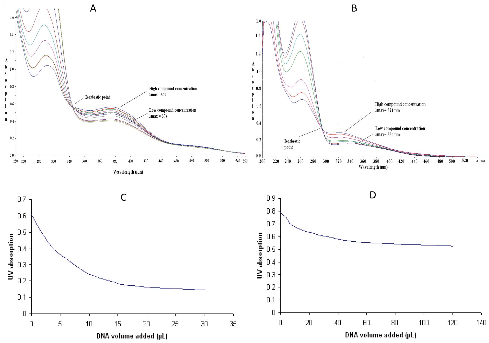
Spectral results of DNA binding with 2MH and 2MJ are
presented in Fig. 2A and B respectively. They show the
various absorption spectra for compounds 2MH and 2MJ before and
after mixing with the calf thymus DNA. Both spectra show
significant shift and decrease in the UV absorbance spectrum of the
compounds following the addition of DNA. Fig. 2A shows the bathochromic
λmax value shift from 323 to 347 nm, and the absorption
at λmax (hypochromic shift) decrease significantly upon
the addition of DNA to 2MH solution. Fig. 2B shows the bathochromic
λmax value for 2MJ shift from 345 to 351 nm, and a
decrease in absorption reading at λmax (hypochromic
shift) during addition of DNA to 2MJ solution. Fig. 2C and D shows the drop in absorption
level upon the addition of DNA to 2MJ and 2MH, respectively.
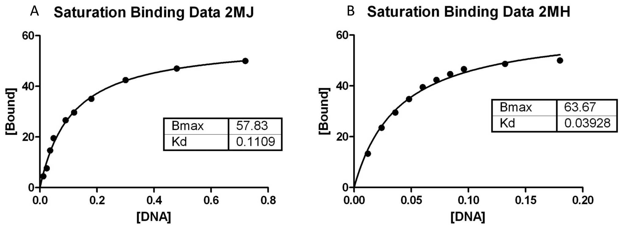
Scatchard equation was applied to find the intrinsic coefficient of
each compound towards the DNA and their strength of binding,
Fig. 4A and B.
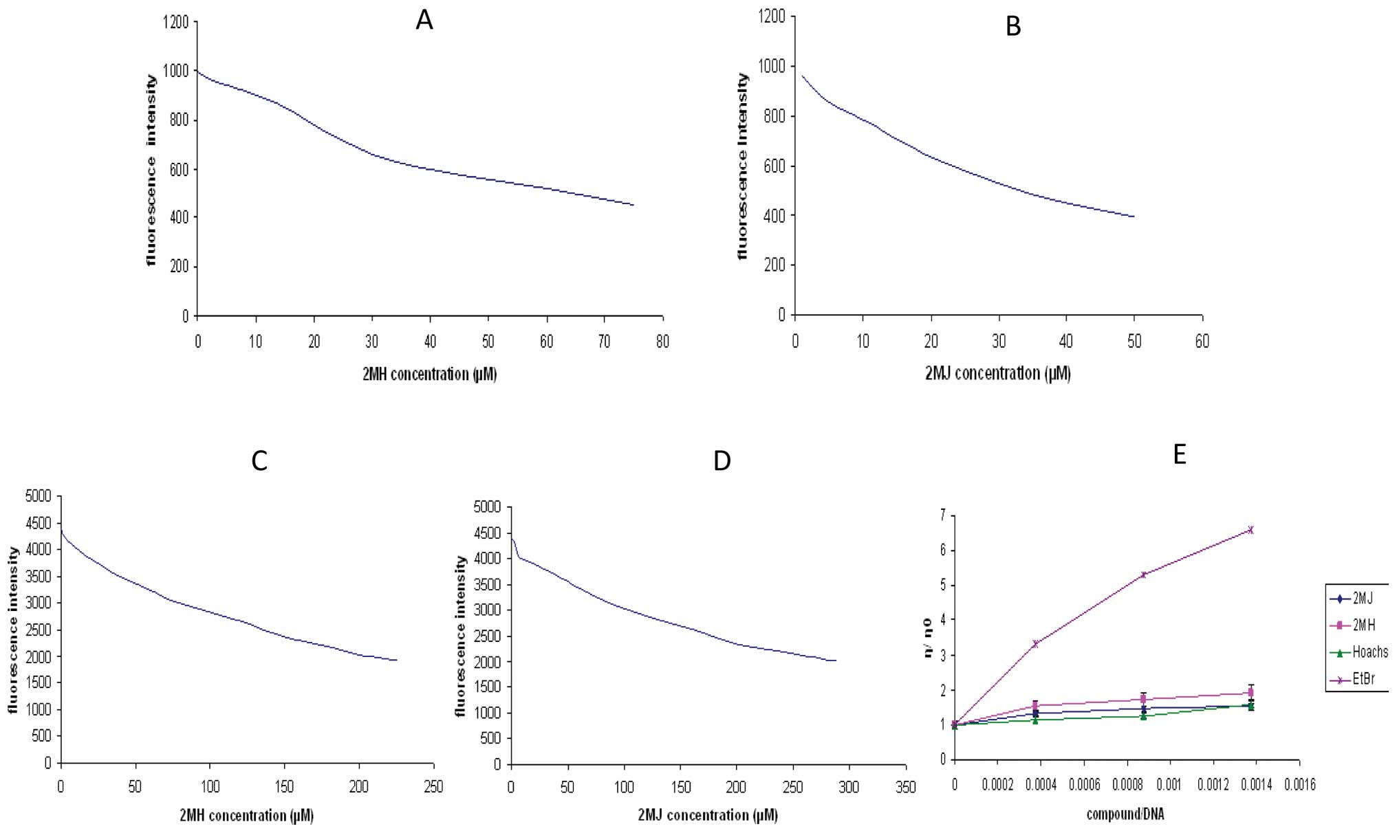
In the competitive binding assay utilizing the
fluorescence technique, each compound caused a decrease in
fluorescence reading of the EtBr-DNA complex. The Q value, a
constant that represents strength of molecule binding, is taken at
a concentration where the initial fluorescent intensity is reduced
by half (21). The Q values were
found to be 173 and 229 μM for 2MH and 2MJ, respectively.
Both 2MJ and 2MH were able to displace the Hoechst 33258 molecule
with 2MH having slightly higher displacement ability than 2MJ, with
their Q values being 37 and 46 μM, respectively. Fig. 3 (A and B) and (C and D) show the
decrease in fluorescence intensity after the addition of 2MJ and
2MH on the EtBr-DNA and Hoechst 33258-DNA mixtures,
respectively.
The results of the viscosity experiment show that
2MH and 2MJ can cause an increase to the DNA solution viscosity.
This indicates a binding interaction between the nucleic acid and
the two compounds. The viscosity was calculated using the following
derived from Poiseuille’s law (35). ηsp =
ηr − 1 = t − to / to.
Where ηsp represents the specific viscosity and to is
the time needed for elution of the solvent alone and t is the
elution time needed for the solution. By this equation, the
viscosity after addition of each compound was calculated. Fig. 3E shows the result of the viscometry
studies for 2MJ and 2MH. The slope measurement for 2MH is
significantly higher than 2MJ indicating stronger DNA binding. The
data are presented as η/η0 vs compound/DNA concentration
ratio. η presents the viscosity for DNA-compound mixture, while
η0 represents viscosity for DNA solution alone. The
results of the viscosity experiments show that 2MH and 2MJ do not
cause significant increase to the DNA solution viscosity compared
to the well established intercalator ethedium bromide which acts as
the control for this experiment (Fig.
3E). Hoechst 33258 reagent is used as a positive control to
represent a minor groove binder. The viscosity reading for the
Hoechst 33258 compound is similar to that of 2MJ and 2MH.
The IC50 for compound 2MJ and 2MH when
exposed to the HCT-116 cell line were 73 and 54 μM,
respectively. In the HepG2 cell line, the IC50 for
compound 2MJ and 2MH were 138 and 98 μM, respectively, while
in the T-D47 breast cancer cells, the values were 205 and 124
μM for 2MJ and 2MH, respectively (Table I).
 | Table IThe IC50 ± SD for each
cell line after treatment with different compounds. |
Table I
The IC50 ± SD for each
cell line after treatment with different compounds.
| Cell lines | Compounds
|
|---|
| 2MJ | 2MH |
|---|
| HCT- 116 | 73±1.5 | 54±0.7 |
| HepG2 | 138±2.2 | 98±0.5 |
| T-D47 | 205±1.8 | 124±1.1 |
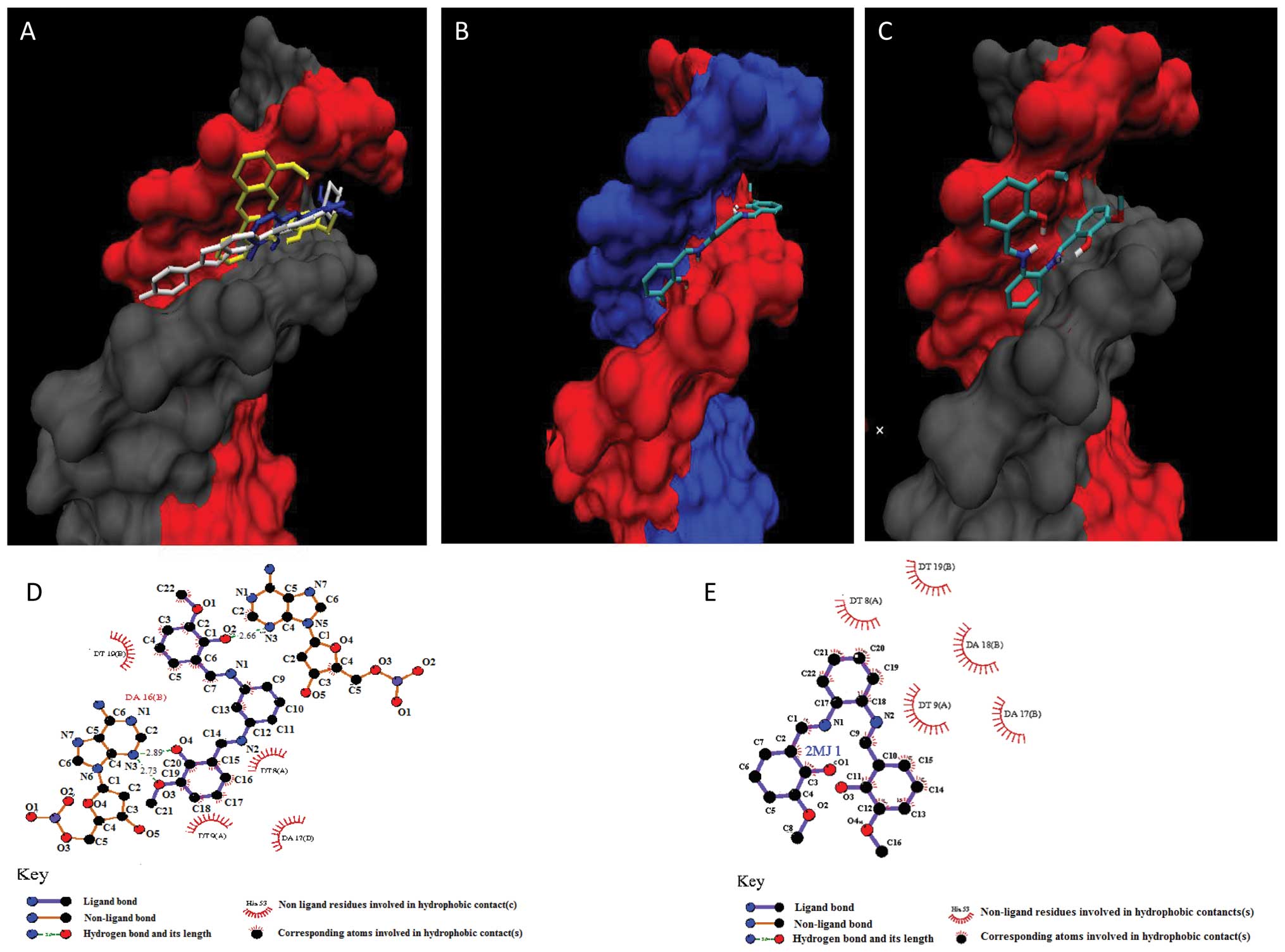
Molecular docking results confirmed the findings in
the spectroscopy analysis. Fig.
5A–C shows that both compounds were able to bind at the same
location where Hoechst resides. The free energy of binding were
−9.61 and −7.38 kcal/mol for 2MH and 2MJ, respectively, indicating
that 2MH has a stronger affinity for the DNA at this site compared
to 2MJ. Fig. 5D and E shows the
result of the docking analysis. The figures reveal that three
hydrogen bonds can be formed between the DNA and 2MH. However, no
hydrogen bond formation occurs between 2MJ and the
deoxyribonucleotide.
Discussion
Since the discovery of minor groove binders, a
number of researchers have developed a variety of alkylating groove
binders by anneling classical chemotherapeutic compounds to MGBs.
This has demonstrated improved potency and better selectivity
(36).
The number of MGBs that have been discovered so far
is limited and there is a constant effort to find better MGBs that
can also be easily synthesized. Schiff bases that harbor metal
complexes have been shown to have the ability to interact with the
nucleic acid (11,12). A number of these Schiff bases
studied, can cause DNA strand breaks (14). However, the study of metal-free
Schiff base interaction with the nucleic acid is still lacking and
research on their anti-tumor potential is not well established. 2MJ
and 2MH are two novel Schiff base structural analogues, which
differ in their 2-hydroxy-3-methoxybenzylidene binding position.
2MJ (Fig. 1A) has its
2-hydroxy-3-methoxybenzylidene substructure located at the 1 and 2
positions of its diaminobenzene unit, while 2MH (Fig. 1B) has this moiety located at the 1
and 3 positions making its structure wider than the former. The
results from the UV spectroscopy and viscometery analysis clearly
indicate that 2MH and 2MJ can bind to the DNA with good binding
strength.
In the displacement assay, 2MJ and 2MH were able to
displace Hoechst 33258 from its site of residence within the DNA,
indicating that these agents are able to bind to the nucleic acid
in its minor groove region. However, based on their calculated Q
values, 2MH shows to be a better MGB than 2MJ with its Q value
being more than 40% lower than that for 2MJ.
To a limited extent, both compounds were also able
to displace EtBr suggesting a trifling intercalation reaction as
well. The lack of significant intercalation reaction is further
evidenced when comparing their overall Q values at the respective
sites. Both compounds were found to have more than 5-fold affinity
towards the minor groove region compared to the intercalation
sites. This suggests that these agents are better MGBs rather than
intercalators. The poor intercalation reaction exhibited by 2MJ and
2MH; with the latter being more prominent, is not a surprise. This
is because they are short of sufficient number of flat aromatic
structures to form adequate electrostatic attraction with the bases
in the narrow spaces between the adjacent DNA base pairs (4). The lack of significant cytotoxicity
activity by 2MH and 2MJ is a typical characteristic of MGBs given
that this region is not frequented by important enzymes such as DNA
polymerase and topoisomerase. However, taken as a whole, the extent
of cytotoxic activity was significantly higher for 2MH compared to
2MJ. This may be due to the stronger DNA binding ability exhibited
by 2MH and its minor intercalative reaction which may interfere
with the functioning of the said regulatory proteins that reside in
the major groove region.
The wide angle of curvature of the 2MH has
demonstrated significantly improved binding to the DNA compared to
its isomeric partner 2MJ. The spectroscopic data, viscometry
analysis and molecular modeling study strongly support this
finding. In the modeling data, the result shows that both 2MJ and
2MH can fit into the minor groove region at the site where Hoechst
33258 can reside (Fig. 5A).
However the finding also shows that part of 2MJ substructure
appears to be protruding out of the groove region as shown in
Fig. 5C. 2MH on the contrary
appears to reside snuggly into the groove with all its structure
lying in parallel within the walls of the minor groove region
(Fig. 5B). The modeling result
also shows that 2MH can form 3 hydrogen bonds at its O4 and O3
atoms with N3 of adenosine 16 of one DNA strand, and its O2 atom
with N3 atom of adenosine 18 of the complementary DNA strand
(Fig. 5D). Moreover, the modeling
data also show that 2MH can form good hydrophobic contact with the
deoxyribonucleotide. However, apart from hydrophobic interaction
with the DNA, 2MJ appears to lack the ability to form any viable
hydrogen linkages with the nucleic acid (Fig. 5E).
The findings of this work support previous studies
which show that good groove binders are crescent in shape and the
ones that have wider angle of curvature are better MGBs (36). However, the angle of curvature of
the molecule must complement the DNA curvature. This can allow
better interaction with hydrogen bond acceptors and donors that
exists at the point of contact between both ligand and DNA.
However, if the curve is too narrow, it may prevent the ligand from
penetrating deep enough into the walls of the groove. This may also
limit hydrophobic interaction hence reducing binding
efficiency.
Although 2MH and 2MJ are relatively neutral MGBs,
they are still potent enough to cause significant DNA binding.
Minor groove regions that are rich in AT sequences emit strong
negative charges due to the presence of phosphate groups. Hence,
positively charged molecules tend to be attracted to the negative
charged AT sequence (37).
However, agents that are devoid of any charge still have the
ability to bind to the MGR but the binding strength is
significantly lower (38).
Taken together, this study reveals diaminobenzene
Schiff base compounds that are devoid of metal cations can bind to
the DNA. The site of ligand binding is mainly via the minor groove
and to lesser extent, the major groove. The annular shape of the
molecule and its degree of curvature influences the DNA-binding
affinity particularly to the minor grove region. The work also
shows that the two Schiff bases, 2MJ and 2MH are non-cytotoxic.
Acknowledgements
We would like to thank Dr Shafida A.
Hamid (Universiti Sains Malaysia) and Dr Salizawati Mohd. Salhimi
(Universiti Sains Malaysia) for their valuable support. This work
was funded by research grants from the Universiti Sains Malaysia
(RU: 1001/PFARMASI/811144).
References
|
1
|
Neidle S: DNA minor-groove recognition by
small molecules. Nat Prod Rep. 18:291–309. 2001. View Article : Google Scholar : PubMed/NCBI
|
|
2
|
Nelson EM, Tewey KM and Liu LF: Mechanism
of antitumor drug action: poisoning of mammalian DNA topoisomerase
II on DNA by 4′-(9-acridinylamino)-methanesulfon-manisidide. Proc
Natl Acad Sci USA. 81:1361–1365. 1984.
|
|
3
|
Sinha R, Islam MM, Bhadra K, Kumar GS,
Banerjee A and Maiti M: The binding of DNA intercalating and
non-intercalating compounds to A-form and protonated form of
poly(rC)·poly(rG): Spectroscopic and viscometric study. Bioorgan
Med Chem. 14:800–814. 2006.PubMed/NCBI
|
|
4
|
Williams TT and Barton JK: Charge
transport in DNA. DNA and RNA Binders: from small molecules to
drugs. Demeunynck M, Bailly C and Wilson WD: Wiley-VCH; Weinheim:
1. pp. 1462003
|
|
5
|
Dadgarnezhad A, Sheikhshoaie I and Baghaei
F: Corrosion inhibitory effects of a new synthetic symmetrical
Schiff-base on carbon steel in acid media. Anti-Corrosion Methods
Materials. 51:266–271. 2004. View Article : Google Scholar
|
|
6
|
Ma H, Chen S, Niu L, Zhao S, Li S and Li
D: Inhibition of copper corrosion by several Schiff bases in
aerated halide solutions. J Appl Electrochem. 32:65–72. 2002.
View Article : Google Scholar
|
|
7
|
Nair R, Shah A, Baluja S and Chanda S:
Synthesis and antibacterial activity of some Schiff base complexes.
J Serb Chem Soc. 71:733–744. 2006. View Article : Google Scholar
|
|
8
|
Morad FM, El Ajaily MM and Gweirif SB:
Preparation, physical characterization and antibacterial activity
of Ni (II) Schiff base complex. J Sci Applicat. 1:72–78. 2007.
|
|
9
|
Hou H, Zhu J, Liu Y and Li Q:
Antibacterial activity of a kind of novel Schiff base and its 3d,4f
complexes. Acta Physicochim Sin. 23:987–992. 2007.
|
|
10
|
Kuz’min VE, Lozitsky VP, Kamalov GL,
Lozitskaya RN, Zheltvay AI, Fedtchouk AS and Kryzhanovsky DN:
Analysis of the structure - anticancer activity relationship in a
set of Schiff bases of macrocyclic 2,6-bis(2- and
4-formylaryloxymethyl) pyridines. Acta Biochim. 47:867–876.
2000.PubMed/NCBI
|
|
11
|
Ye Y, Hu J, He L and Zeng Y:
Surface-enhanced Raman spectroscopy of some Schiff base complexes
and their interaction with DNA. Vibr Spectrosc. 20:1–4. 1999.
View Article : Google Scholar
|
|
12
|
Wang B-D, Yang Z-Y, Qin W, Cai T-K and
Crewdson P: Synthesis, characterization, cytotoxic activities, and
DNA-binding properties of the La(III) complex with naringenin
schiff-base. Bioorg Med Chem. 14:1880–1888. 2006. View Article : Google Scholar : PubMed/NCBI
|
|
13
|
Vijayalakshmi R, Kanthimathi M,
Subramanian V and Nair BU: Interaction of DNA with [Cr(Schi¡
base)(H2O)2]ClO4. Biochim Biophys
Acta. 1475:157–162. 2000.
|
|
14
|
Silveira VCd, Luz JS, Oliveira CC,
Graziani I, Ciriolo MR and Ferreira AMdC: Double-strand DNA
cleavage induced by oxin-dole-Schiff base copper(II) complexes with
potential antitumor activity. J Inorg Biochem. 102:1090–1103. 2008.
View Article : Google Scholar : PubMed/NCBI
|
|
15
|
Al-Douh MH, Al-Fatlawy AA and Abid OH:
Synthesis and characterization of some 2-(N-Benzoyl-N-pyrid-4-yl
amino-benzyl)-aminobarbituric acids via Schiff’s bases. Hadh
Studies Res. 4:37–49. 2003.
|
|
16
|
Al-Douh MH, Al-Fatlawy AA and Abid OH:
Synthesis and characterization of some 2-(N-benzoyl-N-pyrid-2-yl
aminobenzyl)-aminobarbituric acids via N-benzylidene
pyridine-2-amines. J Nat Appl Sci. 8:181–194. 2004.
|
|
17
|
Al-Douh MH, Al-Fatlawy AA and Abid OH:
Synthesis and characterization of 2-(N-benzoyl-N-pyrid-3-yl
aminobenzyl)-aminobarbituric acids via N-benzylidene
pyridine-3-amines. Fac Sci Bull. 16:83–94. 2003.
|
|
18
|
Al-Douh MH, Hamid SA, Osman H, Ng SL and
Fun HK: 6, 6′-dimethoxy-2, 2′-[m-phenylene
bis(nitrilomethylidyne)]diphenol. Acta Crystallogr. 63:O3570–O3571.
2007.
|
|
19
|
Al-Douh MH, Hamid SA, Osman H, Kia R and
Fun HK: 2-amino-N-(2-hydroxy-3-methoxybenzylidene) aniline. Acta
Crystallogr. 64:O1201–O1202. 2008.PubMed/NCBI
|
|
20
|
Stokke T and Steen HB: Multiple binding
modes for Hoechst 33258 to DNA. J Histochem Cytochem. 33:333–338.
1985. View Article : Google Scholar : PubMed/NCBI
|
|
21
|
Fox KR: Drug-DNA Interaction Protocols.
Humana Press; NJ: 1997, View Article : Google Scholar
|
|
22
|
Jenkins TC: Optical absorbance and
fluorescence techniques for measuring DNA-drug interactions.
Drug-DNA Interaction Protocols. 90. Fox KR: Humana Press; NJ: pp.
195–218. 1997, View Article : Google Scholar : PubMed/NCBI
|
|
23
|
Morgan AR, Lee JS, Pulleyblank DE, Murray
NL and Evans DH: Ethidium fluorescence assays. Part 1.
Physicochemical studies. Nucleic Acids Res. 7:1979.PubMed/NCBI
|
|
24
|
Roche CJ, Thomson JA and Crothers DM: Site
selectivity of daunomycin. Biochemistry. 33:926–935. 1994.
View Article : Google Scholar : PubMed/NCBI
|
|
25
|
Haq I, Lincoln P, Suh D, Norden B,
Chowdhry BZ and Chaires JB: Interaction of .delta.- and
.lambda.-[Ru(phen)2DPPZ]2+ with DNA: a calorimetric and equilibrium
binding study. J Am Chem Soc. 117:4788–4796. 1995.
|
|
26
|
Chaires JB, Dattagupta N and Crothers DM:
Studies on interaction of anthracycline antibiotics and
deoxyribonucleic acid: equilibrium binding studies on the
interaction of daunomycin with deoxyribonucleic acid. Biochemistry.
21:3933–3940. 1982. View Article : Google Scholar : PubMed/NCBI
|
|
27
|
Peberdya JC, Malinab J, Khalidc S, Hannond
MJ and Rodger A: Influence of surface shape on DNA binding of
bimetallo helicates. J Inorg Biochem. 101:1937–1945. 2007.
View Article : Google Scholar : PubMed/NCBI
|
|
28
|
Mosmann T: Rapid colorimetric assay for
cellular growth and survival: application toproliferation and
cytotoxicity assays. J Immunol Methods. 65:55–63. 1983. View Article : Google Scholar : PubMed/NCBI
|
|
29
|
Pjura PE, Greskowiak K and Dickerson RE:
Binding of Hoechst 33258 to the minor groove of B-DNA. J Mol Biol.
197:257–271. 1987. View Article : Google Scholar : PubMed/NCBI
|
|
30
|
Accelrys Inc.: Insight II Journal.
2000.
|
|
31
|
Marrone TJ, Luty BA and Rose PW:
Discovering high-affinity ligands from the computationally
predicted structures and affinities of small molecules bound to a
target: a virtual screening approach. Perspect Drug Discov Design.
20:209–220. 2000. View Article : Google Scholar
|
|
32
|
Mehler El and Solmajer T: Electrostatic
effects in proteins: Comparison of dielectric and charge models.
Protein Engineering. 4:903–910. 1991. View Article : Google Scholar : PubMed/NCBI
|
|
33
|
Morris GM, Goodsell DS, Halliday RS, et
al: Automated docking using a Lamarckian genetic algorithm and an
empirical binding free energy function. J Comput Chem.
19:1639–1662. 1998. View Article : Google Scholar
|
|
34
|
Kulys J and Ziemys A: A role of proton
transfer in peroxide-catalyzed process elucidated by substrates
docking calculation. BMC Struct Biol. 1:1–6. 2001. View Article : Google Scholar : PubMed/NCBI
|
|
35
|
Satyanarayana S, Dabrowik JC and Chaires
JB: Tris (phenanthroline) ruthenium (II) enantiomer interactions
with DNA: Mode and specificity of binding. Biochemistry.
32:2573–2584. 1993. View Article : Google Scholar : PubMed/NCBI
|
|
36
|
Neidle S: Nucleic acid Structure and
Recognition. Oxford University Press; New York, NY: 2002
|
|
37
|
Constant J-F and Demeunynck M: Design and
studies of a basic targeting drugs. Small Molecule DNA and RNA
Binders: from synthesis to nucleic acid complex. Demeunynck M,
Bailly C and Wilson WD: Wiley-VCH; Weinheim: 2. pp. 2472003
|
|
38
|
Chen Y-H, Yang Y and Lown JW: Design of
distamicin analogues to probe the physical origin of the
antiparallel side by side oligopeptide binding motif in DNA minor
groove recognition. Biochem Biophys Res Commun. 220:213–218. 1996.
View Article : Google Scholar : PubMed/NCBI
|