Introduction
Intrahepatic cholangiocarcinoma (ICC) is highly
fatal due to early invasion, rich fibrous stroma, and widespread
metastasis. Current therapies for locally advanced or metastatic
disease have little effect on the natural history of this
malignancy. Understanding the molecular mechanisms underlying the
progression of ICC may provide insights allowing the development of
novel anti-neoplastic therapies. Chemokines are major regulators of
cell migration and adhesion especially in the bone marrow
microenvironment and are involved in the trafficking of lymphocytes
and leukocytes during inflammation and immune responses. Recently,
some evidences have been obtained about a role of the chemokine
stromal cell-derived factor-1 (SDF-1) and its specific receptor,
CXCR4, in cell cycle progression, migratory behavior, survival, and
metastasis of tumor cells (1–4).
Carcinoma-associated fibroblasts (CAFs) are thought to promote the
growth of cancerous cells, angiogenesis, and stromal fibrosis by
recruiting endothelial progenitor cells through the interaction
between CXCR4 and SDF-1 released from CAFs (2,5). In
our previous study, we demonstrated that hepatic stellate cell
(HSC) activation by angiotensin II (AngII) facilitated stromal
fibrosis and tumor progression via an interaction with cancer cells
in ICC tissues (6). Tumor
progression and fibrosis are pivotal aspects of malignancy and
several factors, including epidermal growth factor (EGF),
transforming growth factor-β (TGF-β), hepatocyte growth factor
(HGF), vascular endothelial growth factor (VEGF), trypsinogen,
AngII, and chemokines including SDF-1, are regarded as possibly
being involved in cross-talk in tumor-stromal interactions
(2–8).
The epithelial-to-mesenchymal transition (EMT) is a
process initially observed in embryonic development in which cells
lose epithelial characteristics and gain mesenchymal properties
such as increased motility and the capacity for invasion (9,10).
Growth factors such as EGF, TGF-β, HGF and VEGF have been found to
induce EMT (11–14). EMT is also a critical event that is
occasionally observed during solid tumor progression, including
invasion and metastasis, by which cancer cells acquire a more
aggressive phenotype. Under these conditions, EMT is defined as the
occurrence of a variable proportion of tumor cells up-regulating
mesenchymal markers such as vimentin, and down-regulating
epithelial markers such as E-cadherin and β-catenin (14,15).
Expressions of these EMT markers are induced by growth
factor/receptor systems such as the TGF-β/TGF-β receptor system
(12,16). However, little is known about EMT
induced by cytokines and chemokines, including AngII and SDF-1,
despite abundant evidence of their involvement in the diverse
cancer cells. AngII has been known as a growth factor which can
occur EMT in renal epithelial cells, peritoneal mesothelial cells
and alveolar epithelial cells (17–19).
But in the past, the relationship between AngII and SDF-1 in the
micro-environment around cancer tissues and the role of AngII on
EMT of cancer cells has not been reported in detail.
We investigated whether HSCs activated by AngII in
cancerous stromal tissues released SDF-1 into collagen-rich ICC
tissues and hypothesized that the AngII/AngII type 1 receptor
(AT-1) axis and SDF-1/CXCR4 axis activate the fibrogenesis in
cancerous stroma, tumor invasion, and metastasis of ICC cells by
mediating EMT via a synergistic interaction between cancer cells
and CAFs including activated HSCs.
Materials and methods
Human tissue samples
The current study included 22 specimens of primary
ICC (well to poorly-differentiated adenocarcinoma) that were
surgically resected between 1998 and 2009. The average age of ICC
patients was 64 years (range 35–84). The patients had stage I to
IVB disease on the basis of the General Rules for the Clinical and
Pathological Study of Primary Liver Cancer (20). Immediately following surgical
removal, the tissue samples were frozen in liquid nitrogen and
stored at −80°C until the time of the assay for measuring AngII
concentration. For immunohistochemical examination, the materials
employed in this study, 20% formalin-fixed and paraffin-embedded
specimens, were retrieved from the surgical pathology files of the
Pathology Section of Kanazawa University Hospital, Kanazawa,
Japan.
Measurement of angiotensin II in
tissues
The determination of AngII concentration in ICC
tissue was performed as follows. Briefly, tissue samples were
homogenized at 4°C in saline containing 0.1 N HCl and 5%
urinastatin. The homogenate was sedimented at 10000 × g for 30 min
at 4°C, and the supernatant was used for radioimmunoassay of AngII
using the florisil method (florisil absorption and elution with
acetone-hydrochloric acid solution) as described previously
(21). This method is sensitive,
specific and useful for routine clinical investigation. Parallel
frozen tissue samples were homogenized in phosphate-buffered saline
(PBS).
Cell culture
Two human ICC cell lines [HuCCT-1, obtained from the
Cell Resource Center for Biochemical Research, Tohoku University,
Sendai, Japan (22) and CCKS-1,
obtained from the Department of Human Pathology, Kanazawa
University Graduate School of Medicine, Kanazawa, Japan (23)], and a human hepatic stellate cell
line [LI-90, obtained from the Human Science Cell Bank, Saitama,
Japan (24)] were used. ICC cell
lines were maintained at 37°C in a 5% CO2 incubator and
grown in RPMI-1640 medium supplemented with 2 mM glutamine, 10%
fetal bovine serum (FBS), 100 U/l penicillin and 100 μg/ml
streptomycin. LI-90 cell lines were maintained at 37°C in a 5%
CO2 incubator and grown in Dulbecco’s modified Eagle’s
medium (DMEM) supplemented with 2 mM glutamine, 10% FBS, 100 U/l
penicillin and 100 μg/ml streptomycin.
Reagents and antibodies
AngII was used at concentrations of 1, 10, 100 or
1000 nM. An active compound, telmisartan, which is a novel,
long-acting, selective AT-1 receptor antagonist, was purchased from
Sigma-Aldrich (St. Louis, MO, USA) and used at concentrations of
100 and 1000 nM, according to a previous report (6). Human recombinant SDF-1α was purchased
from Sigma-Aldrich and was used at concentrations of 1, 10 or 100
ng/ml according to a previous report (3). A CXCR4 antagonist, AMD3100
octahydrochloride hydrate, was purchased from Sigma-Aldrich and
used at concentrations of 100 or 1000 ng/ml. TGF-β1 was purchased
from Sigma-Aldrich and was used at a concentration of 10 ng/ml. For
primary antibodies, we obtained goat polyclonal SDF-1 antibody
(Santa Cruz Biotechnology, CA, USA), goat polyclonal CXCR4 antibody
(Santa Cruz), mouse monoclonal α-smooth muscle actin (α-SMA)
antibody (Sigma-Aldrich), mouse monoclonal β-actin antibody
(Sigma-Aldrich), rabbit polyclonal glial fibrillary acidic protein
(GFAP) antibody (Dako Cytomation, Glostrup, Denmark), mouse
monoclonal E-cadherin antibody (Zymed Laboratories, CA, USA), mouse
monoclonal β-catenin antibody (Santa Cruz), and mouse monoclonal
vimentin antibody (Santa Cruz).
Immunohistochemistry
The expressions of SDF-1 and CXCR4 in resected ICC
specimens were examined immunohisto-chemically using each primary
antibody by LSAB (labeled streptavidin-biotin) method. To identify
the antigen in tissues, deparaffinized sections were pretreated by
autoclaving in 10% citric acid buffer (pH 8.0) at 120°C for 15 min.
After pretreatment with protein block serum (Dako Cytomation,
Kyoto, Japan) for 10 min and in 2% skim milk for 20 min to block
non-specific reactions, the sections were incubated with each
primary antibody at 4°C overnight. After incubation, the slides
were incubated with biotinylated rabbit anti-goat IgG (Vector
Laboratories, CA, USA) for 1 h at room temperature and then with
LSAB solution (LSAB kit, Dako) for 1 h at room temperature. The
reaction products were developed in 0.02% 3,3′-diaminobenzidine
tetrahydrochloride (DAB) solution containing 0.1%
H2O2. To identify the antigens of α-SMA and
GFAP in HSCs and E-cadherin, β-catenin and vimentin in ICC cells,
we examined using HRP (horseradish peroxidase) method. The
Envision+ polymer solution (HRP-conjugated secondary antibody, Dako
Cytomation) was then applied for 1 h after primary antibody
treatment. The reaction products were developed in DAB solution
containing 0.1% H2O2. The sections were then
lightly counterstained with hematoxylin. The slides were examined
under a fluorescence microscope (Olympus, Tokyo, Japan). Specimens
were classified as positive when >10% of cancer cells were
stained. The intensity of each type of staining was graded as
negative (no stain or weak positive) and positive (intermediate or
strongly positive) microscopically.
Immunocytochemistry
The suspensions of cancer cells were seeded onto a
Lab-Tek Chamber Slide System (2-well) (Nalge Nunc International,
Rochester, NY, USA) and incubated for 24–48 h at 37°C in a humid
atmosphere of 5% CO2/95% air. The coverslips with cells
were then fixed with methanol and acetone 1:1 (v/v). After
pretreatment with protein blocking serum for 10 min to block
non-specific binding, immunostaining for the expressions of CXCR4
in ICC cells was performed using the LSAB kit. Briefly, the slides
were incubated with primary antibody (1:50) at 4°C overnight. After
washing, biotinylated rabbit anti-goat IgG was applied for 1 h and
then LSAB solution for 1 h at room temperature. The reaction
products were visualized via a DAB reaction. The cells were then
lightly counterstained with hematoxylin and examined under a
fluorescence microscope.
Western blot analysis
Western blotting was performed as descrived
previously (6). The polyvinylidene
difluoride (PVDF) membrane were incubated for 2 h at room
temperature with primary antibody against CXCR4 diluted 1:1000 with
washing solution, followed by an HRP-conjugated anti-goat antibody
diluted 1:5000 with washing solution. Chemiluminescence was
detected with the ECL Plus Western Blotting Detection System (GE
Healthcare Bioscience, Japan). We also used anti-SDF-1 antibody for
Western blotting to measure the up- or down-regulation of SDF-1
expressions in LI-90 incubated in medium to which AngII (100 nM) or
TGF-β1 (10 ng/ml) added.
Extraction of RNA and reverse
transcriptase polymerase chain reaction (RT-PCR) for SDF-1, CXCR4
and GAPDH mRNA
After the isolation of mRNA using TRI Reagent
(Sigma-Aldrich) and synthesize first-strand cDNA using RNeasy Mini
Kit (Qiagen, USA) and QIA shredder (Qiagen), PCR for SDF-1, CXCR4
and GAPDH was performed with a TaqMan PCR Kit (Takara, Japan),
according to the manufacturer’s instructions. The SDF-1 forward
primer sequence was 5′-CCGCGCTCTGCCTCAGCGACGGGAAG-3′ and the
reverse was 5′-CTTGTTTAAAGCTTTCTCCAGGTACT-3′ (this pair generated a
227-bp fragment). The CXCR4 forward primer sequence was
5′-TTCTACCCCAATGACTTGTG-3′ and the reverse was
5′-ATGTAGTAAGGCAGCCAACA-3′ (this pair generated a 260-bp fragment).
The GAPDH oligonucleotide primer set (forward
5′-ACCACAGTCCATGCCATCAC-3′, reverse 5′-TCCACCACCCTGTTGCTGTA-3′;
this pair generated a 452-bp fragment) was used as an internal
standard. PCR was performed for 35 cycles (denaturation at 98°C for
15 sec, annealing at 58°C for 30 sec, and extension at 74°C for 45
sec). After PCR, 5 μl samples of the products were subjected
to 2.0% agarose gel electrophoresis and stained with ethidium
bromide.
Enzyme-linked immunosorbent assay (ELISA)
of SDF-1α
The baseline levels of SDF-1α production by LI-90
cells treated with and without different concentrations (0.1–100
ng/ml) of AngII were determined. LI-90 cells were seeded onto 6-cm
dishes at a density of 1×10⁵/ml and cultured for 24 h.
Pretreatment with an antagonist of AngII, telmisartan (1000 nM),
was carried out for 30–60 min before the addition of AngII. After
the medium had been replaced with fresh medium, the cells were
cultured for another 48 h. The concentration of SDF-1α in the
supernatant was measured by ELISA using a human SDF-1α antibody and
an enzyme immunoassay kit (R&D Systems), according to the
manufacturer’s instructions.
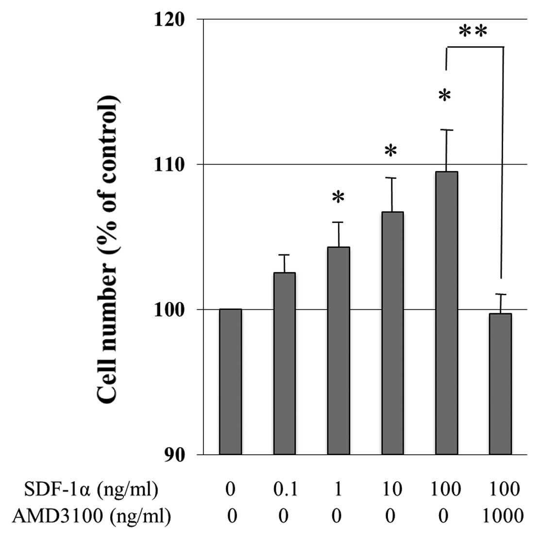
Cell proliferation assay
The proliferative effect of SDF-1α on HSCs was
quantified using an MTT colorimetric assay with Cell Proliferation
Kit I (Roche, Basel, Switzerland), as descrived previously
(6). In brief, LI-90 cells
(5×10³ cells/well) was grown in 96-well flat-bottom
microtiter plates in 100 μl of medium including 1% FBS and
incubated for 48–96 h at 37°C in a humidified atmosphere (e.g.,
37°C, 5% CO2). The medium contained different
concentrations (0.1–100 ng/ml) of SDF-1α. In some experiments,
AMD3100, an antagonist of CXCR4, was added to the SDF-1α treatment
to assure that the proliferative effect caused by SDF-1α occurred
via the CXCR4 receptor. After the incubation period, 10 μl
of the MTT labeling reagent (final concentration 0.5 mg/ml) were
added to each well, the microplate was incubated for 4 h in a
humidified atmosphere (e.g., 37°C, 5% CO2) and then 100
μl of the solubilization solution were added to each well.
We allowed the plate to stand overnight in the incubator in a
humidified atmosphere (e.g., 37°C, 5% CO2), then checked
for complete solubilization of the purple formazan crystals, and
measured the spectrophotometric absorbance value of the samples
using a microplate reader. The wavelength for measuring absorbance
of the formazan product is 595 nm. The experiments were repeated in
triplicate wells. Cell viability was calculated as follows: Cell
number (% of control) = (absorbance of experimental
wells)/(absorbance of control wells) × 100 (%).
Fluorescent immunocytochemistry
LI-90 cells were grown on a Lab-Tek Chamber Slide
System to 40%-60% confluence, serum-deprived overnight (DMEM + 1%
FBS), and then treated with 100 ng/ml of SDF-1α or 10 ng/ml of
TGF-β1 in the presence or absence of pretreatment for 1 h with 1000
ng/ml of AMD3100. CCKS-1 cells were seeded and grown on the Lab-Tek
Chamber Slide System to 40%-50% confluence, serum-deprived
overnight (RPMI-1640 + 1% FBS), and then treated with or without
100 ng/ml of SDF-1α in the presence or absence of pretreatment for
1 h with 1000 ng/ml of AMD3100. Cells were fixed with 3.7% formalin
for 10 min at room temperature. After washing the cells with PBS,
non-specific binding was blocked with Protein Blocking Reagent
(Dako) for 1 h at room temperature. LI-90 cells were incubated with
the primary antibodies (α-SMA and GFAP) and ICC cells were
incubated with each primary antibody (E-cadherin, β-catenin, or
vimentin) overnight at 4°C. Slides were then washed and incubated
with the appropriate Alexa Fluor 488 and 592 nm phalloidin
(Molecular Probes, Inc., Eugene, OR, USA)-conjugated specific
secondary antibodies for double staining for 1 h at room
temperature. Cells were then incubated with Hoechst 33258 for
nuclear staining for 5 min and mounted with propyl gallate
containing phenylenediamine under glass coverslips. Cells were then
visualized for immunofluorescence with a laser scanning Olympus
microscope at ×10, ×20 and ×40 magnification or a Confocal LSM510
microscope (Carl Zeiss Microimaging, Inc., Thornwood, NY, USA) at
×63 magnification.
In vitro migration assays of cultured ICC
cells
The migratory capabilities of both cultured ICC cell
lines were assayed using a BD BioCoat Matrigel invasion chamber
(24-well plate, 8-μm pore) (BD Biosciences, Bedford, MA,
USA). Matrigel invasion chamber used in this study is a growth
factor-reduced type. Medium (0.5 ml) containing 5×10⁵
ICC cells was added to the upper chamber, and 0.5 ml of either
medium alone or medium supplemented with 0.1, 1, 10 or 100 ng/ml of
SDF-1α was added to the lower chamber. AMD3100 was used at 1000
ng/ml. For the migration assay of ICC cells co-cultured with LI-90
cells, medium (0.5 μl) containing 5×10⁵ ICC cells
was added to the upper chamber, and 0.5 ml of either medium alone
or medium containing 4×10⁴ LI-90 cells and 100 nM of
AngII, 100 ng/ml of SDF-1α, or 10 ng/ml of TGF-β1 was added to the
lower chamber with pretreatment of telmisartan or AMD3100. Chambers
were incubated for 48 h at 37°C and 5% CO2. ICC cells on
the upper surface of the filter were removed using a cotton wool
swab, and the cells that had migrated to the lower surface were
stained using 1% toluidine blue after fixation with 100% methanol.
The number of migratory cells was counted in 10 medium power fields
(×20). Each experiment was conducted in triplicate. A migration
index (the ratio of the number of migratory cells in an
experimental group/the number of migratory cells in control groups
with neither chemokines nor cytokines) was calculated in each
experiment.
Statistical analysis
Regression analysis was examineed for a correlation
between AngII concentration in ICC tissues and survival periods.
Statistical analyses were carried out using Student’s unpaired
t-test. Univariate analysis for survival was performed by using
log-rank tests. P<0.05 was considered statistically
significant.
Results
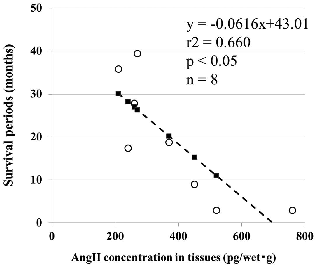
Correlation between AngII concentration
in ICC tissues and survival
The dotted lines indicate the value of each AngII
concentration of initially resected 8 ICC tissues. There is a
significant colleration between AngII concentration of resected ICC
tissues and survival periods after resection. It is noteworthy that
the higher concentration of AngII is, the longer the survival
period of resected patient is. This result confirmed that the
patients whose resected ICC tissues had higher AngII concentration
had significantly poorer prognosis compared with the patients
having lower AngII concentration (Fig.
1).
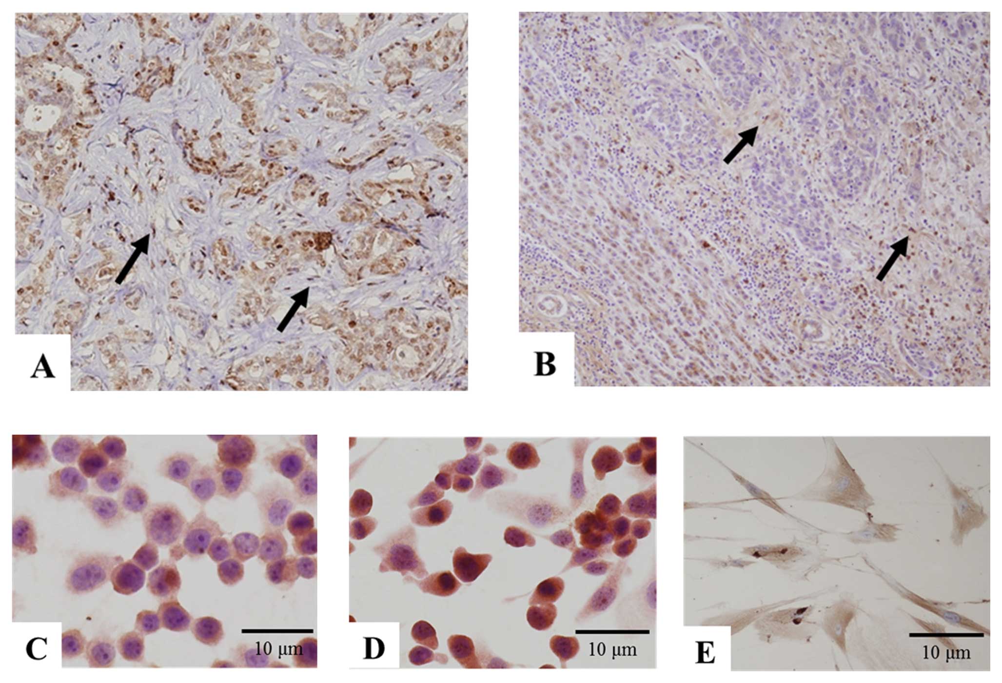
Expression of CXCR4 and SDF-1 in resected
ICC specimens
Immunohistochemistry showed CXCR4 to be expressed in
12 of 16 (75%) surgical specimens of human ICC. Immunoreactivity of
CXCR4 was diffusely positive in the cytoplasm of cancer cells. In
addition, fibroblast-like stromal cells were clearly positive for
CXCR4 in the cancerous stroma (Fig.
2A, arrows). In the surrounding liver, hepatocytes were weakly
positive and lymphoid cells were positive for CXCR4 as well. ICC
cells were weakly and diffusely positive for SDF-1 and
fibroblast-like stromal cells were clearly positive for SDF-1 in
cancerous stromal tissues (Fig.
2B, arrows). In the surrounding liver, a few fibroblasts,
hepatocytes, and bile ducts were positive for SDF-1.
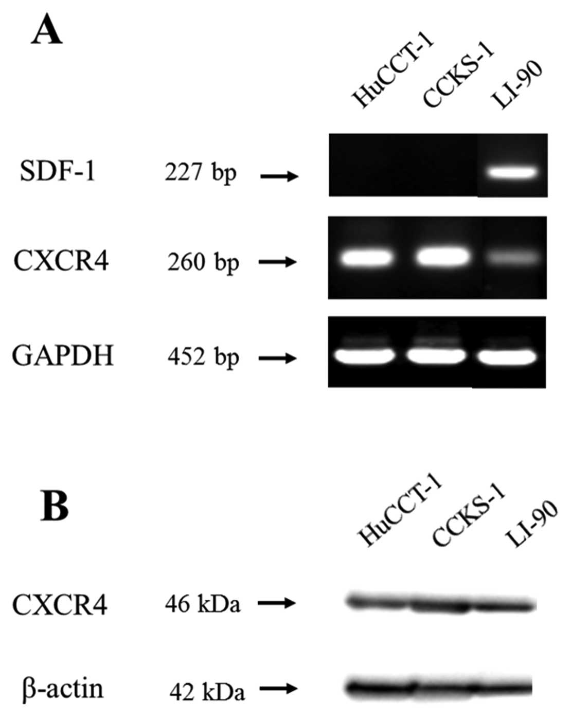
CXCR4 and SDF-1 expressions in human ICC
and HSC cells
Immunoreactivity of CXCR4 receptor was evident in
membranous and granular cytoplasmic patterns in HuCCT-1, CCKS-1 and
LI-90 cells (Fig. 2C–E). CXCR4
expression at the mRNA level was detected in all three cell lines
by RT-PCR (208 bp, Fig. 3A). LI-90
cells expressed SDF-1 mRNA and protein constitutively, whereas no
ICC cells in either cell line expressed SDF-1 mRNA (228 bp,
Fig. 3A). Western blot analysis
demonstrated CXCR4 protein expression in all three cell lines (46
kDa, Fig. 3B).
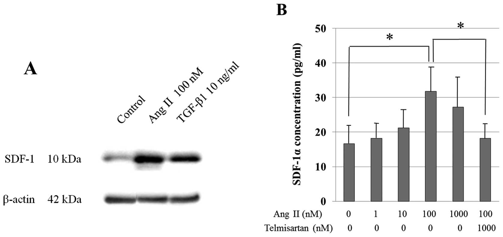
SDF-1α released from activated HSCs
induced cell proliferation at autocrine fashion
LI-90 cells were incubated with 100 nM AngII and 10
ng/ml TGF-β1. Western blotting showed that, SDF-1α protein was more
clearly detectable in cultured LI-90 cells with 100 nM AngII or 10
ng/ml TGF-β1 treatment than in untreated cells (Fig. 4A). ELISA showed the SDF-1α level in
the supernatant of untreated cultured LI-90 cells to be 16.67±5.3
pg/ml. The addition of 1–1000 nM of AngII in the supernatant of the
cultured LI-90 cells induced a dose-dependent increase in SDF-1α
protein level. The addition of 100 nM AngII to the supernatant of
cultured LI-90 cells induced a significant increase in SDF-1α as
compared with untreated cells (31.82±5.3 pg/ml) and pretreatment
with 1000 nM telmisartan significantly inhibited the increase in
the release of SDF-1α protein (18.19±4.5 pg/ml) (Fig. 4B). These results indicated that
SDF-1α released from LI-90 cells into the supernatant was
down-regulated by antagonizing AT-1 receptor. To examine the
proliferative effect of SDF-1α on HSCs, LI-90 cells were treated
with SDF-1α and the proliferative effects were quantified by MTT
assay. After a 48-h incubation, the proliferation of LI-90 cells
was dose-dependently increased by SDF-1α at concentrations from 1
to 100 ng/ml. AMD3100 significantly inhibited the 100 ng/ml
SDF-1α-induced proliferative effect at a concentration of 1000
ng/ml (Fig. 5).
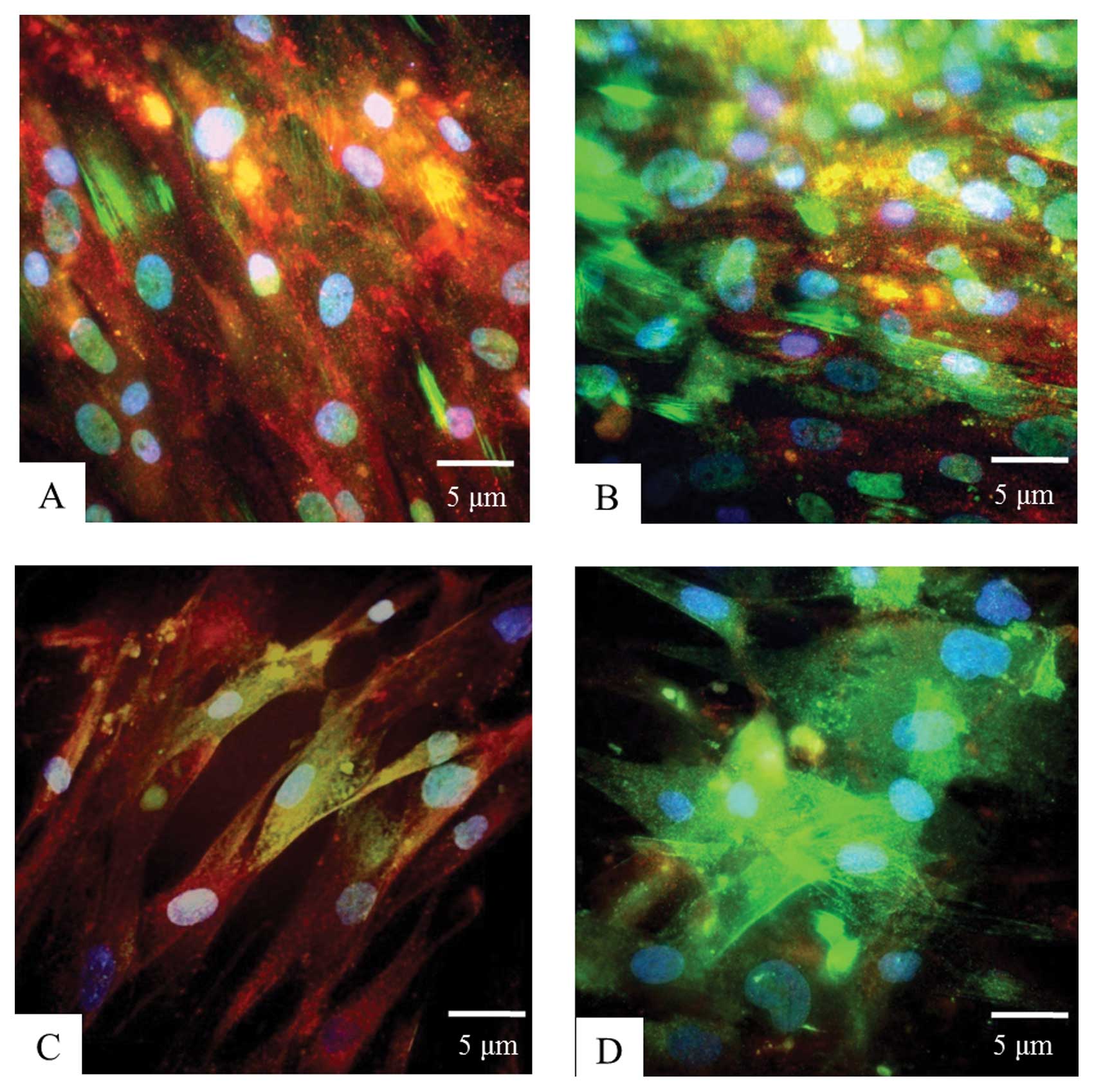
SDF-1α enhanced the activation of
HSCs
GFAP is known to be expressed in quiescent or
partially activated HSCs as a cell-specific marker of HSCs and
α-SMA is known as a cell-specific marker of activated HSCs and
myofibroblasts (25). When the
quiescent HSCs were stimulated by attractants, the
transdifferentiation of HSCs into myofibroblasts and the expression
of α-SMA in the cytoplasm were enhanced. In untreated HSCs, GFAP
expression was ubiquitous in the cytoplasm of nearly all cells,
while only partial α-SMA expression was seen in the cytoplasm.
Fluorescent immunocytochemistry showed that adding 100 ng/ml SDF-1α
to LI-90 cells increased the number of α-SMA-positive LI-90 cells
as compared with control cells (Fig.
6A and B). AMD3100 at 1000 ng/ml inhibited the activation of
LI-90 cells (Fig. 6C). Addition of
10 ng/ml TGF-β1 to LI-90 cells induced the activation and the
transformation into myofibroblasts (Fig. 6D).
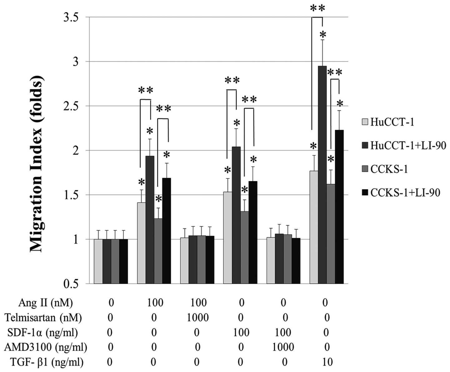
Co-culture with LI-90 cells and the
addition of SDF-1α enhanced migration capability of cultured ICC
cells
When ICC cells were treated with 100 ng/ml of SDF-1α
in the lower chamber, the numbers of migrating HuCCT-1 and CCKS-1
cells significantly increased by 1.79- and 1.4-fold, respectively,
as compared with control cells (Fig.
7). In contrast, more ICC cells migrated when LI-90 cells were
co-cultured in the lower chamber; the migration index was 1.9-fold
in HuCCT-1 and 1.7-fold in CCKS-1 as compared to the control. It is
conceivable that LI-90 cells produce some factors promoting the
migration of cultured ICC cells. When 1000 ng/ml of AMD3100 was
added to the culture medium of both ICC cells co-cultured with
LI-90 cells, migration of ICC cells was significantly suppressed to
the control level (Fig. 8).
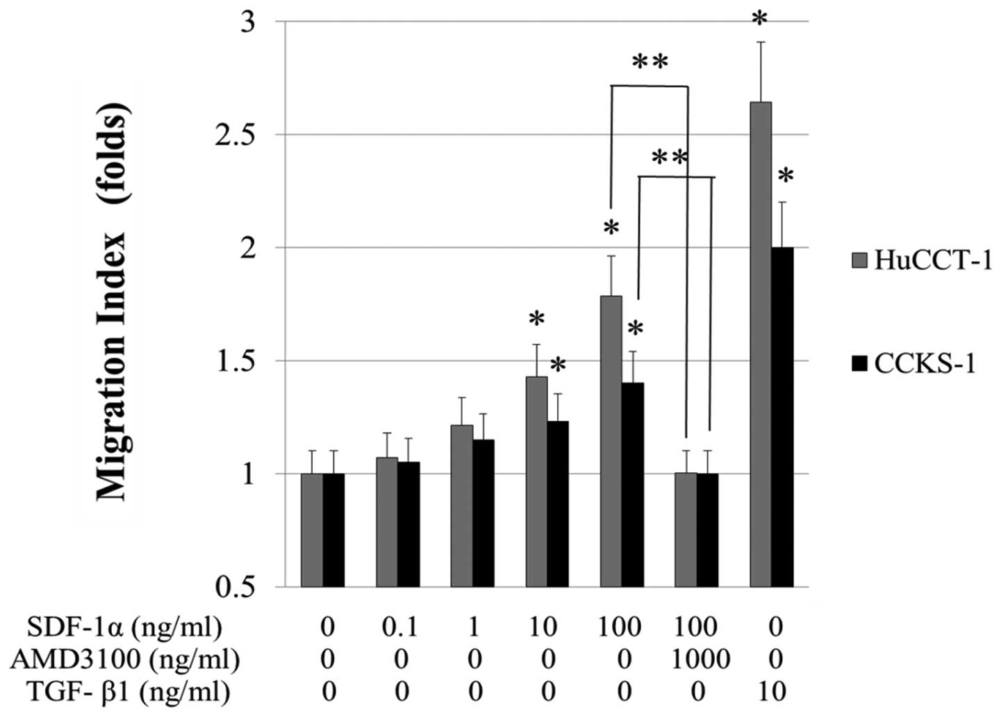 | Figure 7Migration of cultured ICC cells,
increased when 0.1–100 ng/ml SDF-1α was added in the lower chamber
(migration index of HuCCT-1 is 1.07-, 1.21-, 1.42-, 1.79-fold,
respectively, compared with the control. Migration index of CCKS-1
is 1.05-, 1.15-, 1.23-, 1.4-fold, respectively, compared with the
control). Migration capability of HuCCT-1 and CCKS-1 cells
significantly increased when 10 and 100 ng/ml of SDF-1α was added
and the increase was inhibited when 1000 ng/ml AMD3100 was added to
both cell lines. *P<0.05 versus control.
**P<0.05 versus 100 ng/ml SDF-1α. The number of
migrated cells was counted in 10 medium power fields (×20). The
data are provided as the mean ± SD. Migration assay was performed
three times in each experiment. |
AngII and SDF-1α enhanced the expression
of EMT markers
Fluorescent immunocytochemistry revealed the
expression of the epithelial adhesion molecule E-cadherin to be
decreased 48 h after AngII or SDF-1α treatment as compared with
untreated HuCCT-1 and CCKS-1 cells. Furthermore, nuclear expression
of β-catenin was increased 48 h after treatment with AngII or
SDF-1α under serum-starved conditions. Pretreatment of ICC cells
with telmisartan or AMD3100 inhibited the decrease in E-cadherin
expression, while β-catenin expression in the nucleus was decreased
(green, E-cadherin; red, β-catenin; Fig. 9A). Conversely, an increase in the
expression of the mesenchymal cell marker vimentin was observed at
48 h after AngII or SDF-1α treatment. Furthermore, the increase in
vimentin expression was also inhibited by adding 1000 nM
telmisartan or 1000 ng/ml AMD3100 to ICC cells treated with AngII
or SDF-1α (green, vimentin; blue, nucreus; Fig. 9B).
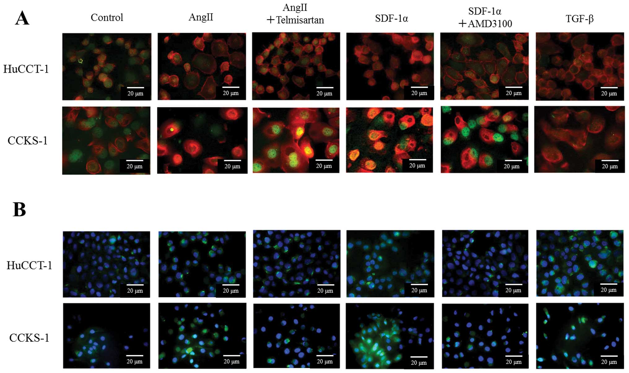 | Figure 9Fluorescent immunocytochemistry of
AngII- or SDF-1α-induced changes in mesenchymal markers of ICC
cells. Addition of AngII or SDF-1α to ICC cells was found to
disorganize, diffuse distribution in the cytoplasm, and
down-regulate E-cadherin expression, whereas β-catenin translocated
to the nucleus [(A): green, E-cadherin; red, β-catenin; blue,
nuclei] and up-regulate vimentin [(B): green, vimentin; blue,
nuclei]. Telmisartan inhibited the down-regulation of E-cadherin,
translocation of β-catenin to the nucleus, and vimentin
up-regulation in ICC cells treated with AngII. AMD3100 also
inhibited the down-regulation of E-cadherin, translocation of
β-catenin to the nucleus, and vimentin up-regulation in ICC cells
treated with SDF-1α. |
Correlation between the expression of
each markers and the prognosis
The patients were visually divided into two groups
by the intensity of each type of staining in immunohistochemistry.
The correlations between the degree of the expression of each
markers and the clinicopathological features with median survival
time (MST) are shown in Table I.
There was no significant difference on MST in the expression of
β-catenin (P=0.059), CXCR4 (P=0.429), and the stage of resected ICC
patients (P=0.116). Significant differences on MST were observed in
the expression of E-cadherin, vimentin, SDF-1, and histological
differentiated type. Negative expression of E-cadherin (P=0.007),
positive expression of vimentin (P=0.011) and SDF-1 (P=0.035), and
poorly-differentiated type were found to have significantly poorer
prognosis (P=0.044).
 | Table ICorrelation between the
clinicopathological features and the expression of each markers
with the prognosis of resected ICC patients.a |
Table I
Correlation between the
clinicopathological features and the expression of each markers
with the prognosis of resected ICC patients.a
| | No. (month) | MST | P-value |
|---|
| E-cadherin | Positive | 11 | 40 | 0.007 |
| Negative | 11 | 9 | |
| β-catenin | Membranous | 9 | 24 | 0.059 |
| Cytoplasmic or
intranuclear | 13 | 19 | |
| Vimentin | Positive | 10 | 17 | 0.011 |
| Negative | 12 | 40 | |
| SDF-1 | Positive | 6 | 21 | 0.035 |
| Negative | 10 | 40 | |
| CXCR4 | Positive | 6 | 24 | 0.429 |
| Negative | 10 | 21 | |
| Histological
type | well-mod | 16 | 30 | 0.044 |
| por | 6 | 9 | |
| Stage | I–III | 10 | 30 | 0.116 |
| IV | 12 | 10 | |
Discussion
In this study, we found that SDF-1, released from
activated HSCs and AngII, contributes to stromal fibrosis,
migration of cancer cells, and tumor progression in ICC tissues via
EMT. It was suggested that AngII was associated with the
synergistic effects on tumor fibrogenesis and tumor progression via
SDF-1/CXCR4 axis. Proliferation and activation of HSCs are the
dominant events in liver injury, inflammation and fibrosis that
render cells responsive to cytokines and other local stimuli
(25,26). The current study demonstrated the
enhancement of SDF-1 secretion from activated HSCs stimulated by
AngII and the role of SDF-1 on the proliferation and activation of
HSCs. Our previous report showed ICC tissues to have higher AngII
concentrations than hepatocellular carcinoma and normal liver
tissues, and we described the role of the AngII/AT-1 axis in cancer
proliferation and cancerous fibrogenesis via the activation of HSCs
(6). Furthermore, it was
previously reported that HSCs may contribute to the desmoplastic
reaction, invasiveness, and cell proliferation associated with
primary and metastatic liver tumors (8,27).
It has also been reported that bone marrow-derived fibroblasts
contribute to tumor stromal reactions and tissue fibrosis and were
recruited into cancer-induced stroma in late tumor stages (28). In terms of the role of chemokines
on cancer cell proliferation, Ohira et al and Leelawat et
al previously demonstrated that SDF-1 had no effect on the
proliferative activity of cultured ICC cells (3,29).
We also examined the proliferative effect of SDF-1α on HuCCT-1 and
CCKS-1 cells by MTT assay, and our result was almost identical to
their results (data not shown). On the other hand, we previously
reported that AngII had a significantly proliferative effect on ICC
cells and AngII receptor blocker, telmisartan, treatment had a
suppressive effect on cell proliferation (6).
EMT, in which tumor cells undergo loss of polarity
and cell-cell junctions and acquire a mesenchymal phenotype and the
capabilities of invading the extracellular matrix and migrating to
distant sites, has been a research focus with local invasion and
metastasis being of particular interest (30,31).
Epithelial cancer cells have been known to transform to the cells
with mesenchymal characteristics via stimulation with several
cytokines or chemokines such as TGF-β1, VEGF, HGF and SDF-1
(9–14). In the current study, we obtained
ample evidence that CXCR4 activation by SDF-1 and AT-1 activation
by AngII contribute to tumor progression by acting as a crucial
mediator of EMT in vitro. We demonstrated that AngII and
SDF-1 induced key events including down-regulation of epithelial
adherens, such as E-cadherin and β-catenin, induction of
mesenchymal marker, vimentin, and enhanced cell invasiveness and
migration. Evidence of EMT in clinical tumor specimens has been
established including loss or delocalization of junctional
E-cadherin, switching to other cadherins (e.g., N-cadherin replaces
E-cadherin), degradation of cell-to-cell adhesion, apicobasal
polarity and tissue architecture alterations, pleiotropic cell
shape, nuclear β-catenin, Snail or Slug expression, and the
otherwise unexpected expressions of mesenchymal markers such as the
intermediate filament protein vimentin (9,32,33).
But in the past, no evidence has been reported on the pivotal
correlation between AngII in the cancer tissues and EMT of cancer
cells.
AngII shares many cellular responses with TGF-β. In
the kidney, ling, liver and vascular, AngII actively participates
in renal fibrosis and in the parts mediated by TGF-β. Many studies
have shown that AngII inhibitors diminish renal TGF-β
overproduction and signaling activation (26,34–41).
A recent study found that AngII directly activates the Smad
signaling system via AT-1 receptor in the kidney and induces the
up-regulation of growth factors, extracellular matrix accumulation
and EMT through the TGF-β/Smad pathway (36,37).
The AT-1 signaling mechanisms are similar to those activated by
cytokines, and include activation of protein kinases, e.g., the
mitogen-activated protein kinase (MAPK) cascade and Rho-kinase
(ROCK). Recent studies have demonstrated that the MAPK pathway
regulates EMT via TGF-β (34,35).
Previous investigations have shown that elucidating the molecular
mechanisms involved in organ fibrosis might lead to improvements in
current clinical treatments for fibrous diseases. Several studies
have shown the MAPK pathway to be involved in EMT and fibrosis.
AngII activates MAPK and through this pathway elicits many cellular
responses. The specific inhibitors of all three MAPKs (p38, JNK and
ERK1/2) prevented phenotypic conversion of cultured human
epithelial cells into myofibroblasts and the loss of E-cadherin
observed after 3 days of treatment with AngII, and markedly
diminished inductions of the EMT markers vimentin and α-SMA. The
MAPK pathway is involved in EMT, fibrosis and cell migration caused
by TGF-β (34–41).
CXCR4 is widely expressed on hematopoietic, brain,
lung, colon, heart, kidney and liver cells. CXCR4 has been
suggested to contribute to the metastatic homing of cancer cells to
various organs and tissues and seems to be an important
pro-invasive factor. Cells expressing functional CXCR4 have been
suggested to migrate and/or invade along SDF-1 gradients (1,2).
Previous reports demonstrated the role of CXCR4 signaling in local
invasion and distant metastasis of breast cancer (42). SDF-1 is constitutively expressed in
several organs including the lungs, liver, skeletal muscle, brain,
kidneys, heart, skin and bone marrow and plays a role in the
mobilization and recruitment of cells expressing CXCR4 to
neoangiogenic niches supporting revascularization of ischemic
tissue and tumor growth. Expression of CXCR4 on tumor cells,
including those from several hemato poietic malignancies, implies
that the SDF-1/CXCR4 pathway may influence cancer biology and play
an important role in directing the metastasis of tumor cells
expressing CXCR4 to organs that express SDF-1 such as the lung,
liver, bone or lymph nodes (1,2,42,43).
Interactions between CAFs including activated HSCs and cancer cells
expressing CXCR4 may induce tumor progression, cancer fibrogenesis,
tumor invasion, and cancer metastasis via EMT (44,45),
and may synergistically make ICC more highly malignant and more
resistant to various therapies. The results shown in Table I are thought to be reasonable to
support a correlation between the expression of SDF-1 and each EMT
markers with the prognosis of ICC patients.
In conclusion, our results suggest that SDF-1
released from activated HSCs and AngII itself can contribute to
stromal fibrosis and migration of cancer cells in collagen-rich
liver tumors, such as ICC. A better understanding of the interplay
between ICC cells and cancerous stroma will be important for
developing strategies to improve tumor therapy that take into
account the influences of the neoplastic microenvironment on tumor
fibrosis, survival, and growth. Additionally, AngII receptor
blockers and CXCR4 antagonists may be useful for treating
collagen-rich tumors. Targeting the SDF-1 and AngII signaling
pathways may be both a pivotal and an efficient strategy for
treating high-grade neoplasms rich in cancerous stroma.
References
|
1
|
Teicher BA and Fricker SP: CXCL12
(SDF-1)/CXCR4 pathway in cancer. Clin Cancer Res. 16:2927–2931.
2010. View Article : Google Scholar : PubMed/NCBI
|
|
2
|
Orimo A, Gupta PB, Sgroi DC,
Arenzana-Seisdedos F, Delaunay T, Naeem R, Carey VJ, Richardson AL
and Weinberg RA: Stromal fibroblasts present in invasive human
breast carcinomas promote tumor growth and angiogenesis through
elevated SDF-1/CXCL12 secretion. Cell. 121:335–348. 2005.
View Article : Google Scholar
|
|
3
|
Ohira S, Sasaki M, Harada K, Sato Y, Zen
Y, Isse K, Kozaka K, Ishikawa A, Oda K, Nimura Y and Nakanuma Y:
Possible regulation of migration of intrahepatic cholangiocarcinoma
cells by interaction of CXCR4 expressed in carcinoma cells with
tumor necrosis factor-α and stromal-derived factor-1 released in
stroma. Am J Pathol. 168:1155–1168. 2006.PubMed/NCBI
|
|
4
|
Marchesi F, Monti P, Leone BE, Zerbi A,
Vecchi A, Piemonti L, Mantovani A and Allavena P: Increased
survival, proliferation, and migration in metastatic human
pancreatic tumor cells expressing functional CXCR4. Cancer Res.
64:8420–8427. 2004. View Article : Google Scholar : PubMed/NCBI
|
|
5
|
Mishra PJ, Mishra PJ, Humeniuk R, Medina
DJ, Alexe G, Mesirov JP, Ganesan S, Glod JW and Banerjee D:
Carcinoma-associated fibroblast-like differentiation of human
mesenchymal stem cells. Cancer Res. 68:4331–4339. 2008. View Article : Google Scholar : PubMed/NCBI
|
|
6
|
Okamoto K, Tajima H, Ohta T, Nakanuma S,
Hayashi H, Nakagawara H, Ohishi I, Takamura H, Ninomiya I, Kitagawa
H, Fushida S, Tani T, Fujimura T, Kayahara M, Harada S, Wakayama T
and Iseki S: Angiotensin II induces tumor progression and fibrosis
in intrahepatic cholangiocarcinoma through an interaction with
hepatic stellate cells. Int J Oncol. 37:1251–1259. 2010. View Article : Google Scholar : PubMed/NCBI
|
|
7
|
Anandanadesan R, Gong Q, Chipitsyna G,
Witkiewicz A, Yeo CJ and Arafat HA: Angiotensin II induces vascular
endothelial growth factor in pancreatic cancer cells through an
angiotensin II type 1 receptor and ERK1/2 signaling. J Gastrointest
Surg. 12:57–66. 2008. View Article : Google Scholar : PubMed/NCBI
|
|
8
|
Amann T, Bataille F, Spruss T, Mühlbauer
M, Gäbele E, Schölmerich J, Kiefer P, Bosserhoff AK and Hellerbrand
C: Activated hepatic stellate cells promote tumorigenicity of
hepatocellular carcinoma. Cancer Sci. 100:646–653. 2009. View Article : Google Scholar : PubMed/NCBI
|
|
9
|
Thiery JP: Epithelial-mesenchymal
transitions in tumour progression. Nat Rev Cancer. 2:442–454. 2002.
View Article : Google Scholar : PubMed/NCBI
|
|
10
|
Buck E, Eyzaguirre A, Barr S, Thompson S,
Sennello R, Young D, Iwata KK, Gibson NW, Cagnoni P and Haley JD:
Loss of homotypic cell adhesion by epithelial-mesenchymal
transition or mutation limits sensitivity to epidermal growth
factor receptor inhibition. Mol Cancer Ther. 6:532–541. 2007.
View Article : Google Scholar
|
|
11
|
Thompson EW, Newgreen DF and Tarin D:
Carcinoma invasion and metastasis: a role for
epithelial-mesenchymal transition? Cancer Res. 65:5991–5995. 2005.
View Article : Google Scholar : PubMed/NCBI
|
|
12
|
Valcourt U, Kowanetz M, Niimi H, Heldin CH
and Moustakas A: TGF-beta and the Smad signaling pathway support
transcriptomic reprogramming during epithelial-mesenchymal cell
transition. Mol Biol Cell. 16:1987–2002. 2005. View Article : Google Scholar : PubMed/NCBI
|
|
13
|
Elliott BE, Hung WL, Boag AH and Tuck AB:
The role of hepatocyte growth factor (scatter factor) in epithelial
mesenchymal transition and breast cancer. Can J Physiol Pharmacol.
80:91–102. 2002. View
Article : Google Scholar : PubMed/NCBI
|
|
14
|
Yang AD, Camp ER, Fan F, Shen L, Gray MJ,
Liu W, Somcio R, Bauer TW, Wu Y, Hicklin DJ and Ellis LM: Vascular
endothelial growth factor receptor-1 activation mediates epithelial
to mesenchymal transition in human pancreatic carcinoma cells.
Cancer Res. 66:46–51. 2006. View Article : Google Scholar
|
|
15
|
Rosanò L, Spinella F, Di Castro V, Nicotra
MR, Dedhar S, De Herreros AG, Natali PG and Bagnato A: Endothelin-1
promotes epithelial-to-mesenchymal transition in human ovarian
cancer cells. Cancer Res. 65:11649–11657. 2005.PubMed/NCBI
|
|
16
|
Andl CD, Fargnoli BB, Okawa T, Bowser M,
Takaoka M, Nakagawa H, Klein-Szanto A, Hua X, Herlyn M and Rustgi
AK: Coordinated functions of E-cadherin and transforming growth
factor beta receptor II in vitro and in vivo. Cancer Res.
66:9878–9885. 2006. View Article : Google Scholar : PubMed/NCBI
|
|
17
|
Chen J, Chen JK and Harris RC: Angiotensin
II induces epithelial-to-mesenchymal transition in renal epithelial
cells through ROS/Src/Caveolin-mediated activation of an EGFR-ERK
signaling pathway. Mol Cell Biol. 32:981–991. 2012. View Article : Google Scholar : PubMed/NCBI
|
|
18
|
Chang J, Jiang Z, Zhang H, Zhu H, Zhou SF
and Yu X: NADPH oxidase-dependent formation of reactive oxygen
species contributes to angiotensin II-induced
epithelial-mesenchymal transition in rat peritoneal mesothelial
cells. Int J Mol Med. 28:405–412. 2011.
|
|
19
|
Buckley ST, Medina C and Ehrhardt C:
Differential susceptibility to epithelial-mesenchymal transition
(EMT) of alveolar, bronchial and intestinal epithelial cells in
vitro and the effect of angiotensin II receptor inhibition. Cell
Tissue Res. 342:39–51. 2010. View Article : Google Scholar
|
|
20
|
Liver Cancer Study Group of Japan: The
general rules for the clinical and pathological study of primary
liver cancer. 5th edition. Kanehara, Tokyo: pp. 17–27. 2008
|
|
21
|
Morimoto T, Aoyama M, Gotoh E and
Shionoiri H: A method of radioimmunoassay of plasma angiotensin II
using Florisil. Folia Endocrinoa Jpn. 59:215–229. 1983. View Article : Google Scholar : PubMed/NCBI
|
|
22
|
Miyagiwa M, Ichida T, Tokiwa T, Sato J and
Sasaki H: A new human cholangiocellular carcinoma cell line
(HuCC-T1) producing carbohydrate antigen 19/9 in serum-free medium.
In Vitro Cell Dev Biol. 25:503–510. 1989. View Article : Google Scholar : PubMed/NCBI
|
|
23
|
Sugawara H, Yasoshima M, Katayanagi K,
Kono N, Watanabe Y, Harada K and Nakanuma Y: Relationship between
interleukin-6 and proliferation and differentiation in
cholangiocarcinoma. Histopathology. 33:145–153. 1998. View Article : Google Scholar : PubMed/NCBI
|
|
24
|
Murakami K, Abe T, Miyazawa M, Yamaguchi
M, Masuda T, Matsuura T, Nagamori S, Takeuchi K, Abe K and Kyogoku
M: Establishment of a new human cell line, LI90, exhibiting
characteristics of hepatic Ito (fat-storing) cells. Lab Invest.
72:731–739. 1995.PubMed/NCBI
|
|
25
|
Salguero Palacios R, Roderfeld M, Hemmann
S, Rath T, Atanasova S, Tschuschner A, Gressner OA, Weiskirchen R,
Graf J and Roeb E: Activation of hepatic stellate cells is
associated with cytokine expression in thioacetamide-induced
hepatic fibrosis in mice. Lab Invest. 88:1192–1203. 2008.PubMed/NCBI
|
|
26
|
Gressner AM and Weiskirchen R: Modern
pathogenetic concepts of liver fibrosis suggest stellate cells and
TGF-β as major players and therapeutic targets. J Cell Mol Med.
10:76–99. 2006.PubMed/NCBI
|
|
27
|
Tien YW, Wu YM, Lin WC, Lee HS and Lee PH:
Pancreatic carcinoma cells stimulate proliferation and matrix
synthesis of hepatic stellate cells. J Hepatol. 51:307–314. 2009.
View Article : Google Scholar : PubMed/NCBI
|
|
28
|
Okabe H, Beppu T, Hayashi H, Horino K,
Masuda T, Komori H, Ishikawa S, Watanabe M, Takamori H, Iyama K and
Baba H: Hepatic stellate cells may relate to progression of
intrahepatic cholangiocarcinoma. Ann Surg Oncol. 16:2555–2564.
2009. View Article : Google Scholar : PubMed/NCBI
|
|
29
|
Leelawat K, Leelawat S, Narong S and
Hongeng S: Roles of the MEK1/2 and AKT pathways in CXCL12/CXCR4
induced cholangiocarcinoma cell invasion. World J Gastroenterol.
13:1561–1568. 2007. View Article : Google Scholar : PubMed/NCBI
|
|
30
|
Gilles C and Thompson EW: The epithelial
to mesenchymal transition and metastatic progression in carcinoma.
Breast J. 2:83–96. 1996. View Article : Google Scholar
|
|
31
|
Ellenrieder V, Hendler SF, Boeck W,
Seufferlein T, Menke A, Ruhland C, Adler G and Gress TM:
Transforming growth factor beta1 treatment leads to an
epithelial-mesenchymal transdifferentiation of pancreatic cancer
cells requiring extracellular signal-regulated kinase 2 activation.
Cancer Res. 61:4222–4228. 2001.
|
|
32
|
Peinado H, Portillo F and Cano A:
Transcriptional regulation of cadherins during development and
carcinogenesis. Int J Dev Biol. 48:365–375. 2004. View Article : Google Scholar : PubMed/NCBI
|
|
33
|
Tomita K, van Bokhoven A, van Leenders GJ,
Ruijter ET, Jansen CF, Bussemakers MJ and Schalken JA: Cadherin
switching in human prostate cancer progression. Cancer Res.
60:3650–3654. 2000.PubMed/NCBI
|
|
34
|
Rodrigues-Díez R, Carvajal-González G,
Sánchez-López E, Rodríguez-Vita J, Rodrigues Díez R, Selgas R,
Ortiz A, Egido J, Mezzano S and Ruiz-Ortega M: Pharmacological
modulation of epithelial mesenchymal transition caused by
angiotensin II. Role of ROCK and MAPK pathways Pharm Res.
25:2447–2461. 2008.PubMed/NCBI
|
|
35
|
Ruiz-Ortega M, Rupérez M, Esteban V,
Rodríguez-Vita J, Sánchez-López E, Carvajal G and Egido J:
Angiotensin II: a key factor in the inflammatory and fibrotic
response in kidney diseases. Nephrol Dial Transplant. 21:16–20.
2006. View Article : Google Scholar : PubMed/NCBI
|
|
36
|
Carvajal G, Rodríguez-Vita J,
Rodrigues-Díez R, Sánchez-López E, Rupérez M, Cartier C, Esteban V,
Ortiz A, Egido J, Mezzano SA and Ruiz-Ortega M: Angiotensin II
activates the Smad pathway during epithelial mesenchymal
transdifferentiation. Kidney Int. 74:585–595. 2008. View Article : Google Scholar : PubMed/NCBI
|
|
37
|
Wolf G: Renal injury due to
renin-angiotensin-aldosterone system activation of the transforming
growth factor-beta pathway. Kidney Int. 70:1914–1919.
2006.PubMed/NCBI
|
|
38
|
Bakin AV, Rinehart C, Tomlinson AK and
Arteaga CL: p38 mitogen-activated protein kinase is required for
TGFbeta mediated fibroblastic transdifferentiation and cell
migration. J Cell Sci. 115:3193–3206. 2002.PubMed/NCBI
|
|
39
|
Rupérez M, Lorenzo O, Blanco-Colio LM,
Esteban V, Egido J and Ruiz-Ortega M: Connective tissue growth
factor is a mediator of angiotensin II-induced fibrosis.
Circulation. 108:1499–1505. 2003.PubMed/NCBI
|
|
40
|
Moustakas A and Heldin CH: Non-Smad
TGF-beta signals. J Cell Sci. 118:3573–3584. 2005. View Article : Google Scholar : PubMed/NCBI
|
|
41
|
Ruiz-Ortega M, Rodríguez-Vita J,
Sanchez-Lopez E, Carvajal G and Egido J: TGF-beta signaling in
vascular fibrosis. Cardiovasc Res. 74:196–206. 2007. View Article : Google Scholar : PubMed/NCBI
|
|
42
|
Lee BC, Lee TH, Avraham S and Avraham HK:
Involvement of the chemokine receptor CXCR4 and its ligand stromal
cell-derived factor 1alpha in breast cancer cell migration through
human brain microvascular endothelial cells. Mol Cancer Res.
2:327–338. 2004.
|
|
43
|
Onoue T, Uchida D, Begum NM, Tomizuka Y,
Yoshida H and Sato M: Epithelial-mesenchymal transition induced by
the stromal cell-derived factor-1/CXCR4 system in oral squamous
cell carcinoma cells. Int J Oncol. 29:1133–1138. 2006.PubMed/NCBI
|
|
44
|
Ishii G, Sangai T, Ito T, Hasebe T, Endoh
Y, Sasaki H, Harigaya K and Ochiai A: In vivo and in vitro
characterization of human fibroblasts recruited selectively into
human cancer stroma. Int J Cancer. 117:212–220. 2005. View Article : Google Scholar : PubMed/NCBI
|
|
45
|
Kalluri R and Zeisberg M: Fibroblasts in
cancer. Nat Rev Cancer. 6:392–401. 2006. View Article : Google Scholar
|