Introduction
Prostate cancer remains the second leading cause of
cancer death for men in the US. According to the National Cancer
Institute, it is estimated that there will be 238,590 new cases and
29,720 deaths from prostate cancer in 2013 (http://www.cancer.gov/cancertopics/types/prostate).
Initially, prostate cancer cells are dependent upon androgen
stimulation for growth and proliferation, and thus sensitive to
hormone therapy such as androgen deprivation, which effectively
blocks androgen-dependent tumor growth. Unfortunately, most
recurrent tumors return within two years exhibiting
castration-resistant growth and a more aggressive, metastatic
phenotype. As of yet, there is no effective treatment for
castration-resistant prostate cancer (CRPC) (1,2).
Therefore, it is of great urgency to elucidate and fully understand
the molecular mechanisms of CRPC with a metastatic phenotype
following androgen deprivation, in order to develop novel
therapeutic strategies or approaches toward CRPC.
Recently, several mechanisms have been proposed for
the development of castration-resistant prostate cancer (3,4)
including mutation, amplification (4,5),
expression of alternative-splice variants (6), proteolytic removal of ligand-binding
domain (LBD) (7) of the androgen
receptor (AR), or the increase of natural testosterone biosynthesis
by cancer cells (8,9). These mechanisms suggest that most
CRPC cells may still depend on AR function, but are adaptive to low
hormone levels or enable AR function for independence of
ligand-binding. However, clinical evidence and basic research study
support the hypothesis that alternative mechanisms may be related
to highly aggressive, metastatic phenotype in AR-negative prostate
cancer cells or cancer-stem cells (10–14).
Calpains are a family of calcium-dependent,
non-lysosomal cysteine proteases, of which 14 human calpains are
well known. Based on structure and function, calpain 1 (μ-calpain)
and calpain 2 (m-calpain) are well characterized (15). There is a plenty of experimental
and clinical evidence to support critical roles of calpains in
normal cellular functions (cell motility, cell cycle, autophagy and
apoptosis), and pathological processes (tumorigenesis, cancer
metastasis and neurodegenerative diseases) (15,16).
In fact, overexpression of calpain 2 has been confirmed in human
colorectal cancer (17), and
increased expression of calpain 1 in schwannomas and meningiomas
has also been reported (18).
However, results for prostate cancer are controversial (19,20).
Filamin A (FlnA), also known as actin-binding protein 280
(ABP-280), is a 280 kDa protein consisting of N-terminal
actin-binding domain (ABD) and a rodlike domain of 24 repeats (each
repeat is approximately 96 amino acids in length), which is
interrupted by two hinge domains denoted as H1 (between repeats 15
and 16) and H2 (between repeats 23 and 24). FlnA is a natural
substrate of calpains 1/2 in cells and cleavage occurs at H1
resulting in two fragments of 170 and 110 kDa, respectively. The
110 kDa fragment containing the H2 domain, can be further cleaved
to yield a 90 kDa fragment which has been confirmed to translocate
to the nucleus and bind AR in androgen-sensitive LNCaP cells, with
nuclear translocation absent in AR-negative PC-3 cells (21). The dimerization domain (65 amino
acids located in repeat 24) of FlnA forms a structure-shaped
homodimer motif to bind or cross-link actin filaments of
cytoskeleton networks through its N-terminal ABD domain. In
addition, FlnA can serve as a molecular scaffold interacting with
multiple proteins such as transmembrane proteins, signaling
molecules and DNA damage repair proteins (22). Recent studies demonstrated that an
increase in both expression and cytoplasmic distribution of FlnA
was related to aggressive, metastatic prostate cancer (23). A new discovery revealed that
calpain 2 can cleave and remove the ligand-binding domain (LBD)
from AR. In turn, the truncated AR retaining the transactivation
domain and DNA-binding domain (DBD), exhibited a constitutively
active AR molecule that translocated into the nucleus and
functioned in an androgen-independent manner (7,24).
Our previous studies revealed the occurrence of two
important events in prostate cancer cells which were the loss of
prostate specific membrane antigen (PSMA) expression and a cell
signal pathway switch from AR to c-Met during long-term androgen
deprivation in an established in vitro model (10,25).
Herein we further explore whether long-term androgen deprivation
can upregulate the expression and activity of calpains 1/2 in
prostate cancer cells, which are correlated with a highly
aggressive, metastatic phenotype (20). Our present data strongly support
the concept that long-term androgen deprivation may push
androgen-sensitive prostate cancer cells evolve into AR-negative,
more aggressive, androgen-independent disease state, with
overexpression of calpain 2 enhancing its activity. Thus, a
combination of calpain 1/2 inhibitor and androgen deprivation may
provide a novel therapeutic strategy to prevent or postpone the
progression of prostate cancer from androgen-sensitive to CRPC.
Materials and methods
Cell lines and reagents
The human prostate cancer cell lines LNCaP and PC-3
were obtained from the American Type Culture Collection (Manassas,
VA, USA). Rabbit polyclonal anti-AR antibody (N-20) was obtained
from Santa Cruz Biotechnology (Santa Cruz, CA, USA). Mouse
monoclonal anti-FlnA (N-terminus) antibody, goat anti-rabbit
secondary antibody-FITC, and rabbit anti-actin antibody were
obtained from Sigma-Aldrich (St. Louis, MO, USA). Protein blocking
solution was obtained from BioGenex (San Ramon, CA, USA). Rabbit
polyclonal anti-calpain 1 and anti-calpain 2 antibodies were
obtained from Cell Signaling Technology (Danvers, MA, USA). Mouse
monoclonal anti-FlnA (C-terminus) antibody and calpeptin were
obtained from EMD Millipore Corporation (Billerica, MA, USA).
Hoechst 33342 were obtained from Invitrogen-Molecular Probes
(Carlsbad, CA, USA). Halt Protease Inhibitor Cocktail 100X was
purchased from Thermo Fisher Scientific (Rockford, IL, USA). All
other chemicals and cell-culture reagents were purchased from
Fisher Scientific (Sommerville, NJ, USA) or Sigma-Aldrich.
Cell culture
LNCaP and PC-3 cells were grown in T-75 flasks with
normal growth media [RPMI-1640 containing 10% heat-inactivated
fetal calf serum (FBS), 100 units of penicillin and 100
μg/ml streptomycin] in a humidified incubator at 37°C with
5% CO2. Otherwise, for androgen-deprivation growth,
cells were cultured with conditioned media [RPMI-1640 containing
10% charcoal-stripped fetal bovine serum, 100 units of penicillin
and 100 μg/ml streptomycin]. Confluent cells were detached
with a 0.25% trypsin 0.53 mM EDTA solution, harvested, and plated
in 2-well slide chambers at a density of 4×10⁴
cells/well. Cells were grown for three days before conducting the
following experiments.
Immunofluorescence detection of AR and
FlnA
The LNCaP cells grown under androgen deprivation
condition over time (5, 10 and 20 passages) were cultured for 3
days on the slides in the conditioned media. For two-day
androgen-deprivation treatment, LNCaP cells were seeded on slides
with normal growth media for 1-day growth, and replaced with
conditioned media for another 2-day growth. Normal LNCaP cells and
PC-3 cells were used for the AR-positive and AR-negative control,
respectively. These cells were seeded on slides with normal growth
media for three days. Slides with cells grown for three days in
normal growth media or conditioned media were washed twice in
phosphate-buffered saline (PBS), fixed in 4% paraformaldehyde in
PBS buffer for 15 min at room temperature, and permeabilized with
pre-cooled methanol for 5 min at −20°C. The permeabilized cells
were blocked in block buffer (0.1% Tween-20, 5% goat normal serum
in PBS buffer) for 2 h at room temperature and incubated with
primary anti-AR antibody and anti-FlnA (C-terminus) antibody in
block buffer overnight at 4°C. After washing, the cells were
incubated with a secondary antibodies (goat anti-rabbit IgG-FITC
and goat anti-mouse IgG-TRITC in 1% BSA, PBS buffer) for 2 h at
room temperature, counterstained with Hoechst 33342, and mounted in
VECTASHIELD® Mounting Medium (Vector Laboratories,
Burlingame, CA, USA) for confocal microscopy.
Confocal laser scanning microscopy
Cells were visualized under 25X water immersion
objective using an LSM 510 META Laser Scanning Microscope. Hoechst
33342 was excited with a Diode Laser (405 nm), and the emission was
collected with a BP420-480 nm filter. AR immunofluorescence (with
goat anti-rabbit IgG-FITC) was excited at 488 nm using an Argon
Laser, and the emission was collected with an LP505 nm filter. FlnA
immunofluorescence (with goat anti-mouse IgG-TRITC) was excited
using 543 nm from a HeNe Laser, and the emission collected with a
BP560-615 nm filter. To reduce interchannel crosstalk, a
multi-tracking technique was used, and images were taken at a
resolution of 1,024×1,024 pixels. Confocal scanning parameters were
set up so that the control cells without treatment did not display
background fluorescence. The imaging colors of the fluorescent
dyes, Hoechst 33342, FITC and TRITC, were defined as blue, green
and red, respectively. The pictures were edited by National
Institutes of Health (NIH) Image J software (http://rsb.info.nih.gov/ij) and Adobe Photoshop
CS2.
Whole cell lysate extraction and western
blot analysis
The controls: PC-3 and LNCaP cells (cultured in
normal growth media) and LNCaP cells under androgen deprivation
over time (2 days, 5, 10 or 20 passages) were collected by
scraping, washed once in ice-cold PBS, re-suspended in 3-fold cell
pellet volumes of lysis buffer (1% NP-40, 20 mM Tris pH 8.0, 137 mM
NaCl, 10% glycerol) (10,25–27)
supplemented with 1X Halt Protease Inhibitor Cocktail for 15 min on
ice, then transferred to Eppendorf tubes for centrifugation at
10,000 × g for 15 min at 4°C, the supernatant was saved as
whole-cell protein extracts. For calpain 1/2 inhibitor block study,
LNCaP cells under androgen deprivation over time (5, 10 passages)
and PC-3 cells were allowed to be cultured for 2 days, then
continued for another 24 h growth in the above mentioned media
containing 40 μM calpeptin or DMSO, prior to cell harvest
and protein extraction. Protein concentrations were determined
using Non-Interfering Protein Assay (G-Biosciences, St. Louis, MO,
USA). Western blot analysis was performed as described previously
with only minor modifications (27,28).
In brief, detergent-soluble proteins (30 μg) were loaded and
separated on a NuPAGE™ 4-12% Bis-Tris Gel (Invitrogen, Carlsbad,
CA, USA) by electrophoresis for 60 min at a constant 200 V under
reducing conditions, and then transferred to a 0.45 μm PVDF
Immobilon-P Transfer Membrane (Millipore Corporation) at 400 mA for
120 min in a transfer apparatus-Owl Bandit VEP-2 (Owl, Portsmouth,
NH, USA) according to the manufacturer’s instructions. Membranes
were incubated with primary antibody at corresponding dilution
overnight at 4°C and then with horse-radish peroxidase
conjugated-second antibody for 1 h at room temperature. The
immunoreactive bands were visualized using Protein Detector TMB
Western Blot kit (KPL, Gaithersburg, MD, USA) following the
manufacturer’s instructions.
Results
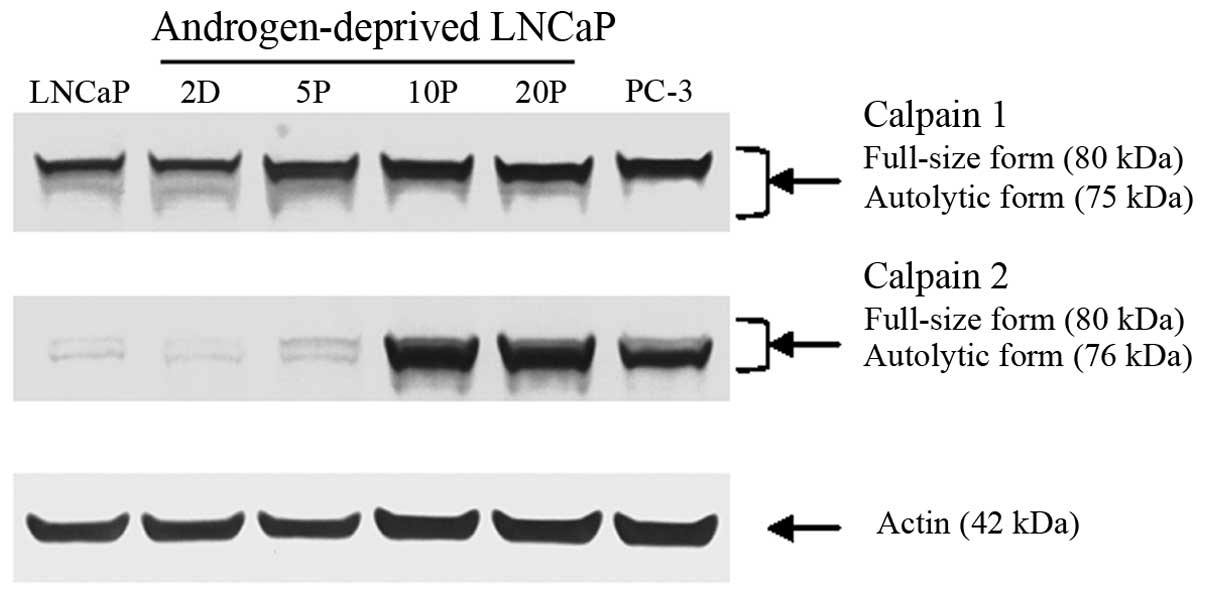
Overexpression of calpain 2, not calpain
1 induced by androgen deprivation
Western blot analysis (Fig. 1) clearly demonstrated that
overexpression of calpain 2 was observed in long-term
androgen-deprived LNCaP cells (10, 20 passages) and PC-3 cells
(AR-negative), of which, AR was confirmed to be no longer
detectable in our previous work (10,25).
In contrast, calpain 1 expression was relatively stable in both
AR-positive and AR-negative prostate cancer cells.
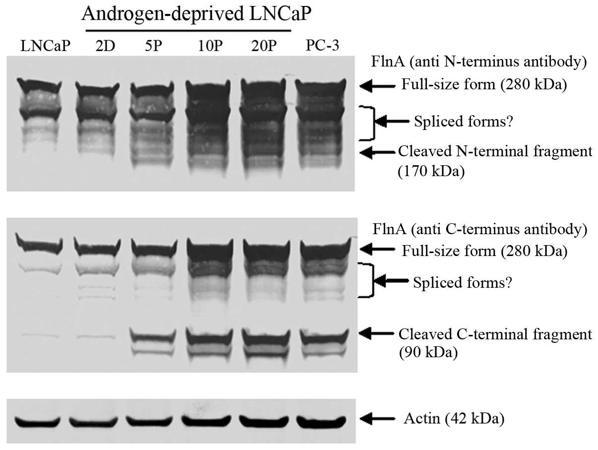
Increased expression and proteolytic
cleavage of FlnA
Western blot analysis also revealed that there was
an apparent increase of FlnA expression and accumulation of its
cleaved fragments (Fig. 2) in
long-term androgen-deprived LNCaP cells (10, 20 passages) and
AR-negative PC-3 cells (10,25).
The increase of cleaved FlnA fragments (Fig. 2) is clearly correlated with
overexpressed calpain 2 in these cells (Fig. 1).
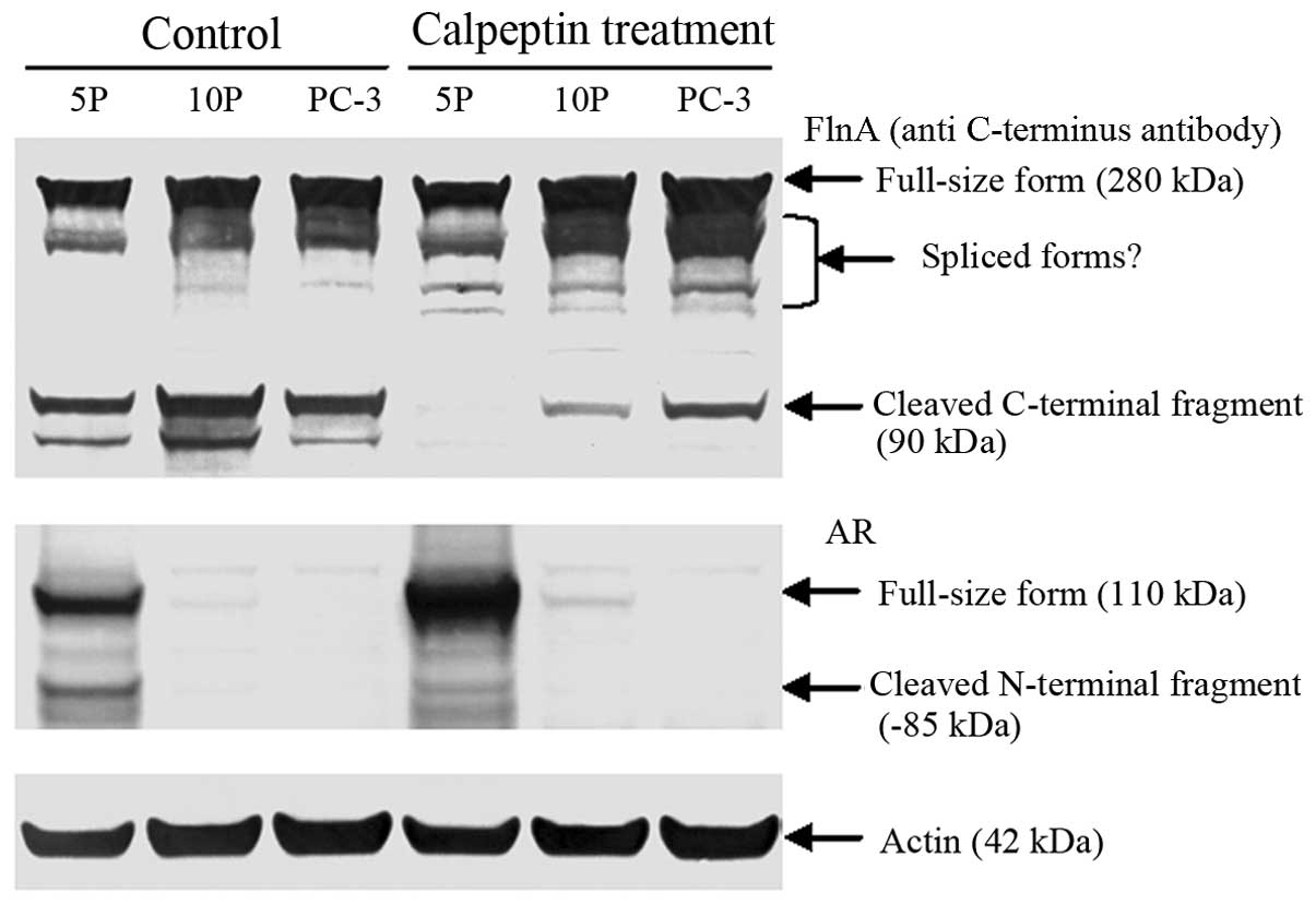
Calpeptin protected FlnA from cleavage in
AR-negative cells
Calpain-block study in cells further confirmed that
calpeptin treatment effectively inhibited calpain 1/2 activities,
and protected FlnA from cleavage (Fig.
3). Through cleavage of FlnA, calpain 2 can dynamically
regulate FlnA mediated cytoskeleton network, and further regulate
cell signal pathways, cell morphology and motility.
Calpeptin protected AR from cleavage
In Fig. 3, compared
to control cells, it was also noticed that AR was clearly protected
from calpain-mediated cleavage by calpeptin treatment in relatively
short-term androgen-deprived LNCaP cells (5 passages). An increase
of 85 kDa truncated AR fragment with removal of ligand-binding
domain in CWR22Rv1 cells, has been suggested to be a mechanism for
androgen independence of prostate cancer cells (7).
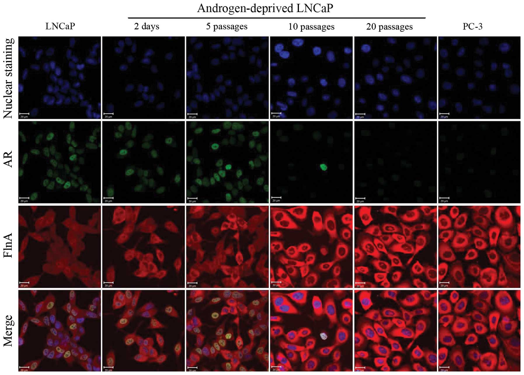
Different distribution of FlnA in
AR-positive and AR-negative cells
A relatively average distribution of FlnA from the
cytoplasm to the nucleus, strong nuclear co-localization of AR and
FlnA, and a slight increase of cytoplasmic distribution of FlnA
over time were observed in AR-positive cells (Fig. 4). Inversely, the cytoplasmic
distribution of FlnA was the highest in AR-negative cells (Fig. 4). It was also noticable that a
minimal amount of FlnA was located within the nucleolus in
AR-negative cells (Fig. 4). The
contrasting distribution of FlnA in the nucleus or in the cytoplasm
is consistent with clinical data in which immunohistochemistry
analysis demonstrated that cytoplasmic localization of FlnA is
highly correlated with metastases of prostate cancer (23).
Discussion
Our previous study suggests that long-term androgen
depletion may induce a signaling pathway switch from AR to c-Met,
which may lead to a diagnostically and therapeutically elusive
androgen-independent disease-state. Following our previous research
studies, our present data further define the key role of calpain 2
and not calpain 1 in prostate cancer progression, especially in
cancer invasion and metastasis with its overexpression and enhanced
activity in AR-negative prostate cancer cells.
Both calpains 1 and 2 are heterodimers, which
consist of a specific 80 kDa catalytic subunit and a common
regulatory 28 kDa subunit shared by calpain 1 and calpain 2. Their
proteolytic activities exhibit dependence on different calcium
concentrations in vitro (15). Therefore, activation of calpain 1
or calpain 2 may be responsible for different signaling pathways
and physiological or pathological processes in cells. Calpastatin
is an endogenous inhibitor of calpain 1 and calpain 2 while
inhibitory action of calpastatin is also dependent on
calcium-induced structural changes of calpain 1/2 (15). Calcium, calpastatin and calpain 1/2
are three components whose concentration, distribution and
interaction determine spatial and temporal regulation of calpain
1/2 activity in cells (15). The
dynamic regulation of calpain activity is necessary for
coordination of cell-substrate adhesion, actin and myosin-mediated
contraction and cell-substrate detachment to control cell movement
(15). There is experimental
evidence that demonstrates calpain 1/2 has several roles in cancer
progression such as cleaving focal adhesion kinase (FAK) to
dynamically regulate integrin-mediated focal adhesion for cell
migration (29,30), regulating activation of
membrane-type matrix metalloproteinase 1 (MT1-MMP or MMP-14) and
matrix metalloproteinase 2 (MMP2) for extra-cellular matrix
remodeling and angiogenesis (31),
and cancer invasion and metastasis (32,33).
Considering the multiple cellular functions of
calpain 2, its abnormal high expression and enhanced activity may
play important roles in prostate cancer progression including
migration, invasion and metastasis during androgen deprivation
therapy. It is reasonable that calpain 2 may be treated as a target
for limiting tumor progression. In fact, inhibition or
downregulation of calpain 2 clearly decreased migration and
invasion of DU-145 prostate cancer cells in vitro and in
vivo (19). Furthermore,
calpain 2 can also cleave AR to generate a truncated, functional AR
without ligand-binding domain in androgen-sensitive prostate cancer
cells, which enables cancer cell adaptation to androgen-independent
growth and proliferation during androgen-deprivation treatment
(7). Our present study further
confirms the above discovery in short-term androgen-deprived LNCaP
cells (5 passages). It should be mentioned that short-term
androgen-deprived LNCaP cells (5 passages) can survive without
androgen in media, but grow and proliferate at very low rates
(10). In contrast, long-term
androgen deprivation caused the most loss of AR expression, with
development of alternative signaling pathways enabling cell growth
and proliferation at high rates (10,25).
In addition, inhibition of calpain activity can enhance cytotoxic
activity of bortezomib in vitro and in vivo against
cancer cells by preventing autophagic survival response (34).
In summary, our present data support the concept
that long-term androgen deprivation promotes overexpression and
enhanced activity of calpain 2 leading to an increase in the
fragmental cleavage of AR and FlnA. The overexpression of calpain 2
and increased expression of FlnA may contribute to the development
of an aggressive phenotype of prostate cancer. It is expected that
the combination of androgen deprivation with inhibition of calpain
2 may provide a new therapeutic strategy to prevent or postpone
appearance of CRPC in patients.
Acknowledgements
The authors extend their gratitude for
technical assistance from C. Davitt and V. Lynch-Holm at the WSU
Franceschi Microscopy and Imaging Center. This study was supported
in part by the National Institutes of Health (R01CA140617).
References
|
1.
|
Gustavsson H, Welen K and Damber JE:
Transition of an androgen-dependent human prostate cancer cell line
into an androgen-independent subline is associated with increased
angiogenesis. Prostate. 62:364–373. 2005. View Article : Google Scholar : PubMed/NCBI
|
|
2.
|
Niu Y, Altuwaijri S, Lai KP, et al:
Androgen receptor is a tumor suppressor and proliferator in
prostate cancer. Proc Natl Acad Sci USA. 105:12182–12187. 2008.
View Article : Google Scholar : PubMed/NCBI
|
|
3.
|
Lee SO, Dutt SS, Nadiminty N, Pinder E,
Liao H and Gao AC: Development of an androgen-deprivation induced
and androgen suppressed human prostate cancer cell line. Prostate.
67:1293–1300. 2007. View Article : Google Scholar : PubMed/NCBI
|
|
4.
|
Saraon P, Jarvi K and Diamandis EP:
Molecular alterations during progression of prostate cancer to
androgen independence. Clin Chem. 57:1366–1375. 2011. View Article : Google Scholar : PubMed/NCBI
|
|
5.
|
Devlin HL and Mudryj M: Progression of
prostate cancer: multiple pathways to androgen independence. Cancer
Lett. 274:177–186. 2009. View Article : Google Scholar : PubMed/NCBI
|
|
6.
|
Watson PA, Chen YF, Balbas MD, et al:
Constitutively active androgen receptor splice variants expressed
in castration-resistant prostate cancer require full-length
androgen receptor. Proc Natl Acad Sci USA. 107:16759–16765. 2010.
View Article : Google Scholar
|
|
7.
|
Libertini SJ, Tepper CG, Rodriguez V,
Asmuth DM, Kung HJ and Mudryj M: Evidence for calpain-mediated
androgen receptor cleavage as a mechanism for androgen
independence. Cancer Res. 67:9001–9005. 2007. View Article : Google Scholar : PubMed/NCBI
|
|
8.
|
Cai C and Balk SP: Intratumoral androgen
biosynthesis in prostate cancer pathogenesis and response to
therapy. Endocr Relat Cancer. 18:R175–R182. 2011. View Article : Google Scholar : PubMed/NCBI
|
|
9.
|
Chang KH, Li R, Kuri B, et al: A
gain-of-function mutation in DHT synthesis in castration-resistant
prostate cancer. Cell. 154:1074–1084. 2013. View Article : Google Scholar : PubMed/NCBI
|
|
10.
|
Liu T, Mendes DE and Berkman CE: From AR
to c-Met: Androgen deprivation leads to a signaling pathway switch
in prostate cancer cells. Int J Oncol. 43:1125–1130.
2013.PubMed/NCBI
|
|
11.
|
Hendriksen PJ, Dits NF, Kokame K, et al:
Evolution of the androgen receptor pathway during progression of
prostate cancer. Cancer Res. 66:5012–5020. 2006. View Article : Google Scholar : PubMed/NCBI
|
|
12.
|
Lorenzo GD, Bianco R, Tortora G and
Ciardiello F: Involvement of growth factor receptors of the
epidermal growth factor receptor family in prostate cancer
development and progression to androgen independence. Clin Prostate
Cancer. 2:50–57. 2003. View Article : Google Scholar : PubMed/NCBI
|
|
13.
|
Maeda A, Nakashiro K, Hara S, et al:
Inactivation of AR activates HGF/c-Met system in human prostatic
carcinoma cells. Biochem Biophys Res Commun. 347:1158–1165. 2006.
View Article : Google Scholar : PubMed/NCBI
|
|
14.
|
Nishida S, Hirohashi Y, Torigoe T, et al:
Prostate cancer stem-like cells/cancer-initiating cells have an
autocrine system of hepatocyte growth factor. Cancer Sci.
104:431–436. 2013. View Article : Google Scholar : PubMed/NCBI
|
|
15.
|
Storr SJ, Carragher NO, Frame MC, Parr T
and Martin SG: The calpain system and cancer. Nat Rev Cancer.
11:364–374. 2011. View
Article : Google Scholar : PubMed/NCBI
|
|
16.
|
Camins A, Verdaguer E, Folch J and Pallas
M: Involvement of calpain activation in neurodegenerative
processes. CNS Drug Rev. 12:135–148. 2006. View Article : Google Scholar : PubMed/NCBI
|
|
17.
|
Lakshmikuttyamma A, Selvakumar P, Kanthan
R, Kanthan SC and Sharma RK: Overexpression of m-calpain in human
colorectal adenocarcinomas. Cancer Epidemiol Biomarkers Prev.
13:1604–1609. 2004.PubMed/NCBI
|
|
18.
|
Kimura Y, Koga H, Araki N, et al: The
involvement of calpain-dependent proteolysis of the tumor
suppressor NF2 (merlin) in schwannomas and meningiomas. Nat Med.
4:915–922. 1998. View Article : Google Scholar : PubMed/NCBI
|
|
19.
|
Mamoune A, Luo JH, Lauffenburger DA and
Wells A: Calpain-2 as a target for limiting prostate cancer
invasion. Cancer Res. 63:4632–4640. 2003.PubMed/NCBI
|
|
20.
|
Rios-Doria J, Day KC, Kuefer R, et al: The
role of calpain in the proteolytic cleavage of E-cadherin in
prostate and mammary epithelial cells. J Biol Chem. 278:1372–1379.
2003. View Article : Google Scholar : PubMed/NCBI
|
|
21.
|
Wang Y, Kreisberg JI, Bedolla RG,
Mikhailova M, deVere White RW and Ghosh PM: A 90 kDa fragment of
filamin A promotes Casodex-induced growth inhibition in
Casodex-resistant androgen receptor positive C4-2 prostate cancer
cells. Oncogene. 26:6061–6070. 2007. View Article : Google Scholar : PubMed/NCBI
|
|
22.
|
Yue J, Huhn S and Shen Z: Complex roles of
filamin-A mediated cytoskeleton network in cancer progression. Cell
Biosci. 3:7–18. 2013. View Article : Google Scholar : PubMed/NCBI
|
|
23.
|
Bedolla RG, Wang Y, Asuncion A, et al:
Nuclear versus cytoplasmic localization of filamin A in prostate
cancer: immunohistochemical correlation with metastases. Clin
Cancer Res. 15:788–796. 2009. View Article : Google Scholar : PubMed/NCBI
|
|
24.
|
Chen H, Libertini SJ, Wang Y, Kung HJ,
Ghosh P and Mudryj M: ERK regulates calpain 2-induced androgen
receptor proteolysis in CWR22 relapsed prostate tumor cell lines. J
Biol Chem. 285:2368–2374. 2010. View Article : Google Scholar : PubMed/NCBI
|
|
25.
|
Liu T, Wu LY, Fulton MD, Johnson JM and
Berkman CE: Prolonged androgen deprivation leads to downregulation
of androgen receptor and prostate-specific membrane antigen in
prostate cancer cells. Int J Oncol. 41:2087–2092. 2012.PubMed/NCBI
|
|
26.
|
Matroule JY, Carthy CM, Granville DJ,
Jolois O, Hunt DW and Piette J: Mechanism of colon cancer cell
apoptosis mediated by pyropheophorbide-a methylester
photosensitization. Oncogene. 20:4070–4084. 2001. View Article : Google Scholar : PubMed/NCBI
|
|
27.
|
Liu T, Wu LY and Berkman CE:
Prostate-specific membrane antigen-targeted photodynamic therapy
induces rapid cytoskeletal disruption. Cancer Lett. 296:106–112.
2010. View Article : Google Scholar : PubMed/NCBI
|
|
28.
|
Liu T, Toriyabe Y and Berkman CE:
Purification of prostate-specific membrane antigen using
conformational epitope-specific antibody-affinity chromatography.
Protein Expr Purif. 49:251–255. 2006. View Article : Google Scholar
|
|
29.
|
Chan KT, Bennin DA and Huttenlocher A:
Regulation of adhesion dynamics by calpain-mediated proteolysis of
focal adhesion kinase (FAK). J Biol Chem. 285:11418–11426. 2010.
View Article : Google Scholar
|
|
30.
|
Franco SJ, Rodgers MA, Perrin BJ, et al:
Calpain-mediated proteolysis of talin regulates adhesion dynamics.
Nat Cell Biol. 6:977–983. 2004. View
Article : Google Scholar : PubMed/NCBI
|
|
31.
|
Kwak HI, Kang H, Dave JM, et al:
Calpain-mediated vimentin cleavage occurs upstream of MT1-MMP
membrane translocation to facilitate endothelial sprout initiation.
Angiogenesis. 15:287–303. 2012. View Article : Google Scholar : PubMed/NCBI
|
|
32.
|
Jang HS, Lal S and Greenwood JA: Calpain 2
is required for glioblastoma cell invasion: regulation of matrix
metalloproteinase 2. Neurochem Res. 35:1796–1804. 2010. View Article : Google Scholar : PubMed/NCBI
|
|
33.
|
Postovit LM, Dutt P, Dourdin N, et al:
Calpain is required for MMP-2 and u-PA expression in SV40 large
T-antigen-immortalized cells. Biochem Biophys Res Commun.
297:294–301. 2002. View Article : Google Scholar : PubMed/NCBI
|
|
34.
|
Escalante AM, McGrath RT, Karolak MR, Dorr
RT, Lynch RM and Landowski TH: Preventing the autophagic survival
response by inhibition of calpain enhances the cytotoxic activity
of bortezomib in vitro and in vivo. Cancer Chemother Pharmacol.
71:1567–1576. 2013. View Article : Google Scholar : PubMed/NCBI
|