Introduction
Neuroendocrine tumors (NETs) are a diverse group of
neoplasms characterized by their endocrine secretion and histologic
features and are often well-differentiated and slow-growing
(1). They most commonly occur in
the gastroenteropancreatic tract, but are also found in the lung
and the rest of the body (1,2).
Pancreatic neuroendocrine tumors (PNETs) are rare neoplams; in
2004, their incidence was 0.22 per 100,000 in the United States
(3). PNETs are classified
clinically as either functioning or non-functioning based on their
clinical endocrine manifestations (4). Most PNETs (90%) are non-functioning
(3). Unlike functioning PNETs such
as insulinomas and gastrinomas, non-functioning PNETs are often
malignant and usually present at a late stage, often including a
huge mass, local invasion, and distant metastases (5–7).
Modern cancer care highly depends on imaging
technologies. For general imaging, positron emission tomography
(PET) using 18F-fluorodeoxyglucose (18F-FDG)
is a powerful and common functional imaging technique that can
provide information about the functional and metabolic
characteristics of malignancies, tumor stage, therapeutic response,
and tumor recurrence (8). The
advent of combined 18F-FDG PET with computed tomography
(18F-FDG PET/CT) allows anatomic correlation and exact
localization of lesions (9).
Unfortunately, FDG PET has limited value for imaging
gastroenteropancreatic NETs and only tumors with low
differentiation and high proliferative activity have demonstrated
an elevated FDG uptake (10–13).
In contrast to functioning PNETs, which are usually well
differentiated and slow growing, non-functioning PNETs are often
found incidentally or through symptoms caused by their enlargement
or metastatic spread; they are often malignant and usually present
at an advanced stage (14).
Therefore, it is reasonable to infer that non-functioning PNETs
could be detected by 18F-FDG PET. However, there is
currently no published evidence of 18F-FDG PET/CT in
evaluating non-functioning PNETs, and generally 18F-FDG
PET/CT is considered to have limited value in assessing
non-functioning PNETs (10–13).
Moreover, 18F-FDG PET/CT is not recommended to detect
non-functioning pancreatic neuroendocrine tumors by the newest
edition of NCCN guideline (15).
In the present study, we evaluated the utility of
whole-body 18F-FDG PET/CT in the detection and staging
of non-functioning PNETs.
Materials and methods
Patients
This study retrospectively reviewed 31 patients with
PNETs referred to Shanghai Cancer Center, Shanghai, China (single
institution) who underwent 18F-FDG PET/CT from January
2010 to February 2014. One patient underwent transcatheter arterial
chemoembolization before PET/CT scan. Tumor size was measured
intraoperatively or by contrast-enhanced CT scan for patients
without operation. The resected or biopsy tissues were prepared for
histological and immunocytochemistry examination (cell
cycle-associated Ki-67 antigen, insulin and gastrin). The
functioning category of PNETs was determined by clinical symptoms
and immunocytochemistry examination. The TNM staging of PNETs was
determined according to the newest edition of the AJCC Cancer
Staging Handbook (15). Tumor
grade was defined as low grade (G1), intermediate grade (G2), and
high grade (G3) according to the NCCN guidelines (15). The study protocol conforms to the
ethical guidelines of the World Medical Association Declaration of
Helsinki, and was approved by the ethics boards of Fudan University
Shanghai Cancer Center. Written informed consent was obtained from
all the patients participating in the study.
The whole-body 18F-FDG PET/CT
protocol
The whole-body FDG PET/CT was performed as
previously described (16).
Briefly, 18F-FDG was made automatically by cyclotron
(Siemens CTI RDS Eclipse ST; Knoxville, TN, USA) using an Explora
FDG4 module. All patients had been fasting for ≥6 h and at the time
of the tracer injection, normal glucose plasma levels were
confirmed. Scanning was started 60 min after intravenous
administration of the tracer (7.4 MBq/kg). The images were acquired
on a Siemens biograph 16HR PET/CT scanner with a transaxial
intrinsic spatial resolution of 4.1 mm. CT scanning was first
initiated from the proximal thighs to the head, with 120 kV, 80–250
mA, pitch 3.6, and rotation time 0.5 sec. A PET emission scan that
covered the identical transverse field of view was carried out
immediately after CT scanning. PET image data were reconstructed
iteratively by using the CT data to attenuate correction.
Image interpretation
The image interpretation was performed as previously
described (16). Two experienced
nuclear medicine physicians independently assessed the data for
each patient. The reviewers reached a consensus in cases of
discrepancy. Image review and manipulation was carried out on a
multimodality computer platform (Syngo; Siemens). Quantification of
metabolic activity was acquired using the standardized uptake value
(SUV) normalized to bodyweight, and the maximum SUV (SUVmax) for
each lesion was calculated. A PET lesion was considered as positive
if the SUVmax was ≥2.5 (16). For
the detection of distant metastases, we adopted the same functional
criteria (SUVmax ≥2.5). Suspicious lesions detected by CT scans,
such as nodules in the liver or the peritoneum, were also included
for follow-up in order to confirm whether they were false-negative
cases of PET/CT. Images were independently interpreted and compared
with the histopathologic findings at surgery (5 patients) or with
serial imaging and clinical follow-up findings (7 patients).
Statistical analysis
Demographics and clinical characteristics of
patients with or without SUVmax of the primary tumor >2.5 were
compared using Fisher’s exact probability test. The statistical
software used was StataSE11.0 (StataCorp, College Station, TX,
USA). A P-value of <0.05 was considered statistically
significant.
Results
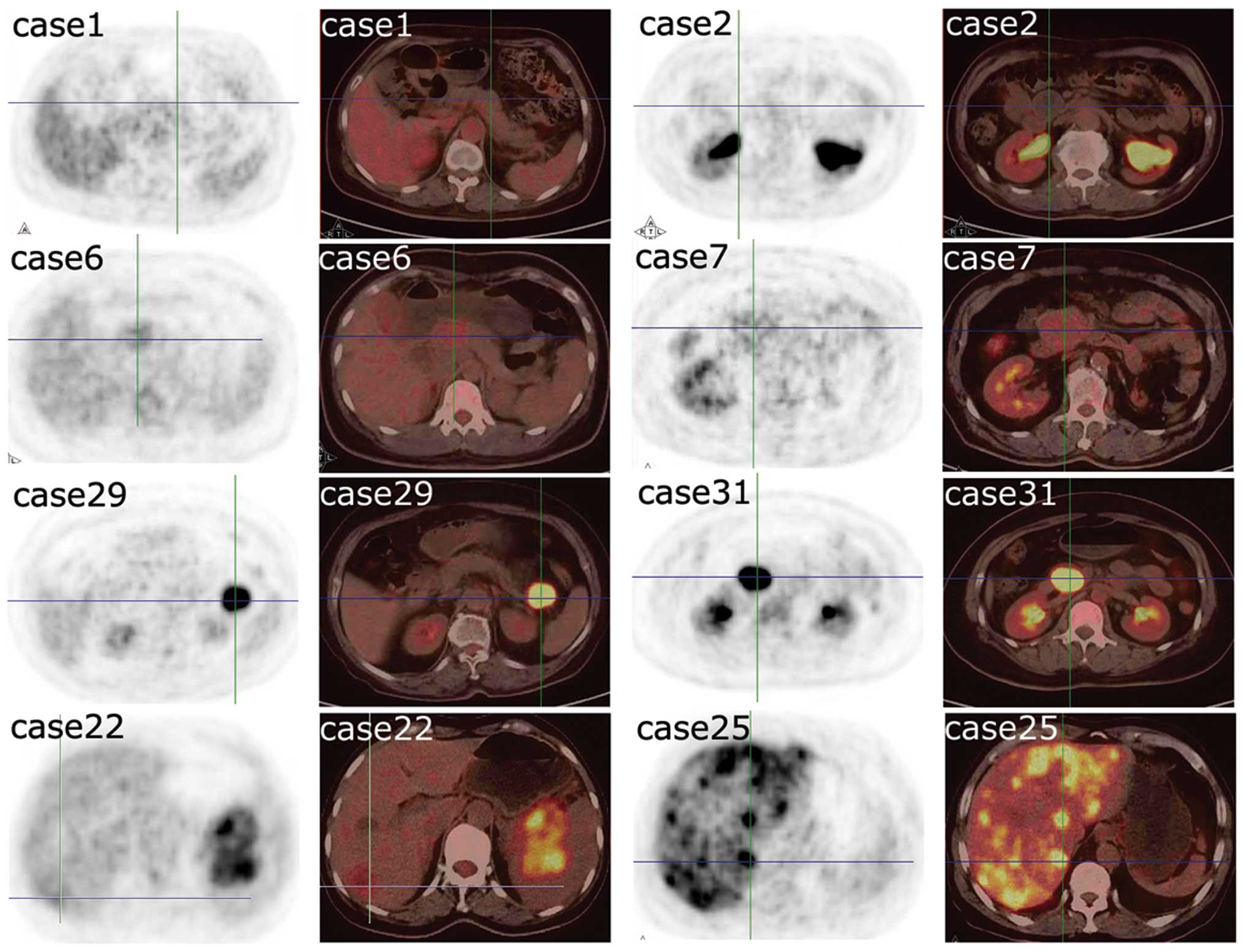
The study group consisted of 31 cases with
non-functioning PNETs and 18F-FDG PET/CT detected 28
(90.3%) primary lesions (Figs. 1
and 2). The study cohort included
16 women and 15 men, age range 25–77 years (median age, 48 years)
(Table I). In the subgroup of
patients aged ≤50 years, 100% patients had SUVmax ≥2.5, which was
similar to that of the subgroup of patients aged >50 years
(78.6%) (Table II). The
percentage of patients with SUVmax ≥2.5 was similar for women and
men (93.8 and 86.7%, respectively). Thirteen lesions were located
at the head of the pancreas and 18 at the body and tail of the
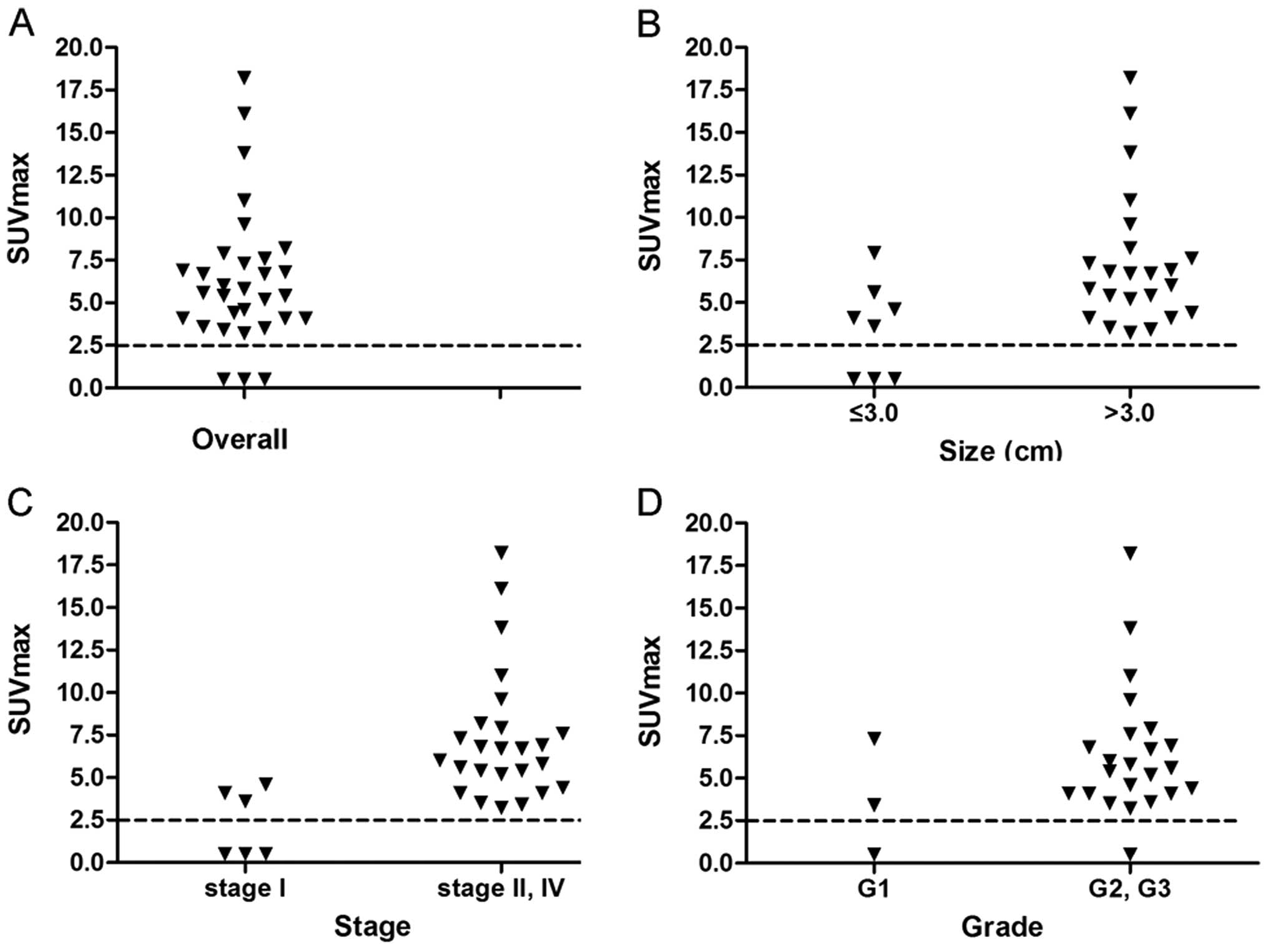
pancreas. Size of lesions ranged from 1.4 to 10.0 cm (mean ± SD,
4.9±2.3 cm). In 8 cases of tumor size ≤3.0 cm, 62.5% of patients
had SUVmax ≥2.5. However, in 23 cases of tumor size >3.0 cm, all
the patients (100.0%) had SUVmax ≥2.5 (Fig. 2B). According to the AJCC TNM
staging system, 6 patients had stage I tumors, 12 had stage II, and
13 had stage IV tumors. All 25 patients with stage II and IV tumors
had SUVmax ≥2.5, compared with only 50.0% of patients with stage I
tumors (Fig. 2C). Of all 26 cases
with grade information, 3 cases had G1 tumors, 16 had G2 tumors,
and 7 cases had G3 tumors. The percentage of SUVmax ≥2.5 in the
cases with G1 tumors was lower than that of the cases with G2 and
G3 tumors (66.7 and 95.7%, respectively) (Fig. 2D).
 | Table IPatient characteristics. |
Table I
Patient characteristics.
| Patient | Age (years) | Gender | Location | Size (cm) | TNM stage | Grade | SUVmaxa |
|---|
| 1 | 62 | F | Body | 2.0 | I | G1 | (−) |
| 2 | 64 | M | Head | 2.0 | I | G2 | (−) |
| 3 | 63 | M | Head | 3.0 | I | Unknown | (−) |
| 4 | 52 | F | Head | 6.0 | II | G2 | 3.2 |
| 5 | 30 | M | Body | 4.0 | II | G1 | 3.4 |
| 6 | 65 | F | Head | 10.0 | IV | G2 | 3.5 |
| 7 | 31 | F | Head | 3.0 | I | G2 | 3.6 |
| 8 | 66 | F | Body | 4.0 | II | G2 | 4.1 |
| 9 | 46 | F | Body | 2.5 | I | G3 | 4.1 |
| 10 | 44 | F | Body | 4.0 | IV | G2 | 4.1 |
| 11 | 29 | F | Body | 5.1 | IV | G2 | 4.4 |
| 12 | 47 | M | Head | 3.0 | I | G2 | 4.6 |
| 13 | 43 | M | Body | 5.0 | II | G3 | 5.2 |
| 14 | 68 | F | Head | 8.0 | IV | G2 | 5.4 |
| 15 | 71 | M | Tail | 3.5 | IV | Unknown | 5.4 |
| 16 | 49 | M | Head | 2.0 | II | G2 | 5.6 |
| 17 | 42 | M | Body | 5.0 | II | G3 | 5.8 |
| 18 | 52 | M | Body | 5.0 | II | G2 | 6 |
| 19 | 42 | M | Head | 5.0 | II | G3 | 6.7 |
| 20 | 41 | M | Head | 5.4 | IV | Unknown | 6.7 |
| 21 | 48 | M | Head | 10.0 | IV | G3 | 6.8 |
| 22b | 48 | F | Tail | 8.0 | IV | G2 | 6.9 |
| 23 | 58 | M | Body | 5.7 | IV | G1 | 7.3 |
| 24 | 56 | F | Tail | 9.0 | IV | G2 | 7.6 |
| 25 | 67 | M | Tail | 1.4 | IV | G3 | 7.9 |
| 26 | 28 | F | Tail | 4.1 | IV | Unknown | 8.2 |
| 27 | 25 | F | Tail | 4.0 | II | G2 | 9.6 |
| 28 | 47 | M | Body | 8.0 | IV | G3 | 11 |
| 29 | 77 | F | Tail | 4.0 | II | G2 | 13.8 |
| 30 | 66 | F | Head | 6.0 | II | Unknown | 16.1 |
| 31 | 45 | F | Head | 5.0 | II | G2 | 18.2 |
| Table IISUVmax of primary tumor, patient
demographics and clinical characteristics. |
Table II
SUVmax of primary tumor, patient
demographics and clinical characteristics.
| Characteristic | SUVmax <2.5 | SUVmax ≥2.5 | % (SUVmax
≥2.5/total) | P-value |
|---|
| Age | | | | 0.081 |
| ≤50 | 0 | 17 | 100.0 | |
| >50 | 3 | 11 | 78.6 | |
| Gender | | | | 0.600 |
| Male | 2 | 13 | 86.7 | |
| Female | 1 | 15 | 93.8 | |
| Location | | | | 0.558 |
| Head | 2 | 11 | 84.6 | |
| Body/tail | 1 | 17 | 94.4 | |
| Size (cm) | | | | 0.012 |
| ≤3.0 | 3 | 5 | 62.5 | |
| >3.0 | 0 | 23 | 100.0 | |
| TNM stage | | | | 0.004 |
| I | 3 | 3 | 50.0 | |
| II, IV | 0 | 25 | 100.0 | |
| Gradea | | | | 0.222 |
| G1 | 1 | 2 | 66.7 | |
| G2, G3 | 1 | 22 | 95.7 | |
| Total | 3 | 28 | 90.3 | |
For all 12 patients with stage IV tumors (liver
metastases of case 15 were resected before PET/CT examination), 42
distant metastatic lesions were found and confirmed by biopsy or
serial imaging and clinical follow-up findings (Table III). Thirty-eight (90.5%) lesions
had a SUVmax ≥2.5 during PET/CT scans and only 4 (9.5%) lesions
demonstrated normal 18F-FDG distribution. Eight of the
31 (25.8%) cases of PNETs had changed their clinical management
based on results of the 18F-FDG PET/CT examinations.
Among these 8 cases, 4 cases underwent one-stage resection for both
the primary tumor and distant metastases and 4 cases abandoned
curative surgery and underwent systematic treatments.
| Table IIISUVmax in distant metastases. |
Table III
SUVmax in distant metastases.
| Patienta | Site of distant
metastasis | SUVmax |
|---|
| 6 | 1 liver | - |
| 10 | 2 liver | - |
| 11 | 3 lung, 3 distant
lymph nodes | 2.9–5.8 |
| 14 | 1 liver | - |
| 20 | 3 liver | 4.3–6.8 |
| 21 | 4 liver, 1 lung, 2
peritoneum | 3.0–6.2 |
| 22 | 2 liver | 2.8,3.1 |
| 23 | 2 peritoneum, 1
distant lymph nodes | 4.5–7.8 |
| 24 | 1 liver | 3.5 |
| 25 | 6 liver | 3.1–12.2 |
| 26 | 3 liver | 3.5–8.6 |
| 28 | 1 liver, 6
peritoneum | 2.5–7.0 |
| % (SUVmax
≥2.5/total) | 90.5% |
Discussion
Imaging technologies are essential to detect
non-functioning PNETs and guide their treatment. At present,
because of the high expression of somatostatin receptors on NET
cells, somatostatin receptor scintigraphy with 111In
octreotide is a standard functional imaging technique in the
detection and staging of NETs (11,17).
However, poorly differentiated or aggressive NETs with high
proliferative rates may not be localized on 111In
octreotide scanning (17). Kisker
et al (17) found that
somatostatin receptor scintigraphy visualized only 4 of 10 cases
with non-functioning PNETs, which are often poorly differentiated
and aggressive. In another study, Rickes et al (18) showed that the sensitivity and
specificity of somatostatin receptor scintigraphy for diagnosing
PNETs was 54 and 81%, respectively. Therefore, additional
functional imaging modalities are needed to detect non-functioning
PNETs.
The application of 18F-FDG PET in the
detection of NETs depends on the grade of tumor differentiation and
the biological characteristics of the tumor (10). NETs are mostly well-differentiated
and low-malignancy tumors and 18F-FDG PET frequently
fails to visualize them (10).
However, 18F-FDG accumulation is often observed in
less-differentiated NETs with high proliferative activity (10,19).
It has been reported that tumors with increased FDG accumulation
appear more aggressive and correlate with poor prognosis (20). Because a great proportion of
non-functioning PNETs are poorly differentiated with high
proliferative activity, it is reasonable to infer that they can be
detected by 18F-FDG PET. However, there has been no
published evidence of 18F-FDG PET in detecting
non-functioning PNETs.
In the present study, we showed that 90.3% of
non-functioning PNETs can be visualized by 18F-FDG
PET/CT, indicating that 18F-FDG PET/CT can be employed
to detect non-functioning PNETs. The implication 18F-FDG
PET/CT in PNETs changed the clinical management of 8 cases. In
addition, 18F-FDG PET/CT was found to accumulate in
90.5% of distant metastatic lesions, suggesting that
18F-FDG PET/CT can be used to stage non-functioning
PNETs. Other PET imaging agents, such as
11C-5-hydroxytryptophan (11C-5-HTP) and
68Ga-DOTA-Tyr3-octreotide, have been used to detect NETs
with promising results (13,21–23).
For example, in 2011, Naswa et al (16) showed that 68Ga-labeled
[1, 4, 7, 10-tetraazacyclododecane-1, 4, 7, 10-tetraacetic
acid]-1-NaI(3)-octreotide
(68Ga-DOTA-NOC) PET/CT had a sensitivity of 78.3 for
primary tumor and 97.4% for metastases in gastroenteropancreatic
NETs, findings that are similar to our study using
18F-FDG PET/CT. However, these imaging agents are still
not widely applied in clinical practice compared with
18F-FDG PET/CT.
Because the uptake of 18F-FDG in NETs is
closely related to differentiation status and tumor stage (10,12,13,19),
we also examined whether tumor size, tumor grade, and TNM stage
affect the uptake of 18F-FDG. We found that the uptake
of 18F-FDG in PNETs was significantly associated with
tumor size and TNM stage, indicating that 18F-FDG PET
can be used to detect and stage non-functioning PNETs. In addition,
although there was no significant association between increased
18F-FDG uptake and tumor grade, we found that FDG-PET/CT
may aid in the diagnosis and staging of PNETs with G2 and G3 tumors
(SUVmax ≥2.5, G1, 66.7%, G2 and G3, 95.7%, respectively,
P=0.222).
In conclusion, 18F-FDG PET/CT can be used
to detect, stage, and conduct surveillance of non-functioning
PNETs. In the detection stage, 18F-FDG PET/CT can be
applied to a fully evaluated tumor burden to guide treatments,
especially for curative and cytoreductive surgery. It also can be
used to assess therapeutic response including cytotoxic
chemotherapy and targeted therapy and monitor disease recurrence.
However, further systematic analyses with even larger study
populations will be needed to confirm the role of
18F-FDG PET/CT in the clinical management of
non-functioning PNETs.
Acknowledgements
This study was supported in part by the Sino-German
Center (GZ857), by the Science Foundation of Shanghai
(13ZR1407500), by the Shanghai Rising Star Extension Program
(12QH1400600), by the Fudan University Young Investigator promoting
program (20520133403) and by the National Science Foundation of
China (grant nos. 81101807, 81001058, 81372649, 81372653 and
81172276).
References
|
1
|
Garcia-Carbonero R, Capdevila J,
Crespo-Herrero G, et al: Incidence, patterns of care and prognostic
factors for outcome of gastroenteropancreatic neuroendocrine tumors
(GEP-NETs): results from the National Cancer Registry of Spain
(RGETNE). Ann Oncol. 21:1794–1803. 2010. View Article : Google Scholar
|
|
2
|
Modlin IM, Oberg K, Chung DC, et al:
Gastroenteropancreatic neuroendocrine tumours. Lancet Oncol.
9:61–72. 2008. View Article : Google Scholar
|
|
3
|
Halfdanarson TR, Rabe KG, Rubin J and
Petersen GM: Pancreatic neuroendocrine tumors (PNETs): incidence,
prognosis and recent trend toward improved survival. Ann Oncol.
19:1727–1733. 2008. View Article : Google Scholar : PubMed/NCBI
|
|
4
|
Burns WR and Edil BH: Neuroendocrine
pancreatic tumors: guidelines for management and update. Curr Treat
Options Oncol. 13:24–34. 2012. View Article : Google Scholar : PubMed/NCBI
|
|
5
|
Bertani E, Fazio N, Botteri E, et al:
Resection of the primary pancreatic neuroendocrine tumor in
patients with unresectable liver metastases: possible indications
for a multimodal approach. Surgery. 155:607–614. 2014. View Article : Google Scholar
|
|
6
|
Nomura N, Fujii T, Kanazumi N, et al:
Nonfunctioning neuroendocrine pancreatic tumors: our experience and
management. J Hepatobiliary Pancreat Surg. 16:639–647. 2009.
View Article : Google Scholar : PubMed/NCBI
|
|
7
|
Li J, Luo G, Fu D, et al: Preoperative
diagnosis of nonfunctioning pancreatic neuroendocrine tumors. Med
Oncol. 28:1027–1031. 2011. View Article : Google Scholar : PubMed/NCBI
|
|
8
|
Czernin J, Weber WA and Herschman HR:
Molecular imaging in the development of cancer therapeutics. Annu
Rev Med. 57:99–118. 2006. View Article : Google Scholar
|
|
9
|
von Schulthess GK, Steinert HC and Hany
TF: Integrated PET/CT: current applications and future directions.
Radiology. 238:405–422. 2006.PubMed/NCBI
|
|
10
|
Adams S, Baum R, Rink T, Schumm-Drager PM,
Usadel KH and Hor G: Limited value of fluorine-18
fluorodeoxyglucose positron emission tomography for the imaging of
neuroendocrine tumours. Eur J Nucl Med. 25:79–83. 1998. View Article : Google Scholar : PubMed/NCBI
|
|
11
|
Slooter GD, Mearadji A, Breeman WA, et al:
Somatostatin receptor imaging, therapy and new strategies in
patients with neuroendocrine tumours. Br J Surg. 88:31–40. 2001.
View Article : Google Scholar : PubMed/NCBI
|
|
12
|
Binderup T, Knigge U, Loft A, et al:
Functional imaging of neuroendocrine tumors: a head-to-head
comparison of somatostatin receptor scintigraphy,
123I-MIBG scintigraphy, and 18F-FDG PET. J
Nucl Med. 51:704–712. 2010. View Article : Google Scholar : PubMed/NCBI
|
|
13
|
Kayani I, Bomanji JB, Groves A, et al:
Functional imaging of neuroendocrine tumors with combined PET/CT
using 68Ga-DOTATATE (DOTA-DPhe1, Tyr3-octreotate) and
18F-FDG. Cancer. 112:2447–2455. 2008. View Article : Google Scholar : PubMed/NCBI
|
|
14
|
Modlin IM, Pavel M, Kidd M and Gustafsson
BI: Review article: somatostatin analogues in the treatment of
gastroenteropancreatic neuroendocrine (carcinoid) tumours. Aliment
Pharmacol Ther. 31:169–188. 2010.PubMed/NCBI
|
|
15
|
National Comprehensive Cancer Network.
NCCN Clinical Practice Guidelines in Oncology. Pancreatic Endocrine
Tumors. 2:14–18. 2014.
|
|
16
|
Naswa N, Sharma P, Kumar A, et al:
Gallium-68-DOTA-NOC PET/CT of patients with gastroenteropancreatic
neuroendocrine tumors: a prospective single-center study. AJR Am J
Roentgenol. 197:1221–1228. 2011. View Article : Google Scholar : PubMed/NCBI
|
|
17
|
Kisker O, Bartsch D, Weinel RJ, et al: The
value of somatostatin-receptor scintigraphy in newly diagnosed
endocrine gastroenteropancreatic tumors. J Am Coll Surg.
184:487–492. 1997.PubMed/NCBI
|
|
18
|
Rickes S, Unkrodt K, Ocran K, Neye H and
Wermke W: Differentiation of neuroendocrine tumors from other
pancreatic lesions by echo-enhanced power Doppler sonography and
somatostatin receptor scintigraphy. Pancreas. 26:76–81. 2003.
View Article : Google Scholar
|
|
19
|
Pasquali C, Rubello D, Sperti C, et al:
Neuroendocrine tumor imaging: can 18F-fluorodeoxyglucose
positron emission tomography detect tumors with poor prognosis and
aggressive behavior? World J Surg. 22:588–592. 1998.PubMed/NCBI
|
|
20
|
Strauss LG and Conti PS: The applications
of PET in clinical oncology. J Nucl Med. 32:623–650.
1991.PubMed/NCBI
|
|
21
|
Gabriel M, Decristoforo C, Kendler D, et
al: 68Ga-DOTA-Tyr3-octreotide PET in neuroendocrine
tumors: comparison with somatostatin receptor scintigraphy and CT.
J Nucl Med. 48:508–518. 2007. View Article : Google Scholar
|
|
22
|
Ambrosini V, Tomassetti P, Castellucci P,
et al: Comparison between 68Ga-DOTA-NOC and
18F-DOPA PET for the detection of
gastro-entero-pancreatic and lung neuro-endocrine tumours. Eur J
Nucl Med Mol Imaging. 35:1431–1438. 2008.
|
|
23
|
Orlefors H, Sundin A, Garske U, et al:
Whole-body (11) C-5-hydroxytryptophan positron emission tomography
as a universal imaging technique for neuroendocrine tumors:
comparison with somatostatin receptor scintigraphy and computed
tomography. J Clin Endocrinol Metab. 90:3392–3400. 2005. View Article : Google Scholar
|