Introduction
Breast cancer is the most common type of cancer in
women, with over one million cases diagnosed worldwide (1,2).
Despite improvements in the development of chemotherapeutic drugs
for breast cancer treatment, cancer related death rates continue to
rise. Over time patients develop multidrug resistance (MDR) to
chemotherapeutic drugs and this is a major factor in the failure of
many forms of therapy, which can ultimately lead to tumor
metastasis, affecting the quality of life of survivors and
contributing to the mortality rate (3,4).
Thus, it is of significant value to investigate the mechanism and
pathways linked to drug resistance and metastasis in order to
improve therapeutic treatments.
The development of resistance to chemotherapeutic
agents occurs frequently in breast cancer patients and is a major
factor for poor prognosis, mortality and morbidity in these
patients (5). Among the strategies
by which cancer cells acquire drug resistance is via the
overexpression of ATP-binding cassette (ABC) transporters (6) which function as an energy-dependent
efflux pump. The ABC transporters family include the P-glycoprotein
(P-gp)/MDR belonging to the ABCB family (7). Included in the ABC superfamily are
the multidrug resistance-protein (MRP) family of transporter
proteins encoded by ABCC1-6, 10–13, the ATP-gated chloride channel,
CFTR (ABCC7) and the ATP-dependent sulfonylurea receptors, SUR
(ABCC8, 9) (6). Of these,
MRP1/ABCC1 is designated as a negative prognostic marker for early
stage breast cancer, with reports linking a strong correlation of
its expression to relapse and overall survival in several cancers
(8–11). Although MRP1 is expressed in normal
tissues, several studies have demonstrated overexpression of MRP1
in multiple solid tumors including breast, lung and prostate
(6,12). Overexpression of MRP1 in breast
cancer lymph nodes has been associated to the metastatic potential
of the disease (13). Expression
pattern of MRP1 makes this an optimal candidate for use as a marker
for prediction of chemoresistance. Although the structure and
function of ABC transporters have been explored in detail,
developing effective agents against these transporters have not
been translated into a clinical target (14).
Controlling cancer at the initial stage is vital in
managing the disease. Tumor progression and metastasis within the
body results in chemoresistance or an increase in chemotherapeutic
dosage which ultimately causes cardiac toxicity (15). Hence, conventional therapeutic
options have not been successful in halting the progression of the
disease, and thus understanding the role of key proteins in the
dissemination and metastasis of tumor cells will be essential in
identifying effective and innovative drug alternative to treat the
disease at its initial or later stages. One of the diagnostic
markers of metastatic progression is the overexpression of a
transmembrane protein called Mucin 1 (MUC1) which has been
implicated in reduced survival rate. Among the many functions
associated with mucins, MUC1 is involved in cell growth, cell
adhesion, motility and cell survival (16,17).
MUC1 has also been implicated in chemoresistance and studies have
shown that cancers overexpressing MUC1 are unresponsive to
therapeutic chemotoxic drugs (18–20).
A link between metastasis and chemoresistance has been observed in
pancreatic cancer cells, whereby MUC1 has been shown to upregulate
MRP1, lowering sensitivity to chemotherapeutic agents (18). This study from Nath et al
(18) provides a mechanistic
insight on the correlation between chemoresistance and metastasis
in pancreatic cells. However, little is known on the relationship
between MUC1 and MRP1 in mammary carcinoma cells. Targeting both
MRP1 and MUC1 may provide an opportunity to intervene with drug
resistance, and ultimately reduce the migration of cancer cells to
other parts of the body, while augmenting the efficacy of
chemotherapeutic drugs such as doxorubicin (DOX) against breast
cancer cells.
Due to the cardiac toxicity associated with
chemotherapeutic drugs such as DOX (15), various approaches to enhance
chemoresponse in cancer models have been explored, including
combining standard chemotherapy agents with natural products
(21,22). Honokiol, isolated from the bark of
the magnolia tree, has been found to have anti-oxidant (23), anti-inflammatory (24) and anticancer properties (25,26),
promoting its potential for targeting various diseases, including
cancer, arthritis, and diabetes (25–29).
In addition to the multifaceted effects of honokiol, it has
improved the actions of conventional chemotherapies to promote
growth suppression or overcome drug resistance in a variety of
preclinical models of human cancer, including skin cancer (30), prostate cancer (31) and multiple myeloma (32).
The study was aimed to investigate the relationship
between the process of chemoresistance and cancer progression in
mammary carcinoma cells by characterizing the effect of honokiol on
MRP1 and MUC1 gene expression, two proteins directly involved in
these activities. In this study, we demonstrate that honokiol
suppresses the expression of MUC1 and MRP1 in mammary carcinoma
cells. Based on these data, we provide mechanistic evidence that
honokiol enhances the efficacy of DOX by regulating the expression
of MUC1, which directly downregulates MRP1. Thus, understanding the
mechanism by which honokiol promotes its anti-carcinogenic effects
and prevents drug resistance may provide mechanistic insights into
the underlying antitumor effects of honokiol, alone or in
combination with chemotherapeutic agents, in breast cancer.
Materials and methods
Reagents
Antibodies against MUC1 were obtained from Santa
Cruz Biotechnology (Santa Cruz, CA, USA) and β-tubulin was
purchased from Sigma Aldrich Co (St. Louis, MO, USA). Antibodies
for MRP1 were obtained from Abcam (Cambridge, MA, USA). Anti-mouse
and anti-rabbit immunoglobulin horseradish peroxidase-conjugated
antibodies were from Bio-Rad (Hercules, CA, USA), and anti-goat
immunoglobulin was from Santa Cruz. Honokiol was obtained from LKT
Laboratories (St. Paul, MN, USA) and DOX was purchased from Sigma.
MTT reagent (3-(4,5-dimethylthiazol-2-yl)-2,5-diphenyl tetrazolium
bromide) was purchased from Sigma. MUC1 and control siRNA were
purchased from Santa Cruz Biotechnology.
Cell lines
MCF-7, MCF10A and MDA-MB-231 cells were maintained
in Dulbecco's modified Eagle's medium (DMEM) supplemented with 10%
fetal bovine serum (FBS) with antibiotics-antimycotics. DMEM was
obtained from Life Technologies (Grand Island, NY, USA). FBS and
antibiotics-antimycotics were purchased from Atlanta Biologicals
(Flowery Branch, GA, USA). MCF-7 and MDA-MB-231 cells were a kind
gift of Dr Ming Tan (Mitchell Cancer Institute, Mobile, AL, USA).
MCF10A was purchased from American Type Culture Collection (ATCC,
Manassas, VA, USA).
Honokiol and DOX preparation
Honokiol was prepared at a stock concentration of 10
mM in dimethyl sulfoxide (DMSO) and cells were treated at the
appropriate concentration, using DMSO as control. DOX was prepared
at a stock concentration of 2 mM in water and diluted at the
appropriate concentration for the treatment of cells.
Western blot analyses
Cells were cultured in 100-mm plates and treated
with honokiol for either 24 or 48 h. DMSO was used as control.
Cells were lysed in ice-cold buffer containing 150 mM NaCl, 10 mM
Tris, pH 7.2., 0.1% SDS, 1% Triton X-100, 1% deoxycholate, 5 mM
EDTA, and 1 mM PMSF. Cells were lysed on ice for 1 h and protein
concentration was determined by the Bradford assay. Cell lysate was
resolved by SDS-PAGE and blotted onto a nitrocellulose membrane.
The membrane was blocked in 10% bovine serum albumin in Tris-buffer
saline containing 0.05% tween for 1 h at room temperature. The
membrane was then incubated with primary antibody, MUC1 or MRP1 at
a dilution of 1:1,000 overnight at 4°C. For equal loading,
β-tubulin (1:1,000) was incubated with the membrane for 1 h at 4°C.
The antigen-antibody complex was visualized with SuperSignal West
Pico Chemiluminescent Substrate (Fisher, Hanover Park, IL,
USA).
Quantitative real-time polymerase chain
reaction (qRT-PCR)
Cells were treated with honokiol for 48 h, and RNA
was extracted using TRIzol (Life Technologies). As described in the
high capacity RNA to cDNA kit from Applied Biosystems, 2 μg total
RNA was reverse transcribed into cDNA. To determine expression of
MUC1 and ABCC1, qRT-PCR was carried out by using commercially
available Taqman Chemistry and Assay on Demand Probes (Applied
Biosystems). Glyceraldehyde-3-phosphate dehydrogenase (GAPDH) was
used for normalization. Detection and data analysis were carried
out on the ABI Step One Plus Real-Time PCR System. Relative quatity
of gene expression was performed using 2−ΔΔCt method
(33).
Cell viability assay
For cell proliferation assays, 5,000 cells of
MDA-MB-231 and MCF-7 were seeded in a 96-well plate and allowed to
adhere overnight. Cells were then treated with honokiol in the
presence or absence of DOX for 48 h. Controls were treated with or
without DMSO depending on the drug treatment. After 48 h, 5 μg/ml
MTT reagent was added directly to the cells for 3 h, or until
crystals formed. The media was carefully removed from the plate,
leaving the cells intact and the cells were then resuspended in 150
μl of 0.04 M HCl in isopropanol. Absorbance was read at 570 nm to
determine cell proliferation.
Cell transfection
MCF-7 and MDA-MB-231 cells were transfected
according to the protocol provided by Invitrogen (Grand Island, NY,
USA). Briefly, 180 pmol of control or MUC1 siRNA was mixed with 2
ml Gibco Opti-MEM® I medium without serum by Life
Technologies in a 100-mm plate. Lipofectamine™ RNAiMAX (25 μl) was
added to the diluted RNAi mix and left for 20 min at room
temperature. Cultured MCF-7 or MDA-MB-231 cells were subdivided and
2×10⁶ cells were diluted in the media. Cells were added
to the plates containing the RNAi duplex/Lipofectamine™ mixture
labeled with control or MUC1 siRNA. The cells were incubated for 48
h at 37°C in a CO2 incubator. After 48 h, cells were
lysed, protein was extracted, quantitated and protein extract was
loaded onto a gel for SDS-PAGE. The blot was probed for MUC1 with
MUC1 antibody to assay knockdown of the protein, and using the same
extract, MRP1 protein expression was probed with MRP1 antibody.
Trypan blue exclusion assay
As described above, MCF-7 and MDA-MB-231 cells were
transfected with control or MUC1 siRNA overnight, followed by
treatment with 1 μM DOX for 48 h. After 48 h, cells were
trypsinized, collected and counted using trypan blue. Live cells
are presented as number of cells/ml.
Statistical analysis
Statistical significance of differences between
treatments was determined using two tailed Student's t-test and
p-values were noted. Differences between groups were considered
statistically significant at p<0.05.
Results
Honokiol enhances the efficacy of DOX in
mammary carcinoma cells
One of the advantages of honokiol is its lack of
toxicity in non-tumorigenic cells, promoting its use with
conventional chemotherapeutic agents (34). To compare the effects of honokiol
on mammary carcinoma cells (MCF-7 and MDA-MB-231) with normal human
mammary epithelial cells, we treated the cells with varying
concentrations of honokiol. As observed previously (35,36),
honokiol had moderate toxicity with a 50% decrease in cell
proliferation at 25 and 28 μM honokiol in MCF-7 and MDA-MB-231
cells, respectively, while MCF10A were less sensitive to the
anti-proliferative effects of honokiol (Fig. 1A).
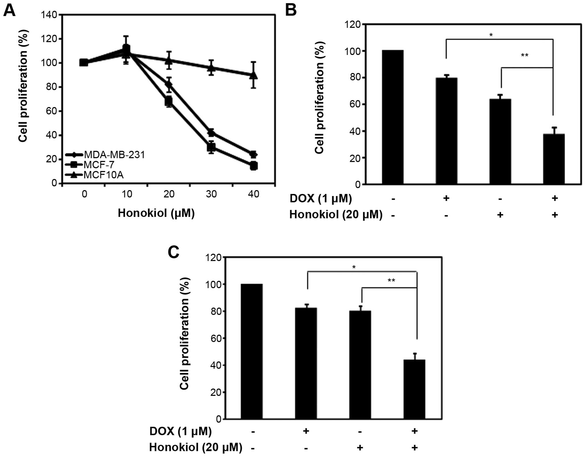 | Figure 1Honokiol enhances the efficacy of DOX
in mammary carcinoma cells. (A) MDA-MD-231, MCF-7 and MCF10A cells
were plated in 96-well plates, treated with varying dose of
honokiol for 48 h. The control was DMSO treated according to the
concentration of the cells treated with honokiol. The percentage
(%) cell proliferation for each of the treatment with honokiol for
the designated concentration was calculated with respect to the
treatment with DMSO for the corresponding dose. Data are mean of ±
SE (n=3). (B) MCF-7 cells were plated in 96-well plates, and
treated with 1 μM DOX in the presence or absence of 20 μM honokiol
for 48 h. The percentage of cell proliferation for each of the
treatment (DOX, honokiol or both) was calculated relative to their
solvent, water, DMSO or the combination, respectively. The controls
were set at 100%. Data are mean of ± SE (n=3). *p=0.01;
**p=0.008. (C) MDA-MD-231 cells were plated in 96-well
plates, and treated with 1 μM DOX in the presence or absence of 20
μM honokiol for 48 h. The percentage (%) cell proliferation for
each of the treatment (DOX, honokiol or both) was calculated
relative to their solvent, water, DMSO or the combination,
respectively. The controls were set at 100%. Data are mean of ± SE
(n=3). *p=0.02; **p=0.002. |
Previous studies have shown that combining honokiol
with chemotherapeutic agents in a variety of cancer cell lines
increases the efficacy of chemotherapeutic agents by augmenting
cell growth inhibition or inducing apoptosis (30–32,34,35).
We evaluated the ability of honokiol to enhance the efficacy of
DOX-mediated growth suppression in MCF-7 and MDA-MB-231. Given that
20 μM honokiol suppressed mammary carcinoma cell growth by ~33 and
20% in MCF-7 and MDA-MB-231 cells, respectively, we treated mammary
carcinoma cells with 1 μM of DOX with a fixed 20 μM concentration
of honokiol and analyzed cell proliferation using MTT assay. As
shown in Fig. 1B and C, 1 μM DOX
suppressed MCF-7 and MDA-MB-231 cell growth, respectively. As
observed previously (Fig. 1A),
honokiol at 20 μM reduced cell growth in MCF-7 and MDA-MB-231 cells
(Fig. 1B and C). However, the
combination of DOX with honokiol further increased the cell growth
inhibitory properties of DOX and improved the potency of DOX in
MCF-7 and MDA-MB-231 cells (Fig. 1B
and C). This indicates that honokiol improves the efficacy of
DOX, ultimately reducing the toxic effects of DOX.
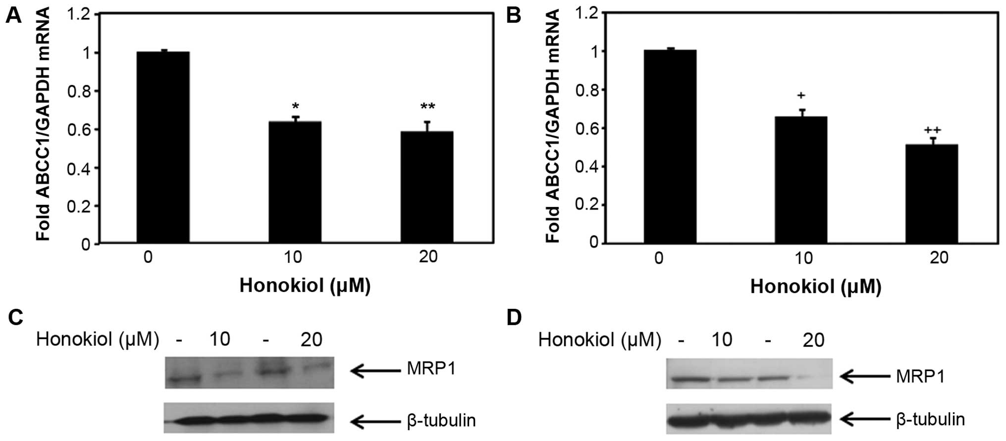
Honokiol regulates the expression of
ABCC1/MRP1 in mammary carcinoma cells
DOX is a substrate of MRP1 (37). To investigate whether the enhanced
efficacy of DOX in mammary carcinoma cells (MCF-7 and MDA-MB-231)
treated with honokiol was likely due to the associated changes of
ABCC1 expression, we sought to assess the effect of honokiol on
ABCC1 mRNA and protein expression in the non-metastatic MCF-7 cells
and the highly aggressive MDA-MB-231 cells. To determine whether
honokiol regulates the expression of ABCC1 in these mammary
carcinoma cells, we treated MCF-7 and MDA-MB-231 mammary carcinoma
cells with 10 and 20 μM honokiol for 48 h, and the levels of ABCC1
mRNA and protein were determined by qRT-PCR and western blotting,
respectively. As shown in Fig. 2A,
10 and 20 μM honokiol equally suppressed ABCC1 mRNA levels in MCF-7
cells, without a dose response. Similarly, both 10 and 20 μM
honokiol reduced ABCC1 mRNA expression in MDA-MB-231 cells by ~40
and 60%, respectively (Fig. 2B).
The levels of the respective protein expression of ABCC1, MRP1 was
also analyzed in these cells and consistent with ABCC1 mRNA
expression in MCF-7 cells, 10 and 20 μM of honokiol reduced MRP1
protein expression in MCF-7 cells (Fig. 2C). To determine whether the
inhibitory effects of honokiol on ABCC1 mRNA expression in
MDA-MB-231 cells resulted in the suppression of MRP1 protein
expression in MDA-MB-231 cells, western blotting was conducted. As
shown in Fig. 2D, 20 μM honokiol
suppressed MRP1 protein expression in MDA-MB-231 cells, while 10 μM
honokiol had a moderate effect. These results indicate that
honokiol suppresses ABCC1 mRNA and the expression of its respective
protein, MRP1 in two different breast cancer cell lines, suggesting
that the regulation of MRP1 by honokiol is a global effect among
mammary carcinoma cells, emphasizing the importance of using
honokiol to target drug resistance gene MRP1 in breast cancer.
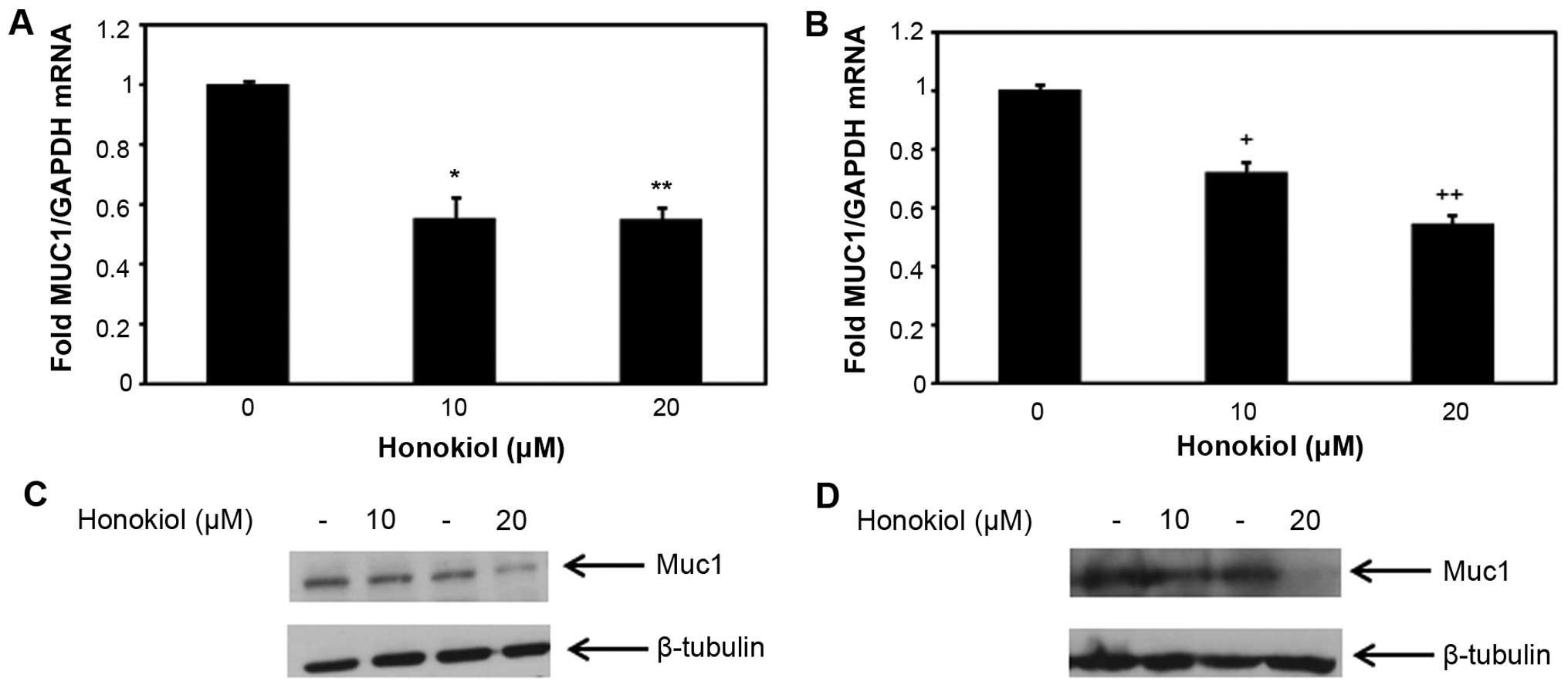
MUC1 is regulated by honokiol in mammary
carcinoma cells
Several studies have suggested the role of MUC1 in
conferring drug resistance in cancer cells (18–20),
while a study in pancreatic cancer cells specifically demonstrated
regulation of MRP1 by MUC1 (18).
In order to determine whether honokiol regulates MUC1 in mammary
carcinoma cells, MCF-7 and MDA-MB-231 cells were treated with
honokiol (10 and 20 μM) for 48 h and MUC1 mRNA expression level was
analyzed. As shown in Fig. 3A and
B, 10 and 20 μM honokiol suppressed MUC1 mRNA expression in
both MCF-7 and MDA-MB-231 cells (Fig.
3A and B). Concomitantly, we examined the protein expression
level of MUC1 in MCF-7 and MDA-MB-231 cells treated with honokiol
and observed the downregulation of MUC1 protein in honokiol treated
cells (Fig. 3C and D). These
results demonstrate that honokiol suppresses MUC1 expression level
in MCF-7 and MDA-MB-231 cells, suggesting the regulation of MUC1 by
honokiol may be a global effect observed in breast cancer
cells.
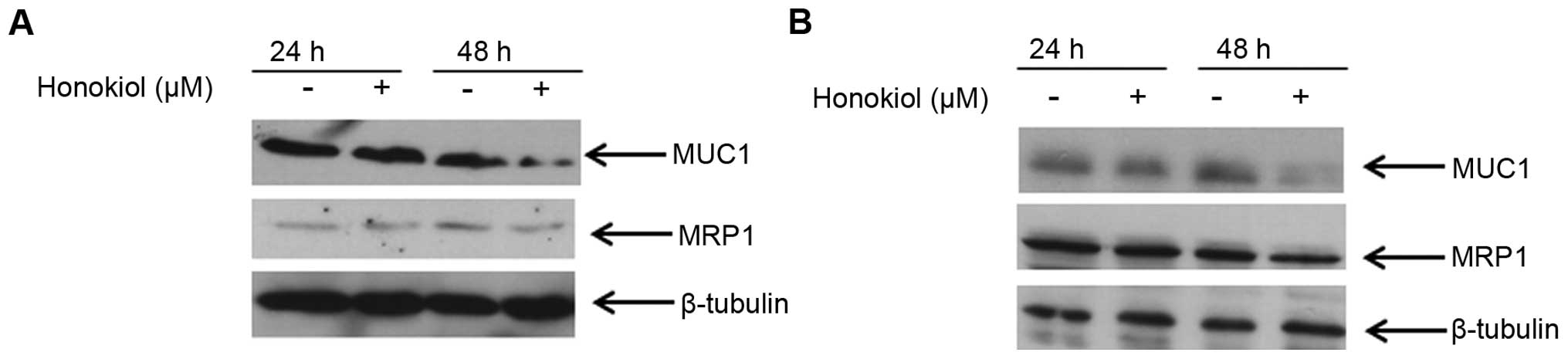
Relationship between MUC1 and MRP1
protein expression in mammary carcinoma cells
To further understand the relationship between MUC1
and MRP1 in mammary carcinoma cells, we specifically examined their
protein expression level at 24 and 48 h of treatment with honokiol.
Because significant downregulation of these proteins was observed
with 20 μM honokiol, we treated MCF-7 and MDA-MB-231 cells with
this concentration for 24 and 48 h, and analyzed the expression of
these proteins at these two different time-points. Results from
western blot analysis showed that 20 μM honokiol did not reduce
either MUC1 or MRP1 expression at 24 h in either MCF-7 or
MDA-MB-231 cells (Fig. 4).
However, at 48 h, 20 μM honokiol reduced MUC1 expression and
concomitantly MRP1 protein expression was downregulated in these
cell lines (Fig. 4). These data
suggest that there may be a correlation between the expression
level of MUC1 and MRP1, and in fact reducing MUC1 suppresses MRP1
in mammary carcinoma cells.
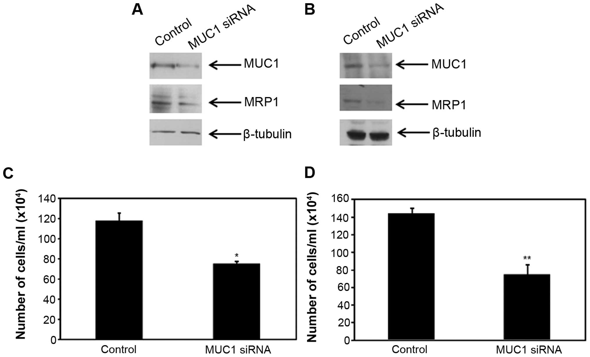
Suppression of MUC1 reduces the
expression level of MRP1 and enhances the efficacy of DOX in
mammary carcinoma cells
To investigate whether regulation of MRP1 by
honokiol is directly dependent on MUC1 pathway, we silenced MUC1 in
mammary carcinoma cells (MCF-7 and MDA-MB-231) with MUC1 siRNA
using Lipofectamine RNAimax. For control, we used a non-specific
control siRNA. Equal amounts of cell lysates from control and MUC1
knockdown cells were lysed and probed for MUC1 protein expression.
As shown in Fig. 5, MUC1 was
silenced in both MCF-7 and MDA-MB-231 cells, respectively.
Concomitantly, we examined the protein expression level of MRP1 in
the control and MUC1 siRNA transfected cells. Knock-down of MUC1
protein in MCF-7 cells suppressed MRP1 protein expression in these
cells (Fig. 5A). Similarly, in
MDA-MB-231 cells, MRP1 protein expression was suppressed in MUC1
silenced cells (Fig. 5B). With
direct evidence to corroborate the findings from Fig. 4, these results suggest that MUC1
directly regulates MRP1 in mammary carcinoma cells.
Since MUC1 regulates MRP1, we next assessed the
growth and inhibitory effects of DOX in mammary carcinoma cells
transfected with control and MUC1 siRNA using trypan blue. MCF-7
and MDA-MB-231 cells were transfected with control or MUC1 siRNA,
treated with 1 μM DOX for 48 h and cells were counted using trypan
blue. The sensitivity to DOX in MUC1 siRNA transfected MCF-7 and
MDA-MB-231 cells was significantly increased compared to the cells
treated with the control siRNA (Fig.
5C and D). Taken together our results indicate that silencing
MUC1 suppresses MRP1 which in turn enhances the efficacy of
DOX-mediated growth suppression in mammary carcinoma cells.
Discussion
The role of MUC1 in promoting breast cancer
development and its overexpression leading to mammary gland
hyperplasia in mouse models is well established (38,39).
MUC1 is well known to induce chemoresistance in several cancer
types (18–20). To understand the relationship
between MUC1 and MRP1 in breast cancer cells, we examined the
effect of honokiol on these two proteins, while gaining insights on
the functional consequence of regulating these genes. This study
shows that honokiol downregulates the expression of MUC1 and MRP1
in breast cancer cell lines and increases the efficacy of
DOX-mediated growth suppression. Silencing MUC1 gene expression in
breast cancer cells reduces the expression of MRP1, and improves
the potency of DOX to suppress cell growth. By examining the
protein expression of MUC1 and MRP1 in honokiol-treated mammary
carcinoma cells, we have observed that honokiol reduces MRP1
contingent upon the reduction of MUC1 expression. This report
provides mechanistic data that regulation of MRP1 is dependent on
MUC1 in mammary carcinoma cells, and combining honokiol with DOX
reduces the toxic effects of this chemotherapeutic agent by
modulating MUC1-mediated MRP1 expression.
Though MUC1 is well known for its metastatic
properties, several mechanisms by which MUC1 confers
chemoresistance have been reported and these mechanisms may be
cancer type-specific or dependent on the chemotherapeutic agent.
For instance, in breast, colon and thyroid cancer, MUC1 has been
involved in blocking apoptosis induced by genotoxic or
chemo-therapeutic agents such as cisplatin by controlling the
release of cytochrome c (20,40,41).
In pancreatic cancer cells, previous study demonstrated that
overexpression of MUC1 decreases sensitivity to chemotherapeutic
drugs by increasing the expression of MRP1 (18). MUC1 immmuotherapy, including
antibodies, vaccines or inhibitors against MUC1, have long been
sought as a target of investigation, however, none of these
therapies for MUC1 are under clinical applications (42). In this study, natural product
honokiol, which is not toxic to normal breast epithelial cells,
suppresses MRP1 and MUC1 expression at both the mRNA and protein
level, suggesting a novel therapeutic inhibitor to block both
chemoresistance and cancer progression in breast cancer. However,
it is beyond the scope of this study to determine how honokiol
regulates MUC1 and future studies will be initiated to investigate
this question.
Here, we provide evidence that honokiol-mediated
down-regulation of MRP1 is directed by suppression of MUC1. Thus,
MUC1 regulates MRP1 in mammary carcinoma cells. Though new
chemosensitizers and novel ways such as RNA interference and
epigenetic regulation are emerging as targets to overcome MDR,
honokiol may provide an alternative means to regulate drug
resistance and metastasis by suppressing the expression of the two
proteins involved in this phenomenon, MRP1 and MUC1, respectively.
Although honokiol has been shown to suppress MDR gene product
P-glycoprotein (43), this study
focuses on the effect of honokiol on MRP1, which has been
associated to shorter disease-free survival of breast cancer
patients (8,13). A significant correlation between
high expression levels of MRP1 and response to treatment have been
observed in breast tumors (8,13).
Having observed that honokiol suppresses MRP1 in triple-negative
breast cancer cell line, MDA-MB-231 cells and estrogen
receptor-positive mammary carcinoma cell line, MCF-7 promotes
further investigation of using honokiol, in combination with
traditionally used chemotherapeutic agents in breast cancer.
To extend these observations in breast cancer cells,
we evaluated the relationship between MUC1 and MRP1 in the mammary
carcinoma cells MDA-MB-231 and MCF-7. We have shown that regulation
of MRP1 is directly dependent on MUC1 expression in the estrogen
receptor-positive and triple-negative breast cancer cell lines.
This suggested that downregulation of MRP1 by MUC1 would likely
sensitize mammary carcinoma cells to chemotherapeutic agents. In
light of the mounting evidence that there is a correlation between
MUC1 and MRP1, we explored the relationship between MUC1 and MRP1
in regulating resistance to chemotherapeutic agent, DOX, a
substrate of MRP1. Accordingly, abrogation of MUC1 improved the
responsiveness of mammary carcinoma cells to DOX. Interestingly,
our data also showed that downregulation of MUC1 and MRP1 by
honokiol enhances the efficacy of DOX-mediated growth suppression.
We provide mechanistic evidence that downregulation of MUC1 by
honokiol suppresses MRP1 which increases the potency of DOX.
Toxicity is an issue with chemotherapeutic drugs and
limiting the dosage is a way to circumvent this problem. However,
breast cancer patients may require higher dosages or may ultimately
become resistant to chemotherapy. Along with existing drug
treatments for breast cancer, the focus of clinicians and
researchers have shifted towards exploring natural products.
Various approaches to enhance chemoresponse in chemoresistant
cancer models have been explored, including combining standard
chemotherapy agents with natural products (44–46).
Because of the toxicity associated with chemotherapeutic agents,
combination with natural products that do not affect normal cells
may improve the efficacy of chemotherapeutic drugs, while reducing
the cardiac toxicity implicated with these agents. As shown in this
study, the advantage of honokiol is that it does not affect normal
breast cells and requires a high dose of honokiol (>50 μM) to
even have a growth inhibitory effect in normal breast cancer cells.
Combining DOX with honokiol improves the efficacy of DOX-mediated
growth suppression in mammary carcinoma cells. By reducing the
dosage of DOX by combinatorial treatment with honokiol, toxicity
associated with chemotherapeutic agents such as DOX can be
reduced.
Mechanistically, we provide evidence that honokiol
improves the efficacy of DOX by modulating the interplay between
MUC1 and MRP1. The interaction of multiple proteins in cancer
progression poses a therapeutic challenge in controlling and
managing the disease. Combinatorial treatment of chemotherapeutic
regimen with agents that target different mechanisms of action in
cancer progression, such as drug resistance and metastasis are
warranted, especially in reducing the toxicity of standard
chemotherapeutic agents. Future studies will further dissect the
mechanism by which honokiol regulates MUC1 and its translational
effect in cancer transformation. Nevertheless, this study
demonstrates that honokiol enhances the efficacy of DOX by
intervening in the crosstalk between MUC1 and MRP1 in mammary
carcinoma cells.
Acknowledgements
This study was supported by the start up funds from
the College of Allied Health Professions at University of South
Alabama. The authors would like to thank the Department of
Pharmacology, University of South Alabama for use of their film
developer.
Abbreviations:
|
MRP
|
multidrug resistance protein
|
|
MUC1
|
Mucin 1
|
|
MDR
|
multidrug resistance
|
|
ABC
|
ATP-binding cassette
|
|
P-gp
|
P-glycoprotein
|
|
DOX
|
doxorubicin
|
|
MTT
|
(3-(4,5-dimethylthiazol-2-yl)-2,5-diphenyl tetrazolium bromide
|
|
DMSO
|
dimethyl sulfoxide
|
|
qRT-PCR
|
quantitative real-time polymerase
chain reaction
|
|
GAPDH
|
glyceraldehyde-3-phosphate
dehydrogenase
|
References
|
1
|
Weigelt B, Peterse JL and van 't Veer LJ:
Breast cancer metastasis: Markers and models. Nat Rev Cancer.
5:591–602. 2005. View
Article : Google Scholar : PubMed/NCBI
|
|
2
|
Ripperger T, Gadzicki D, Meindl A and
Schlegelberger B: Breast cancer susceptibility: Current knowledge
and implications for genetic counselling. Eur J Hum Genet.
17:722–731. 2009. View Article : Google Scholar
|
|
3
|
McCubrey JA, Abrams SL, Fitzgerald TL,
Cocco L, Martelli AM, Montalto G, Cervello M, Scalisi A, Candido S,
Libra M, et al: Roles of signaling pathways in drug resistance,
cancer initiating cells and cancer progression and metastasis. Adv
Biol Regul. 57:75–101. 2015. View Article : Google Scholar
|
|
4
|
Kerbel RS, Kobayashi H and Graham CH:
Intrinsic or acquired drug resistance and metastasis: Are they
linked phenotypes? J Cell Biochem. 56:37–47. 1994. View Article : Google Scholar : PubMed/NCBI
|
|
5
|
Lønning PE: Molecular basis for therapy
resistance. Mol Oncol. 4:284–300. 2010. View Article : Google Scholar : PubMed/NCBI
|
|
6
|
Munoz M, Henderson M, Haber M and Norris
M: Role of the MRP1/ABCC1 multidrug transporter protein in cancer.
IUBMB Life. 59:752–757. 2007. View Article : Google Scholar : PubMed/NCBI
|
|
7
|
Deeley RG, Westlake C and Cole SP:
Transmembrane transport of endo- and xenobiotics by mammalian
ATP-binding cassette multidrug resistance proteins. Physiol Rev.
86:849–899. 2006. View Article : Google Scholar : PubMed/NCBI
|
|
8
|
Nooter K, de la Riviere GB, Klijn J,
Stoter G and Foekens J: Multidrug resistance protein in recurrent
breast cancer. Lancet. 349:1885–1886. 1997. View Article : Google Scholar : PubMed/NCBI
|
|
9
|
Berger W, Setinek U, Hollaus P, Zidek T,
Steiner E, Elbling L, Cantonati H, Attems J, Gsur A and Micksche M:
Multidrug resistance markers P-glycoprotein, multidrug resistance
protein 1, and lung resistance protein in non-small cell lung
cancer: Prognostic implications. J Cancer Res Clin Oncol.
131:355–363. 2005. View Article : Google Scholar : PubMed/NCBI
|
|
10
|
Bagnoli M, Beretta GL, Gatti L, Pilotti S,
Alberti P, Tarantino E, Barbareschi M, Canevari S, Mezzanzanica D
and Perego P: Clinicopathological impact of ABCC1/MRP1 and
ABCC4/MRP4 in epithelial ovarian carcinoma. BioMed Res Int.
2013:1432022013. View Article : Google Scholar : PubMed/NCBI
|
|
11
|
Larbcharoensub N, Leopairat J, Sirachainan
E, Narkwong L, Bhongmakapat T, Rasmeepaisarn K and Janvilisri T:
Association between multidrug resistance-associated protein 1 and
poor prognosis in patients with nasopharyngeal carcinoma treated
with radiotherapy and concurrent chemotherapy. Hum Pathol.
39:837–845. 2008. View Article : Google Scholar : PubMed/NCBI
|
|
12
|
Taheri M and Mahjoubi F: MRP1 but not MDR1
is associated with response to neoadjuvant chemotherapy in breast
cancer patients. Dis Markers. 34:387–393. 2013. View Article : Google Scholar : PubMed/NCBI
|
|
13
|
Zöchbauer-Müller S, Filipits M, Rudas M,
Brunner R, Krajnik G, Suchomel R, Schmid K and Pirker R:
P-glycoprotein and MRP1 expression in axillary lymph node
metastases of breast cancer patients. Anticancer Res. 21(1A):
119–124. 2001.PubMed/NCBI
|
|
14
|
Kathawala RJ, Gupta P, Ashby CR Jr and
Chen ZS: The modulation of ABC transporter-mediated multidrug
resistance in cancer: A review of the past decade. Drug Resist
Updat. 18:1–17. 2015. View Article : Google Scholar : PubMed/NCBI
|
|
15
|
Chatterjee K, Zhang J, Honbo N and
Karliner JS: Doxorubicin cardiomyopathy. Cardiology. 115:155–162.
2010. View Article : Google Scholar :
|
|
16
|
Kufe DW: Mucins in cancer: Function,
prognosis and therapy. Nat Rev Cancer. 9:874–885. 2009. View Article : Google Scholar : PubMed/NCBI
|
|
17
|
Hollingsworth MA and Swanson BJ: Mucins in
cancer: Protection and control of the cell surface. Nat Rev Cancer.
4:45–60. 2004. View
Article : Google Scholar
|
|
18
|
Nath S, Daneshvar K, Roy LD, Grover P,
Kidiyoor A, Mosley L, Sahraei M and Mukherjee P: MUC1 induces drug
resistance in pancreatic cancer cells via upregulation of multidrug
resistance genes. Oncogenesis. 2:e512013. View Article : Google Scholar : PubMed/NCBI
|
|
19
|
Deng M, Jing DD and Meng XJ: Effect of
MUC1 siRNA on drug resistance of gastric cancer cells to
trastuzumab. Asian Pac J Cancer Prev. 14:127–131. 2013. View Article : Google Scholar : PubMed/NCBI
|
|
20
|
Ren J, Agata N, Chen D, Li Y, Yu WH, Huang
L, Raina D, Chen W, Kharbanda S and Kufe D: Human MUC1
carcinoma-associated protein confers resistance to genotoxic
anticancer agents. Cancer Cell. 5:163–175. 2004. View Article : Google Scholar : PubMed/NCBI
|
|
21
|
El-Senduny FF, Badria FA, El-Waseef AM,
Chauhan SC and Halaweish F: Approach for chemosensitization of
cisplatin-resistant ovarian cancer by cucurbitacin B. Tumour Biol.
Aug 5–2015.(Epub ahead of print). PubMed/NCBI
|
|
22
|
Yiannakopoulou EC: Interaction of green
tea catechins with breast cancer endocrine treatment: A systematic
review. Pharmacology. 94:245–248. 2014. View Article : Google Scholar : PubMed/NCBI
|
|
23
|
Hu H, Zhang XX, Wang YY and Chen SZ:
Honokiol inhibits arterial thrombosis through endothelial cell
protection and stimulation of prostacyclin. Acta Pharmacol Sin.
26:1063–1068. 2005. View Article : Google Scholar : PubMed/NCBI
|
|
24
|
Kim BH and Cho JY: Anti-inflammatory
effect of honokiol is mediated by PI3K/Akt pathway suppression.
Acta Pharmacol Sin. 29:113–122. 2008. View Article : Google Scholar
|
|
25
|
Kumar A, Kumar Singh U and Chaudhary A:
Honokiol analogs: A novel class of anticancer agents targeting cell
signaling pathways and other bioactivities. Future Med Chem.
5:809–829. 2013. View Article : Google Scholar : PubMed/NCBI
|
|
26
|
Arora S, Singh S, Piazza GA, Contreras CM,
Panyam J and Singh AP: Honokiol: A novel natural agent for cancer
prevention and therapy. Curr Mol Med. 12:1244–1252. 2012.
View Article : Google Scholar : PubMed/NCBI
|
|
27
|
Lee YJ, Lee YM, Lee CK, Jung JK, Han SB
and Hong JT: Therapeutic applications of compounds in the Magnolia
family. Pharmacol Ther. 130:157–176. 2011. View Article : Google Scholar : PubMed/NCBI
|
|
28
|
Wang L, Waltenberger B, Pferschy-Wenzig
EM, Blunder M, Liu X, Malainer C, Blazevic T, Schwaiger S,
Rollinger JM, Heiss EH, et al: Natural product agonists of
peroxisome proliferator-activated receptor gamma (PPARγ): A review.
Biochem Pharmacol. 92:73–89. 2014. View Article : Google Scholar : PubMed/NCBI
|
|
29
|
Munroe ME, Arbiser JL and Bishop GA:
Honokiol, a natural plant product, inhibits inflammatory signals
and alleviates inflammatory arthritis. J Immunol. 179:753–763.
2007. View Article : Google Scholar : PubMed/NCBI
|
|
30
|
Chilampalli C, Zhang X, Kaushik RS, Young
A, Zeman D, Hildreth MB, Fahmy H and Dwivedi C: Chemopreventive
effects of combination of honokiol and magnolol with α-santalol on
skin cancer developments. Drug Discov Ther. 7:109–115.
2013.PubMed/NCBI
|
|
31
|
Shigemura K, Arbiser JL, Sun SY, Zayzafoon
M, Johnstone PA, Fujisawa M, Gotoh A, Weksler B, Zhau HE and Chung
LW: Honokiol, a natural plant product, inhibits the bone metastatic
growth of human prostate cancer cells. Cancer. 109:1279–1289. 2007.
View Article : Google Scholar : PubMed/NCBI
|
|
32
|
Ishitsuka K, Hideshima T, Hamasaki M, Raje
N, Kumar S, Hideshima H, Shiraishi N, Yasui H, Roccaro AM,
Richardson P, et al: Honokiol overcomes conventional drug
resistance in human multiple myeloma by induction of
caspase-dependent and -independent apoptosis. Blood. 106:1794–1800.
2005. View Article : Google Scholar : PubMed/NCBI
|
|
33
|
Livak KJ and Schmittgen TD: Analysis of
relative gene expression data using real-time quantitative PCR and
the 2(−Delta Delta C(T)) method. Methods. 25:402–408. 2001.
View Article : Google Scholar
|
|
34
|
Hu J, Chen LJ, Liu L, Chen X, Chen PL,
Yang G, Hou WL, Tang MH, Zhang F, Wang XH, et al: Liposomal
honokiol, a potent anti-angiogenesis agent, in combination with
radiotherapy produces a synergistic antitumor efficacy without
increasing toxicity. Exp Mol Med. 40:617–628. 2008. View Article : Google Scholar
|
|
35
|
Liu H, Zang C, Emde A, Planas-Silva MD,
Rosche M, Kühnl A, Schulz CO, Elstner E, Possinger K and Eucker J:
Anti-tumor effect of honokiol alone and in combination with other
anti-cancer agents in breast cancer. Eur J Pharmacol. 591:43–51.
2008. View Article : Google Scholar : PubMed/NCBI
|
|
36
|
Park EJ, Min HY, Chung HJ, Hong JY, Kang
YJ, Hung TM, Youn UJ, Kim YS, Bae K, Kang SS, et al:
Down-regulation of c-Src/EGFR-mediated signaling activation is
involved in the honokiol-induced cell cycle arrest and apoptosis in
MDA-MB-231 human breast cancer cells. Cancer Lett. 277:133–140.
2009. View Article : Google Scholar : PubMed/NCBI
|
|
37
|
Hooijberg JH, Jansen G, Kathmann I,
Pieters R, Laan AC, van Zantwijk I, Kaspers GJ and Peters GJ:
Folates provoke cellular efflux and drug resistance of substrates
of the multidrug resistance protein 1 (MRP1). Cancer Chemother
Pharmacol. 73:911–917. 2014.PubMed/NCBI
|
|
38
|
Li Y, Liu D, Chen D, Kharbanda S and Kufe
D: Human DF3/MUC1 carcinoma-associated protein functions as an
oncogene. Oncogene. 22:6107–6110. 2003. View Article : Google Scholar : PubMed/NCBI
|
|
39
|
Schroeder JA, Masri AA, Adriance MC,
Tessier JC, Kotlarczyk KL, Thompson MC and Gendler SJ: MUC1
overexpression results in mammary gland tumorigenesis and prolonged
alveolar differentiation. Oncogene. 23:5739–5747. 2004. View Article : Google Scholar : PubMed/NCBI
|
|
40
|
Raina D, Kharbanda S and Kufe D: The MUC1
oncoprotein activates the anti-apoptotic phosphoinositide
3-kinase/Akt and Bcl-xL pathways in rat 3Y1 fibroblasts. J Biol
Chem. 279:20607–20612. 2004. View Article : Google Scholar : PubMed/NCBI
|
|
41
|
Siragusa M, Zerilli M, Iovino F,
Francipane MG, Lombardo Y, Ricci-Vitiani L, Di Gesù G, Todaro M, De
Maria R and Stassi G: MUC1 oncoprotein promotes refractoriness to
chemotherapy in thyroid cancer cells. Cancer Res. 67:5522–5530.
2007. View Article : Google Scholar : PubMed/NCBI
|
|
42
|
Rivalland G, Loveland B and Mitchell P:
Update on Mucin-1 immunotherapy in cancer: A clinical perspective.
Expert Opin Biol Ther. 15:1773–1787. 2015. View Article : Google Scholar : PubMed/NCBI
|
|
43
|
Xu D, Lu Q and Hu X: Down-regulation of
P-glycoprotein expression in MDR breast cancer cell MCF-7/ADR by
honokiol. Cancer Lett. 243:274–280. 2006. View Article : Google Scholar : PubMed/NCBI
|
|
44
|
Wang X, Beitler JJ, Wang H, Lee MJ, Huang
W, Koenig L, Nannapaneni S, Amin AR, Bonner M, Shin HJ, et al:
Honokiol enhances paclitaxel efficacy in multi-drug resistant human
cancer model through the induction of apoptosis. PLoS One.
9:e863692014. View Article : Google Scholar : PubMed/NCBI
|
|
45
|
Arora S, Bhardwaj A, Srivastava SK, Singh
S, McClellan S, Wang B and Singh AP: Honokiol arrests cell cycle,
induces apoptosis, and potentiates the cytotoxic effect of
gemcitabine in human pancreatic cancer cells. PLoS One.
6:e215732011. View Article : Google Scholar : PubMed/NCBI
|
|
46
|
Leeman-Neill RJ, Cai Q, Joyce SC, Thomas
SM, Bhola NE, Neill DB, Arbiser JL and Grandis JR: Honokiol
inhibits epidermal growth factor receptor signaling and enhances
the antitumor effects of epidermal growth factor receptor
inhibitors. Clin Cancer Res. 16:2571–2579. 2010. View Article : Google Scholar : PubMed/NCBI
|